
Abstract
Objectives
To evaluate levels of CD44 standard variant (CD44s), CD44 variant exon 3 (CD44v3) and CD44 variant exon 6 (CD44v6) protein in breast cancer tissue, and investigate their relationships with clinicopathological characteristics of the disease.
Methods
Immunohistochemistry for CD44s, CD44v3 and CD44v6 was retrospectively performed on formalin-fixed paraffin wax-embedded breast cancer tissue samples.
Results
Tumour tissue samples from 60 patients with breast cancer were included. There was a significant relationship between CD44s positivity and tumour diameter and lymph node involvement. CD44v6 positivity was significantly associated with tumour–node–metastasis (TNM) stage and lymph node involvement. There were significant negative correlations between CD44s immunopositivity, tumour diameter and TNM stage, and significant positive correlations between CD44v6 immunopositivity, tumour diameter and TNM stage.
Conclusions
CD44s and CD44v6 appear to play opposing roles in the development of breast cancer, but their precise functions and mechanisms of action remain unclear.
Introduction
Breast cancer is the most prevalent malignancy and the second most common cause of cancer-related mortality in women worldwide. 1 Improvement in the clinical management of breast cancer is dependent on having a greater understanding of known prognostic factors and the identification of indicators that may help to assess tumour behaviour.
The multifunctional and multistructural transmembrane glycoprotein CD44 was originally characterized as a hyaluronan (HA) receptor and lymphocyte homing receptor. 2 CD44 plays a pivotal role in the prognosis of malignancies including breast cancer.3–5 CD44 exists as both the standard form (CD44s) and a number of isoforms generated by alternative splicing of variant exons (CD44v). 6
The standard form, CD44s, is related to the proliferation, infiltration, angiogenesis, metastasis and prognosis of breast cancer.6,7 CD44s expression is significantly higher in gastric cancer than in normal gastric tissue, and this increased expression is associated with adenocarcinoma tumourigenesis, metastasis and clinically aggressive behaviour. 8 In contrast, loss of CD44s has been found to correlate with lymph node metastasis and unfavourable outcome in patients with breast carcinoma. 9 The role of CD44s in cancer remains to be fully elucidated.
The CD44 exon 3 variant (CD44v3) plays a role in breast cancer development. 10 CD44v3 promotes tumourigenesis in noma of the head and neck, and might be an effective tumour marker for targeted therapy.10,11 In addition, CD44v3 is associated with relapse and reduced overall survival in people with vulvar cancer. 12 CD44 exon 6 variant (CD44v6) is responsible for the regulation of tumour invasion, progression and metastasis in rat carcinoma cells. 13 CD44v6 is also correlated with prognosis in breast, 14 gastric, 15 colorectal, 16 ovarian, 17 bladder 18 and liver cancer. 19
The mechanisms by which CD44 isoforms exert their effects in cancer are unclear. The present study used immunohistochemistry to evaluate CD44s, CD44v3 and CD44v6 levels in breast cancer tissue, and investigate their relationships with clinicopathological characteristics.
Patients and methods
Study population
The study included samples of tumour tissue from female patients with breast cancer who underwent surgical resection at Department of Radiation Oncology, The First People’s Hospital of Xuzhou, Xuzhou, China, between January 2011 and June 2012. Patient diagnoses were confirmed by two independent pathologists (G. L. and Y. X.), and tumours were classified according to the tumour–node–metastasis (TNM) system of the International Union against Cancer (1988). 20 No patient received any radiotherapy or chemotherapy prior to enrolment. The Medical Ethics Committees of Soochow University and Nanjing Medical University approved the study and all patients provided written informed consent prior to enrolment.
Immunohistochemistry
Formalin-fixed, paraffin wax-embedded tumour tissue and adjacent normal mucosa samples were sliced into 4 µm-thick sections. Immunohistochemistry for CD44s, CD44v3 and CD44v6 was performed using ultrasensitive immunohistochemical kits (Maxim Co., Ltd, Fuzhou, China), according to the manufacturer’s instructions.
Immunoreactivity for CD44 proteins was observed as brown, granular staining on the cytoplasmic membrane of both cancerous and stromal cells. Protein staining was evaluated in arbitrarily selected visual fields. Staining was classified as: negative (−), 0–<10% positive cells; weak positive (+), 10–25% positive cells; positive (++), 26–50% positive cells; or strong positive (+++), ≥51% positive cells. For the purposes of this study, all positive staining levels (+, ++ and +++) were defined as positive.
Statistical analyses
Data were presented as n. Analysis of variance, Wilcoxon’s rank sum test and Spearman’s rank correlation analysis were used to analyse the relationships between clinicopathological features and CD44s, CD44v3 and CD44v6 immunopositivity. Statistical analyses were performed using SPSS® version 11.0 (SPSS Inc., Chicago, IL, USA) for Windows®. P-values < 0.05 were considered statistically significant.
Results
The study included tissue samples from 60 female patients with breast cancer (42 infiltrating ductal carcinomas, seven intraductal carcinomas, six mucinous adenocarcinomas, one medullary carcinoma, three papillary adenocarcinomas and one lobular infiltrating carcinoma). Patients were aged 32–81 years (median age, 51 years).
Positive staining rates of CD44s, CD44v3 and CD44v6 in breast cancer tissue samples were 78.3%, 75.0% and 78.3%, respectively. The cellular location of CD44 proteins varied, with CD44s found both on the cytoplasmic membrane and in the cytoplasm, and CD44v3 and CD44v6 mainly found in the cytoplasm and only occasionally on the cytoplasmic membrane (Figure 1).
Representative light photomicrographs showing immunohistochemical staining of breast cancer tissue samples for CD44s (standard variant), CD44 variant exon 3 (CD44v3) and CD44 variant exon 6 (CD44v6). Original magnification× 200. The colour version of this figure is available online at www.sagepub.com.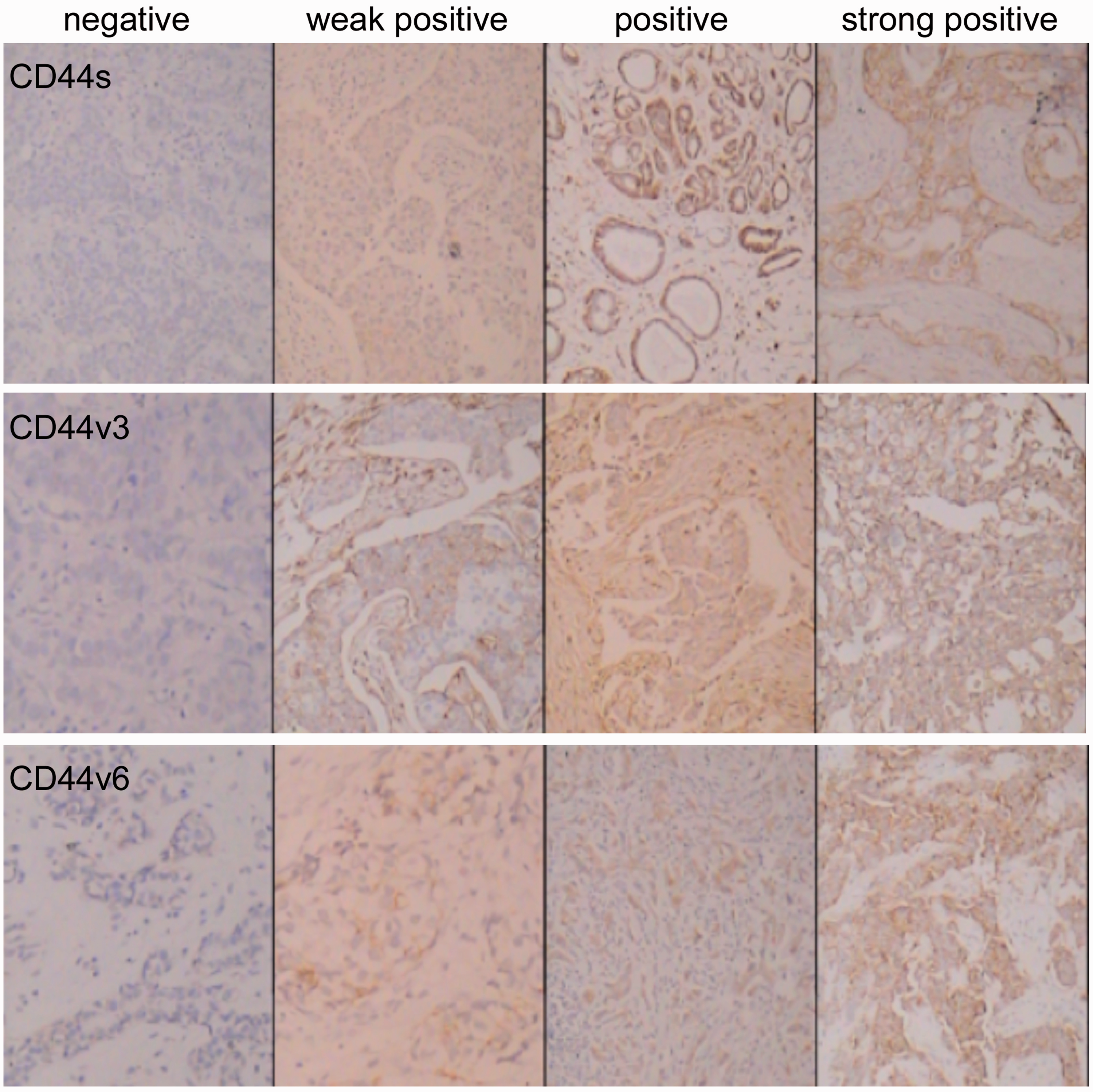
Relationship between immunopositivity for standard and variant forms of CD44 in tumour tissue samples and clinicopathological characteristics of breast cancer (n = 60).

Data presented as n.
Staining: −, 0–<10% positive cells; +, 10–25% positive cells; ++, 26–50% positive cells; +++, ≥51% positive cells.
CD44s, CD44 standard variant; CD44v3, CD44 variant exon 3; CD44v6, CD44 variant exon 6; NS, not statistically significant (P ≥ 0.05, analysis of variance); TNM, tumour–node–metastasis. 20
There were significant negative correlations between CD44s immunopositivity in breast cancer tissue and tumour diameter (r = –0.338, P < 0.01) and stage (r = −0.298; P < 0.05), and significant positive correlations between CD44v6 immunopositivity and tumour diameter (r = 0.257; P < 0.05) and stage (r = 0.383; P < 0.01). There were no significant correlations between CD44v3 and tumour diameter or stage.
Discussion
The molecular and cellular processes underlying breast cancer development are poorly understood, and there are few definitive biological markers. The present study was designed to determine the value of CD44 molecules as tumour markers for breast cancer. We found that CD44s was located to the cytoplasmic membrane, whereas CD44v3 and v6 were found in the cytoplasm. This suggests that CD44s may be the more useful of these molecules as a therapeutic target.
The present study observed that CD44s and CD44v6 had identical rates of immunopositivity, but their cellular location and biological function were different. This identical rate of positive staining is likely to be a coincidence, but larger numbers of samples are required to confirm this.
Both CD44s and CD44v6 positivity were associated with tumour stage and diameter in the present study, indicating that these molecules may be useful tumour markers. In addition, CD44s and CD44v6 were both correlated with lymph node involvement. In contrast to our findings, others have found significant associations between CD44v3 and tumour development or prognosis.10–12 This inconsistency may be due to heterogeneity within the tumour itself, different standards for evaluating staining, or variations in antibody specificity or sensitivity.
Interestingly, tumour diameter and stage were negatively correlated with CD44s but positively correlated with CD44v6 in the present study, suggesting that these molecules have opposing roles in breast cancer development. Patients with CD44v6-positive/CD44s-negative tumours may have an increased risk of tumour invasion and metastasis. Others have found that normal ductal epithelial cells of mammary glands and proliferative mammary cells do not express CD44 variants. 21 In addition, CD44v6-positive breast cancers were more likely to have lymph node involvement than CD44v6-negative tumours. 21 It is possible that transcriptional alterations in CD44 result in functional and structural abnormalities, impacting on recognition, adhesion and information transfer among cells, ultimately altering biological behaviour. Moreover, these adhesion factors could be important molecular biological markers that predict the invasion and metastatic prognosis of breast cancer. Members of the CD44 family were found to be biological markers in colorectal cancer. 22
It is well known that CD44 is the major HA receptor, and that HA-bound CD44 participates in tumour progression, metastasis and proliferation. 6 CD44 has been demonstrated to play a role in promoting cell-to-extracellular matrix interaction, 23 binding to HA,24,25 binding to matrix metalloproteinase, 24 and erbB receptor tyrosine kinases. 24
The present study has several limitations, notably the small sample size. In particular, only five patients had stage IV tumours. Future studies should include at least 100 participants in order to meet statistical requirements as well as nontumour control tissues. A further limitation was that reverse transcription–polymerase chain reaction was not performed. This would allow direct assessment of gene expression and enable the investigation of its correlation with protein levels. Finally, we included a limited number of clinicopathological variables. Data regarding histological grade, proliferation index, and oestrogen receptor/HER2 status should be included in future studies.
In conclusion, CD44s and CD44v6 appear to play opposing roles in the development of breast cancer, but their precise function and mechanisms of action remain unclear.
Footnotes
Declaration of conflicting interest
The authors declare that there are no conflicts of interest.
Funding
This work was supported by a grant from the Jiangsu Health International Exchange Supporting Program provided by Dr Xiao-Dong Li.