Increase in cerebral blood flow on brain magnetic resonance angiogram following correction of cervical lordosis
E Katz1, 2, S Katz1, C Fedorchuk1, 2 and D Lightstone1, 2
1Institute For Spinal Health and Performance
2CBP Nonprofit
Abstract
Background: Loss of cervical lordosis is associated with decreased vertebral artery hemodynamics. Vertebral arteries merge forming the basilar artery which continues to the circle of Willis and cerebral arteries. A close anatomical relationship exists between the cervical spine, vertebral arteries, and cerebral vasculature.
Aim: Evaluate cerebral blood flow changes on brain MRA in patients with loss of cervical lordosis prior to and following correction of cervical lordosis.
Method: This study is a retrospective consecutive case series. Cervical lordosis of seven patients (five females, two males, 28 to 58 years) was measured on lateral cervical radiographs ranging from −13.1 to 19.0 degrees (ideal is −42.0 degrees). Brain MRAs were analyzed for pixel intensities representing blood flow. Pixel intensity of the cerebral vasculature was quantified, and percent change determined.
A student’s t-test established significance of percent change in cerebral blood flow between pre- and post-cervical lordosis adjustment images. Regression analysis was performed. An a priori analysis determined correlation between cervical lordosis and change in MRA pixel intensity. The statistician was blinded to cervical lordosis.
Results/Conclusions: Pixel intensity increased 23.0 to 225.9% and a student’s t-test determined the increase was significant (p < 0.001). Regression analysis of change in pixel intensity versus cervical lordosis showed that as deviation from a normal cervical lordosis increases, percent change in pixel intensity on MRA decreases.
These results indicate that correction of cervical lordosis may be associated with immediate increase in cerebral blood flow. Further studies are needed to confirm these findings and understand clinical implications.
Cervical Lordosis and Cerebral Blood Flow Quantifications of Participants for Pre and Post Cervical Lordosis Adjustment MRA.
Participant
Normal ARA C2–C7 (°)
ARA C2-C7 (°)
Deviation from idealARA C2–C7 (°)
Pre-Adjustment MRA area over threshold (in2, )
Post-Adjustment MRA area over threshold (in2, )
Pre-adjustment MRA total pixel intensity
Post-adjustment MRA total pixel intensity
Percent change in total pixel intensity (%)
Pre-adjustment MRA threshold
1
−42.0
19.0
61
0.444
0.525
453094
557461
23.0
76
2
−42.0
6.7
48.7
0.257
0.311
190537
240016
26.0
60
3
−42.0
3.7
45.7
0.104
0.273
84668
249567
194.8
70
4
−42.0
3.0
45
0.304
0.427
300480
444834
48.0
79
5
−42.0
−11.8
30.2
0.190
0.256
170810
256071
49.9
70
6
−42.0
−13.1
28.9
0.333
0.655
386883
841061
117.4
90
7
−42.0
−20.4
21.6
0.235
0.607
176260
574491
225.9
65
Mean
−42.0
−1.8
40.2
1.721
2.815
251819
451929
97.9
73
66
Intermittent exercise ameliorates maladaptive effects of compensatory limb use in a mouse model of stroke
A Kerr, H Scheller and M Harris
Illinois Wesleyan University
Abstract
Background: Reliance on the less-impaired limb (i.e., compensatory limb use) after stroke results in poor recovery of the impaired limb in humans and animals. It has been demonstrated in a mouse model of stroke that voluntary exercise (24-hour wheel access) ameliorates the maladaptive effects of compensatory limb use and preserves the rehabilitative potential of the impaired limb. However, this exercise regimen is significantly more intensive than is feasible for an adjunctive aerobic therapy in humans.
Aim: In an effort to establish a more clinically relevant model of adjunctive exercise therapy, the current study investigated a lower intensity exercise regimen during early compensatory limb use.
Method: Mice were trained on a skilled reaching task before receiving unilateral photothrombotic stroke. Following a brief recovery period, mice were divided into three equal groups: control (no training/wheel access), compensatory training (training of the less-impaired limb), and exercise (training of the less-impaired limb with 12-hours of wheel access every other night). Following a 14-day training period, all mice were assessed on the skilled reaching task using their impaired limb.
Results/Conclusions: All groups displayed impaired reaching performance at initial impaired limb assessment. Over 7 days of impaired limb training, performance of mice with intermittent access to running wheels improved. These findings suggest that intermittent wheel access during compensatory limb use is sufficient to preserve the rehabilitative potential of the impaired limb and point to aerobic exercise as a potential adjunctive therapy to permit early compensatory limb use without sacrificing the ultimate recovery of the impaired limb.
68
An attempt to early detection and early treatment of spinal cord ischemia using clinical MRI
N Shingo1, S Masahiro2, A Koji3, T Hitoshi4 and U Tetsuro1
1Second Department of Surgery, Yamagata University, school of medicine
2Sendai University, Faculty of Sport Science
3National Center of Neurology and Psychiatry
4Yamagata University Health Administration Center
Abstract
Background: In aortic surgery, paraplegia due to spinal cord ischemia is an important complication that terribly reduces the patient’s quality of life. However, there is currently no reliable treatment or early detection method that can be applied clinically beyond the realm of basic research.
Aim: The aim of this study is to establish a reliable method for early detection and treatment of spinal cord ischemia, and as an initial step, we analyzed signal changes in the ischemic spinal cord of rabbits using clinical magnetic resonance imaging (MRI).
Method: A rabbit spinal cord infarction model was prepared and divided into four groups according to the time since preparation: 8 hours (A: n = 6), 24 hours (B: n = 6), 48 hours (C: n = 6), and no intervention (D: n = 6). MRI signal changes were assessed by the region of interest index of the spinal cord, especially in the anterior horn of the spinal cord where motor neurons are located, at the height of the inferior end of the scapula and the height of the superior end of the iliac crest.
Results/Conclusions: The median region of interest index was 1.1 (1.06–1.43) at A, 2.4 (1.9–2.6) at B, and 1.3 (−) at C, 0.98 (0.96–0.98) at D. The signal intensity of the infarcted spinal cord increased with time peaked at 24 hours, and then significantly decreased. The results of this study were similar to the course of delayed paraplegia death.
70
Improvement in post-concussion symptoms/disabilities and concussion frequency in a rugby player following cervical spine correction
S Strauss1, 2, D Lightstone1, 2 and C Fedorchuk1, 2
1Institute for Spinal Health and Performance
2CBP Nonprofit
Abstract
Background: Loss of cervical lordosis is associated with decreased vertebral and cerebral artery hemodynamics which play a role in concussion pathophysiology.
Aim: Report on improvements in post-concussion symptoms and disabilities and reduction in concussion frequency in a 27-year-old male professional rugby player following correction of cervical lordosis and frontal plane alignment.
Method: A 27-year-old male presented with a six-year history of 20 concussions as a starting professional rugby player and associated post-concussion symptoms/disabilities which persisted despite treatments involving physical therapy and chiropractic adjustments. Initial cervical radiographs showed loss of C2–C7 cervical lordosis measuring −13.5° (ideal is −42.0°) and C2–T3 right translation measuring −22.7 mm (ideal is 0 mm). The patient was treated 30 times over 16 days using Chiropractic BioPhysics® spinal rehabilitation exercises, adjustments, and spinal tractions.
Results/Conclusions: Re-exam at 16 days revealed improvements in post-concussion symptoms/disabilities and radiographs showed correction of C2-C7 cervical lordosis to −37.4° and in C2-T3 right translation to −11.3 mm. Two-year follow-up exam showed maintained improvements in post-concussion symptoms/disabilities and radiographs showed maintained correction of C2–C7 cervical lordosis measuring −36.7° and C2–T3 right translation measuring −11.7 mm. Six-year follow-up assessment revealed the patient maintained improvements in post-concussion symptoms/disabilities and only sustained 2 more concussions while maintaining the same intensity/frequency of participation as a starting professional rugby player.
This case reveals that correction of cervical lordosis and frontal spinal alignment using Chiropractic BioPhysics® protocol may improve post-concussion symptoms/disabilities and may help to prevent/minimize concussions in contact sports.
Figure 1. NLC radiographs with actual sagittal alignment (red) and ideal alignment (green). (a) Initial NLC radiograph; C2–C7 lordosis measuring −13.5° (ideal is −42.0°); (b) 16-day Re-exam NLC radiograph; C2–C7 lordosis measuring −37.4°; (c) 2-year Follow-up NLC radiograph; ARA C2–C7 measuring −36.7°.
Figure 2. APLC radiographs with actual frontal alignment (red) and ideal alignment (green). a. Initial APLC radiograph; C2-T3 translation measuring −22.7 mm (ideal is 0 mm); b. 16-day Re-exam APLC radiograph; C2-T3 translation measuring −11.3 mm; c. 2-year Follow-up APLC radiograph; C2-T3 translation measuring −11.7 mm.
Pre and post treatment outcome assessments for post-concussion symptoms and disabilities.
Outcome assessment
Initial exam
Re-exam
Quadruple Visual Analog Scale For Neck Pain (0-10, 0 = no pain, 10 = max)
Now
10
0
Best
2
0
Average
7
2
Worst
10
5
Time in pain (%)
100
<5
Quadruple Visual Analog Scale For Headache Pain (0–10, 0 = no pain, 10 = max)
Neck Disability Index (0-100%, 0% = no disability, 2% = minimal disability, 21% = moderate disability, 41% = severe disability, 61% = crippled, 81% = complete disability, 100% = max)
52
4
Short-Form 36-Item Health-Related Quality of Life Questionnaire (0–100, 0 = worst quality of life, 100 = best quality of life)
General health
85
100
Physical functioning
45
95
Role physical limitations
0
100
Role emotional limitations
100
100
Energy/fatigue
85
100
Emotional health
92
92
Social well-being
100
100
Pain
22.5
90
Change in health status
50
100
74
Deep learning to decipher the progression and morphology of hemin-induced axonal degeneration
A Palumbo1, 2, 3, 4, 5, P Grüning6, S Landt1, 2, L Heckmann1, 2, L Bartram1, A Pabst1, 2, C Flory1, 2, M Ikhsan1, 2, 3, 7, S Pietsch1, 2, 8, R Schulz9, C Kren10, N Koop10, J Boltze1, 2, 11, A Madany Mamlouk6 and M Zille1, 2, 3, 12
1Fraunhofer Research and Development Center for Marine and Cellular Biotechnology EMB
2Institute for Medical and Marine Biotechnology, University of Lübeck
3Institute for Experimental and Clinical Pharmacology and Toxicology, University of Lübeck
4Department of Cancer Biology, Dana-Farber Cancer Institute
5Department of Neurobiology, Harvard Medical School
6Institute for Neuro- and Bioinformatics, University of Lübeck
7Faculty of Medicine, Malikussaleh University
8Department of Neonatology, Universitätsklinikum Leipzig
9Wissenschaftliche Werkstätten, University of Lübeck
10Medical Laser Center Lübeck GmbH
11School of Life Sciences, The University of Warwick, Gibbet Hill Campus
12Department of Pharmaceutical Sciences, Division of Pharmacology and Toxicology, University of Vienna
Abstract
Background: Axonal degeneration (AxD) is characterized by the formation of axonal swellings and axonal fragments. However, how hemolysis products such as hemin affect axons after hemorrhagic stroke remains unknown.
Aim: Deciphering the axonal morphology in hemin-induced AxD.
Method: We evaluated the progression of hemin-induced AxD in spatially isolated axons using a novel microfluidic device together with a deep learning tool that we developed for the enhanced-throughput analysis of AxD on phase-contrast microscopic images.
Results/Conclusions: The performance of the convolutional neural network to segment axons, axonal swellings, and axonal fragments sensitively and specifically exceeded that of human evaluators. We detected a time-dependent degeneration of axons upon exposure to hemin. While the axon area decreased starting at 11.5 h, the axonal swelling and axonal fragment areas increased starting at 6 and 9 hours, respectively. As axonal swellings preceded axonal fragmentation, this finding suggests that axonal swellings may be reliable predictors of AxD. Using a recurrent neural network, we identified four morphological patterns of AxD (granular, retraction, swelling, and transport degeneration). The morphological patterns of AxD occurred concomitantly and correlated with increasing hemin concentrations.
Taken together, our data suggests that hemin-induced AxD is a time-dependent and morphologically heterogeneous process. Our EntireAxon platform enables the systematic analysis of axons and AxD and unravels a so-far unknown intricacy of AxD in context of hemorrhagic stroke.
Figure 1. Progression and morphology of hemin-induced AxD. a, Analysis of hemin-induced AxD over time in vitro. b, Schematic illustrations and time lapse recordings of the four morphological patterns of AxD.
75
Repeated administration of 2-hydroxypropyl-β-cyclodextrin (HPβCD) attenuates the chronic inflammatory response to experimental stroke
D Becktel, J Zbesko, J Frye, A Chung, T Nguyen and K Doyle,
University of Arizona
Abstract
Background: Globally, more than 67 million people are living with the effects of ischemic stroke. Importantly, many stroke survivors develop a chronic inflammatory response that may contribute to cognitive impairment, a common and debilitating sequela of stroke that is insufficiently studied and currently untreatable. 2-hydroxypropyl-β-cyclodextrin (HPβCD) is an FDA-approved cyclic oligosaccharide that can solubilize and entrap lipophilic substances.
Aim: The goal of the present study was to determine whether the repeated administration of HPβCD curtails the chronic inflammatory response to stroke by reducing lipid accumulation within stroke infarcts in a distal middle cerebral artery occlusion mouse model of stroke.
Method: To achieve this goal, we subcutaneously injected young adult and aged male mice with vehicle or HPβCD three times per week, with treatment beginning one week after stroke. We evaluated mice at 7 weeks following stroke using immunostaining, RNA sequencing, lipidomic, and behavioral analyses.
Results/Conclusions: Chronic stroke infarct and peri-infarct regions of HPβCD-treated mice were characterized by an upregulation of genes involved in lipid metabolism and a downregulation of genes involved in innate and adaptive immunity, reactive astrogliosis, and chemotaxis. Correspondingly, HPβCD reduced the accumulation of lipid droplets, T lymphocytes, B lymphocytes, and plasma cells in stroke infarcts. Repeated administration of HPβCD also preserved NeuN immunoreactivity in the striatum and thalamus and c-Fos immunoreactivity in hippocampal regions. Additionally, HPβCD improved recovery through the protection of hippocampal-dependent spatial working memory and reduction of impulsivity. These results indicate that systemic HPβCD treatment following stroke attenuates chronic inflammation and secondary neurodegeneration and prevents post-stroke cognitive decline.
78
Severe intracerebral hemorrhage is associated with activated pro-inflammatory signaling pathways in human peripheral blood
B Knepp1, M Durocher1, A Yee1, G Jickling1, 2, H Hull1, N Alomar1, K Ng1, X Zhan1, P Carmona-Mora1, H Amini1, M Hakoupian1, B Ander1, F Sharp1 and B Stamova1
1University of California Davis School of Medicine
2University of Alberta Department of Medicine
Abstract
Background: Intracerebral Hemorrhage (ICH) is caused by bleeding into brain parenchyma and is associated with high morbidity and mortality. The specific mechanisms of brain damage are not well characterized and few treatments proven to limit damage or improve recovery exist.
Aim: We aimed to identify genes and gene-networks in peripheral blood associated with ICH severity.
Method: Whole-genome gene expression of 21,175 genes in 17 ICH patients was analyzed at the gene-network and per-gene levels. Weighted Gene Co-Expression Network Analysis identified co-expressed gene modules associated with ICH severity (log2-trasformed NIHSS, offset = 1, average time post ictus = 58.1 hours), p < 0.05. At the per-gene level, genes associated (p < 0.05) with vascular risk factors were excluded to examine the ICH response irrespective of comorbidities. Of the remaining 15,378 genes, a Pearson correlation identified genes associated with ICH severity (p < 0.05, q < 0.2).
Results/Conclusions: Six modules (5 positively correlated, 1 negatively) (Figure 1(a)) and 596 genes from the per-gene analysis were associated with ICH severity. Positively correlated modules were enriched in Monocyte and Granulocyte (mostly Neutrophil) specific genes. The per-gene list was also enriched in Monocyte specific genes (Figure 1(b)). For patients with worse severity, these lists were enriched and predicted activated in many pro-inflammatory pathways including Neuroinflammation, Inflammasome, Leukocyte Extravasation, Fcγ Receptor-mediated Phagocytosis in Macrophages and Monocytes, Production of NO and ROS in Macrophages, IL-(1,2,6,8,17,23) Signaling, and IL-15 Production. These results show pro-inflammatory responses associate with worse ICH severity. These genes and pathways could be novel treatment targets to improve outcomes.
79
Treatment with gut microbial-related fibre can improve post-stroke recovery in mice
A Peh1, 2, E Dinakis1, M Nakai1, B Broughton2 and F Marques1, 3
1Hypertension Research Laboratory, School of Biological Sciences, Monash University
2Cardiovascular & Pulmonary Pharmacology Group, Department of Pharmacology, Monash University
3Heart Failure Research Group, Baker Heart and Diabetes Institute
Abstract
Background: Evidence supports that the gut microbiota contributes to ischemic stroke pathobiology via the gut-brain axis. We hypothesised that high fibre (HF) diets, which are prebiotic and feed the gut microbiota, may be beneficial for post-stroke recovery.
Aim: To determine the role of prebiotic fibre in an experimental mouse model of ischaemic stroke.
Method: Male C57BL/6J mice (8–9-weeks old, n = 8/group) were randomized and treated with a control (AIN93G) or nutrient-matched diets containing low fibre (LF) or HF for 4 weeks prior to photothrombotic stroke surgery. Post-stroke, mice were either fed the same diet or HF for 7 days. Infarct volume and functional testing were determined at day 1 (D1) and D7 post-surgery. Gut barrier integrity and cell infiltration were examined via immunofluorescence at D7. P < 0.05 was considered significant.
Results/Conclusions: HF treatment improved D7 infarct recovery rate (mean ± SEM: control: 73.83 ± 2.36%; HF: 82.57 ± 2.88%; LF: 64.93 ± 2.94%, P = 0.0007) and wire-hanging grip strength (control: 160.40 ± 33.22s; HF: 230.20 ± 29.69s; LF: 36.83 ± 7.99s, P = 0.0001). To determine the importance of HF after stroke, LF mice were placed on HF diet immediately post-surgery. This improved D7 brain recovery (P = 0.0014) and wire-hanging grip strength (P < 0.0001). Compared to LF, HF-treated mice had more CD3+ T cells (P = 0.0009) and DCX+/Tuj1+ neuroblasts (P = 0.0005) in the ischemic hemisphere, with a positive correlation between DCX+/Tuj1+ and recovery rate post-stroke (R2, = 0.18, P = 0.0355). HF-treated mice also had significant improved gut barrier integrity post-stroke (P = 0.0001). In conclusion, prebiotic fibre reduced stroke-induced injury and improved functional recovery, which likely occurs via neurogenesis and the suppression of the immune response.
80
miR-193a-5p exerts neuroprotection via reprogramming neutrophil phenotypic transformation partially through UBE2V2 mediated-ubiquitination of PPARγ
L Li1, 2, Z Han1, 2, F Yan1, H Zhao1 and Y Luo1, 2
1Institute of Cerebrovascular Diseases Research, Xuanwu Hospital, Capital Medical University
2Department of Neurology, Xuanwu Hospital,Capital Medical University
Abstract
Background: Circulating neutrophils are activated shortly after stroke and in turn impact the fate of ischemic brain tissue. microRNAs participate in regulating neuroinflammation.
Aim: To probe the role of neutrophilic microRNA (miR-193a-5p) in ischemic stroke.
Method: Two cohorts of acute ischemic stroke (AIS) patients were recruited for neutrophilic miRNA sequencing, validation, and evaluation of miRNA’s clinical significance. Experimental I/R model and neutrophils were transfected with agomiR-193a-5p/antagomiR-193a-5p for in vivo and in vitro study. TTC staining and neurological function scoring were performed to evaluate the severity of I/R injury. Flow cytometry and ELISA were adopted for detecting N2 neutrophils and inflammatory factors. We pooled the mRNA targets with bioinformatic analyses and explored miRNA-protein association with Fluorescence-In-Situ-Hybridization and RNA-Binding-Protein-Immunoprecipitation. RT-qPCR and western-blotting were adopted to detect the mRNA and protein expression. Immunoprecipitation and immunofluorescence were performed to investigate the relation between UBE2V2 and PPARγ.
Results: i) miR-193a-5p is decreased in circulating neutrophils of AIS patients.
ii) Higher neutrophilic miR-193a-5p is associated with better prognosis of AIS patients.
iii) miR-193a-5p protects the brain against I/R injury in vivo and in vitro.
iv) Protective role of miR-193a-5p may correlate with neutrophils N2 transformation.
v) AgomiR-193a-5p may induce neutrophils N2 transformation via UBE2V2.
vi) UBE2V2 may target neutrophilic phenotype-associated switch-PPARγ.
Conclusions: miR-193a-5p protected against cerebral ischemic injury by restoring neutrophil N2 phenotype associated neuroinflammation reduction. In mechanism, the effect of miR-193a-5p is likely induced, in part, through UBE2V2 mediated ubiquitination of PPARγ.
81
Lomitapide ameliorates MCAO induced cerebral ischemia/reperfusion injury via promoting neuron autophagy and inhibiting microglia migration
y hu1, 2, f yan1, 2, z han1, 2, y luo1, 2, 3 and y zheng1, 2
1Institute of Cerebrovascular Disease Research and Department of Neurology, Xuanwu Hospital of Capital Medical University
2Beijing Geriatric Medical Research Center and Beijing Key Laboratory of Translational Medicine for Cerebrovascular Diseases
3Beijing Institute for Brain Disorders, Capital Medical University
Abstract
Background: Stroke is a high incidence and disabling condition that can lead to severe cognitive, motor and sensory dysfunction. However, there is still a lack of specific drugs for the treatment of stroke up to now. Lomitapide is a kind of lipid-lowering drug, and it is non-toxic or low toxic to human body. This is the first study to find that it has a protective effect on ischemic nerve injury in vitro and in vivo.
Aim: In this study, we take advantage of a drug repurposing strategy to research the neuroprotective effect of lomitapide on focal ischemic brain injury and explore its possible mechanism.
Method: Experimental cerebral ischemia was induced by middle cerebral artery occlusion (MCAO) in male adult C57BL/6 mice. The adhesive removal testing and beam walking testing was used to assess neurological functions. Via western blot and immunofluorescent staining, autophagy flux were evaluated by analyzing the expression profiles of LC3, P62 and LAMP2. In addition, microglial migration was assessed by oxygen-glucose deprivation (OGD) N2a-BV2 cocultivation.
Results/Conclusions: We found that lomitapide significantly increased survival rate, reduced brain tissue loss, and improved neurological function after MCAO. Further, lomitapide could increase the expression of LC3, reduce the expression of p62 and LAMP2 and promote autophagic flux, and inhibit apoptosis by increasing the expression of apoptosis-associated proteins Bcl-2 and inhibiting the expression of Bax. In addition, lomitapide inhibits the migration of pro-inflammatory microglia. Taken together, our results suggest that lomitapide can be repositioned as a potential therapeutic drug for stroke treatment.
84
Nicotine patch alters patterns of cigarette smoking-induced dopamine release: Patterns relate to treatment response biomarkers
Y Zakiniaeiz, H Liu, H Gao, S Najafzadeh, J Ropchan, N Nabulsi, Y Huang, D Matuskey, M Chen, K Cosgrove and E Morris
Yale University
Abstract
Background: Tobacco smoking is a major public health burden. The first line pharmacological treatment for tobacco smoking is nicotine replacement therapy (e.g., the nicotine patch (NIC)). Nicotine acts on nicotinic-acetylcholine receptors on dopamine terminals to release dopamine in the ventral and dorsal striatum encoding reward and habit formation, respectively.
Aim: To better understand treatment efficacy, a naturalistic experimental design combined with a kinetic model designed to characterize smoking-induced dopamine release in vivo, was used.
Method: Thirty-five tobacco smokers (16 female) wore a nicotine patch (21mg, daily) for 1-week and a placebo patch (PBO) for 1-week in a randomized, counter-balanced order. Following 1-week under NIC and then overnight abstinence, smokers participated in a 90-minute [11, C]raclopride positron emission tomography scan and smoked a cigarette while in the scanner. Identical procedures were followed for the PBO scan. A time-varying kinetic model was used at the voxel level to model transient dopamine release peaking instantaneously at the start of the stimulus and decaying exponentially. Magnitude and spatial extent of dopamine release were estimated. Smokers were subcategorized by nicotine dependence level and nicotine metabolism rate.
Results/Conclusions: Dopamine release magnitude was enhanced by NIC in ventral striatum and diminished by NIC in dorsal striatum. More-dependent smokers activated more voxels than the less-dependent smokers under both conditions. Under PBO, fast metabolizers activated more voxels in ventral striatum and fewer voxels in dorsal striatum compared to slow metabolizers. These findings demonstrate that the model captured a pattern of transient dopamine responses to cigarette smoking which may be different across smoker subgroup categorizations.
85
Safety and protein content of blood derived extracelluar vesicles in an intracerebral hemorrhage rat model
F LasoGarcía1, L Casado-Fernández1, M Gómez-de Frutos1, D Piniella-Alcalde1, L Otero-Ortega1, S Bravo2, B Fuentes1, E Díez-Tejedor1, M Gutiérrez-Fernández1, de Alonso and M Leciñana1
1IdiPAZ Health Research Institute
2Health Research Institute of Santiago de Compostela (IDIS)
Abstract
Background: Our group has previously demonstrated the efficacy of blood-derived extracellular vesicles (EVs) administration in improving functional recovery in a rat intracerebral hemorrhage (ICH) model.
Aim: We thereafter analyzed safety, brain tissue integrity, and proteins content of circulating EVs from ICH rats treated with EVs.
Methods: Allogenic and xenogenic (human) EVs were obtained from the blood of subjects with spontaneous good outcome after ICH to be used as a treatment. We studied four groups of Sprague-Dawley rats: healthy (N = 10), ICH controls (N = 20), ICH treated with i.v. allogenic EVs (AEVs, N = 10), or xenogenic EVs (XEVs, N = 10) at 24 h. For safety studies, circulating inflammatory cytokines and neoformations in organs (H&E) were analyzed. Brain tissue integrity was analyzed by Diffusion Tensor Imaging. Protein content of circulating EVs at 28d after treatment was analyzed by quantitative mass spectrometry (DIA SWATH).
Results/Conclusions: Neither immune response nor tumor formation after treatment was observed. Both treatments improved tissue integrity compared to controls at 28d (p < 0,05). Notably, among 1646 proteins identified in EVs, values of RASH and SYT1 (related with angiogenesis and trafficking of synaptic vesicles) were higher in AEVs group at 28d; and FA7, LSAMP, and PCSK9 (related with angiogenesis, axon targeting, and neuronal growth) in XEVs group at 28d compared with ICH control group at 28d. In conclusion, the administration of blood-derived EVs, regardless of origin, is safe. Higher values of proteins involved in biological processes potentially related to brain repair may account for the improvement of tissue integrity and functional evolution of the treated subjects.
86
Image derived input function to perform non-invasive quantitative analysis of myelin imaging with [11C]MeDAS PET
C Van Der Weijden1, 2, J Meilof1, 2, 3, R Dierckx1, 2, A van der Hoorn1, 2
A Willemsen1, 2, A Lammertsma1, 2 and E de Vries1, 2
1UMCG
2University of Groningen
3Martini Ziekenhuis
Abstract
Background: Multiple sclerosis (MS) is an inflammatory demyelinating disease. New treatment strategies aiming to repair myelin damage are under development, but require an accurate biomarker for efficacy evaluation. We have recently shown that quantification of myelin density with [11, C]MeDAS PET using pharmacokinetic modelling is possible. However, this method uses an arterial input function (AIF), which requires arterial blood samples. The invasive nature of arterial sampling reduces clinical applicability due to logistical complications and patient discomfort.
Aim: To investigate whether an image derived input function (IDIF) can be used as a non-invasive input function for pharmacokinetic modelling of [11, C]MeDAS PET data.
Method: Six healthy controls and 11 MS patients underwent MRI and [11, C]MeDAS PET with arterial blood sampling. The IDIF was created from either the hottest 2, 4, 6, or 12 voxels of the carotid artery, or an iso-contour region-of-interest from the summed images of 0–70 seconds. This was followed by blood-to-plasma conversion and metabolite correction of the IDIF.
Results/Conclusions: All IDIFs had a sharper and higher peak in the blood curves than the AIF, probably due to dispersion during blood sampling. All IDIF methods provide outcome parameters (Ki) with a high precision (mean r = 0.95–0.98), but a highly variable accuracy across subjects (mean slope = 0.90–1.09). The reproducibility of kinetic modelling using an IDIF is fairly high for both brain regions (TRTV = 12.1–18.0%) and MS lesions (TRTV = 12.3–23.5%).
Although pharmacokinetic modelling of [11, C]MeDAS PET using an IDIF gave precise and reproducible results, the highly variable accuracy across subjects limits its use for [11, C]MeDAS PET quantification.
87
Increasing the efficiency of cerebrovascular autoregulation-guided personalized management for traumatic brain injury patients
E Chaleckas1, V Putnynaite1, V Petkus1, M Deimantavicius1, K Boere1, T Tamosuitis2, M Kavaliauskas3, S Rocka4, A Preiksaitis4 and A Ragauskas1
1Health Telematics Science Institute, Kaunas University of Technology
2Department of Intensive Care, Academy of Medicine, Lithuanian University of Health Sciences
3Department of Applied Mathematics, Kaunas University of Technology, Lithuania
4Clinic of Neurology and Neurosurgery, Faculty of Medicine, Vilnius University, Lithuania
Abstract
Background: Optimal cerebral perfusion pressure (CPPopt) targeted treatment of traumatic brain injury (TBI) requires 4 h or longer multi-modal monitoring data accumulation to identify CPPopt for individual patients. Nevertheless, even with a long 4-h data accumulation window, the CPPopt value can be identified in about 50–60% of monitoring time.
Aim: To increase the efficiency of CPPopt targeted treatment by minimizing the time of monitoring data accumulation needed to choose the appropriate individualized clinical therapy.
Method: A retrospective analysis of multi-modal physiological monitoring data of 87 severe TBI patients was performed by representing cerebrovascular autoregulation (CA) indexes in relation to CPP, arterial blood pressure (ABP) and intracranial pressure (ICP) separately in order to improve the existing CPPopt identification algorithms. Machine learning (ML) based algorithms were developed for automatic identification of selected informative data segments that were used for reliable CPPopt, ABPopt, ICPopt or lower/upper limits of CA (LLCA/ULCA) identification.
Results/Conclusions: The reference datasets of the informative data segments, artifact-distorted segments, and datasets of different clinical situations were used for training ML-based algorithms allowing to choose the appropriate individualized CPP, ABP or ICP management in 79% of full monitoring time of studied population data.
Developed ML-based algorithms allow to identify CPPopt (or ABPopt or LLCA/ULCA) values within 24 min (10-times shorter comparing to 4 h of existing in clinical practice data accumulation time) in the cases when informative physiological ABP/ICP variations are detected. Prospective clinical studies are needed in order to prove the efficiency of developed algorithms.
89
Establishing oxygen-glucose deprivation model for cerebral ischemia in a developing country: The experience in Panama
B Denis1, 2 and R Gittens1, 2
1INDICASAT AIP
2University of Panama
Abstract
Background: Cerebrovascular diseases occupy the 2nd place in mortality1, and 3rd in disability adjusted life years around the world.2, For Panama, in Central America, the situation with cerebral ischemia, the most prevalent type of cerebrovascular disease, seems to be worsening, moving from 4th3, to 2nd place in mortality in the last 8 years.4,
At this moment two therapies are approved for treatment of this disease: thrombectomy and thrombolysis.5,6, However, only some patients in urban settings have access to this therapy7, and less than 20% arrive to the hospital in time to be eligible.8, For these reasons is important that countries in the Central American region strengthen their research capabilities to understand this disease and develop new therapies.
Aim: Establish the first oxygen-glucose deprivation (OGD) model in Panama. The model will allow pathophysiological studies and the evaluation of natural product molecules isolated from its vast biodiversity that can be targeted as possible therapies for this disease.
Method: Primary neurons from Sprague Dawley embryos were cultured in neurobasal media supplemented with Ara-C at 48 h. The cultures were subjected to oxygen deprivation using a ProOxmodel 110 hypoxia chamber (Biospherix) and glucose deprivation using a glucose free media. Hypoxia mortality of control and experimental groups was evaluated by Lactate Dehydrogenase and Immunocytochemistry.
Results/Conclusions: Three exposure times were evaluated (1, 1.5 and 2 hrs) and we found that 1.5 h was enough to reach a 40 to 60% mortality, in accordance with the accepted values for an OGD model.
References
OMS. Global health estimates summary tables: projection of deaths by cause, age and sex, by WHO region.
Geneva, Switzerland:
Author, 2018.World Health Organization. WHO | Metrics: Disability-Adjusted Life Year (DALY). Geneva, Switzerland, www.who.int/healthinfo/global_burden_disease/metrics_daly/en/ (2018, accessed 30 August 2020).a-2a-75Ministerio de Salud. Indicadores basícos de país.
Panamá:
Author, 2014.
Ministerio de Salud. Indicadores de Salud Básicos, Panamá, www.minsa.gob.pa/sites/default/files/publicacion-general/indicadores_basicos_de_salud_2017_.pdf (2017, accessed 30 August 2020).HackeWKasteMBluhmkiE, et al.
Thrombolysis with Alteplase 3 to 4.5 hours after acute ischemic stroke. N Engl J Med2008;
359: 1317–1329.SmithWS.
Endovascular stroke therapy. Neurotherapeutics. 2019;
16: 360–368.FujiwaraKOsanaiTKobayashiE, et al.
Accessibility to tertiary stroke centers in Hokkaido, Japan: use of novel metrics to assess acute stroke care quality. J Stroke Cerebrovasc Dis. 2018;
27: 177–184.KleindorferDKisselaBSchneiderA, et al.
Eligibility for recombinant tissue plasminogen activator in acute ischemic stroke. Stroke2004;
35: ▪.
91
Boosting regulatory T cells promotes white matter repair after stroke
C Yuan1, L Shi1, Z Sun1, F Xu1, 2, Q Ye1, 2, D Sun1, 2 and X Hu1, 2
1University of Pittsburgh
2Geriatric Research, Education and Clinical Center, Veterans Affairs Pittsburgh Health Care System
Objectives: We recently report that regulatory T cells (Treg) improve white matter integrity at late stage of stroke. The low frequency of Tregs restricts their clinical utility as a cell therapy. IL-2/IL-2-antibody complex (IL-2/IL-2Ab) has been used to expand the number of Tregs in vivo for stroke treatment. The current study evaluated the effect of IL-2/IL-2Ab in white matter repair at late stage after stroke.
Methods: Adult male C57/BL6 mice were randomly received IL-2/IL-2Ab or isotype IgG (i.p) for 3 consecutive days starting 6h or 5d after transient (60 min) middle cerebral artery occlusion (tMCAO), and repeated once a day on day 10, 20 and 30. White matter integrity and sensorimotor functions were assessed up to 35d after stroke. Tregs were cocultured with lysophosphatidyl choline (LPC)-treated organotypic cerebellar cultures to evaluate the direct effect of Tregs on remyelination.
Results: The long-term IL-2/IL-2Ab treatment regimen significantly increased the number of Tregs in blood and spleen after tMCAO. Longitudinal in vivo and ex vivo diffusion tensor imaging (DTI) analysis revealed a significant increase in fractional anisotropy (FA) value at 28d and 35d after stroke in the IL-2/IL-2Ab-treated mice compared to isotype-treated mice, while their FA values at 14d after tMCAO remained similar, suggesting a delayed improvement in white matter integrity. IL-2/IL-2Ab treatment also greatly improved sensorimotor (rotarod test and adhesive removal test) and cognitive (water maze test) functions after stroke. There were positive correlations between the white matter integrity and behavior performance. Immunostaining of myelin basic protein and luxol fast blue staining confirmed the beneficial effects of IL-2/IL-2Ab on white matter structures 35d after tMCAO. In addition, IL-2/IL-2Ab treatment starting as late as 5d after stroke also improved white matter integrity 21d after tMCAO, suggesting long-term salutary effects of Tregs on late-stage tissue repair. LPC exposure for 16h induced demyelination in organotypic cultures, followed by gradual spontaneous remyelination upon removal of LPC. Co-culture with Tregs accelerated remyelination in organotypic cultures at 7d after LPC.
Conclusion: IL-2/IL-2Ab treatment expands Treg population in vivo and promotes white matter repair after stroke.
Acknowledgement
This work was supported by the NIH/National Institute of Neurological Disorders and Stroke (NINDS) grants NS094573 and by a VA grant (I01 BX003651).
92
Hypocapnia with indocyanine green kinetics detects the effect of staged carotid angioplasty to avoid hyperperfusion
I Nakagawa, M Kotsugi, S Yokoyama, T Furuta, H Sasaki, R Maeoka, S Yamada and H Nakase
Nara Medical University
Abstract
Background: Cerebral hyperperfusion syndrome (CHS) is a serious complication following carotid artery stenting (CAS). Staged angioplasty (AP) could potentially prevent CHS and hyperperfusion phenomenon (HPP) after revascularization. However, methods for measuring the effects of staged AP on cerebral hemodynamic reserve have not been established.
Aim: We evaluated whether indocyanine green kinetics and near-infrared spectroscopy (ICG-NIRS) with hypocapnia induced by hyperventilation can detect the effects of staged AP on hemodynamic reserve to prevent CHS after CAS.
Methods: Participants comprised 44 patients at high risk of CHS, whose ipsilateral cerebrovascular reactivity (CVR) was impaired on preoperative single-photon emission computed tomography (SPECT). Patients were divided into a staged AP group (n = 13) and a regular CAS group (n = 31).
Results/Conclusions: In the staged AP group, stenting was performed 3 weeks after staged AP. In the regular CAS group, 16 cases (52%) showed HPP, and five (16%) presented with CHS after CAS, while no HPP or CHS occurred in the staged AP group (p = 0.001). Changes in blood flow index (BFI) and time to peak (TTP) ratio during hypocapnia calculated from ICG-NIRS indicated a significant linear relationship with preprocedural CVR on SPECT (r = −0.710, 0.632, respectively; p < 0.0001 each). BFI and TTP ratio during hypocapnia were significantly improved after staged AP (p < 0.001 each). Furthermore, significant linear correlations were observed between BFI and TTP ratio during hypocapnia and postoperative asymmetry index AI (r = 0.405, −0.475, respectively; p < 0.01 each). Hypocapnia induced by hyperventilation under ICG-NIRS appears useful for detecting the effects of staged AP on hemodynamic reserve in patients at high risk of CHS.
93
Simultaneous quantification of glucose and oxygen metabolism in the cortex during task processing
A Bose1, 2, S Epp1, 2, K Kurcyus1, A Ranft3, A Hechler1, 2, I Yakushev4, C Preibisch1, G Castrillón1, 5 and V Riedl1
1Neuroradiology Klinikum rechts der Isar Technical University
2Ayudas diagnósticas SURA
3Klinik für Anästhesiologie und Intensivmedizin Klinikum rechts der Isar Technical Unversity
4Klinik und Poliklinik für Nuklearmedizin Klinikum rechts der Isar Technical University
5Graduate School of Systemic Neurosciences Ludwig-Maximilians-Universität
Abstract
Background: The human brain depends on a constant supply of glucose and oxygen. While most brain imaging studies rely on relative BOLD signal changes as a proxy for neuronal activity, only a few studies have attempted to quantify cerebral metabolic rates of glucose (CMRglc) and oxygen (CMRO2).
Aim: We established a setup at an integrative PET/MR-scanner to simultaneously measure CMRO2 and CMRglc with multiparametric, quantitative BOLD (mqBOLD; Bulte et al., 2012) and 18F-FDG-functional-PET (fPET) in the same subjects across multiple conditions (Villien et al., 2014).
Method: In the scanner, healthy participants were alternately shown a fixation cross and a moving checkerboard. We acquired fPET data while continuously infusing 18F-FDG intravenously. Arterial blood sampling was performed continuously to define the arterial input function. Simultaneously, we acquired BOLD-fMRI data and mqBOLD data. CMRO2 quantification is shown in Figure 1.
Results/Conclusions: Preliminary single-subject results of the three modalities (fPET, mqBOLD, BOLD) show a consistent increase in metabolic rates and relative BOLD. Task-related increase of glucose uptake is predominant in visual processing networks. For mqBOLD data, we found significant increases in T2*, OEF, CBF and CMRO2 in visual stimulation regions (group level). We are currently integrating the modalities to examine consistencies across the cortex.
This study, for the first time, simultaneously quantifies CMRglc and CMRO2 across experimental conditions within one imaging session. This allows us to address ongoing discussions about the relevance of non-oxidative metabolism in certain regions of the brain and during task processing.
Figure 1. Overviese over data acquisition and preprocessing.
94
Neuroprotection by taurine or N-acetylcysteine in high-fat diet-fed mice involves recovery of hippocampal energy metabolism
A Garcia-Serrano1, 2 and J Duarte1, 2
1Dep. Experimental Medical Science, Lund University
2Wallenberg Centre for Molecular Medicine, Lund University
Abstract
Background: Type 2 diabetes (T2D) is a risk factor for memory impairment. T2D models with memory impairment show brain metabolic alterations, including increased levels of hippocampal taurine. In contrast, treatments with taurine, or with N-acetylcysteine (NAC) that stimulates endogenous taurine synthesis, were suggested to enhance memory performance.
Aim: We aimed at studying metabolic mechanisms by which taurine and NAC improve memory performance in obesity-associated T2D.
Method: Ten-week-old female C57Bl/6J mice (n = 10/group) were fed either a control (CD, 10% fat) or a high-fat diet (HFD, 60% fat), and received regular water, water supplemented with 3% taurine (CD + T or HFD + T) or water with 3% NAC (CD + NAC or HFD + NAC). After 2 months, hippocampal metabolites were analysed by proton magnetic resonance spectroscopy (MRS) using STEAM, and memory performance was evaluated by novel location and novel object recognition tasks.
Results/Conclusions: Both taurine and NAC increased insulin secretion, and partially ameliorated HFD-associated glucose intolerance. HFD-fed mice showed memory impairment, but not when treated with taurine or NAC. MRS in the hippocampus showed decreased concentration of the neuronal marker N-acetylaspartate, lactate and phosphocreatine in HFD-fed mice compared to controls. While both taurine and NAC normalized N-acetylaspartate concentration to control levels, only NAC promoted the recovery of lactate concentration and of the creatine-to-phosphocreatine ratio in the hippocampus of HFD-fed mice. Interestingly, both treatments increased hippocampal levels of glutamate in CD- and HFD-fed mice. These results suggest that recovery of memory impairment in HFD-fed mice by taurine and NAC involves improvement of energy metabolism and N-acetylaspartate production.
96
Paediatric brain MRI findings following congenital heart surgery: A systematic review
F Alablani
University of Leicester
Objective: This systematic review aimed to establish the relative incidence of new post-operative brain MRI findings following paediatric congenital cardiac surgery.
Design: To distinguish perioperative changes from pre-existing MR findings, our systematic search strategy focused on identifying original research studies reporting both pre- and post-surgery brain MRI scans. Patient demographics, study methods, and brain MR findings were extracted.
Results: Twenty-one eligible publications, including two case-control, and one randomised controlled trial were identified. Pre-existing brain MRI findings were noted in 43% (513/1205) of neonates prior to surgery; mainly white matter injuries (WMI). Surgery was performed at a median age of 8 days with comparison of pre- and post-operative MR scans revealing additional new post-operative findings in 51% (550/1075) of patients; mainly WMI. Four studies adopted a brain injury scoring system, but the majority did not indicate the severity or time course of findings. In a sub-group analysis approximately 32% of patients with pre-existing lesions went on to develop additional new lesions post-surgery. Pre-existing findings was not found to confer a higher risk of acquiring brain lesions post-operatively. No evidence was identified linking new MR findings with neurodevelopmental delay.
Conclusion: This review and meta-analysis suggests that surgery approximately doubles the number of patients with new brain lesions.
100
Regional changes of cerebral oxidative stress in early Alzheimer’s disease
H Okazawa1, M Ikawa1, 2, T Tsujikawa1, T Mori1, A Makino1, Y Kiyono1 and H Kosaka3
1Biomedical Imaging Research Center, University of Fukui
2Department of Neurology, Faculty of Medical Sciences, University of Fukui
3Department of Psychiatry, Faculty of Medical Sciences, University of Fukui
Background/Aim: Oxidative stress imaging using radioactive copper labeled diacetyl-bis(N4, -methylthiosemicarbazone) (Cu-ATSM) has been established and applied to the studies of neurodegenerative diseases. The method was applied to early Alzheimer’s disease patients (eAD) and age-matched healthy controls (HC) to evaluate brain regional changes.
Methods: Ten AD (66 ± 9y) and 10 HC (71 ± 12y) were studied using PET/MRI with [C-11]PiB and [Cu-64]ATSM on separate days. The PET/MRI scans were performed with dynamic acquisition for 70min (PiB) and 40min (CuATSM). Multiple MRI sequences were acquired during the PET scans including 3D-T1WI, diffusion tensor imaging (DTI) and resting-state functional MRI (rs-fMRI). To evaluate brain oxidative stress, three Cu-ATSM parameters, standardized uptake value (SUV), influx rate (Kin) and k3 values were compared. Regional values obtained by regions of interest (ROIs) were statistically compared, as well as SPM12 using SUV and Kin images to reveal regional differences.
Results/Conclusions: All eAD patients were found to have positive cortical PiB accumulation, while HC subjects had negative accumulation. MMSE scores showed significant difference between eAD (23.2 ± 2.3) and HC (29.3 ± 0.9). Cortical Cu-ATSM accumulation tended to be greater in eAD than in HC for all SUV, Kin and k3. Kin of the posterior cingulate cortex (PCC) and k3 of the hippocampus showed significantly higher values in eAD than those in HC. Kin images of the eAD group showed significant reduction in the right parahippocampal gyrus and the anterior cingulate cortex (ACC). The latter was also observed in rs-fMRI analysis. [Cu-64]ATSM PET/MRI is useful to evaluate cerebral oxidative stress and MRI parameters simultaneously.
101
Melatonin-induced postconditioning suppresses NMDAR currents through opening of MPTP and melatonin receptor in mouse neurons
H Sasaki, T Furuta, I Nakagawa, S Yokoyama, Y Morisaki, Y Saito and H Nakase
Department of Neurosurgery and Neurophysiology, Nara Medical University
Abstract
The ischemic postconditioning (PostC) phenomenon is known that a mild ischemic load applied after a lethal ischemic insult reduces the subsequent ischemia-reperfusion injury, and the involvement of mitochondrial membrane potential regulation through mitochondrial permeability transition pore (mPTP) in its mechanism has been reported. Melatonin is known to be an endogenous hormone that regulates circadian rhythms, and its neuroprotective effects via mitochondrial melatonin receptors (MTs) have recently attracted attention, however, the detailed neuroprotective mechanism has not been clarified. We tested the involvement of the MTs and the mPTP in the melatonin-induced Post C mechanism similar to that of ischemic PostC. Hippocampal CA1 pyramidal cells from C57BL mice were used. We measured changes in spontaneous excitatory post-synaptic currents (sEPSC) after ischemic challenge using the whole-cell patch clamp technique and measured the frequency of sEPSC between the melatonin-treated group and the control group after 7.5 min of ischemic challenge, and the number of dead neurons after ischemia between the two groups was also histologically measured. Changes in intracellular calcium concentration, mitochondrial membrane potential, and NMDA receptor current were measured. Melatonin significantly suppressed the increase in sEPSC and intracellular calcium concentrations, and the number of histological death neurons due to ischemic insult was significantly suppressed. NMDA receptor currents were significantly suppressed by melatonin and MT agonist, however, the suppressive effect was cancelled by mPTP inhibitors and MT antagonist. Melatonin-induced PostC suppressed NMDA receptor induced by partial depolarization of mitochondrial membrane potential through opening of the mPTP and MTs.
104
Human neutrophil peptides preceding ischaemic stroke contribute to worsened stroke pathology
L Roberts1, 2, K South1, 2, G Coutts1, 2, B Dickie1, 2, C Smith1, 2and S Allan1, 2
1University of Manchester
2Geoffrey Jefferson Brain Research Centre
Abstract
Background: Human neutrophil peptides (HNPs, alpha-defensins) are within the first mediators released by activated neutrophils in response to infection or ischaemic stroke. HNPs effect thrombo-inflammatory pathways, potentially exacerbating stroke pathology.
Aim: Investigate the roles of HNPs preceding ischaemic stroke on subsequent pathology.
Methods: HNPs half-life was determined by intravascular HNP injection and plasma HNP measured over 2 h by ELISA.
Acute (2h) effects of HNPs on cerebrovascular endothelial activation were assessed using magnetic resonance imaging (MRI) with von Willebrand Factor (VWF) antibody-conjugated microparticles of iron oxide contrast agent.
To determine HNPs effects on subsequent stroke, 500 ng/mL HNP was administered intravascularly and, at 2 h, a stroke induced using the ferric chloride model of distal middle cerebral artery occlusion. At 24 h, MRI T2-weighted and arterial spin labelling scans were conducted measuring lesion volume and cerebral blood flow (CBF) respectively. At 48 h, plasma cytokines were measured by LEGENDplex and immune cell populations investigated using flow cytometry.
Results/Conclusions: HNP’s plasma half-life was 18 min and returned to baseline by 2 h.
500 ng/mL HNP induced significant cerebrovascular VWF upregulation compared to control (P = 0.02). No difference in 24 h plasma VWF concentration suggests VWF maintenance at the endothelium.
In stroked mice, HNPs caused a trend towards increased lesion volume at 24 h compared to control, alongside HNPs decreasing CBF in the ipsilateral hemisphere, penumbra and lesion. With HNP, plasma pro-inflammatory cytokines decreased 48h after stroke but the concentration of anti-inflammatory interleukin-10 did not differ. Immune cell populations were investigated and differences between populations may explain the differences in cytokines.
108
Perivascular flow dynamics studied with two photon microscopy
M Tanaka, I Hasegawa, Y Hirayoshi, S Minatani, A Takeda and Y Itoh
Department of Neurology, Osaka City University
Abstract
Background: Perivascular flow in the brain may function as a washout mechanism of large molecules, including amyloid beta in Alzheimer’s disease. Flow dynamics of perivascular space was evaluated with live imaging system using two photon microscopy.
Method: A closed cranial window was installed in a Tie2-GFP mouse (287 Sato/J). TRITC-labeled dextran solution of 100 micro-M was applied over the cortex and dynamic images of perivascular space were obtained repeatedly. To differentiate convective flow from diffusion, different size of dextran was used, i.e. 4.4 kD (n = 19), 40kD (n = 13) and 155 kD (n = 5). Furthermore, combined solution of FITC-dextran and HiLyte Fluor647 beta-amyloid (1–40) (MW 5315.4) was administered over the cortex in C57BL/6 mice (n = 20) to compare the dynamics of different type of molecules. The study was approved by the Animal Ethics Committee.
Results: Small size dextran was increasingly observed along the penetrating arteries and veins as well as in the brain parenchyma. Diffusion of the dextran through the parenchyma was observed from the surface down to 150 micro-m deep. Middle to large size dextran was exclusively found along the penetrating vessels. The flow was rarely observed in the entire circumference of the vessels but lateralized flow in a crescent shape was more often found. Double lumen suggestive of intra vessel wall flow was found quite rarely.
Conclusions: Perivascular gap may function as a space of transportation flow for middle to large size molecules. The flow is often lateralized and may not even found around penetrating vessels. Small molecule may easily diffuse from the space.
109
Gamma frequency activation of inhibitory neurons acutely after stroke enhances restorative brain plasticity processes
C Wang1, Y Zhao2 and M Balbi1
1The University of Queensland, Queensland Brain Institute
2The University of Fudan
Abstract
Background: Changes in oscillatory brain patterns have been observed in several neurological disorders, including stroke. Evoked neural oscillations in the gamma range have recently shown the ability to restore and maintain intrinsic homeostatic processes in the brain. However, the causal relationship between stimulation and restoration of function is not well understood.
Aim: Here, we aimed to determine the mechanisms behind the observed neuroprotective effects following optogenetic stimulation at 40 Hz by characterising electrophysiology and genomic profiling of inhibitory neurons.
Method: We used optogenetic stimulation together with laser speckle imaging, electrophysiology, and RNA sequencing in a photothrombotic stroke model in awake mice, to investigate the effects of gamma-waves modulation in the acute phase after stroke.
Result/Conclusion: We show that optogenetic stimulation at 40 Hz drives activation of inhibitory neurons specifically. We found that following stroke induction in the motor area (M1), 40 Hz stimulation enhances interregional communication between M1 and the parietal association area (PTA), and interestingly this is still present 24 hours after stimulation. Cross-correlogram analysis indicates that optogenetic stimulation of inhibitory neurons in the gamma range leads to an increase in functional synaptic plasticity observed 24 hours after stroke induction. Our results suggest that modulation of cortical oscillatory dynamics may serve as a target for neuroprotection after stroke by modulating synaptic connections.
111
Propofol modulates neural dynamics of thalamo-cortical system in rats: Role of reticular thalamic nucleus
j zhang
Fudan University Shanghai Cancer Center
Abstract
Background: The electrocephalographic studies suggest that the thalamocortical system plays an important role in creation and maintenance of consciousness. However, the role of reticular thalamic nucleus (RTN) located between thalamus and cerebral cortex in modulation of conscious states caused by propofol administration is unknown.
Methods: We simultaneously recorded the local field potentials (LFP) in RTN and ventroposteromedial nuclei (VPM), the electrocorticographical (ECoG) activities in frontal and occipital cortex in freely moving rats (n = 11). With propofol administration, we analyzed the changes in thalamic and cortical local spectral power as well as thalamocortical connectivity, which was measured with phase-amplitude coupling (PAC), coherence and multivariate Granger causality, in the states of baseline, intravenous infusion of propofol 20, 40, 80 mg/kg/h and recover of righting reflex.
Results: The spectral power in thalamus was significantly higher than power in cortex throughout the experiment (p < 0.014 for all bands, repeated measures ANOVA). Propofol induced a significant decrease for spectral power both in thalamus and cortex (p < 0.001 for all frequency bands, repeated measures ANOVA). The cross-frequency PAC increased by propofol, which was characterized by gradually stronger ‘trough-max’ pattern in RTN and stronger ‘peak-max’ pattern in cortex. The cross-region PAC analysis indicated increase in coupling mainly concentrated in the phase of RTN modulating the amplitude of cortex and in the local coupling of RTN. The functional connectivity (measured by coherence) between RTN and cortex for alpha/beta bands also significantly increased (p < 0.040, repeated measures ANOVA), with increased directional connectivity (measured by multivariate Granger causality) from RTN to cortex under propofol anesthesia. In contrast, the corticocortical functional connectivity significantly decreased (p < 0.047, repeated measures ANOVA), with decrease in directional connectivity from frontal to occipital cortex. However, the thalamothalamic functional connectivity remained largely unchanged, and directional connectivity kept nearly balanced during propofol anesthesia.
Conclusions: The neural activities in RTN may play a key role in modulating thalamocortical oscillations that are associated with propofol-induced altered conscious states. In addition, an individual’s conscious state could be distinguished through these neural dynamics profiles.
112
Pentoxifylline alleviates ischemic white matter injury through up-regulating Mertk-mediated myelin clearance
L Zheng1, J Jia1, Y Chen1, 2, 3, 4, R Liu5, R Cao1, M Duan6, M Zhang1, 2, 3, 4 and Y Xu1, 2, 3, 4
1Department of Neurology, Drum Tower Hospital
2Jiangsu Key Laboratory for Molecular Medicine, Medical School of Nanjing University
3Jiangsu Province Stroke Center for Diagnosis and Therapy
4Nanjing Neuropsychiatry Clinic Medical Center
5Department of Radiology, The Affiliated Drum Tower Hospital of Nanjing University Medical School
6Department of Anesthesiology, Affiliated Jinling Hospital, Medical School of Nanjing University
Abstract
Background: Vascular dementia (VAD) is the second most common type of dementia lacking effective treatments. Pentoxifylline (PTX), a nonselective phosphodiesterase inhibitor, displays protective effects in multiple cerebral diseases.
Aim: This study aims to investigate the therapeutic effects and potential mechanisms of PTX in VAD.
Method: Bilateral common carotid artery stenosis (BCAS) mouse model was established to mimic VAD. Mouse behavior was tested by Open field test, Novel object recognition test, Y maze and Morris water maze tests. Histological staining, MRI Diffusion tensor imaging (DTI) and electron microscope were used to define white matter integrity. The impact of PTX on microglia phagocytosis, peroxisome proliferators-activated receptors-γ (PPAR-γ) activation and Mertk expression was assessed by immunofluorescence, western blotting and flow cytometry with the application of microglia specific Mertk knockout mice, Mertk inhibitor and PPAR-γ inhibitor.
Results/Conclusions: Here, we found that PTX treatment improved cognitive impairment in behavior tests. Furthermore, PTX alleviated white matter injury in corpus callosum and internal capsule areas by histological staining and MRI DTI analysis. BCAS model with PTX treatment presented with thicker myelin sheath than vehicle group by electron microscope. Mechanistically, PTX facilitated microglial phagocytosis of myelin debris by upregulating the expression of Mertk in BCAS model and primary cultured microglia. Importantly, microglia specific Mertk knockout blocked the therapeutic effects of PTX in BCAS model. Moreover, Mertk expression was regulated by the nuclear translocation of PPAR-γ. Through modulating PPAR-γ, PTX enhanced Mertk expression.
113
Cerebrovascular imaging with fluorescently-tagged magnetic protein nanoparticles
S Mishra1, P Herman1, J Walsh2, A Akif2 and F Hyder1, 2
1Radiology and Biomedical Imaging, Yale University
2Biomedical Engineering, Yale University
Abstract
Background: Real-time angiography with magnetic resonance angiography (MRA) offers a non-invasive approach that provides information about larger vessels for diagnosis of cardiovascular and cerebrovascular diseases. The contrast enhanced MRA is commonly used with Gd3+, -based agents for T1 effect and SPIO-NPs for T2 effect, but extremely small paramagnetic iron oxide nanoparticles (FeMNPs) of <5 nm have been explored as T1 agents. Optical imaging has emerged as an indispensable tool in preclinical some applications in clinical practice.
Aim: Design, synthesis and evaluation of FeMNPs based protein nanoparticles for longer blood circulation and multimodal cerebrovascular imaging through ultra-high field MRI (11.7T) and optical imaging.
Method: FeMNPs were synthesized through a solvothermal decomposition reaction, then were coated with citric acid to produce water-soluble c-FeMNPs. The c-FeMNPs were chemically conjugated with albumin protein, and thereafter, FeMNPs core magnetic protein NPs (MPNPs) were synthesized through a desolvation process. These MPNPs were then conjugated with a fluorescent material, i.e. Alexa Fluor 488 (f-MPNPs), for both optical and MRI multimodal high-resolution angiography, suitable for dual fluorescent and MRI imaging in rodent brain.
Results/Conclusions: The c-FeMNPs had average size of 3.8 ± 0.7 nm. After conjugation with albumin protein, the f-MPNPs showed r1 value of 2.18 mM−1, s−1, and r2/r1 ratio of 2.88 at 11.7T, which generates high positive MRI contrast. In vivo cerebral angiography with f-MPNPs (90 μM(Fe)/kg.body.weight) 5-minutes post-injection in rodents enabled detailed microvascular contrast enhancement, ∼10% for optical and ∼50% for MRI. This innovation in MRI-optical probe design will allow deep insights into vessel-borne lesions, from strokes to haemorrhages.
114
HSPB2 facilitates white matter recovery after TBI through selective autophagic clearance of axonal damage-related proteins
Y Huang, Z Shi, Y Zhang, M Shan, Y Lyu, X Jia and Y Gao
State Key Laboratory of Medical Neurobiology, MOE Frontier Center for Brain Science, and Institutes of Brain Science, Fudan University, Shanghai, China;
Abstract
Background: Heat shock proteins (HSPs) play vital roles in central nervous systems (CNS) injuries. HSPB2 is an important member of small molecular heat shock proteins, whose expression and function after traumatic brain injury (TBI) remain elusive.
Aim: This study investigated the cell-specific role of HSPB2 in neuron responses and the mechanism of its protection after TBI.
Methods: the role of HSPB2 in TBI was assessed by using immunofluorescence, immunoblotting, behavioral tests, electrophysiology, T2/DTI-MRI, virus transfection, BDA projection tracing, and immunoprecipitation.
Results/Conclusions: HSPB2’s level was elevated after TBI in neurons. The transgenic mice of HSPB2-conditional overexpression in neuron (HSPB2-TG) promoted long-term sensorimotor recovery, alleviated tissue loss, and relieved hydrocephalus. Furthermore, HSPB2-TG helped axonal damage-related protein clearance including accumulated βAPP and enhanced white matter functional integrity. Meanwhile, HSPB2-TG increased autophagy-related proteins and the number of autophagosomes and autolysosomes. HSPB2-TG exhibited the effect on axonal βAPP clearance via its extraordinary autophagy-promoting potential, which was inhibited by CQ. Besides, DTI-MRI revealed HSPB2’s beneficial role in white matter repair. HSPB2-TG also facilitated axon sprouting at cervical segment of spinal cord at 49 days after TBI. Interestingly, acute-stage injection of CQ impeded HSPB2-TG long-term protective and reparative function, which indicated that the importance of acute-stage autophagy affecting long-term axon regeneration. Mechanismly, HSPB2-TG participated in selective autophagy via composing HSPB2/BAG3/SQSTM1 complex thus guiding axonal erroneously-accumulated proteins’ clearance. These results unveil a beneficial role for HSPB2 in the repair of white matter and functional recovery after TBI and the therapeutic potential of enhancing neural regeneration through regulating autophagy.
115
The effects of blood pressure on brain tissue pulsation amplitude
J Nicholls1, 2, J Ince1, 2, P Turner1, G de Vries2, M Oura3, K Ebirim1, E Pallett2, A Lecchini-Visintini1, K Ramnarine2 and E Chung1, 2
1University of Leicester
2University Hospitals of Leicester
3Nihon Kohden Corporation
Abstract
Background: The precise mechanism and determinants of brain tissue pulsations (BTPs) are poorly understood and the impact of blood pressure (BP) on BTPs relatively unexplored.
Aim: This study aimed to quantify the relationship between BP parameters, mean arterial pressure (MAP) and pulse pressure (PP), and BTP amplitude.
Method: The impact of MAP, PP, heart rate (HR), and end-tidal CO2 (EtCO2) on BTPs were explored by obtaining and analysing time-series physiological measurement data at rest and during a lower-leg-raise exercise from 20 healthy participants, using a transcranial tissue Doppler (TCTD) prototype. A phantom brain model generating artery-induced BTPs was developed to observe BP changes in the absence of confounding variables and cerebral autoregulation feedback processes.
Results: The lower-leg-raise manoeuvre induced an increase in all variables. An increase in PP of 3.7 mmHg (95% CI: 1.5, 5.8; p = 0.0019) and an increase in MAP of 2.7 mmHg (95% CI: 0.3, 5.0; p = 0.0273) during the lower-leg-raise was associated with an increase in median bulk BTP amplitude of 1.9 µm (measured from 19 depths). BTP amplitude significantly increased with arterial PP, but not with MAP, in a phantom (measured from 30 depths); a 1 mmHg increase in PP in the phantom was associated with an increase in BTP amplitude of 0.34 µm.
Conclusions: Changes in BP significantly affects BTP amplitude in both healthy volunteers and a phantom model. We recommend future papers reporting BTP activity also report BP PP data as there appears to be a relationship between the two.
116
The effect of HDAC3 inhibition in microglia on white matter repair after ischemic stroke
Y Zhang, J Li, Y Huang, Z Shi, D Chen and Y Gao
Fudan University
Abstract
Background: Microglia play a dual role at both the onset and progression stage of ischemic stroke. Activation of histone deacetylase (HDAC3) reported to modulate inflammatory responses in microglia and macrophages upon stimulation. The role of HDAC3 in microglial responses in neurological disorders remains elusive.
Aim: This study aimed to investigate the effect and mechanism of microglia-specific HDAC3 on white matter injury and repair after ischemic stroke.
Methods: Constructing the transgenic mice of conditional microglia-specific knockout of HDAC3 (HDAC3-cKO). Mice suffered tMCAO at one month after tamoxifen injection. A series of cell biological, molecular, and biochemical approaches were used. These approaches include behavioral tests, flow cytometry, RT-qPCR, histological immunostaining, protein microarray, electron microscopy, DTI-MRI, electrophysiology.
Results: HDAC3 expression was increased only in microglia after tMCAO. HDAC3-cKO attenuated long-term sensorimotor and cognitive deficits in mice after tMCAO. HDAC3-cKO changed the microglial heterogeneity and alleviated cerebral inflammation after tMCAO. HDAC3-cKO improved white matter integrity, which manifested by accelerated compound action potential (CAP) conduction in the corpus collosum (CC) and external capsure (EC), decreased SMI32/MBP ratio in striatum and EC and g-ratio in CC at 35 days after tMCAO. Furthermore, DTI-MRI was performed serially days 3, 14, and 35 after tMCAO. T2W images showed HDAC3-cKO significantly decreased the brain lesion volume. HDAC3-cKO enhanced FA, reduced RD than the WT-mice in the 35 day, but not 3,14 day after tMCAO. Finally, HDAC3-cKO promoted the oligodendrocytes differentiation and maturation. These results further revealed HDAC3-cKO may promotes white matter repair.
117
Designing drug occupancy studies with PET neuroimaging: Sample size, occupancy ranges and analytical methods
G Laurell1, P Plavén-Sigray1, C Svarer1, R Ogden2, 3, G Knudsen1, 4 and M Schain1, 5
1Neurobiology Research Unit
2Department of Biostatistics, Columbia University
3Molecular Imaging and Neuropathology Area, New York State Psychiatric Institute
4Department of Clinical Medicine, Faculty of Health and Medical Sciences, University of Copenhagen
5Antaros Medical AB, BioVenture Hub
Abstract
Background: PET has the potential to cut costs and condense timelines in CNS drug development, e.g., by quantifying key drug interaction parameters, such as the IC50.
Aim: We aim to optimize the design of, and analysis of data from, a dose-occupancy PET study, to improve IC50 estimation.
Method: Using a [11, C]Cimbi-36-based simulation set-up, we evaluated how the IC50 estimate was affected when changing the (i) range of administered doses, (ii) number of subjects, and (iii) noise in the measured plasma drug level (CP). Occupancies were calculated both using the Lassen plot (Lassen, 1995; Cunningham, 2010) and Likelihood Estimation of Occupancy (LEO; Schain, 2018). Additionally, we introduce a new likelihood-based estimator (LEA) for calculation of IC50 directly from PET distribution volumes.
Results/Conclusions: Subjects administered doses corresponding to less than 40% occupancy contributed negligibly to IC50 estimation (Figure 1(a)). Within the range of typical sample sizes for a PET occupancy study, increasing the number of subjects clearly improved IC50 estimation. This improvement plateaued after approximately 20 subjects for LEO and LEA, and approximately 30 subjects for Lassen (Figure 1(b)). LEA was more robust to very high levels of noise in CP than LEO and Lassen (Figure 1(c)). Throughout, likelihood-based approaches performed better than the Lassen plot.
118
Remarkable neuroprotection with PAF-receptor antagonism plus enhancement of anti-inflammatory signaling in experimental ischemic stroke
L Belayev1, P Mukherjee1, M Reid1, A Obenaus2, C Roque3, L Khoutorova1, N Petasis4, R Oria3, N Bazan1, A Resano5 and M Barajas5
1Louisiana State University Health Sciences Center
2University of California Irvine, Department of Pediatrics School of Medicine
3Federal University of Ceara School of Medicine Laboratory of the Biology of Tissue Healing, Ontogeny and Nutrition, Department of Morphology and Institute of Biomedicine
4University of Southern California, School of Medicine
5Department of Health Science, Public Univ
Abstract
Background: Acute ischemic stroke triggers complex neurovascular, neuroinflammatory, and synaptic alterations. We examined the neuroprotective efficacy of moderate doses of LAU-0901, an antagonist of PAF-R that blocks activated pro-inflammatory signaling and aspirin-triggered neuroprotectin D1 (AT-NPD1), which activates cell-survival pathways in the brain and their combination after middle cerebral artery occlusion (MCAo) in rats.
Aim: To define the therapeutic window within this therapy confers neuroprotection.
Method: Male Sprague-Dawley rats were subjected to 2 h of MCAo. Animals were treated with vehicle, LAU (60 mg/kg, IP), AT-NPD1 (222 µg/kg, IV), LAU+AT-NPD1 which were administered at 3, 4, 5, and 6 h after onset of MCAo. The behavior was evaluated on days 1, 2, 3, and 7, and ischemic core and penumbra were computed from T2WI on day 7.
Results/Conclusion: All physiological variables showed no significant differences among groups. Treatments with LAU-0901 and AT-NPD1 alone improved total neurological scores on day seven by 44 and 30%, respectively, compared to the vehicle group. Combinatory treatment with LAU-0901 + AT-NPD1 significantly improved behavior on day 7 when administered at 3h by 50%, at 4 h by 56%, at 5 h by 33% at 6 h by 26%, compared to the vehicle group and reduced T2WI total lesion volumes by 93, 90, 82, and 84% compared to vehicle. We concluded that combination therapy with LAU-0901 and AT-NPD1 is more effective than the single therapy, affording synergistic neuroprotection with improved neurological recovery when administered up to 6 h after focal cerebral ischemia in rats.
This study was supported by NIH, NINDS grant R01NS104117, R01109221 (NGB and LB), and Brazilian CAPES (88881.311939/2018-01) (CRR)
120
Intranasally administered elovanoid precursors improve behavioral function, protect the ischemic penumbra, and enhance neurogenesis
M Reid1, S Hong2, P Mukherjee1, A Obenaus3, L Khoutorova1, K Shelvin1, N Desai1, B Jun1, L Belayev1, N Bazan1, A Resano4 and M Barajas4
1Louisiana State University Health Sciences Center
2University of Texas Health Sciences Center at Houston, Mcgovern Medical School
3Department of Pediatrics, University of California Irvine
4Department of Health Science, Public Univ
Abstract
Background: Ischemic stroke triggers oxidative and biochemical responses that disrupt cell homeostasis. We previously demonstrated that Elovanoids (ELVs), a novel class of homeostatic lipid mediators, reduced infarct volumes and improved neurologic function when administered IV following middle cerebral artery occlusion (MCAo).
Aim: To assess the neuroprotective effect and potential cellular targets of ELV precursors administered intranasally (IN) after MCAo.
Method: Male SD rats received 2 h of the MCAo. Treatments with C-32:6, C-34:6 (1 µg/µl, 20 µg per rat), or vehicle were administered at 1, 24, and 48h after onset of MCAo. Behavior testing was performed on days 1, 2, 3, and 7. Ischemic core and penumbra were computed from T2WI on day 7 followed by immunohistochemistry.
Results/Conclusion: C-32:6 and C-34:6 improved behavioral function on days three by (41% and 20%) and seven by (45% and 24%); T2WI total lesion volumes were reduced by (87% and 61%), respectively, compared to the vehicle. In the ischemic penumbra C-32:6 and C-34:6 reduced TMEM119+, microglia by (38% and 29%), increased NeuN+, neurons by (87% and 110%), reduced IgG staining by (54% and 25%), and increased the number of SMI-71+, vessels by (50% and 26%). In the core C-32:6 and C-34:6 reduced TMEM119+, microglia by (12% and 15%), increased NeuN+, neurons by (251% and 89%) and increased the number of SMI-71+, vessels by (31% and 70%). We concluded that IN administered C-32:6 C-34:6 improved behavior, protected the ischemic penumbra, increased neurogenesis, and angiogenesis, and reduced the number of microglia cells.
This study was supported by NIH, NINDS grant R01NS104117, R01109221 (NGB and LB)
121
Leptomeningeal collaterals regulate blood flow recovery in stroke and rescue the brain from reperfusion injury
N Binder1, A Bertolo2, 3, M El Amki1, W Middleham1, M Wyss5, T Deffieux2, C Orset4, M Tanter2, D Vivien4, B Weber5 and S Wegener1
1Department of Neurology, University Hospital Zurich and University of Zurich
2Physics for Medicine, INSERM U1273, ESPCI Paris, CNRS UMR 8063, PSL Research University, 17 rue Moreau
3Iconeus, 6 rue Jean Calvin
4Normandie University, UNICAEN, INSERM, UMR-S U1237, Physiopathology and Imaging of Neurological Disorders (PhIND), GIP Cyceron
5Institute of Pharmacology and Toxicology, Experimental Imaging and Neuroenergetics, University of Zurich
Abstract
Background: Leptomeningeal collaterals (LMC) are important determinants of ischemic stroke severity. However, the underlying mechanism of protection is still unknown.
Aim: Here, we propose that LMCs lead to a controlled gradual reperfusion after thrombolytic treatment preventing reperfusion injury.
Method: We compared three mouse strains with differences in their collateral network (Balb-C, C57BL/6, Rabep2−/−, ). Stroke was induced by injecting thrombin into the M2 bifurcation of the right middle cerebral artery (MCA). Thirty minutes later, thrombolysis was initiated through recombinant tissue plasminogen activator (rt-PA). CBV was monitored using laser speckle imaging and ultrafast ultrasound for 2 hours during stroke and reperfusion. In a subset of animals, functional deficits were assessed during the chronic phase and on day 7, infarct volumes were quantified using Triphenyl-tetrazolium chloride (TTC).
Results and Conclusions: In mice with no (Balb-C) and poor (Rabep2−/−, ) LMCs, rt-PA led to a significant reperfusion in the area supplied by the MCA-M4 segment. In mice with rich (C57BL/6) LMCs, irrespective of rt-PA administration, CBV did not recover in this area. However, regions more proximal (MCA-M3) showed considerable reperfusion in all three strains. While C57BL/6 displayed a gradual reperfusion reaching 100% of baseline, Balb-C and Rabep2−/−, showed steep reperfusion resulting in an uncontrolled hyperemic response. In the chronic phase after stroke (7d), only mice with no and poor LMCs suffered hemorrhages and mortality. In addition, these animals had more extensive lesions than mice with rich LMCs. Our findings suggests that LMC allow a controlled, gradual reperfusion after thrombolysis.
122
Novel PET approach to measure target engagement of a dopamine D1 receptor positive allosteric modulator
J Mercier1, C Vermeiren1, A Valade1, F Bouzom1, C Constantinescu2, O Barret2, 3, M Gillard1 and R Maguire1
1UCB Pharma
2Invicro LLC
3CEA, MIRCen
Abstract
Background: In the context of biomarker strategy to support a dopamine D1 receptor positive allosteric modulator (D1 PAM) research program, we developed a novel PET imaging approach to measure target engagement of a D1 PAM via the allosteric potentiation of a radiolabelled D1 receptor orthosteric agonist tracer.
Aim: The objective of this study was to validate whether PET imaging could quantify the change in binding potential of a novel D1 receptor orthosteric agonist tracer (Compound 1) upon administration of a D1 PAM.
Method: Potentiation of [³H]Compound 1 binding by a D1 PAM was measured in vitro by autoradiography on non-human primate (NHP) brain slices, using the striatum as region of interest. In vivo potentiation, measured by PET imaging in NHPs, was defined as the percent change in binding potential of [18, F]Compound 1 following intravenous administration of a D1 PAM.
Results/Conclusions: To improve the sub-optimal metabolism of the existing D1 receptor orthosteric agonist PET tracers, Compound 1 was developed based on a non-catechol chemotype, providing a best-in-class tracer devoid of brain penetrant radiometabolites.
Following treatment with a D1 PAM, a dose-dependent potentiation of Compound 1 binding, up to 100% of baseline, was measured in vitro on brain slices, and in vivo in the NHP striatum.
In conclusion, we demonstrated D1 receptor target engagement by a D1 PAM in vitro and in vivo and report the measurement of dose-dependent potentiation of a D1 agonist tracer binding through an allosteric mechanism, using a novel PET imaging approach.
123
High-density diffuse optical tomography for perioperative neuromonitoring of neonatal cortical hemodynamics and functional brain networks
K King1, T George1, D Tallchief2, A Magee3, A Sharma4 and A Eggebrecht1, 3
1Mallinckrodt Institute of Radiology, Washington University School of Medicine in St. Louis
2Department of Anesthesiology, Washington University in St. Louis
3Department of Biomedical Engineering, Washington University in St. Louis
4Benioff Children’s Hospital, University of California-San Francisco
Abstract
Background: Congenital heart defects (CHD) are associated with myriad neurological injuries and neurodevelopmental deficits. Despite risks of neurologic sequelae, current options for bedside neuromonitoring each suffer from low sensitivity and specificity,1 and little data exists on the impact of surgical factors. Therefore, novel tools that provide measurements of brain health with sensitivity to network disruptions from hypoxic and ischemic insults are urgently needed.
Aim: Developments of high-density diffuse optical tomography (HD-DOT) have demonstrated fMRI-comparable image quality with bedside utilization for imaging neonates.1,2, Herein, we aim to establish the feasibility of HD-DOT for investigating cerebral hemodynamics and cortical networks in neonates throughout cardiac surgery.
Method: Data were collected during surgical procedures using an HD-DOT system optimized for infants. After quantitative assessment, data epochs with low motion were retained. An age-appropriate atlas and patient-specific fiducials were used to model light propagation and assess functional connectivity throughout superficial cortex.
Results/Conclusions: Functional connectivity analyses in a cardiopulmonary bypass patient revealed intricate alterations in spatial-temporal patterns of low-frequency brain network correlations over the surgical timecourse. These results demonstrate that HD-DOT presents tremendous opportunity for perioperative neuromonitoring in acutely ill neonates. This investigation motivates further study of the relationships between functional connectivity and physiological and surgical events in infants with CHD using HD-DOT.
Figure 1. Details on data collection and analysis for one example subject. a Timelines of perioperafive data collection, including I) time of day. II) surgical and data collection-related events. ill) all DOT data, with boxes representing runs of DOT data collected, and iv) low motion DOT data, with purple boxes representing epochs of low-motion data retained for further analysis, and red boxes labeling epochs of focus for the functional connectivity maps. b HD-DOT cap position on neonatal patient. c Anatomical atlas for infants aged 0–2 months. d HD-DOT optode array placed on head model according to subject-specific fiducial points measured during the scanning session. e Subject-specific sensitivity matrix demonstrating depth sensitivity of the HD-DOT cap. f Lateral view demonstrating HO-DOT cap field of view, coveting bilateral temporal. parietal. occipital, and prefrontal areas. g Maps of low-frequency temporal correlation (functional connectivity) relative to 5mm radius spherical regions of interest in left visual (visL) and right visual (visR) cortex for oxygenated hemoglobin (HbO). deoxygenated hemoglobin (HbR), and total hemoglobin (HbT). Each map corresponds to low-motion epochs of surgery highlighted by red boxes in aiv, demontrating the complex spatio-tem-poral patterns in the low-frequency correlation structure of cerebral hemodynamics during the surgical time course.
References
FerradalSL, et al.
Functional imaging of the developing brain at the bedside using diffuse optical tomography. Cereb Cortex. 2016;
26(4):1558–68.EggebrechtAT, et al.
Mapping distributed brain function and networks with diffuse optical tomography. Nat Photonics. 2014;
8(6):448–54.a-0a-1
124
Prior social isolation stress increases stroke severity in middle-aged rats in a sex-dependent manner
M Zardeneta1, 2, T Branyan2, 3 and F Sohrabji2, 3
1Department of Neuroscience and Experimental Therapeutics, Medical Sciences Program, Texas A&M College of Medicine
2Department of Neuroscience and Experimental Therapeutics, Women’s Health in Neuroscience Program, Texas A&M College of Medicine
3Texas A&M Institute for Neuroscience
Abstract
Background: Patients often experience social isolation after stroke due to persistent disability and/or post-stroke changes in mood and affect. Experimental studies show that this isolation delays recovery. Considering the long periods of social distancing imposed by the current pandemic, it is likely that prior social isolation might also significantly affect stroke outcome.
Aim: We aim to elucidate the physiological effects of social isolation stress (SIS) and subsequent vulnerability to stroke. We hypothesize that pre-stroke social isolation stress will increase stroke severity in middle-aged rats due to increased neuroinflammation.
Method: Middle-aged male and female rats were single or double housed for 30–35 days and then subjected to middle cerebral artery occlusion. Rats were terminated 5 days post-stroke, and infarct volume, survival, and sensorimotor performance was assessed. Serum was analyzed for inflammatory cytokines and the bacterial metabolite lipopolysaccharide (LPS).
Results/Conclusions: SIS increased stroke-induced mortality (p = 0.0224) in females but not males. Infarct volume (p = 0.0181) in surviving SIS females was increased compared to SIS males. No significant difference in sensorimotor deficit was observed. Females demonstrated increased circulating levels of LPS (p = 0.0023) and the pro-inflammatory factor, RANTES, (p = 0.0037). Additionally, there was an interaction effect of sex and SIS on the pro-inflammatory factor MIP-1α. Increased LPS after stroke suggests greater gut permeability, and coupled with elevated cytokine expression, could impact blood brain barrier permeability, resulting in more severe stroke outcomes in females. Our data indicate a sex-specific effect of SIS on the gut-immune response to stroke.
125
Transient disruption of neural functional connectivity in a mouse model of acute septic encephalopathy
L Brier, A Sherafati, A Bice, J Lee and J Culver
Washington University in St. Louis School of Medicine
Abstract
Background: Septic encephalopathy leads to major and costly burdens for a large percentage of admitted hospital patients. Approximately 30% of general hospital and up to 80% of ICU patients are effected, with the elderly and those with dementia at increased risk. Current treatments are aimed at sedation to combat mental status changes and are not aimed at the underlying cause of encephalopathy. Indeed, the underlying pathology linking together peripheral infection and altered neural function has not been established, largely because good, acutely accessible readouts of encephalopathy in animal models do not exist. This can largely be attributed to the fact that behavioral testing in animals lasts multiple days, outlasting the time frame of acute encephalopathy.
Aim: Here, we propose optical fluorescent imaging of neural functional connectivity (FC) as a readout of encephalopathy in a mouse model of acute sepsis.
Method: Wide-field optical GCaMP imaging and basic behavioral assessment was performed at baseline, Hr8, Hr24, and Hr72 following injection of either lipopolysaccharide (LPS) or phosphate buffered saline (PBS).
Results/Conclusions: Neural FC strength decreased at Hr8 and returned to baseline by Hr72 in somatosensory and parietal cortical regions. Additionally, neural fluctuations transiently declined at Hr8 and returned to baseline by Hr72. Both neural FC strength and fluctuation tone correlated with behavioral neuroscore indicating this imaging methodology is a sensitive and acute readout of septic encephalopathy. Additionally, the spatially specific effects in somatosensory/parietal regions implies altered sensory processing corresponding with peak encephalopathy.
126
Neutrophil extracellular trap (net) targeting protects against ischemic damage after fibrin-rich thrombotic stroke despite non-reperfusion
A García-Culebras1, 2, 3, C Peña-Martínez2, 3, 4, V Durán-Laforet2, 3, 5, M Cuartero1, 3, M Moro1, 3 and I Lizasoain2, 3, 6
1Neurovascular Pathophysiology Group. Centro Nacional de Investigaciones Cardiovasculares (CNIC)
2Unidad de Investigación Neurovascular, Dpto. Farmacología y Toxicología, Facultad de Medicina, Universidad Complutense de Madrid (UCM)
3Instituto Universitario de Investigación en Neuroquímica (IUIN), Universidad Complutense de Madrid (UCM)
4Instituto de Investigación Hospital 12 de Octubre (i+12)
5Department of Neurobiology and the Brudnik Neuropsychiatric Institute, University of Massachusetts Medical School, Lazare Research Building
6USF Health Byrd Alzheimer’s Center and Neuroscience Institute; Department of Molecular Medicine, Morshani College of Medicine
Abstract
Background: Stroke is one of the most prevalent diseases worldwide caused primarily by a thrombotic vascular occlusion that leads to cell death. To date, t-PA (tissue-type plasminogen activator) is the only thrombolytic therapy approved which targets fibrin as the main component of ischemic stroke thrombi. However, due to its highly restrictive criteria, t-PA is only administrated to less than 10% of all stroke patients. Furthermore, the research in neuroprotective agents has been extensive with no translational results from medical research to clinical practice up to now.
Aim: Since we first described the key role of NETs (Neutrophil Extracellular Traps) in platelet-rich thrombosis, we asked, first, whether NETs participate in fibrin-rich thrombosis and, second, if NETs modulation could prevent neurological damage after stroke.
Method: To this goal, we have used the thromboembolic in situ stroke model, which produces fibrin-rich thrombotic occlusion and the permanent occlusion of the middle cerebral artery by ligature.
Results/Conclusions: Our results demonstrate that NETs do not have a predominant role in fibrin-rich thrombosis and, therefore, DNase-I lacks lytic effects on fibrin-rich thrombosis. Importantly, we have also found that NETs exert a deleterious effect in the acute phase of stroke in a platelet-TLR4 dependent manner and, subsequently, that its pharmacological modulation has a neuroprotective effect. Therefore, our data strongly support that the pharmacological modulation of NETs in the acute phase of stroke, could be a promising strategy to repair the brain damage in ischemic disease, independently of the type of thrombosis involved.
128
Gene expression in early peripheral whole blood correlates with functional outcome following human ischemic stroke
H Amini1, B Knepp1, F Rodriguez1, P Carmona-Mora1, J Khoury2, A Pancioli3, E Jauch4, B Ander1, F Sharp1 and B Stamova1
1University of California, Davis, School of Medicine
2Cincinnati Children’s Hospital Medical Center
3University of Cincinnati Medical Center
4Mission Research Institute
Abstract
Background: The peripheral immune system plays a crucial role in post-ischemic stroke (IS) repair, damage, and outcome. Little is known about gene expression (GE) correlates to outcome.
Aim: To identify gene co-expression networks associated with functional outcome.
Method: RNA from 108 samples from three peripheral blood draws (≤3h – before thrombolytic treatment; 5h and 24h – post-treatment) from 36 CLEAR-trial subjects were analyzed. ANCOVA included hypercholesterolemia, hypertension, diabetes, age, gender and 90-day mRS outcome (Good: 0–2; Poor: 3–5). Differentially expressed genes between Good and Poor outcome (p < 0.05, fold-change >|1.3|) were considered significant. Weighted Gene Co-Expression Network Analysis at each time identified co-expressed gene modules associated with mRS (p < 0.05).
Results/Conclusions: Three-hour GE revealed 7 modules associated with 90-day mRS; 5h GE had 5 and 3 significant modules, respectively; 24h GE had 1 and 3 significant modules, respectively (Figure 1). Cell-specific gene enrichment analysis indicated neutrophil-specific genes and hubs were enriched in positive-regression-beta-modules (higher expression associated with Poor outcome); T cell-specific genes – in negative-regression-beta-modules (higher expression associated with Good outcome). Three-hour T cell-specific hubs were enriched in leukocyte migration, apoptosis, and T-cell specific pathways, and predicted suppressed in subjects with Poor outcomes. Neutrophil-specific hubs were enriched in cell death, autophagy, and inflammatory signaling pathways. Several hubs have been implicated in stroke outcomes (STAT4, SLC6A6, and SKAP1). STAT4 is involved in atherosclerosis by regulating T cell differentiation. SLC6A4 polymorphisms associated with stroke outcomes. SKAP1 encodes a T cell adapter promoting adhesion and degranulation. These outcome-associated networks/genes could be investigated as potential treatment targets for improving outcomes.
129
STAT3 induces hypoxic preconditioning against oxidative stress in neural stem cells
N Fukuda, H Yoshioka, T Wakai, K Hashimoto, T Tateoka and H Kinouchi
Department of Neurosurgery, University of Yamanashi
Abstract
Background: Hypoxic preconditioning (HP) of neural stem cells (NSCs) is a promising strategy for the improvement of efficacies of transplantation therapy, but the mechanisms are obscure. Activation of signal transducer and activator of transcription 3 (STAT3) plays a pivotal role in hypoxia/ischemia-induced preconditioning in various type of cells, but its role in HP of NSCs has not been elucidated.
Aim: We investigated the role of STAT3 and its downstream signals in hypoxic preconditioned NSCs.
Method: NSCs isolated from neonatal C57BL/6 mice were exposed to 5% hypoxia for 24 hours as HP, and H2O2 (100 μM) for 12 hours as lethal oxidative stress. Cell injury was assessed with TUNEL staining. The expression of STAT3 and its downstream signals (VEGF and cyclin D1) were analyzed.
Results/Conclusions: HP decreased cell injury of NSCs after H2O2-mediated lethal oxidative stress (n = 6, p < 0.05). The phosphorylation of STAT3 (pSTAT3) was increased after HP (n = 7, p < 0.01) along with increased expression of VEGF and cyclin D1 (n = 7, p < 0.05). Administration of STAT3 inhibitor suppressed the activation of STAT3 and the downstream signals after HP (n = 7, p < 0.01), and reversed the protective effects of HP. Our results suggest that the activation of STAT3 could induce HP in NSCs against oxidative stress, which might be due to protective function of VEGF and cell proliferative effects of cyclin D1. Activation of this pathway could be a treatment strategy to improve the efficacy of cell therapy in stroke. (249/250 words)
130
Cognitive function was associated with Iodine-123-Iomazenil SPECT before and after revascularization in Moyamoya disease
H Yoshioka, T Wakai, K Hashimoto, T Tateoka, N Fukuda and H Kinouchi
University of Yamanashi
Abstract
Background: Recent studies indicate 123, I-iomazenil (123, I-IMZ) SPECT could demonstrate a neuronal viability. Although cognitive dysfunction has been recognized as an important issue in adult moyamoya disease (MMD), standard neuroradiological methods to define such condition are not established.
Aim: Relationship between cognitive function and 123, I-IMZ SPECT before and after revascularization in MMD was firstly examined.
Method: Fifteen patients whose cerebrovascular reactivities (CVRs) were decreased only on the treatment sides were studied. Cognitive function was examined using mini-mental scale examination (MMSE, cutoff 27) and frontal assessment battery (FAB, cutoff 16) before and 6 months after surgery. 123, I-IMZ and 123, I-iodoamphetamine SPECT with acetazolamide challenge were performed at the same timings. Radioreactivities of 123, I-IMZ SPECT at the regions with decreased CVR were investigated using affected-to-contralateral side ratio (ACR).
Results: Eleven patients showed normal cognitive dysfunction before surgery (MMSE 29.5, FAB 18). In their preoperative 123, I-IMZ SPECT, there were no regions with decreased uptakes (ACR 0.97). The cognitive functions and 123, I-IMZ SPECT were not worsened after surgery (MMSE 29.9, FAB 18, ACR 0.99). By contrast, preoperative images of four patients with cognitive dysfunction (MMSE 23.8, FAB 14) showed decrease of 123, I-IMZ uptakes, and the preoperative ACRs (0.82) were significantly lower than those of the normal group. Cognitive functions and 123, I-IMZ uptakes of these four patients tended to ameliorate after revascularization (MMSE 27.5, FAB 15.5, ACR 0.92).
Conclusion: Cognitive functions of MMD patients were associated with 123, I-IMZ uptakes, and could improve after surgery with recoveries of neuronal viabilities.
131
NETs as markers of ischemic stroke prognosis
A Moraga, B Diaz-Benito, L Alzamora, C Marcos, C Peña-Martinez, F Ostos, A Martinez-Salio, M Moro and I Lizasoain
Doce De Octubre Hospital, Complutense University
Abstract
Background: Neutrophil extracellular traps (NETs) are a double-edged sword component of innate immunity, aiding in host defence against pathogens yet contributing to the deleterious effects of inflammation, immunothrombosis and tissue damage following stroke. NETs are released during stroke following neutrophil activation by DAMPs and are mainly composed of cytosolic and granule proteins on a decondensed chromatin scaffolding.1
Aim: The goal of our study is to explore the role of NETs in the prognosis of ischemic stroke, as potential markers of neuroinflammation and brain damage.
Method: 250 patients who suffered acute ischemic stroke were recruited. Data regarding age, sex, cardiovascular risk factors, stroke etiology and severity (NIHSS) and previous functional status (mRS) were collected. Only previously “independent” patients were included (mRS ≤2). NIHSS was reassessed at 24 hours following admission as an indicator of short-term prognosis while new mRS scores at 3 months were used as reference for long-term prognosis. Specific ELISAs were performed using plasma retrieved upon admission to quantify levels of NETs markers: myeloperoxidase, elastase, histone, and cell-free DNA.
Results/Conclusions: Here we show that increased levels of plasma NETs upon admission are linked to worse outcomes at 24 hours and 3 months in patients who suffered ischemic stroke. Our results demonstrate that different plasma markers of NETosis play distinct roles in the prediction of short- and long-term prognosis in stroke depending on etiology (atherothrombotic or cardioembolic). Further research is warranted to determine the exact underlying pathophysiological mechanisms.
Reference
PapayannopoulusV. Nat Rev Immunol2018a-3
133
Quantitative study of [18F]-FP-DTBZ PET image using single modality segmentation method
Y Pan1, S Liu2, Y Zeng1, C Ye3, H Qiao2, T Song2, H Lv4, P Chan2, J Lu2 and T Ma1, 2, 3
1Harbin Institute of Technology At Shenzhen
2Xuanwu Hospital Capital Medical University
3Peng Cheng Laboratory
4Mindsgo Life Science Shenzhen Co. Ltd
Abstract
Background: Increasing evidence suggests that positron emission tomography (PET) imaging using [18F]9-fluoropropyl-(+)-dihydrotetrabenazin ([18F]-FP-DTBZ) was helpful to assess the degeneration of nigrostriatal dopaminergic neurons. To avoid the high expense of the MR-based quantitative method, single modality ROI segmentation method was important to further improve the clinical workflow.
Aim: In this study, we proposed a single modality [18F]-FP-DTBZ PET image segmentation method for Parkinson’s disease quantification assessment.
Method: A total of 84 subjects (30 healthy controls and 54 PD patients) from Xuanwu Hospital of Capital Medical University were included in this study. Data of 20 subjects were used to generate normalized PET atlases with ROI information. Based on atlas selection and fusion strategy, a single modality segmentation method was developed especially for striatum. SUVR and corresponding lateral index for [18F]-FP-DTBZ PET of different striatal subregions were measured. Classification analysis between healthy controls and PD patients was performed.
Results/Conclusions: Our quantification results (Figure 1) showed that PD patients had generally lower SUVRs than healthy controls in all of the striatal subregions (An average reduction of 36.2%, all ANOVA p < 0.001). The median and posterior putamen performed best in discriminating between healthy controls and PD patients with an accuracy of 100%. Compared to healthy controls, PD patients presented significant higher lateral indexes in bilateral subregions (An average of 241.1%). These findings indicate that the single modality [18F]-FP-DTBZ PET image segmentation method could provide reliable quantification for Parkinson’s disease assessment.
134
Magnetic resonance imaging measurement of brain tissue motion in humans: A systematic review
A Almudayni1, 2, M Alharbi1, 3, J Ince1, 4, F Alablani1, 2, A Chowdhury4, A Lecchini-Visintini5 and E Chung1, 4, 6, 7
1Cerebral haemodynamics in Ageing and Stroke Medicine (CHiASM) research group, Department of Cardiovascular Sciences, University of Leicester, Leicester, UK
2College of Applied Medical Sciences, Prince Sattam bin Abdulaziz University for Health Sciences, Riyadh, Saudi Arabia
3College of Applied Medical Sciences, King Saud bin Abdulaziz University for Health Sciences, Riyadh, Saudi Arabia
4University Hospitals of Leicester NHS Trust, Leicester, LE1 5WW, UK
5School of Engineering, University of Leicester, Leicester, UK
6National Institute for Health Research Leicester Biomedical Research Centre, University of Leicester, Leicester, UK
7School of Life Course and Population Science, King’s College London, London, UK
Abstract
Background: Recent advances in magnetic resonance image (MRI) acquisition and analysis have allowed study of the biomechanics of the brain in unprecedented detail. As neurovascular pathology have potential to affect tissue biomechanical properties, the measurement of brain tissue pulsations (BTPs) may be used in the future for diagnosing pathology and determining prognosis.
Aim: This review summarises existing MR techniques for exploring cardiac-induced brain movement to identify future directions for research into brain tissue motion in health and disease.
Methods: Two databases were searched on the 10th of May 2021 using MEDLINE and SCOPUS, with no date limit. The search strategy included a prospectively registered protocol, with predefined study selection criteria, quality evaluation, and data extraction by two reviewers. The findings of the review were reported according to the PRISMA scoping review reporting guidelines.
Results/Conclusions: Our systematic search retrieved 18 eligible articles investigating cardiac-induced brain tissue motion using MRI. Four main MRI techniques were identified for visualising and/or quantifying BTP in humans; most studies are feasibility studies involving small numbers of participants. MR techniques agree that brain tissue motion varies regionally, with larger displacements closest to the centre of the brain compared to peripheral brain regions. A pooled analysis suggests that there was a statistically significant difference in pulsation amplitude [ΔP] for the cerebellar tonsillar region of 0.31 mm [95% CI: 0.23, 0.38, p < 0.0001] between CM-I patients and healthy controls. Further larger studies are required to obtain normative values for comparison with data from patients with conditions such as stroke.
136
Global hypoperfusion model of bilateral common carotid artery stenosis induces hippocampus-dependent memory deficits
E Fraga1, 2, 3, 4, V Medina1, 2, 3, M Cuartero1, 2, 3, 4, C Torres1, 2, 3, 4, M Galán-Ganga1, 2, 3, 4, J Hernández-Matarazzo1, 2, 3, 4, I Lizasoain2, 3, 4 and M Moro1, 2, 3, 4
1Centro Nacional De Investigaciones Cardiovasculares
2Facultad de Medicina, Universidad Complutense de Madrid
3Instituto Universitario de Investigación en Neuroquímica (IUIN), Universidad Complutense de Madrid
4Instituto de Investigación Hospital 12 de Octubre (i+12)
Abstract
Background: Chronic cerebral hypoperfusion due to carotid artery stenosis is one of the main causes of vascular cognitive impairment (VCI), the second cause of dementia behind Alzheimer’s disease (AD; Iadecola 2013). Bilateral carotid artery stenosis (BCAS) in rodents is a well-established model of cerebral hypoperfusion in which most studies have focused on white matter pathology and subsequent cognitive deficit. Several reports have recently highlighted the importance of adult neurogenesis and neuroinflammation in the development of dementia (Hort et al. 2019, Moreno-Jimenez et al. 2019).
Aim: Our objective is studying the implication of these processes in the BCAS model and its relationship with cognitive hippocampal deficits.
Method: Mice were subjected to cerebral hypoperfusion by using the BCCAS procedure, and hippocampal memory and neurogenesis were assessed after 3 months. Cognitive function was evaluated using the Novel Object Location test (NOL). Hippocampal neurogenesis and neuroinflammation were evaluated by immunohistochemical methods. Hypoperfusion was assessed by arterial spin labelling-MRI.
Results/Conclusions: Our data demonstrate that hypoperfused mice displayed deficits in pattern separation demonstrated by a lower recognition index in the NOL than sham control animals. Along with the cognitive deficit, neurogenesis assessed by the number of doublecortin-positive cells showed a significant decrease in BCCAS-mice and their analysis also suggest an altered morphology. Finally, our results also showed that hypoperfusion promotes a reduction in microglia branches. Therefore, we can conclude that the hypoperfusion originated by BCCAS mouse model in brain lead to cognitive impairment concomitant to impaired hippocampal neurogenesis and morphological alterations in the microglia.
137
Diagnostic value of striatal [18F]-FP-DTBZ PET in Parkinson’s disease
X Liu1, S Liu1, 2, 3, O Barret4, G Tamagnan2, 5, H Qiao6, 7, T Song6, 7, J Lu6, 7 and C Piu1, 2, 8
1Department of Neurology and Neurobiology, Xuanwu Hospital, Capital Medical University
2National Clinical Research Center of Geriatric Diseases, Xuanwu Hospital,Capital Medical University
3Chinese Institute for Brain Research(CIBR)
4University Paris-Saclay, CEA, CNRS, MIRCen, Laboratoire des Maladies Neurodegeneratives
5Xingimaging LCC
6Department of Radiology, Xuanwu Hospital, Capital Medical University
7Beijing Key Laboratory of Magnetic Resonance Imaging and Brain Informatics, Capital Medical University
8Advanced Innovation Center for Human Brain Protection, Capital Medical University
Abstract
Background: [18F]-FP-DTBZ has been proven as a biomarker for quantification the density of presynaptic vesicular monoamine transporter 2 (VMAT2).
Objective: To detect the difference of dopaminergic integrity between PDs and healthy controls (HC) using [18F]-FP-DTBZ PET in vivo and to obtain the diagnostic value of standardized uptake value ratios (SUVRs) using Receiver Operating Characteristic (ROC) curve.
Methods: A total of 34 PD and 31 HC participants were enrolled in the PET/MR derivation cohort; while90 PD and 10 HC participants were recruited in the PET/CT validation cohort. The Hoehn-Yahr Scale and the third part of the MDS-Unified Parkinson’s Disease Rating Scale (MDSUPDRS-III) were used to evaluate the disease staging and severity. All assessment and PET scanning were performed in drug-off states. We segmented the striatum of each side into five subregions as following: caudate, anterior dorsal putamen, anterior ventral putamen, posterior dorsal putamen, posterior ventral putamen using automatic pipeline built with the PMOD software (version 4.105). The SUVRs of the targeted subregions were calculated using bilateral occipital cortex as the reference region.
Results: ROC curve showed that the diagnostic value of contralateral posterior ventral putamen (SUVR = 2.98) had the best diagnostic accuracy (AUC = 0.977; P < 0.001), with a sensitivity of 97.1% (95%CI: 82.9–99.8%), specificity of 100% (95%CI: 86.3–100%), positive predictive value of 100% (95%CI: 87.0–100%), negative predictive value of 96.9% (95%CI: 82.0–99.8%) and an accuracy of 98.5% for the diagnosis of PD. Blind validation was done using the PET/CT cohort, and participants with a SUVR of the posterior ventral putamen less than 2.98 were defined as PD. Kappa test showed a consistency of 0.701 (P < 0.001) between clinical diagnostic and imaging diagnosis, with a sensitivity of 91.8% (95%CI: 81.1–94.6%) and a specificity of 100% (95%CI: 65.5–100%).
Conclusions: Our results showed the SUVR of 2.98 in the posterior ventral putamen could effectively distinguish PD participants from HC.
138
Resveratrol preconditioning attenuates major excitotoxic events during cerebral ischemia and protects against synaptic dysfunction
I Escobar, J Xu, I Saul, M Lopez, C Jackson and M Perez-Pinzon
University of Miami
Abstract
Background: Cognitive impairments often manifest after cerebral ischemia (CI), likely resulting from early/delayed perturbations in synaptic function. Resveratrol preconditioning (RPC) offers a promising means to mitigate pathological mechanisms of ischemic injury as evidenced by its neuroprotective capacity against CI-induced cellular injury/death. However, its effect on synaptic dysfunction remains unclear.
Aim: In this study, we aimed to identify CI-induced excitotoxic events regulated by RPC and examine the effects of RPC-induced ischemic tolerance on synaptic transmission and plasticity.
Method: We measured changes in cytosolic calcium levels spectrophotometrically using Fura-PE3 AM and performed extracellular field potential recordings in acute hippocampal slices utilizing ex vivo—oxygen and glucose deprivation (OGD)—and in vivo—transient middle cerebral artery occlusion (tMCAo)—models of CI. Forty-eight hours prior to the induction of ischemia, 8–12 week-old male mice were injected intraperitoneally with trans-resveratrol (10 mg/kg) or vehicle.
Results/Conclusions: RPC-derived hippocampal slices exposed to OGD exhibited an increased latency to anoxic depolarization (9.74 ± 0.69 vs 7.63 ± 0.47 minutes; p = 0.0214), decreased accumulation of cytosolic calcium (treatment effect: F(1, 54) = 53.97; p < 0.0001), and were resistant to aberrant increases in synaptic transmission (treatment effect: F(1, 33) = 5.754; p = 0.0223). OGD significantly impaired hippocampal long-term potentiation (LTP) 1 hour after exposure, which was preserved by RPC (treatment effect: F(1,15) = 15.76; p = 0.0012). RPC also rescued hippocampal LTP deficits 7 days post tMCAo (surgery*treatment interaction: (F(1, 46) = 4.89, p = 0.032), suggesting RPC may ameliorate long-term synaptic impairments on a global level. Altogether, these findings demonstrate that RPC attenuates excitotoxic mechanisms and preserves synaptic function within the hippocampus following CI.
141
Effect of neutrophil circadian rhythms in ischemic stroke
S Vázquez-Reyes1, 2, M Cuartero-Desviat1, 2, A García-Culebras1, 2, F De Castro-Millán1, I Ballesteros-Martín1, A Rubio-Ponce1, T Jareño-Flores1, A Hidalgo-Alonso1, I Lizasoain-Hernanádez2, 3, 4 and M Moro-Sánchez1, 2
1Centro Nacional De Investigaciones Cardiovasculares (CNIC)
2Instituto Universitario de Investigaciones Neuroquímicas (IUIN)
3Universidad Complutense de Madrid (UCM)
4Hospital Universitario 12 de Octubre
Abstract
Background: Ischemic stroke is a devastating disease that is currently the second cause of death in the world. Circadian rhythms are postulated as one of the possible causes responsible for the translational failure in most neuroprotective strategies that are developed. Due to the key role played by the neutrophils in the pathophysiology of this disease.
Aim: This work has studied the influence of this immune cells and its phenotypic heterogeneity, partially as a result of their circadian rhythmicity, on the susceptibility to cerebral ischemia.
Method: For this purpose, an experimental model of ischemic stroke in mice has been performed by permanent middle cerebral artery occlusion (pMCAO), to study the differential response to ischemia after 24 h, through magnetic resonance imaging, flow cytometry and immunofluorescence, in their active period (ZT13-ZT15) versus their inactive period (ZT5-ZT7).
Results/Conclusions: During the active period, we observe that there is a reduction in the infarcted area, however, they present a greater infiltration of neutrophils. Interestingly, only during the inactive period there is an increase in blood neutrophilia, both in young and mature neutrophils, in response to ischemia. In addition, the number of neutrophils in different organs, as possible reservoirs of these cells, was studied, observing a differential response in the lungs and in the bone marrow of the tibia and the skull between the two activity periods. Therefore, our results are indicative that circadian rhythms promote a differential response in neutrophils to cerebral ischemia in different organs, including the brain, between both periods of activity.
146
Characterisation of the thrombin model of stroke and reperfusion in rats
W Middleham1, 2, N Binder1, 2, M Wyss1, 3, A Luft1, 2, B Weber1, 3, M El Amki1, 2 and S Wegener1, 2
1Neuroscience Centre Zürich, University of Zürich
2Department of Neurology, University Hospital Zürich
3Institute of Pharmacology and Toxicology, Experimental Imaging and Neuroenergetics, University of Zürich
Abstract
Background: Animal models are an essential part of preclinical research. However, there is still an urgent need for animal models of stroke that are clinically relevant and therefore provide more translational research. The thrombin model of stroke, which is well described in mice, offers the possibility of thrombolysis and recanalization of the occluded vessel using recombinant tissue plasminogen activator (rt-PA). So far, there is little data from this model in rats.
Aim: Our aim is to characterise and establish the thrombin model of stroke and rt-PA treatment in rats.
Method: Induction of ischemia was achieved by injecting thrombin into the exposed left middle cerebral artery (MCA) of Sprague Dawley rats. After 30 minutes, saline was given intravenously, as a control, or thrombolysis was achieved through a bolus and subsequent infusion of rt-PA. Laser speckle imaging was used to monitor CBF changes during stroke and reperfusion. Furthermore, MRI was performed on days 1 and 7, post-stroke, to assess the stroke lesion evolution. On day 7, the rats were perfused, and their brains harvested for histological analysis.
Results/Conclusions: A reliable stroke was achieved when CBF dropped to around 20–25% of baseline values and remained under 50% for 30 minutes. First results show that CBF in rats treated with rt-PA recovered to around 70–80% of baseline, compared to only 50% when saline was given. A reduction of infarct size for the rats administered with rt-PA was observed on both days 1 and 7. Further experiments are currently being performed to investigate microvascular failure.
147
Activation of CD8+ T cells induced by antigen presenting microglias aggravates demyelination during perioperative stroke
Y Zhou, P Li and W Yu
Renj Hospital, School of Medcine, Shanghai Jiaotong University
Abstract
Background: Perioperative stroke is emerging as a devastating health concern due to its high incidence among the elderly surgical patients. However, limited therapeutic strategy is available to improve long term neurological deficit.
Aim: We sought to examine the impact of perioperative stroke on the brain injury after cerebral ischemic stroke and further explore the mechanism that underlies the exacerbated long-term neurological deficit after perioperative stroke.
Methods: We established a perioperative stroke mice model, then infarct volume, immune cell infiltration, microglia activation and myelin loss were measured by immunofluorescence staining, flow cytometry, single-cell sequencing and Western blot.
Results: Surgery exacerbated infarct volume 3 days after stroke and deteriorated the sensorimotor deficits up to 28 days after stroke. Perioperative stroke mice also exhibited aggravated demyelination with a significant reduction of myelin basic protein (MBP) and Olig2 expression and increased activated CD8+, T cells in the ischemic brain. Neutralization of CD8+, T cells hindered (oligodendrocyte precursor cell) OPC differentiation and reversed the exacerbated white matter injury after perioperative stroke. Furthermore, single-cell sequencing unraveled the occurrence of the antigen-presenting microglia cluster, which presented with a significantly increased expression of antigen presenting genes 3 days after stroke. Perioperative stroke mice also showed increased resident MHC-I+, microglia compared with stroke-only-mice 7 days after stroke, which could facilitate the activation state of CD8+, T cells. Modulation of the antigen-presenting microglia cluster or suppressing the activation of CD8+, T cells could serve as novel therapeutic strategies for to reduce the brain injury after perioperative stroke.
148
Circulating Syndecan-1 levels for prognostic significance after intravenous thrombolysis in acute ischemic stroke
F Zhao
Xuanwu Hospital of Capital Medical University
Abstract
Background: This study used Syndecan-1 level as a marker of glycocalyx injury after thrombolysis in acute ischemic stroke (AIS), aiming to examine its role as a biomarker in the diagnosis and prognosis of patients with acute ischemic stroke.
Methods: A total of 109 thrombolytic patients with acute cerebral infarction and 47 controls were recruited. The modified Rankin Scale was used to evaluate patient prognosis at 3 months; patients were divided into poor and good prognosis groups. Data on the patients’ clinical characteristics, including plasma Syndecan-1 levels, were collected at admission. Univariate and multivariate logistic regression analyses were used to determine risk factors affecting prognosis. Risk prediction models were divided into new (with Syndecan-1) and old (without Syndecan-1) models, and presented as nomograms. The prediction model is evaluated by fitting curve, ROC curve, NRI, IDI, and decision curve analyses.
Results: The plasma levels of Syndecan-1 was significantly increased in patients with poor prognosis at 3 months, and it positively correlated with the neutrophil-to-lymphocyte ratio (p < 0.05). Univariate analysis showed that higher levels of Syndecan-1 were more likely to occur in patients with poor prognosis than in those with good prognosis (t = −4.273, p < 0.001). Syndecan-1 alone and in combination with other factors predicted patient outcomes after thrombolysis in AIS. After adjusting for confounding factors, Syndecan-1 levels remained associated with poor prognosis (odds ratio [OR], 1.024; 95% confidence interval [CI], 1.010–1.038). The predictive risk model was presented as a nomogram. The new model had good fit with the area under the ROC curve of 0.884 (95% CI, 0.815–0.954); the corresponding values for the old model were 0.935 (95% CI, 0.888–0.981). The NRI and continuous NRI values were greater than 0. The IDI value was 0.111 (95% CI, 0.049–0.174, p < 0.001). DCA findings revealed that the new model was more clinically valuable than the old model.
Conclusions: The plasma levels of Syndecan-1 may be a prognostic marker for thrombolytic patients with AIS. The model that included Syndecan-1 levels showed clinical value. Syndecan-1 may contribute to the pathophysiology of intravenous thrombolysis in AIS and may be a potential target molecule in the treatment and prognosis of this patient group.
149
Sirt5 restores mitochondrial metabolism of perivascular macrophages and prevents inflammation in MCAO mice
Q Hu and X Zhang
Renji Hospital, Shanghai Jiao Tong University School of Medicine
Abstract
Background: Perivascular macrophages (PVM) play an important role in blood-brain barrier (BBB) disruption and inflammatory responses following ischemic stroke. However, the mechanisms remain to be ascertained.
Aim: We aimed to investigate the metabolic reprogramming of PVM and its phenotype transition after ischemic stroke and the underlying mechanism.
Method: The middle cerebral artery occlusion (MCAO) model was applied in Male C57BL/6 or Sirt5−/−, mice. The accumulation of macrophages and neutrophils, the concentration of succinate, and blood-brain barrier permeability and neurobehavioral deficits were assessed 24 hours after MCAO. The bone marrow-derived macrophages (BMMs) were subjected to oxygen-glucose deprivation (OGD), protein lysine succinylation, cell metabolism and M1/M2 phenotype was studied.
Results/Conclusions: PVM depletion by clodronate reduced the infiltration of inflammatory cells and restored the integrity of BBB in MCAO mice. MCAO induced a sharp increase of succinate in the brain tissue and peripheral blood, followed by increased expression of Sirt5 and succinylation of the PKM2, which inhibited tricarboxylic acid cycle. In BMMs, OGD increased the succinylation of PKM2 and activated HIF1α to promote M1 macrophages transition. Knockdown of SIRT5 or inhibition its activity increased the succinylation of PKM2, leading to reduced oxygen consumption rate. The impaired mitochondrial metabolism can be restored by activation of Sirt5 with resveratrol, leading to the shift toward M2 phenotype and protected the brain against ischemia. This study revealed that PVM contributed greatly to the local inflammatory response in the very early stage of ischemic stroke, which is partially regulated by Sirt5/PKM2/HIF1α pathway.
151
Resveratrol preconditioning protects against PARP1-mediated neuronal death following oxygen glucose deprivation
C Jackson
University of Miami
Abstract
Background: Following stroke, one major pathology is DNA damage. Single-strand breaks (SSB) in the DNA are detected by the enzyme PARP1, which is overactivated when SSBs are abundant. Overactivation in turn; depletes NAD+, overproduces PAR, and induces AIF-mediated cell death. A prophylactic attenuation of PARP1, that prevents overactivation but maintains homeostatic function, would be highly neuroprotective. Preconditioning paradigms, like Resveratrol Preconditioning (RPC), instill ischemic tolerance. It has yet to be determined whether RPC attenuates PARP1 overactivation.
Aim: This study investigated whether RPC instills ischemic tolerance against Oxygen Glucose Deprivation by protecting against PARP1-overactivation.
Method: E17-18 rat primary cortical neurons were treated with 50 uM RPC and 48-hours later, 3.5-hours OGD, to investigate PARP1 overactivation. Cytotoxicity was quantified through Propidium Iodide staining, 24-hours post-OGD. 1-hour post-OGD, PARP1 protein was assessed via Western Blot. Features of PARP1-overactivation were assessed 12-hours post-OGD, NAD+, depletion via colorimetric assay, PAR production via Immunocytochemistry, and AIF translocation via Subcellular Fractionation.
Results/Conclusions: 50uM RPC reduced cytotoxicity, 24-hours post-OGD, by 24% (p < 0.0001). PARP1 protein increased 2.6-fold (p = 0.0006), 1-hour post-OGD, while RPC prevents a significant increase in PARP1. The studies 12-hours post-OGD found RPC protects NAD+, levels by 47.6% (p = 0.008) and NADH levels by 55% (p = 0.048), compared to DMSO. OGD induced a 4.12-fold (p = 0.008) increase in nuclear AIF, where RPC prevents a significant increase. Nuclear PAR signals are prevalent post-OGD, however, RPC reduces this signal. Collectively, these results show that RPC instills ischemic tolerance by protecting against OGD-induced PARP1 overactivation.
152
Dual-tracer assessment of CBF and early amyloid uptake of Alzheimer’s disease
K Chen1, 2, M Zhao2, E Mormino2, M Khalighi2 and G Zaharchuk2
1National Taiwan University
2Stanford University
Abstract
Background: Reduced CBF and amyloid update measured by PET imaging have been reported among Alzheimer’s disease (AD) patients. Evidence suggested that early amyloid update may reflect the underlying CBF information.
Aim: In this pilot study, we aimed to compare whether early uptake amyloid PET images can be used to estimate CBF, by comparing them with gold-standard 15, O-water PET CBF images.
Methods: Data were collected from an AD patient (female, 74 years) and a normal subject (female, 75 years) using a simultaneous 3T PET/MRI system and injection of 775 MBq of 15, O-water followed by 330 MBq of the amyloid radiotracer 18F-florbetaben. CBF was computed by fitting the reconstructed PET data to a one-compartment pharmacokinetic model using an image-derived AIF. The amyloid PET data were reconstructed from time of injection to 1 and 5 minutes post-injection yielding two short early duration uptake images, with the OSEM reconstruction parameters set as 2 iterations, 28 subsets, and a 4 mm Gaussian post-reconstruction filter. All images were transformed to the MNI-152 2 mm standard space using non-linear registration.
Results/Conclusions: The figure shows the measured CBF and amyloid PET maps of the patient and normal subject. In the AD patient, a CBF deficit is present in the posterior biparietal regions (indicated by the arrows), in contrast with other regions. This characteristic CBF deficit is not seen in the normal subject. With further investigation and quantification in various tracers and participant populations, using early dynamic data acquired with amyloid PET tracers could be a potential tool for measuring CBF.
153
Bacterial lipopolysaccharide (LPS) and lipoteichoic acid (LTA) associate with different causes of stroke
M Hakoupian1, E Ferino1, H Amini1, B Stamova1, H Hull1, P Carmona-Mora1, B Ander1, B Knepp1, N Alomar1, G Jickling1, 2, F Sharp1 and X Zhan1
1Department of Neurology, University of California Davis School of Medicine
2Department of Medicine, University of Alberta
Abstract
Background: Infection and inflammation have been shown to increase the incidence of stroke. C-reactive protein (CRP) levels after stroke correlate with stroke severity. There is a whole genome immune response after stroke that differs for each stroke cause. This response includes TNF, IL1, IL6 and other cytokines downstream of TLR4 and TLR2 pathways.
Aim: We determined if plasma levels of lipopolysaccharide (LPS) or lipoteichoic acid (LTA) are associated with different causes of stroke since TLR4 is the receptor for Gram-negative bacterial LPS and TLR2 is the receptor for Gram-positive bacterial LTA. In addition, we explored if LPS and LTA levels correlate with acute phase proteins including CRP and LPS-binding protein (LBP), WBC count and the NIH stroke scale (NIHSS).
Methods: Ischemic stroke (cardioembolic (CE), large artery atherosclerosis (LAA), small vessel occlusion (SVO)), intracerebral hemorrhage (ICH), transient ischemic attack (TIA) and control subjects were compared (n = 205). Plasma LPS, LBP, LTA, and CRP levels were quantified by enzyme-linked immunosorbent assay (ELISA).
Results/Conclusions: LPS and CRP levels were elevated in ischemic strokes (CE, LAA, SVO) and ICH compared to controls. LBP levels were elevated in ischemic strokes (CE, LAA) and ICH compared to controls. LTA levels were lower in CE stroke and TIA compared to controls. LPS levels but not LTA levels correlated with CRP and LBP levels in stroke and TIA. LPS, LBP and CRP levels positively correlated with the NIHSS and WBC count. These results suggest that higher LPS levels were associated with greater inflammatory responses and with worse stroke outcomes.
154
AhR deletion reduces amyloid plaque accumulation in the APP NL-F knock-in Alzheimer’s mouse model
M Cuartero1, C Nieto-Vaquero1, A García-Culebras1, I Bravo-Ferrer2, S Sacristán1, O Hurtado and M Moro1
1CNIC
2UK Dementia Research Institute
Abstract
A interaction between intrinsic and environmental factors probably contributes to the molecular processes that drive Alzheimer Disease (AD). Although variation in specific genes increases the risk of AD, one of the main risk factor is age. However, how molecular processes of aging predispose to AD, or become deregulated in AD, still remains to be understood. Studies in different organisms from invertebrates to humans show that the Aryl Hydrocarbon Receptor (AhR) that integrate environmental stimuli (from pollulant or diet agonist) into trancriptional changes, is implicated in the aging process and therefore, may play a pivotal role in age-associated neurodegeneration. Here, we crossed the APPNLF, knock-in mouse model of Alzheimer disease whit an Aryl Hydrocarbon Receptor Knockout Mice (AHR−/−, ). Histological characterization of plaque development, soluble and insoluble Aβ loading, and tandem mass tagging (TMT)-based quantitative proteomics analysis of cortex samples were carried out for investigating the potential role of AhR in AD development. Our results demonstrated that the absence of AhR reduces amyloid plaque formation, Aβ load and plaque-associated dystrophic neurites. Importantly, correlation network analysis and functional enrichment from proteomic data identified a set of pathways associated with mitochondrial metabolism, neuron projection and synaptic vesicles among others. Therefore, we can conclude that AhR play a pivotal role in the development and progression of AD and suggests that the AhR pathway and/or its modulation by exogenous or endogenous agonists can be explored for AD therapy.
155
The spreading depolarization continuum in spontaneously hypertensive stroke prone rats
C Lemale1, 2, B Balança3, 4, E Kang1, 2 and J Dreier1, 2, 5
1Center for Stroke Research Berlin, Charité – Universitätsmedizin Berlin
2Department of Experimental Neurology, Charité – Universitätsmedizin Berlin
3Hospices Civils de Lyon, department of neuro-anesthesiology and neuro-intensive care
4Lyon Neuroscience Research Center, team TIGER
5Department of Neurology, Charité – Universitätsmedizin Berlin
Abstract
Background: Spreading depolarization (SD) is a pathological process in cerebral gray matter that originates from metabolically compromised areas and induces neuronal cytotoxic edema with the risk of neuronal death. Using electrodes for electrocorticography (ECoG), SD is observed as a negative direct current (DC) shift (frequency range: <0.05Hz). In electrically active tissue, SD typically induces spreading depression of activity, quantified in the alternate current (AC)-ECoG range (>0.5Hz). Notably, SD induces tone alterations in resistance vessels, causing either transient hyperperfusion in normal tissue or severe hypoperfusion in tissue at risk for progressive injury.
Aim: Stroke incidence in spontaneously hypertensive stroke-prone rats (SHRsp) exceeds 80% at 30 weeks of age and they represent a model disease for endothelial dysfunction. We investigated whether variables such as the frequency, duration, and amplitude of SDs, duration of activity depression, and hemodynamic response to SD differ in SHRsp rats compared with Wistar-Kyoto (WKY) control rats.
Method: The bilateral carotid artery occlusion model of stroke was performed in 144 male SHRsp and WKY rats. The protocol included 4 steps: 1-baseline recording, 2-ipsilateral carotid artery occlusion, 3-bilateral carotid artery occlusion and 4-reperfusion.
Results/Conclusions: Already during baseline, the incidence of KCl-induced SD was 22% in WKY (n = 69) but 89% in SHRsp (n = 72) (Chi2, -test p ≤ 0.001). SDs were significantly longer in SHRsp than WKY (104sec versus 71sec, t-test p ≤ 0.001). Accordingly, depression durations were also longer in SHRsp (222sec versus 178sec, t-test p ≤ 0.001). Under ischemic conditions, the differences between SHRsp and WKY became progressively more pronounced.
157
Small and low-grade glioma detections using serum-based ATR-FTIR
A Theakstone1, P Brennan2, M Jenkinson3, R Goodacre3 and M Baker4
1University of Strathclyde
2University of Edinburgh
3University of Liverpool
4Dxcover Limited
Abstract
Background: This study focuses on investigating the link between brain tumour volume and the spectroscopic classification between patients with known gliomas and asymptomatic controls. Discrimination of brain cancer vs. non-cancer patients using serum-based ATR-FTIR diagnostics was first developed by Hands et al. achieving sensitivity and specificity values of 92.8% and 91.5% respectively. Cameron et al. then went on to stratifying between specific brain tumour types and was successful in providing a sensitivity of 90.1% and a specificity of 86.3%.
Aim: Expanding on these studies, it is vital to determine if the size of a tumour has a direct effect on test accuracy and whether or not it was only the larger tumours that were being identified as cancerous.
Method: A cohort of 90 patients whose tumour volumes were calculated using their MRI images (either T1-weighted contrast enhanced, T2-weighted or FLAIR images), including patients with high-grade glioblastoma multiforme (GBM), and low-grade gliomas such as anaplastic astrocytoma, astrocytoma, oligoastrocytoma and oligodendroglioma, were used for investigation. Utilising ATR-FTIR spectroscopy coupled with machine learning algorithms these tumour patients were stratified against 87 asymptomatic controls and were classified as either cancer or non-cancer.
Results/Conclusions: From these initial findings’ sensitivities, specificities and balanced accuracies were greater than 88% and cancer patients with tumour volumes as small as 0.2 cubic cm were correctly identified, demonstrating that classifications are not affected by tumour volume. Both small and low-grade gliomas were identified which shows great promise for deployment of this technique for early detection and diagnosis.
159
Mouse rsfMRI connectome fingerprinting recovers subject and genetically encoded Ca2+ indicator loci
F Mandino1, C Horien2, X Shen1, D O’Connor3, X Ge4, P Herman1, A Qu1, J Onofrey1, 3, 5, M Crair6, 7, X Papademetris1, 3, T Constable1, 8 and E Lake1
1Department of Radiology and Biomedical Imaging, Yale University School of Medicine
2Interdepartmental Neuroscience Program, Yale School of Medicine
3Department of Biomedical Engineering, Yale University School of Medicine
4Department of Physiology, University of California
5Department of Urology, Yale School of Medicine
6Kavli Institute for Neuroscience, Yale School of Medicine
7Department of Neuroscience, Yale School of Medicine
8Department of Neurosurgery, Yale School of Medicine
Abstract
Background: FMRI data are well suited to translational research. To this end, it is important to understand how fMRI measures and analyses differ across species, and how the manipulations we introduce into animal models – e.g. the use of clones or the introduction of fluorophores – impact analyses. ‘Fingerprinting’ or connectome-based identification (CB-ID) – the ability to identify a person using fMRI connectivity data – hints at potential applications in individualized medicine for fMRI measures of connectivity. We investigate CB-ID in mice.
Aim: How do CB-ID rates differ across species, and how are genetically encoded calcium indicators (GECI) reflected in CB-ID?
Method: Longitudinal murine (N = 48, 3 time-points), and human data (N = 825, 2 time-points – from the human connectome project) are analysed. Mice belong to five genotype groups distinguished by their GECI loci. Data from both species are used to compute connectomes for each subject at each time point. For both species, we compute time-point-to-time-point connectome similarity (Pearson’s Correlation). CB-ID of (1) subject (human/mouse), or (2) genotype (mouse-only), is made based on greatest inter-session connectome similarity (Pearson’s Correlation). We investigate network-specific characteristics of GECIs by computing the deviation of GECI-specific connectomes from the group-average.
Results/Conclusions: (1) CB-ID works in both species, albeit with different success rates (human outperforming mouse). (2) In mice, the connectome can also be used to identify genotype. Mice with different GECIs show different networks which distinguish them from the group-average. These findings hint at some inter-species differences in fMRI CB-ID analyses which may have implications for translational research in individualized medicine.
161
Detection of TSPO upregulation with PET in an ICH model three days post lesion induction
C Wimberley1, 2, C Kirby1, 3, C Alcaide-Corral2, 4, T Morgan2, 5, C Lucatelli2, S Pimlott6, A Sutherland7, B McColl1, 3, 4 and A Tavares2, 5
1Centre for Clinical Brain Sciences, University of Edinburgh
2Edinburgh Imaging, University of Edinburgh
3UK Dementia Research Institute, University of Edinburgh
4Centre for Discovery Brain Sciences, University of Edinburgh
5BHF-University of Edinburgh Centre for Cardiovascular Science, University of Edinburgh
6West of Scotland PET Centre, Greater Glasgow and Clyde NHS Trust
7School of Chemistry, University of Glasgow
Abstract
Background: The 18 kDa translocator protein (TSPO) is a marker of glial cell activation and is a biomarker of interest for preclinical models of stroke, however PET scanning in such models is challenging due to the lesion and associated blood brain barrier disruption.
Aim: To establish whether TSPO upregulation is detectable using [18, F]LW223 TSPO-PET in a stroke model in the mouse 3 days post lesion induction.
Method: Intracerebral haemorrhage was induced by intrastriatal injection of bacterial collagenase in six C57Bl6/J mice. The accumulation and phenotype of mononuclear phagocytes were manipulated using a CSF1R agonist. 1mg/kg of CSF1R agonist (n = 3) or vehicle (n = 3) subcutaneously administered daily for three days. After three days, each animal underwent 18, F-LW223 PET scanning over 2 hours with [18, F]LW223 (6.7 ± 3.6 MBq). Time activity curves were extracted from left and right striata, along with an image derived input function used in two-tissue compartmental modelling and SUV (90–120min). Voxel-wise simplified reference tissue model was executed. Paired t-tests were performed.
Results/Conclusions: No significant difference was found between the lesioned (R) and non-lesioned (L) striatum for SUV nor VT. The average BPND was significantly higher in the lesioned striatum than non-lesioned. K1 is reduced in the lesioned striatum (not significant). The effect of treatment was not detectable. This pilot study shows that it is possible to detect increases in TSPO tracer binding at early time points post lesion induction despite changes in tissue integrity. It is necessary to use quantification that allows separation of tracer binding and tracer transfer into tissue.
164
Brain metabolism is altered for at least 12 weeks following stroke
S Loppi, M Tavera-Garcia, R Schnellmann and K Doyle
University of Arizona
Abstract
Background: Stroke is a major cause of disability worldwide. The majority of strokes are ischemic, caused by the narrowing or blockage of a brain blood vessel. This causes an acute and severe perturbation in brain metabolism. Cells within the core of the ischemic territory die within minutes, and cells bordering the ischemic territory die in the next hours, days, and weeks.
Aim: The aim of this study was to determine how long it takes for brain metabolism to be restored after stroke, with the goal of identifying if there is a chronic perturbation that contributes to cell death in the weeks and months after stroke.
Method: To complete this study, we performed liquid chromatography-mass spectroscopy (LC-MS) global metabolomics on bulk tissue from C57BL/6 mice, aged 18–19 months. Mice were sacrificed at 1 day, and 2, 4, 8, and 12 weeks after stroke and compared to naïve and sham operated mice.
Results/Conclusions: We discovered that glycolysis is significantly decreased for over 12 weeks after stroke. This correlated with a significant increase of itaconate, a metabolite synthesized from aconitate by the enzyme aconitate decarboxylase 1 (Acod1). Itaconate is almost exclusively expressed in myeloid cells and is known to inhibit glycolysis as a means of modulating myeloid cell effector function. Acyl carnitines associated with inflammation and mitochondrial dysfunction were also significantly elevated, especially in the early timepoints. These data demonstrate that stroke alters brain metabolism for at least 3 months after stroke, and suggest that it may be caused by changes in immunometabolism.
165
Assessing DCE for optimizing FUS energy dose to open BBB of rabbit model of AD
B Hou1, J Carpenter1, T Arsiwala2, P Wang1, D Wang3, B Schreurs3 and P Lockman2
1Neuroradiology, WVU
2Pharmacy, WVU
3RNI, WVU
Abstract
Introduction: We had two aims in this study. We first intended to have an optimizing fitting model for the DCE data applied for our second aim, which was for applying the DCE results to evaluate the opening blood brain barrier (BBB) of a cholesterol-fed rabbit model of AD by using MRI-guided focused ultrasound (FUS). Especially we in this study evaluated the received energy dose effect of FUS that result in reliable and optimized BBB opening restricted to the hippocampus by the detection of gadolinium after the FUS on four healthy rabbits which should be used for the cholesterol-fed rabbit model of AD. Our hypotheses were that the best fitting model for this DCE data should be a two-compartment model, and the fitting results could expose that if MRI-guided FUS with an optimized energy dose could open BBB in hippocampus of the rabbits.
Materials and Methods: Four heathy rabbits (i.e., the pre-cholesterol-fed rabbit model of AD) were studied. Exablate 4000 Series 2 system of FUS1, with the assistance of the systemic infusion of microbubbles was applied to open the BBB in hippocampus of the rabbits. The received energy doses in the different regions of interested (ROIs) of the left and right hippocampus of the rabbits were measured and recorded. Dynamic contrast enhanced (DCE) MRI analysis with kinetic modeling were performed to acquire in vivo quantitative information regarding BBB integrity post BBB opening. The data acquisition protocol for the DCE data was performed in a 3-T Siemens Prisma MRI scanner with a 64 channels head matrix coil. First we run a 3D gradient echo (3D flash) pulse sequence sequentially with 5 variable flip angles of 2, 10, 15, 25, 40 for generating T1 maps used of the DCE parameter maps such as area under fitting curve (AUC) and Ktrans. The major parameters are TR/TE/ = 5.7 ms/2.69 ms, and the special resolution is 2*2*4 mm. Then this 3D flash (i.e., the DCE) pulse sequence was run with 80 timing points (i.e., the total scanning volume numbers of 80) for discovering the BBB opening voxels from the Ktrans and AUC maps. The temporal resolution for the DCE pulse sequence was 6.04 seconds, and the total scanning time was 484 seconds. After 20 seconds of this DCE scan, Gadovist (i.e., contrast agent) was injected to the rabbits. After the DCE MRI, we run a high resolution T1 weighted fast spin echo 2D pulse sequence with the major parameters of TR/TE/echo train length = 600ms/8.8mm/3 with the 1mm isotropic special resolution for detecting the signal enhancement areas. The data processing was offline by using “NordicICE”2, software. The MRI DCE timing-intensity data for each voxel were processed first by noise threshold, motion correction, and special and temporal smoothing before modeling fits. For comparing the effect of the received energy doses on difference locations in the hippocampus of the rabbits, a t-test was applied with a P value as 0.05 for being significant.
Results: After evaluating five fitting models, which were Tofts-Kermode (2 parameter model), Patlak (2 parameter model), two-compartment exchange (TCx) (4-parameter model), Expended Tofts (3-parameter model), and Incremental (also a 3 parameter) model,3–7, on a typical DCE data from a ROI in the hippocampus (see Figure 1 for the ROI and 5 fitting curves), we concluded (see the Ktrans and goodness-of-fit in table 1) that the best fitting model for the DCE data is either the Incremental model with using Ktrans, Ve and Vp or Extended Tofts (3-parameter model), which produced the values of Goodness-of-fit (chi^2) as 0.0182 and 0.0231, and the Ktrans values were 0.017 and 0.016, respectively. However Extended Tofts (3-parameter) model assumes a rapid flow (perfusion) relative to the sampling rate of the MRI sequence so that the Contrast agent’s transit time through tissue is essentially instantaneous. This should be the case for the BBB opening by applying the FUS method. Then this two-compartment (one is plasma and another is cell space) and fast perfusion model (i.e., Extended Tofts 3 parameters) was applied for exposing the optimizing received energy dose of FUS on the ROIs (see Figure 2: The energy dose distribution map) of the rabbits. The results were listed in the Table 2 and Figure 3 (the average energy dose Vs Ktrans). The average Ktrans and standard deviation for the left hippocampus posteriorly (LHP), left hippocampus anteriorly (LHA), right hippocampus posteriorly (RHP), and right hippocampus anteriorly (RHA) were 0.006525 ± 0.002845, 0.015175 ± 0.025282, 0.009875 ± 0.003328, 0.00275 ± 0.003961, respectively. There were not significant differences for the Ktrans values among all these 4 locations in the hippocampus of the rabbits. The data in Figure 3 suggested there was a weak (not significant) Ktrans’ increase with the received FUS energy doses in the ROIs.
Figure 2. The received FUS energy doses in the ROIs of the rabbits.
Figure 3. The average received FUS energy doses Vs the Ktrans in the ROIs/
Summary: From the study, we concluded that the best fitting model for the DCE data was a two-compartment and fast perfusion model, and the optimized energy dose for the BBB opening on the hippocampus could be approximate as 1.0.
Immune and nervous system-derived extracellular vesicles (EVs) provide information on clinical status in multiple sclerosis
G TorresIglesias1, M Gutiérrez-Fernández1, D Piniella-Alcalde1, M Fernández-Fournier1, I Puertas Muñoz1, A Montero-Calle2, B Chamorro1, A TallónBarranco1, E Díez-Tejedor1 and L Otero-Ortega1
1Neurology and Cerebrovascular diseases Research Group. Department of Neurology. IdiPAZ Health Research Institute. La Paz University Hospital. Universidad Autónoma de Madrid
2Functional Proteomics Unit, Chronic Disease Programme (UFIEC), Instituto de Salud Carlos III
Abstract
Background: Blood-derived Extracellular Vesicles (EVs) largely reflect processes that take place in their parent cells during relapsing remitting Multiple Sclerosis (RRMS).
Aim: Analyze whether characterizing the cellular origin of the blood-derived EVs could provide information on disease state and neurological dysfunction in RRMS patients.
Methods: Blood-derived EVs from 47 RRMS patients were isolated from neurons, oligodendrocytes, B and T lymphocytes by immunoprecipitation. The levels and size of EVs have been studied by Nanosight. We analyzed the possible correlation between EVs with disease activity [relapses or new lesions in magnetic resonance imaging (MRI) in the last year], and neurological dysfunction [disability by Expanded disability Status scale (EDSS), cognitive impairment by Symbol Digit Modalities Test (SDMT) and upper extremity motor function by nine-hold peg test (9HPT)].
Results/Conclusions: Both patients with relapse or those with new MRI lesions showed higher levels of T Lymphocyte-derived EVs compared to patients with stable disease (p = 0,005 and p = 0,009 respectively). While no correlation was found when analyzing any cellular origin-derived EVs with EDSS, higher size of oligodendrocyte-derived EVs correlated with worse score in both the cognitive impairment test by SDMT (P = 0,01 R = −949) and the motor function test 9HPT (using dominant p = 0,009, R = 991 and non-dominant hand p = 0,022, R = 977). In conclusion, levels and size of immune system-derived EVs may reflect disease activity, while oligodendrocyte-derived EVs inform about cognitive and motor dysfunction in RRMS patients.
167
Circulating extracellular vesicles as treatment response biomarker in multiple sclerosis
G TorresIglesias, M Gutiérrez-Fernández, D Piniella-Alcalde, M Fernández-Fournier, I Puertas Muñoz, M Gómez-de Frutos, B Chamorro, A TallónBarranco, E Díez-Tejedor and L Otero-Ortega
Neurology and Cerebrovascular diseases Research Group. Department of Neurology. IdiPAZ Health Research Institute. La Paz University Hospital. Universidad Autónoma de Madrid
Abstract
Background: The past two decades are known “the treatment era” in Multiple Sclerosis (MS), as several disease modifying treatments (DMT) have been developed for modulation of the immune system. However, given that the natural disease course of MS is unpredictable, the benefit of each treatment for individual patient is unknown. Thus, a biomarker that predict the response or failure to treatment could help with treatment decisions for precision medicine and would have a huge positive impact on MS patients.
Aim: Analyze the role of circulating extracellular vesicles (EVs) as biomarker for treatment response.
Methods: EVs from blood of 47 patients with recurrent remittent MS (21 treated patients with different DMTs and 26 naïve controls) were isolated using Exoquick ultra kit. From total blood-EVs, we isolate EVs specifically derived from B and T lymphocytes by immunoprecipitation using the surface biomarkers CD3 and CD20, respectively and their levels and diameter were studied using Nanosight. We analyzed treatment effects on circulating EVs.
Results/Conclusions: Patients treated with different disease modifying treatments showed lower levels of B lymphocyte-derived EVs than naive patients (p = 0,002). Both B and T lymphocyte-derived EVs were larger in treated patients (p = 0,001 and 0 = 0,046, respectively). Larger T lymphocytes-derived EVs were found in those patients treated with DMT whose mechanism of action is based on the modulation of T cell activation (p = 0.013). In conclusion, treatments for MS modify levels and size of immune system-derived EVs, suggesting that these EVs may play an important role as a marker of treatment response.
168
Are total blood-derived extracellular vesicles specific biomarkers of multiple sclerosis?
G TorresIglesias1, M Gutiérrez-Fernández1, M Fernández-Fournier1, I Puertas Muñoz1, F Laso-García1, A Montero-Calle2, B Chamorro1, M Miranda-Carús3, E Díez-Tejedor1 and L Otero-Ortega1
1Neurology and Cerebrovascular diseases Research Group. Department of Neurology. IdiPAZ Health Research Institute. La Paz University Hospital. Universidad Autónoma de Madrid
2Functional Proteomics Unit, Chronic Disease Programme (UFIEC), Instituto de Salud Carlos III
3Department of Rheumatology, La Paz University Hospital. IdiPAZ Health Research Institute
Abstract
Background: Blood-derived extracellular vesicles (EVs) are heterogenous groups of membrane-bound particles secreted by cells that contain information on the pathological processes of Multiple Sclerosis (MS). However, when analyzing total blood EVs, small changes occurring in EVs could be masked by dilution.
Aim: Address blood EVs originating from the nervous and immune system cells to obtain information about the processes taking place in MS.
Methods: We isolated total EVs from the blood of 47 MS patients, 46 controls non-neurological autoimmune disease [rheumatoid arthritis (RA)], 23 controls with non-immune-triggered white matter lesions [subcortical stroke (SS)] and 29 healthy controls (HCs). We pulled-out neuron-derived EVs (NEVs), oligodendrocytes-derived EVs (OEVs), T and B lymphocytes-derived EVs (TLEVs and BLEVs) by immunoprecipitation using the surface biomarkers L1CAM, MOG, CD3 and CD20, respectively. EV levels and diameter were analyzed by Nanosight.
Results/Conclusion: Neither levels nor diameter of total blood-derived EVs showed differences between groups. When analyzing cellular origin, bigger diameter of OEVs (p = 0,003) and NEVs (p = 0,013) were found in MS compared to RA patients. Higher levels of BLEVs (p = 0,001) were found in MS comparing to SS patients. Bigger diameter of TLEVs, NEVs and OEVs were found in MS compared to HCs (p = 0,010, p = 0,021, p = 0,034). In conclusion, nervous system-derived EV subpopulation was able to reflect the white matter involvement of MS compared to no-neurological autoimmune disease and HCs, while B and T-lymphocytes EVs highlight the implication of immune system in MS in contrast to non-immune-triggered white matter lesions and HCs. This information was diluted when analyzing total blood-EVs.
169
Oxidative stress impairs hippocampal vascular function during chronic hypertension
A Johnson, S Tremble and M Cipolla
University of Vermont Larner College of Medicine
Abstract
Background: Chronic hypertension causes cerebrovascular disease and cognitive decline. However, little is known about its impact on arterioles supplying deep brain regions involved in memory and cognition, such as the hippocampus, that may contribute to vascular dementia.
Aim: To investigate the effect of chronic hypertension and age on hippocampal arteriole (HA) function and determine the role of oxidative stress in hypertension-induced HA dysfunction.
Methods: HAs were studied isolated and pressurized from male Wistar rats and spontaneously hypertensive rats (SHR) at 4 mo. and compared to 12 mo. old SHR without and with 2 mo. antioxidant treatment with apocynin (SHR + Apo, 1.5mM in drinking water starting at 10 mo., n = 6/group). Reactivity to mediators of vasodilation were determined, including nitric oxide (NO), adenosine, and small- and intermediate-conductance calcium-activated potassium (SKCa/IKCa) channels. Data are mean ± SEM. Two-way ANOVAs were used to determine effects of hypertension and treatment on HA vasoreactivity.
Results/Conclusions: Chronic hypertension impaired vasodilation of HAs to a NO donor, as vasoreactivity was blunted in HAs from SHR (F1,18 = 9.32; p < 0.01) that did not worsen with age, but was restored by apocynin (F2,18 = 14.93; p < 0.01; Figure 1). Similarly, vasoreactivity to adenosine was 33–42% in SHR compared to 70–72% in Wistar and SHR+Apo (F2,18 = 12.08; p < 0.01). SKCa/IKCa channel activation only elicited 61 ± 7% vasoreactivity in arterioles from 12 mo. SHR that was in contrast to ∼85–95% reactivity in 4 mo. SHR, SHR + Apo and Wistar rats (F2,18 = 5.97; p < 0.01). These findings suggest oxidative stress during chronic hypertension causes HA dysfunction that worsens with age and may contribute to hypertension-induced cognitive decline.
172
Metabolic changes surrounding intracerebral hemorrhage in the rat collagenase model
S Proesmans1, R Raedt1, V Baekelandt2, C Germonpré1, E Christiaen3, B Descamps3, P Boon1, C Vanhove3 and V De Herdt1
14Brain Lab, Ghent University
2Laboratory for Neurobiology and Gene Therapy, Leuven Brain Institute, KU Leuven
3IBiTech MEDISIP-Infinity Lab, Ghent University
Abstract
Background: Metabolic changes surrounding intracerebral hemorrhage (ICH) are a point of controversy in literature.
Aim: We investigated the nature, extent and localisation of perihematomal glucose uptake changes in the acute phase following ICH and investigated the cellular changes associated with these metabolic alterations by histology.
Method: Sprague-Dawley rats were injected with 0.6U collagenase (n = 16) for striatal ICH induction or with saline (n = 10) as control. 18, F-FDG-PET and T2w-MRI were performed one day before and one day after ICH. A subset of animals was also scanned on day 3 and 7. After histogram-based normalization, post-ICH PET images were compared voxel-per-voxel to the baseline mean and overlayed on T2w-images. Neurons, astrocytes and microglia were stereologically quantified using NeuN, GFAP and Iba-1 stainings on day 2 and 8 in the piriform cortex.
Results/Conclusions: A decreased glucose uptake was observed in the perihematomal region of all animals, and this was most pronounced in the piriform cortex at day 1 and 3 (volume D1vs.D7 and D3vs.D7: p < 0.05). The ipsilateral piriform cortex showed 33–36% less neurons, 40–72% more astrocytes and 110–142% more microglia compared to contralateral at both day 2 and 8 post-ICH (p < 0.05).
In conlcusion, we found decreased glucose uptake in the perihematomal region in the rat collagenase model, which was maximal in volume at day 1 and 3 post-ICH in the piriform cortex. Neuronal loss may play a role in the acutely decreased PET-signal. Further research is needed to elucidate the mechanisms involved in the partial recovery of the metabolic deficit by day 7.
173
CD11c+ microglia promote white matter recovery after ischemic stroke
J Jia, L Zheng, M Zhang and Y Xu
The Affiliated Drum Tower Hospital of Nanjing University Medical School
Abstract
Background: White matter damage after ischemic stroke leads to neurological function impairment, cognitive disorders, and is related to long-term prognosis. Whether microglia are involved in white matter repair after stroke and the underlying mechanism remain unclear.
Aim: This study aims to find a post-stroke-specific microglia subset that regulates white matter repair.
Method: The white matter damage was analyzed by MRI and immunofluorescence staining on different time points after middle cerebral artery occlusion (MCAO) surgery of mice. Microglia in acute and convalescent phases of MCAO were sorted for RNA-seq to evaluate the molecular dynamic changes. Flow cytometry and qPCR were used to verify the expression level of CD11c+, microglia. In vitro phagocytic ability of CD11c+, microglia was observed by fluorescently labeling myelin debris. CD11c expression was down-regulated with stereotactic injection of adeno-associated virus (AAV), then the behavioral tests and pathological methods were actualized to assess white matter repair.
Results/Conclusions: We observed that microglia are distributed around the nerve fibers in the ischemic striatum areas, during the white matter repair process after MCAO. RNA sequencing showed that the expression of CD11c-relative molecules was significantly increased on the 7th to 14th day after MCAO. The number of CD11c+, microglia increased and the CD68 expression in CD11c+, group was higher. In vitro experiments indicated enhanced phagocytic ability of microglia after myelin debris stimulation as well. CD11c-cre mice injected with AAV presented worse motor and sensory function after MCAO. In conclusion, CD11c+, microglia can clear myelin debris and promote white matter repair after ischemic stroke.
178
A subcommissural organ-spondin-derived peptide (NX210c) improves the recovery of synaptic transmission after in vitro ischemia
M Sourioux1, S Lemarchant1, J Le Douce1, S Hugues2, M Farinelli2 and Y Godfrin1, 3
1Axoltis Pharma
2E-Phy-Science
3Godfrin Life-Sciences
Abstract
Background: Cerebral ischemia is characterized by a transient deprivation of oxygen and nutrients to the brain leading to neuronal dysfunction, cell death, and subsequent motor and cognitive impairments. Hence, there is a substantial need for solutions to prevent this condition or treat its consequences.
Aim: The aim of this study was to evaluate the efficacy of a subcommissural organ-spondin-derived peptide (NX210c) to restore synaptic transmission in an in vitro model of cerebral ischemia.
Method: Mouse hippocampal brain slices were submitted to oxygen-glucose deprivation (OGD) to mimic ischemia in vitro. NX210c (250 μg/mL) or vehicle was bath-applied either from the beginning or the end of OGD exposure. Functional recovery was assessed by recording field excitatory postsynaptic potentials (fEPSPs) evoked at Schaffer collateral-CA1 synapses.
Results/Conclusions: While OGD causes synaptic depression, concurrent administration of NX210c improves recovery. Indeed, fEPSP slopes were significantly higher in NX210c-treated than in vehicle-treated slices within 30–60 minutes from OGD induction (fEPSP slope expressed as a % of baseline: 47.2 ± 4.5 and 61.7 ± 5.0 for vehicle and NX210c respectively, p < 0.05) and this beneficial effect persisted for 80–90 minutes post-OGD (72.4 ± 2.9 and 92.6 ± 2.4 for vehicle and NX210c respectively, p < 0.001). Staggeringly, NX210c was efficient even if added after OGD exposure (88.0 ± 1.9 and 77.1 ± 2.7 for vehicle and NX210c respectively, 90–120 minutes post-OGD, p < 0.01). According to complementary studies, this beneficial effect of NX210c on the restoration of synaptic transmission could be explained by its preferential action on GluN2A subunits. Finally, NX210c is a promising drug-candidate to improve neurological outcomes after ischemic insult.
181
Spreading depolarization is implicated in insufficient reperfusion in the ischemic mouse cerebral cortex
R Toth1, 2, A Törteli1, 2, S Berger2, S Samardzic2, F Bari3, Á Menyhárt1, 2 and E Farkas1, 2
1Hungarian Centre of Excellence for Molecular Medicine – University of Szeged, Cerebral Blood Flow and Metabolism Research Group
2Department of Cell Biology and Molecular Medicine, University of Szeged
3Department of Medical Physics and Informatics, University of Szeged
Abstract
Background: Despite successful recanalization, reperfusion failure is often observed in ischemic stroke care. In fact, the efficiency of reperfusion might be determined by secondary pathophysiological events that evolve due to reduced collateral circulation in the ischemic brain. As the major principle of secondary brain injury, spreading depolarization (SD) triggers vasoconstriction and lesion maturation.
Aim: We hypothesize that incomplete collateral anastomoses predict SD evolution under ischemia, which establishes reperfusion failure despite successful recanalization.
Method: Male C57BL/6 mice (n = 15) were anesthetized with isoflurane (0.6–0.9%). After 10 min of baseline, transient (45 min) bilateral carotid artery occlusion (2VO) was followed by 60 min reperfusion. Intrinsic optical and cerebral blood flow (CBF) changes were captured using green light reflectance and laser speckle contrast imaging. Anatomical features of the circle of Willis were examined after carbon black ink perfusion.
Results/Conclusions: Low CBF (<25%) during ischemia favored SD evolution (SD vs. no SD; <25% vs. >35%). SDs occurred in both hemispheres (bilateral) in 33%, in one hemisphere (unilateral) in 54% and in neither hemisphere (no-SD) in 13% of mice. In concert, reperfusion was insufficient in 33%, partial in 54%, and complete in 13% of the animals (28.74 ± 9.89 vs. 55.12 ± 13 vs. 96.88 ± 5.33% CBF, bilateral vs. unilateral vs. no SD). Accordingly, absence of the P1 segment of the posterior cerebral artery (PCA) was detected at the hemispheres affected by SD. Reperfusion failure is usually attributed to out-of-time intervention or localization of the infarct. We demonstrate that decreased collateral circulation fosters SD occurrence and insufficient reperfusion in ischemic stroke.
182
The inhibition of spreading depolarization may prevent reperfusion failure after ischemic stroke
A Torteli1, 2, R Tóth1, 2, S Samardzic2, S Berger2, F Bari3, E Farkas1, 2 and Á Menyhárt1, 2
1Hungarian Centre of Excellence for Molecular Medicine – University of Szeged, Cerebral Blood Flow and Metabolism Research Group
2Department of Cell Biology and Molecular Medicine, Albert Szent-Györgyi School of Medicine and Faculty of Science and Informatics, University of Szeged
3Department of Medical Physics and Informatics, Albert Szent-Györgyi School of Medicine and Faculty of Science and Informatics, University of Szeged
Abstract
Background: Despite successful recanalization therapies (i.e., systemic thrombolysis, endovascular thrombectomy), insufficient reperfusion is often observed in ischemic stroke care. In fact, the efficiency of reperfusion might be determined by spreading depolarizations (SDs), secondary pathological events that trigger perfusion deficit and lesion maturation.
Aim: Here, we aimed to improve reperfusion by the pharmacological blockade of SDs.
Method: Male (n = 12) C57BL/6 mice were anesthetized with isoflurane (0.6–0.9%). A baseline of 10 min was followed by a transient (45 min) bilateral common carotid artery occlusion (2VO) and a subsequent 60 min reperfusion. Cerebral blood flow (CBF) variations were captured using green light reflectance and laser speckle contrast imaging. The irreversible NMDA receptor antagonist MK801 was applied intraperitoneally (0.3 mg/kg). Post stroke neurological deficit was classified after 24 hours on a Composite Garcia Neuroscore scale (0–21).
Results/Conclusions: Low CBF early under ischemia favored SD evolution (SD vs. no SD; <25% vs. >35%). MK801 treatment reduced SD incidence (6/6 vs. 10/6 SD/animal; MK801 vs. Control) and the size of the cortical area invaded by SD (67.12 ± 11.3 vs. 85.32 ± 14.32%; MK801 vs. Control). In concert, MK801 improved the level of reperfusion (92.1 ± 12.37 vs. 54.5 ± 33.2%; MK801 vs. Control) and its slope (2.33 ± 0.78 vs. 0.78 ± 0.12%/s; MK801 vs. Control). Accordingly, MK801 treatment was associated with higher neuroscore values (18.33 ± 1.08 vs. 16.33 ± 0.57; MK801 vs. Control). Our data demonstrate that SD inhibition improves reperfusion after ischemic stroke. We propose that the pharmacological inhibition of SDs may facilitate sufficient reperfusion after recanalization in ischemic stroke care.
184
Sexually dimorphic amygdala dysfunction in a mouse model of global ischemia
J Vigil1, N Chalmers1, E Tiemeier1, P Herson2 and N Quillinan1
1University of Colorado Anschutz Medical Campus
2University of Ohio
Introduction: Modern medicine has greatly increased chances of survival following traumatic events such as cardiac arrest. With more people recovering from this ischemic insult, it is becoming apparent that survivors experience long-term effects as it relates to brain function. Despite clinical and animal model evidence of emotional dysfunctions following global ischemia, no study has attempted to identify amygdala dysfunction after Cardiac Arrest and subsequent Cardiopulmonary Resuscitation (CA/CPR). Utilizing a mouse model of CA/CPR-induced global ischemia, we have identified amygdala dysfunctions that are dependent on biological sex.
Hypothesis: Global ischemia results in deficits in amygdala dependent learning tasks and circuit specific deficits of LTP in the basolateral amygdala (BLA).
Methods: Seven days post CA-induced global ischemia, male and female adult C57BL/6 mice underwent amygdala dependent delay fear conditioning (DFC). In a separate cohort of animals acute-transverse slices were prepared seven days post CA/CPR and extracellular field potential recordings were collected from the BLA.
Results/Conclusions: We have revealed a sexually dimorphic deficit in amygdala dependent fear learning and memory. Only male mice display a diminished fear response during DFC (56.4 ± 7.2% sham freezing vs. 31 ± 6.7% in CA/CPR, n = 7). Similarly, plasticity involving cortical inputs to the basolateral amygdala is also disrupted in a sexually dimorphic manner, with only males displaying diminished LTP (143.6 ± 5.1% of BL in controls, n = 8 vs. 110.1 ± 6% of BL in CA/CPR, n = 8). In the future, we will continue to unravel the mechanisms by which these impairments occur and identify therapeutic targets.
188
Endovascular treatment effects on recovery of hemodynamics and cognition in asymptomatic carotid artery stenosis
S Kaczmarz1, 2, 3, J Göttler1, 2, N Sollmann1, 4, J Petr5, L Schmitzer1, J Kufer1, K Weiss3, M Hansen6, K Mouridsen6, C Zimmer1, F Hyder2 and C Preibisch1
1School of Medicine, Department of Neuroradiology, Technical University of Munich (TUM)
2MRRC, Yale University
3Philips GmbH Market DACH
4Department of Radiology, University Ulm Medical Center
5Institute of Radiopharmaceutical Cancer Research, Helmholtz-Zentrum Dresden-Rossendorf
6Institute of Clinical Medicine, Aarhus University
Abstract
Background: Internal carotid-artery stenosis (ICAS) accounts for approximately 10–20% of all strokes.1, While effective endovascular treatment is available by carotid artery stenting (CAS) or carotid endarterectomy (CEA), they come with substantial risks.2, Those competing risks complicate treatment decisions especially in asymptomatic ICAS and create the need for improved postoperative outcome evaluations. Moreover, revascularization effects on cognitive impairments are widely unknown.3,
Aim: This MRI-study evaluated endovascular treatment efficacy in asymptomatic ICAS by clinically applicable hemodynamic imaging. We evaluated capillary transit-time heterogeneity (CTH) and relative cerebral blood volume (rCBV) with additional visual attention testing.
Method: 16 asymptomatic unilateral high-grade ICAS-patients (age = 71.4 ± 5.8y) and 17 age-matched healthy controls (HC,age = 70.8 ± 5.3y) underwent MRI on a 3T Philips Ingenia twice (10.5 months mean follow-up time). White matter lesions (WML) were derived from FLAIR,4, CTH and rCBV from dynamic susceptibility contrast5, and their lateralization compared within grey matter of the middle cerebral arterial circulation.6, Cognitive testing with inter-hemispheric sensitivity was based on the theory of visual attention.7,
Results/Conclusions: Ipsilateral increases of CTH by +21.4% (p < 0.01, Figure 1(a)) and rCBV by +4.3% (p < 0.03, Figure 1(a)) indicate microvascular impairments5 and chronic vasodilation8, in ICAS at baseline. After endovascular treatment, hemodynamics recovered by reduced lateralization of ∼80% (p < 0.04, Figure 1(a)) in line with known short-term effects.9,10, However, visual attention deficits persisted (Figure 1(b)).10, Here, improved CTH11, might counteract with cognitive decline by micro emboli,12, as postoperative WML increased slightly but non-significantly (Figure 1(c)). In conclusion, hemodynamics recovered after asymptomatic ICAS revascularization, whereas cognitive impairments seem irreversible.
Figure 1. Endovascular treatment effects on hemodynamics, cognition and structural damage in ICAS. Parameters were evaluated before (orange) and after treatment (green) comparing Iv’ IR.1-based CTH & rCBV (a). Cognitive testing revealed persisting visual attention lateralization (b). And whole brain white matter lesion volume was compared (c). Parameter changes after treatment are noted on top of each panel. Two-sample West p-values pre- vs. post-treatment are noted at the bottom and asterisks indicate significant differences (p < 0.05).
Partial pressure of oxygen in the brain of freely moving rats during hypoxemia
Z Redzic1, R Barakat1, G Al-Khaledi2, M Turcani1 and H Al-Sarraf1
1Department of Physiology, Faculty of Medicine, Kuwait University Kuwait University
2Department of Pharmacology, Faculty of Medicine, Kuwait University Kuwait University
Abstract
Background: Brain hypoxia occurs either due to insufficient cerebral blood flow (CBF) or due to hypoxemia.
Aim: To explore changes of partial pressure of oxygen (PO2) in the cerebral cortices of freely moving rats during hypoxemia.
Methods: PO2 or CBF sensors were implanted in the cerebral cortices of SD rats and, after the recovery, connected to the Oxylite or OxyFlow systems, respectively (Oxford Optronics). The jugular vein was catheterized. Measurements (1/sec) were recorded during normoxia (breathing normal air) for 30 min, and then during 48h hypoxemia (8%O2 in N2 in O2 Control In-Vivo Cabinet, Coy Laboratories) with (n = 7) or without (n = 5) CO2 scrubbing procedure. Blood, CSF and brain samples were collected at various time points (n = 5–7/time point) to estimate lactate concentrations. Data (mean ± SE) was compared by unpaired t-test; p < 0.05 was set as a level of significance.
Results/Conclusions: PO2 during normoxia was 49.5 ± 0.52 mmHg; it rapidly decreased in hypoxemia, then steadily partially recovered (Figure 1). CBF did not change significantly. Severe hyperventilation occurred, which was likely enabled by an increase in CO2 in the cabinet (>3% after 24h-48h). Lactate concentrations in all samples after 24–48h hypoxemia were 2.5–6-fold higher than during normoxia. When CO2 in the cabinet was maintained at <0.3%, hyperventilation was limited and PO2 for every 1-hour-period was significantly lower during 5h-48h hypoxemia than for the corresponding periods when no CO2 scrubbing was performed (Figure 1). The ability of the rat brain to adapt to hypoxemia was related to the rate of hyperventilation, rather than to the increase in the CBF.
195
Brain pericytes and perivascular fibroblasts are stromal progenitors that coordinate cerebrovascular regeneration after stroke
L Bernier1, J Hefendehl2, W Scott1, L Tung1, H Soliman1, L Dissing-Olesen1, J Hofmann2, F Rossi1, M Underhill1 and B MacVicar1
1University of British Columbia
2Goethe University Frankfurt
Abstract
Background: Functional revascularization is key to stroke recovery and requires remodeling of the vascular wall, the brain’s only stromal compartment. Stromal progenitor cells (SPC) are critical for tissue regeneration following injury in many organs, yet their identity in the brain remains elusive despite implications in neovascularization and scar formation.
Aim: Here we show that pericytes and perivascular fibroblasts form a perivascular niche of brain SPCs that help regenerate the cerebral microvasculature following stroke.
Results/Conclusions: The ischemic injury triggers amplification of pericytes and perivascular fibroblasts in the infarct region where they associate with endothelial cells inside a reactive astrocyte border. Fate-tracking of Hic1+, SPCs uncovers a transient functional and transcriptional phenotype of stroke-activated pericytes and perivascular fibroblasts, where both SPC populations remain segregated, displaying angiogenic and fibrogenic profiles, respectively. In the adult brain, pericytes and perivascular fibroblasts are therefore distinct subpopulations of stromal progenitors that coordinate revascularization and scar formation after injury.
196
Cognition uniquely maps on the metabolic and hemodynamic connectomes
K Voigt1, E Liang1, B Misic2, G Egan1 and S Jamadar1
1Monash University
2McGill University
Abstract
Background: A major challenge in current cognitive neuroscience is how functional brain connectivity gives rise to human cognition. Functional magnetic resonance imaging (fMRI) measures connectivity based on cerebral oxygenation (hemodynamic connectivity), whereas [18 F]-fluorodeoxyglucose functional positron emission tomography (FDG-fPET) measures connectivity based on cerebral glucose uptake (metabolic connectivity). How these two modalities differ in their contribution to cognition is unclear.
Aim: We used simultaneous resting-state FDG-fPET/fMRI to investigate how hemodynamic connectivity and metabolic connectivity relate to cognitive function.
Method: Participants (n = 28) completed a cognitive assessment including Hopkins Verbal Learning, Symbol-Digit Modalities, Stroop, Controlled Oral Word Association Tasks and Centre for Epidemiological Studies Depression Inventory-Revised. Participants were scanned at rest using simultaneous PET/MR with constant infusion of [18F]-FDG over 95-minutes. PET data were reconstructed with 16-sec frames to create a timeseries of FDG uptake (fPET). fMRI data were acquired with a 2.45sec repetition time (TR). fPET and fMRI data were preprocessed and parcellated into 82 regions. Region-wise correlations were calculated to create fPET and fMRI connectomes. Cognitive data, fPET and fMRI connectomes were submitted to a partial least squares (PLS) analysis to map orthogonal patterns of brain-behaviour relationships.
Results/Conclusions: Results revealed that for both modalities the frontoparietal anatomical subdivisions related the strongest to cognition. The haemodynamic connectome was related to executive functioning, episodic memory, and depression, while the metabolic connectome exclusively mapped on executive functioning. These findings demonstrate the unique advantages that simultaneous FDG-PET/fMRI has to provide a comprehensive understanding of the neural mechanisms that underpin cognition
198
PET imaging of a transgenic tau rat model with [18F]AV1451
N Kuzhuppilly Ramakrishnan1, Z Zhao1, S Thompson1, S Milicevic Sephton1, M Vazquez Rodriguez1, V Mate2, T Smolek2, N Žilka2 and F Aigbirhio1
1Molecular Imaging Chemistry Laboratory, Wolfson Brain Imaging Centre, Department of Clinical Neurosciences, University of Cambridge
2Axon Neuroscience R&D Services SE
Abstract
Background: The development of positron emission tomography (PET) radiotracers that selectively bind to neurofibrillary tangles (NFTs) has been a major breakthrough in the study of neurodegenerative diseases. However, a critical translational link between laboratory research and utility in human disease is missing as PET imaging of tau pathology has yet to be established in a rat model, which represents a major limitation for translational research.
Aim: The SHR24 rat model is one of the few rat models that present human tau pathology exclusive to other misfolded proteins. The present study evaluated the utility of the SHR24 tau rat model for translational PET imaging.
Methods: The binding profile of the most widely validated tau radioligand [18, F]AV1541 was characterised on the SHR24 rats via PET-CT with full kinetic modelling and in vitro autoradiography studies. AT8 immunohistochemistry and staining with T557, a fluorescent dye that labels NFTs, were performed on the same slides to validate the in vitro autoradiography results.
Results/Conclusion: Significant NFTs were imaged in SHR24 brain compared to wild types using both in vitro and in vivo techniques, with the frontal cortex (VT = 5.2 vs 4.2, P < 0.0001) being the most significant brain region presenting tau pathology. Logan graphical analysis was the best kinetic model for the analysis of PET data from this rat model.
This is the first tau PET scan performed in rats where significant binding could be detected using kinetic modelling, which corroborates the in vitro data. The SHR24 tau rat model could be used for translational PET imaging research.
199
Efficacy and safety of novel thrombolysis with spot heating and ultrasound irradiation
R Morihara, T Yamashita and K Abe
Okayama University
Background/Aim: The feasibility of transcranial sonothrombolysis has been demonstrated, although little is known about the relationships between thermal or mechanical mechanisms and thrombolytic outcomes. Therefore, the present study aims to reveal the effect and safety of temperature and ultrasound through in vitro and in vivo thrombolysis models.
Method: Artificial clots in microtubes were heated in a water bath or sonicated by ultrasound irradiation, and then the change in weight of clots was measured. In an in vitro model to mimic thrombotic occlusion, clots were exposed to a spot heater until recanalization occurred. In an in vivo study, the left common carotid artery (CCA) of rats was exposed to a spot heater or sonication. Immunohistochemistry of brain and CCA were performed at 1 and 7 days.
Results: Clot weight decreased with rising temperature and sonication time. In the in vitro thrombotic occlusion model, based on spot heating, clot volume was reduced and clots moved to the distal side, followed by recanalization of the occlusion. After heating or sonicating the CCA, no brain infarct, brain blood barrier disruption, astrocyte responses, or microglial activation was shown, but endothelial junctional dysintegrity and an inflammatory response in the carotid artery were detected.
Conclusions: The present spot heating and ultrasound irradiation models seem to be effective for disintegrating clots in vitro, but the safety of the in vivo model was not fully supported by the data. However, the data indicates that a shorter time exposure could be less invasive than a longer exposure.
201
Cerebrovascular disease affects resting-state functional connectivity and neurovascular coupling, but not neuronal activity and memory
M Bourourou1, M Khouider1, F Lesage2 and E Hamel1
1Montreal Neurological Institute, Mcgill University
2Ecole Polytechnique de Montréal, Department of Electrical Engineering
Abstract
Background: Hemodynamic signals (at baseline and evoked) form the basis of functional brain imaging techniques, such as fMRI, and are often used to infer changes in resting-state functional connectivity (RSFC) and neuronal activity in Alzheimer’s disease (AD) and other dementias associated with cerebrovascular disease. The latter, however, could impinge the reliability of hemodynamic signals as a surrogate of neuronal activity.
Aim: Determine the long-term impact of cerebrovascular disease on RSFC and whisker-evoked neurovascular coupling using hemodynamic-based signals in a mouse model of cerebrovascular pathology.
Methods: Wild-type (WT) and transforming growth factor-β1 (TGF) transgenic mice with chronic cranial windows were recorded acutely or longitudinally (3, 6 and 9 months). In a subset of mice, changes in neuronal activity were measured by electrocorticography. Hemodynamic signals were recorded awake at baseline (7 min) and following whisker stimulation (8 Hz, 10 sec) using optical imaging of intrinsic signals (OIS). Selected cortical areas were used for seed-based correlation analysis of RSFC. Spatial (Barnes maze) and executive (novel object recognition) memories were assessed.
Results/Conclusions: TGF mice of all ages displayed reduced and delayed whisker-evoked hemodynamic responses in oxy- and deoxyhemoglobin compared to WT mice. These deficits were exacerbated by aging and not accompanied by any reduction in evoked neuronal activity. RSFC was altered early in TGF mice, with decreases in bilateral connectivity in the retrosplenial, cingulate and somatosensory cortices detected at 3 months, these occurred without any cognitive deficits. We conclude that cerebrovascular disease precludes the reliance of hemodynamic signals as proxy for neuronal activity changes.
202
Investigating the longitudinal effect of vasodilating agents on vascular alterations in Alzheimer’s disease using SD-OCT
J Lee1, S Stefan1, J Nie1, T Yang2 and J Lee1
1Brown University
2Korea University
Abstract
Background: Vascular dysfunction shows its high prevalence in Alzheimer’s disease (AD), but much is still unknown for vascular alterations in relation to key AD pathological changes such as cognitive impairment, amyloid beta plaque accumulation, and neuronal loss.
Aim: We aim to demonstrate a label-free optical coherence tomography (OCT) imaging as a tool for near-lifespan, longitudinal tracking of vascular alterations in AD transgenic model mice (3xTg). We also used the tool to investigate the effect of early pharmacological intervention (nicorandil, NC; and memantine, MM).
Method: We acquired a set of imaging data including OCT angiogram (OCTA) and Doppler OCT (DOCT) to quantify long-term, relative changes in vessel diameter and blood flow for a wide range of vessels from pial vessels and penetrating arterioles/venules to capillaries. The acquisition started at 11 weeks of age (WOA) then repeated every 4 weeks until the mice reached 57 WOA (OO mice/group for sham, NC-treated, and MM-treated groups). The drugs were administered via drinking water starting at 15 WOA. Both a behavioral test and post-mortem histology were conducted for validating cognitive deficits and amyloid burden, respectively.
Results/Conclusions: The penetrating arterioles showed early decrease in diameter in the sham group around 20 WOA, but the decrease was not observed in the drug-treated groups. For blood flow, both penetrating arterioles and venules exhibited decaying tendency with rapid change occurring at early stage before 20 WOA in the sham group, while little change in the drug-treated groups (Figure 1). Also, only the AD sham group showed cognitive impairment at 40 WOA. In contrast, the MM and NC groups showed similar time courses as wild-type mice for both vascular alterations and cognitive test, implying early vasodilating intervention may suppress later cognitive impairment.
204
Protective effects of edaravone on white matter pathology in a novel Alzheimer’s disease mice model
T Yamashita, T Feng, R Sasaki, K Tadokoro and K Abe
Okayama University
Abstract
Background: White matter lesions (WMLs) caused by cerebral chronic hypoperfusion (CCH) may contribute to the pathophysiology of Alzheimer’s disease (AD). However, the underlying mechanisms and therapeutic approaches have yet to be totally identified.
Aim: In the present study, we investigated a potential therapeutic effect of the free radical scavenger edaravone (EDA) on WMLs in our previously reported novel mouse model of AD (APP23) plus CCH with motor and cognitive deficits.
Method: Relative to AD with CCH mice at 12 months (M) of age, EDA strongly improved CCH-induced WMLs in the corpus callosum of APP23 mice at 12 M by improving the disruption of white matter integrity, enhancing the proliferation of oligodendrocyte progenitor cells, attenuating endothelium/astrocyte unit dysfunction, and reducing neuroinflammation and oxidative stress.
Results/Conclusions: The present study demonstrates that the long-term administration of EDA may provide a promising therapeutic approach for WMLs in AD plus CCH disease with cognitive deficits.
207
Metabolic coupling between astrocytes and neurons may underlie miR-20a-3p mediated neuroprotection after stroke
T Branyan1, 2, K Kosel1 and F Sohrabji1, 2
1Department of Neuroscience & Experimental Therapeutics, Texas A&M Health Science Center
2Texas A&M Institute for Neuroscience
Introduction: Adult female rats (5–7 mos) typically sustain much smaller infarcts following MCA occlusion compared to middle-aged rats (10–12 mos). We found that the microRNA, miR-20a-3p, is profoundly upregulated (>240,000-fold) after stroke in astrocytes in adult but not middle-aged females. Intravenous miR-20a-3p mimic 4 h post-stroke reduced infarct size and improved sensory motor function in middle-aged rats, but exogenous miR-20a-3p was preferentially taken up by neurons. Moreover, rAAV-mediated miR-20a-3p expression in astrocytes versus neurons demonstrated that neuronal miR-20a-3p expression results in more robust neuroprotection post-stroke.
Aim: Bioinformatics indicated that miR-20a-3p regulates many mitochondrial genes; therefore, we compared miR-20a-3p’s effects on neuronal versus astrocytic mitochondria during ischemia.
Methods: Human astrocytes and neurons were cultured until confluent and assigned to normoxia (21% oxygen, 25 mM glucose) or ischemia (1% oxygen, 0 mM glucose). Cells were treated with miR-20a-3p, scrambled miR or vehicle. Mitochondrial function was assessed by Fluorescent Recovery After Photobleaching (FRAP) or Seahorse XFe96 analysis.
Results/Conclusions: MiR-20a-3p treatment increased fluorescent recovery in astrocytes and neurons. Faster fluorescent recovery is a surrogate marker for mitochondrial membrane continuity; therefore, these results indicate that miR-20a-3p treatment increases mitochondrial fusion. Moreover, miR-20a-3p treatment significantly reduced oxygen consumption rates in astrocytes but not in neurons. Reduced oxygen consumption is likely neuroprotective during ischemia due to limited oxygen availability, suggesting that reduced oxygen consumption in astrocytes, the cell type synthesizing miR-20a-3p, frees up oxygen for neurons, the cell type taking up miR-20a-3p, to promote neuronal survival and acute stroke recovery.
Supported by RFAG042189 to FS and 1F31NS118970-01A to TEB.
208
Multiresolution metabolic profile of functional hubness in the resting human brain
J Soucy1, 2, F Razavipour2, O Bin Ka’B Ali2, K Lee3, S Blinder1, 2, S Grimault2, H Benali2, C Gauthier2 and C Grova2
1Montreal Neurological Institute
2PERFORM Centre – Concordia University
3Dept. of Psychiatry, Yale University
Abstract
Background: Brain regions are highly heterogenous, notably expressing varying levels of functional connectivity within brain networks. Characterizing the relationship between functional hubness (H) and oxygen/glucose metabolism is of great interest.
Aim: Our objective was to assess how an increase in functional hubness could be associated with increase in cerebral metabolic rate of glucose (CMRGlc) and oxygen (CMRO2), within large-scale resting-state networks of healthy human brains.
Method: We recruited 19 healthy subjects who underwent resting state and calibrated fMRI followed by Fluoro Deoxy Glucose PET acquisitions. Resting state fMRI data were considered to measure whole-brain sparse H maps (Lee 2016). Calibrated fMRI data, using gas-inhalation challenges, were used to estimate CMRO2 maps, together with Cerebral Blood Flow (CBF) and Oxygen Extraction Fraction (OEF) maps (Gauthier 2012). CMRGlc maps were estimated from PET data. Data were analyzed at two levels of spatial resolution of brain networks (7 or 20 networks) (Urchs, 2019), whereas the relationship between H and metabolism was assessed by fitting a Bayesian multilevel power-law model (Gelman, 2020).
Results/Conclusions: At the whole-brain-scale, we found strong evidence of a power law increase of CMRGlc/CMRO2/CBF as a function of H, whereas OEF remained mainly constant. At the 7-networks-scale, the evidence of a power law increase was moderate-to-strong for CMRGlc/CMRO2/CBF, showing slightly different trends for CMRGlc versus CMRO2/CBF. At the 20-networks-scale, results were exhibiting more variability.
In conclusion, we demonstrated that glucose and oxygen metabolism were associated with a non-linear increase of functional hubness, comparing, for the first time, FDG PET and calibrated fMRI data.
209
Altered neuronal network activity and neurovascular coupling following 5-HT2A receptor agonism in mice
J Padawer-curry, J Seigel, J McCall, G Nicol, A Bauer andX Wong
Washington University at Saint Louis
Abstract
Background: Psychedelics are appealing potential therapeutics for neuropsychiatric conditions because of their rapid, sustained results. The effects of these drugs appear to be mediated by serotonin (5-hydroxytryptamine, 5-HT) receptor agonism, especially the 5-HT2A receptor(5-HT2AR). Serotonin is a potent vasoconstrictor and the 5-HT2AR plays several other physiological roles including regulating neuronal plasticity and arousal. Recent human functional neuroimaging studies evaluating acute effects of psychedelics have shown dramatic changes in functional network organization and glucose metabolism that depend on 5-HT2AR agonism. However, these studies have not accounted for acute neurovascular effects of 5-HT2AR activation, nor how non-hallucinogenic 5-HT2AR agonists affect brain function.
Aim: Determine whether 5-HT2AR activation (using the non-hallucinogenic 5-HT2AR agonist lisuride) affects cortical neuronal and hemodynamic activity, and neurovascular coupling(NVC).
Methods: Eight Thy1-jRGECO1a mice were imaged for 60-minutes under awake, resting-state conditions using wide-field optical neuroimaging(Figure 1(a)). Brain function was evaluated before and after compound injection through measures of power spectral density, functional connectivity(FC), and NVC.
Results/Conclusions: 5-HT2AR activation significantly reduced infraslow (p = 0.0053) and delta-band(p = 0.0029) neural activity and infraslow hemodynamics(p = 0.034) (Figure 1(b)). After lisuride injection, homotopic FC maps exhibit region- and frequency-band-specific changes, with global reduction in delta-band neuronal FC strength (p = 0.036) (Figure 1(c)). Delayed NVC was observed with lisuride (p = 0.006) but not saline (Figure 1(d)). Results suggest that 5-HT2AR agonists differentially alter neuronal vs. hemodynamic activity, FC, and NVC. Lisuride has affinity for other receptors, and psychotomimetic effects of psychedelics are not limited to 5-HT2AR activity. Future work is needed to differentiate the effects of hallucinatory and non-hallucinatory 5-HT2AR activation and examine how activity at other 5-HTRs affects brain function.
210
Modelling microglial migration in stroke with microfluidic technology
P Aruvornlop, M Zagnoni1 and H Carswell2
1Strathclyde Institute of Pharmacy and Biomedical Sciences, University of Strathclyde
2Centre for Microsystems and Photonics, Department of Electronic and Electrical Engineering, University of Strathclyde
Abstract
Background: Microglial migration is a frontline response to brain injuries, induced by chemotaxis towards gradients of danger-associated molecular patterns (DAMPs). Modulation of microglia toward anti-inflammatory phenotypes reduces stroke damage and modifies their migration. However, novel in vitro stroke models that mimic the complex in vivo stroke microenvironment are needed to quantify microglial migration.
Aims: To establish a microglial migration assay and robust chemotaxis parameters using microfluidic technology.
A. To design a new generation microfluidic device with spatiotemporal control of neuronal hypoxia for stroke modelling and microglial migration.
Method: Chemoattractant gradients of DAMPs (or vehicle control) were generated in microfluidic devices (Figure 1(a)) to establish parameters that differentiate microglial chemotaxis from chemokinesis. Spatiotemporally controlled induction of neuronal hypoxia by oxygen scavenger (Na2SO3 1 M) allowed spontaneous diffusion of endogenous DAMPs for microglial migration (Figure 1(b)). Time-lapse imaging of microglial migration was quantified in a blinded fashion using ImageJ and Chemotaxis and Migration Tool software (Figure 1(c)).
Results/Conclusions: Forward migration index (FMI) was identified as the best parameter of microglial chemotaxis in response to glutamate, ADP, or serum (p-value = 0.014, 0.012, <0.001, respectively, FMI parallel versus perpendicular to the gradient direction, n = 50 cells, randomly selected). Mild neuronal hypoxia was achieved in a spatially controlled manner by indirect Na2SO3 perfusion. In combination with glucose deprivation, microglial chemotaxis will be investigated as proof-of-concept of the new stroke model that may be useful in assessing immunomodulatory treatments for stroke and vascular dementia.
211
Orbitofrontal metabolism, volume, and thickness in relation to social anhedonia in depression: A multimodal study
C DeLorenzo1, 3, B Donnelly2, D Hsu3, J Gardus3, J Yang4, J Wang4 and R Parsey3
1Stony Brook University Department of Biomedical Engineering
2Hofstra University
3Stony Brook University Department of Psychiatry and Behavioral Health
4Stony Brook University Department of Family, Population & Preventive Medicine
Abstract
Background: Social anhedonia is common within major depressive disorder (MDD) and associated with worse treatment outcomes.1 The orbitofrontal cortex (OFC) is implicated in both reward (medial OFC) and punishment (lateral OFC) in social decision making.2
Aim: To understand the biology of social anhedonia in MDD, OFC metabolism, volume, and thickness were examined in participants with MDD before and after treatment.
Method: 85 medication-free participants with MDD were assessed with Wisconsin Schizotypy Scales for social anhedonia severity and were imaged with simultaneous positron emission tomography (PET) and magnetic resonance imaging (MRI) before and after treatment. Participants were treated with an 8-week randomized placebo-controlled double-blind course of escitalopram. The Patlak modeling approach and Simultaneous Estimation was utilized to estimate the metabolic rate of glucose uptake (MRGlu) from 60-minute PET time activity curves in each region. Blood glucose was used as a correction.
Results/Conclusions: Pretreatment social anhedonia severity was positively associated with pretreatment lateral OFC metabolism only, driven by the male participants (Figure 1). Improvement in depression with treatment was not significantly associated with change in any of these variables.
Longitudinal studies are needed to confirm effects of social anhedonia on the lateral OFC function, and potentially identify a target for prevention of MDD.
Dendrite degeneration is induced by the innate inflammatory response via cofilin-actin rod formation
G Uruk, S Won, E Mocanu and R Swanson
University of California San Francisco
Abstract
Background: The post-stroke inflammatory response contributes to neuronal death after stroke. It is unknown whether inflammation might also cause degeneration of neurites (i.e. dendrites and axons) in neurons that survive ischemic stress.
Aims: To determine whether (1) the innate immune response can cause neurite degeneration, and (2) the mechanisms by which this may occur.
Methods: The innate immune response was induced in vivo by intracortical injections of the alarmin S100B into mice, and in cell culture by addition of S100B to the medium. The cell cultures were comprised of primary mouse neurons plated onto mixed glia (astrocytes plus microglia). The formation of cofilin/actin rods and neurite survival were assessed at several time points following S100B administration.
Results/Conclusions: Neurons in cell cultures exposed to S100B exhibited a time- and dose- dependent formation of cofilin/actin rods, followed by a loss of neurites that far exceeded loss of neuronal cell bodies. Both rod formation and neurite degeneration was prevented in cultures containing either NADPH-oxidase deficient microglia (which cannot produce superoxide) or cofilin-1 – deficient neurons. Studies performed in vivo showed parallel results: S100B caused cofilin/actin rod formation followed by neurite degeneration that far exceeded neuronal death. Both rod formation and neurite degeneration were attenuated in mice deficient in NADPH oxidase or cofilin-1. These findings demonstrate that (1) inflammation can cause neurite degeneration far out of proportion to neuronal death, and (2) the neurite degeneration is caused by cofilin/actin rod formation in response to microglial superoxide production.
213
Patient-specific numerical modeling of cerebrospinal fluid flow
E Misiulis1, A Barkauskiene2, V Ratkunas3, A Preiksaitis2, S Lukosevicius3, R Alzbutas4, T Iesmantas4, M Serpytis2, A Dziugys1 and V Petkus4
1Lithuanian Energy Institute
2Vilnius University Hospital Santaros Klinikos
3Lithuanian University of Health Science
4Kaunas University of Technology
Abstract
Background: Knowledge on cerebrospinal fluid (CSF) flow patterns in subarachnoid space (SAS) can provide valuable insights on the clinical course on various cerebral diseases (hydrocephalus, traumatic brain injuries, subarachnoid hemorrhage).
One of the techniques allowing prediction of CSF flow patterns is numerical modelling. However, results obtained in the extremely complex geometry of SAS, could be hard to interpret, therefore, spatial partitioning of specific anatomical regions would be beneficial for CSF flow visualization and quantitative analysis.
Aim: Our goal is to create a modelling tool allowing to simulate and analyze patient-specific CSF flow patterns within anatomical regions of SAS based on patients’ computed tomography (CT) and magnetic resonance images (MRI).
Method: The CT/MRI images of healthy and subarachnoid hemorrhage patients are used in the ongoing development of the numerical model of CSF flow within segmented patient-specific SAS regions. The CSF hydrodynamic pressure and velocity distributions in the SAS are obtained by solving Darcy’s equations. By binning the obtained CSF flow data into histograms and focusing on the highest change in flow field values we obtained bounds allowing us to extract anatomical regions of SAS.
Results/Conclusions: Developed model allows analyzing and visualizing patient-specific CSF flow data within selected anatomical SAS regions (Figure 1). Further development is necessary to include automatic partitioning of anatomical SAS regions in order to adapt modelling tool in clinical practice.
Figure 1. Extracted regions for visualization and data analysis.
Research received funding from European Regional Development Fund under grant agreement (No.01.2.2-LMT-K-718-03-0094) with the Research Council of Lithuania.
214
A novel dual-module velocity-selective arterial spin labeling (dm-VSASL) method for robust and delay-insensitive perfusion imaging
J Guo
Bioengineering, University of California Riverside
Abstract
Background: Arterial spin labelling (ASL) MRI is a non-invasive and repeatable perfusion imaging technique. Velocity-selective ASL (VSASL)1, is insensitivity to inhomogeneous arterial transit times (ATTs), in contrast to conventional ASL methods; however, it is susceptible to errors and signal fluctuations caused by eddy current and diffusion effects, resulting in unsatisfactory temporal signal-to-noise ratio (tSNR) in practice.
Aim: To improve the robustness and quantification accuracy of VS labelling.
Method: The new strategy used two sym-BIR-8 VS saturation (VSS)2, or sinc-modulated VS inversion (VSI)3, modules. Five healthy subjects (2F, age 21–38) were scanned on a 3T Siemens MRI scanner. Different VSASL labeling strategies were compared, including: 1) single-module VSS; 2) previous dual-module (dm-) VSS; 4) new dm-VSS; 5) VSI; 5) dm-VSI; and 6) pulsed ASL (PASL) as the reference. The ASL signal and tSNR in gray matter (GM) were measured.
Results/Conclusions: Examples of ASL signal and tSNR maps from Sub1, and averaged GM ASL signal and tSNR are shown below. Compared to the single-module counterparts, the new dm-VSS and dm-VSI improved the tSNR by 107% (p < 0.05) and 115% (p < 0.05), respectively.
The new dm-VSASL labelling strategy doubles the tSNR of VSASL. Combined with its insensitivity to ATTs, dm-VSASL should be an excellent imaging tool for baseline and functional perfusion mapping.
Figure 1. Examples of (a) ASL maps, (b) tSNR maps, (c) averaged GM ASL signal, and (d) averaged GM tSNR across subjects.
Extracellular DJ-1 plays a pivotal role in the induction of sterile inflammation after ischemic stroke
K Nakamura1, 2, S Sakai2, J Tsuyama2 and T Shichita2
1Graduate School of Frontier Sciences, The University of Tokyo
2Stroke Renaissance Project, Tokyo Metropolitan Institute of Medical Science
Objective: Post-ischemic inflammation plays a pivotal role in the progression of ischemic stroke pathologies. However, the detailed molecular mechanisms underlying the activation of infiltrating immune cells which trigger sterile post-ischemic inflammation have not been sufficiently clarified. We tried to identify the previously unknown damage-associated molecular patterns (DAMPs), inflammatogenic self-molecules derived from damaged tissue.
Methods: Among the candidate proteins which were detected from brain lysate by mass spectrometry, recombinant proteins were generated and added to the culture of bone marrow-derived macrophages (BMMs) to examine the expression of inflammatory cytokines. To determine the important peptide sequence for DAMP activity, deletion mutant peptides were generated. We finally examined the extracellular release of candidate DAMPs by using a mouse model of transient middle cerebral artery occlusion (MCAO). Neutralizing antibodies or KO mice were used for compromising DAMP activity in ischemic stroke.
Results: We successfully identified DJ-1 (Also known as Park7) as a novel DAMP in brain lysate. Recombinant DJ-1 protein activated BMMs only through TLR2 and TLR4. The expression of inflammatory cytokines was induced in a DJ-1 dose-dependent manner in vitro. DJ-1 had a unique peptide sequence, which was not related to its antioxidant activity, to trigger the production of inflammatory cytokines.
In the ischemic brain, we observed the induction of DJ-1 expression within only ischemic neuronal cells 6 to 12 hours after stroke onset. Twenty-four hours after stroke onset, DJ-1 was passively released into extracellular space from necrotic brain cells and directly contacted with the surface of infiltrating myeloid cells. DJ-1 deficiency significantly reduced the expression of inflammatory cytokines after the stroke. Administration of DJ-1-neutralizing antibody suppressed the expression of inflammatory cytokines and reduced the infarct volume, and improved neurological deficits.
Conclusion: DJ-1 was released into extracellular space in the ischemic brain and functioned as the previously unknown DAMP that directly activated infiltrating myeloid cells and induced sterile inflammation. Thus, extracellular DJ-1 would be a prominent therapeutic target for ischemic stroke.
221
Brain metabolism changes and their association with cardiovascular risk factors in middle-age
C Tristao-Pereira1, R Toribio-Fernandez1, 2, J Sanchez-Gonzalez3, J Gispert1, 4, B Ibanez1, 2, M Cortes-Canteli1, V Fuster1, 5 and PESA-Study Team1
1Centro Nacional de Investigaciones Cardiovasculares (CNIC)
2Fundación Jiménez Dias
3Philips Healthcare Iberia
4BarcelonaBeta Brain Research Center
5Icahn School of Medicine at Mount Sinai
Abstract
Background: The association between Alzheimer’s disease and atherosclerosis has been well documented in the elderly, sharing multiple risk factors (Cortes-Canteli & Iadecola. JACC 2020). However, the underlying mechanisms at asymptomatic stages remain unclear.
The Progression of Early Subclinical Atherosclerosis (PESA) study is a 10-year longitudinal study following >4000 asymptomatic middle-aged subjects screened for subclinical atherosclerosis and cardiovascular risk factors (CVRFs) (Fernandez-Ortiz et al. AHJ 2013). Subjects with high burden of atherosclerosis underwent FDG-PET scans at baseline and at a 5-year follow-up visit.
At baseline, participants with higher levels of subclinical atherosclerosis in the carotids or at a higher cardiovascular risk showed brain hypometabolism in areas typically affected in Alzheimer’s disease (Cortes-Canteli & Gispert et al. JACC 2021).
Aim: Here, we aim to study 5-year changes in brain metabolism in relation to the progression of CVRFs.
Method: Baseline and follow-up images of 374 cognitively unimpaired subjects (mean age 50 at baseline) were spatially normalized to a group template in the MNI space. FDG uptake was normalized to the pons and images were subtracted, yielding a measure of brain metabolism change per year.
Voxelwise analyses were carried out using multiple linear regression and ANOVA models, correcting for age, sex and glucose. The 5-year change of CVRFs such as hypertension, dyslipidemia, obesity and smoking was studied in association to brain metabolism change.
Results/Conclusions: The results will disclose the association between brain metabolism progression and CVRFs known to impact cognitive function in an asymptomatic middle-aged cohort, revealing an important timeline for their interaction.
223
Microspheres resembling thrombectomy debris down to 15 micrometer induce ischemia and infarction in rats
T Georgakopoulou, A van der Wijk, E Bakker and E vanBavel
Amsterdam Umc, Location Amc
Abstract
Background: Ischemic stroke patients who undergo endovascular treatment (EVT) frequently show poor clinical outcome despite successful recanalization. Microemboli released during the thrombus removal may contribute to poor reperfusion and unfavorable outcome. Optimization of EVT procedures and devices requires quantitative data on the size-dependent effects of microemboli on brain tissue viability.
Aim: To study the relationship between embolus size and ischemia, hypoxia, and infarction volume.
Method: In a rat model of microembolization we infused microspheres of either 15 µm (25000 spheres), 25 µm (5500) or 50 µm (1000) in diameter, via the left common carotid artery (n = 6 animals per group). Neurological impairment was assessed. A 500 µm thick 3D image of the left brain hemisphere, based on serial sectioning and confocal imaging, was segmented based on the presence of ischemia, hypoxia and infarction.
Results/Conclusions: Microspheres of all tested sizes caused stroke symptoms (2/6 in groups 15 µm and 25 µm and 4/6 in group 50 µm) and IgG leakage in the brain parenchyma. Total affected volume was similar between the groups. Mean segmented ischemic and infarcted volume increased with larger particle size. Murray’s law predicts V∼d3, with d the sphere diameter and V the downstream ischemic volume. However, V was only 5x rather than the predicted 37x smaller for 15 versus 50 microspheres. This implies a synergistic effect between the smaller but numerous microspheres. Taken together, 15 µm microspheres cause smaller mean segmented ischemic volumes, but can still lead to infarction and stroke symptoms.
224
Quantifying cerebral energy metabolism in response to varying levels of systemic glucose availability
A Bose1, 2, J Köhler3, S Haschka3, R Iakoubov3 and V Riedl1
1Neuroradiology Klinikum rechts der Isar Technical University
2Graduate School of Systemic Neurosciences Ludwig-Maximilians-Universität
3Klinik und Poliklinik für Innere Medizin II Klinikum rechts der Isar Technical University
Abstract
Background: Studies have shown an association between healthy energy metabolism and intact brain function (e.g. Rhee, 2017). Despite studies reporting relative changes of brain imaging signals during hypo-/hyperglycemia (Anderson, 2006), the absolute impact of the availability of energy substrates on brain metabolism is unknown.
Aim: This study quantifies the effect of blood glucose levels on cerebral energy metabolism in healthy subjects, using multiparametric quantitative MR imaging (mqBOLD), a novel approach to measure the cerebral metabolic rate of oxygen (CMRO2).
Method: For two of the three sessions, subjects (n = 15) underwent a 50-minute hyperinsulinemic-glucose clamp, targeted at a euglycemic (euart; 90mg/dl) or hypoglycemic (hypo; 55 mg/dl) level. During clamping, participants were infused insulin and glucose while being monitored via blood glucose sampling. For the third session they were solely infused NaCl (eunat; 90 mg/dl. We were thereby able to examine insulin effects on cerebral metabolism when comparing these data to euart data.
Subsequently, subjects underwent a 30-minute MR-scan during which clamping was continued and mqBOLD data were acquired. The mqBOLD protocols allowed voxel-wise CMRO2 quantification (Figure 1) in response to varying systemic levels of glucose and insulin.
Results/Conclusions: Preliminary analyses show a significant widespread reduction in CMRO2 during hypoglycemia. The regional profile of metabolic changes and insulin effects on CMRO2 are currently being analyzed in detail.
The present study provides first quantitative insights into the impact of systemic energy substrate levels on brain energy metabolism. This has relevance in identifying the impact of systemic metabolism on brain function in healthy as well as metabolically-disordered populations.
225
Deletion of endothelial CD36 reduces brain injury via attenuating monocyte infiltration and BBB disruption
I Kim1, H Ju1, J Minkler1, G Fardella1, M Febbraio2 and S Cho1, 3
1Burke Neurological Institute
2University of Alberta
3Weill Cornell Medicine
Abstract
Background: The multifunctional receptor CD36 regulates innate immunity, inflammation, and vascular function. We previously reported that CD36 KO mice protects from stroke-induced brain injury. However, CD36 expresses in several types of cells, including monocytes and microvascular endothelial cells.
Aim: This study is to define the role of endothelial cell CD36 (EC-CD36) on stroke pathology in normal and obese conditions.
Method: Mice with endothelial deletion of CD36 (ECCD36−/−, ) and CD36Flox, controls (3m-old males and females) were fed normal- or high-fat diet for 8-weeks and subjected to transient MCAO. At 3 days post-MCAO, we determined infarct size/swelling, the extent of monocyte trafficking, CCR2 mRNA, BBB integrity by IgG, and dextran staining.
Results/Conclusions: Compared to the control mice, ECCD36−/−, mice showed reduced infarct size (35.9 ± 15.8 mm3, vs 17.4 ± 3.2 mm3, n = 6–8/group, p < 0.05) and brain swelling (22.3 ± 7.1% vs 10.3 ± 6.5%, p < 0.01). In ipsilateral hemisphere, CD45High, monocytes were significantly reduced in ECCD36−/−, mice compared to control (5.1% ± 2.5% vs 2.5 ± 1.1%, p < 0.001). CD45Low, subsets were similar between the groups (ns). ECCD36−/−, mice displayed reduced CCR2 mRNA expression, and attenuated BBB leakage. Unlike control mice that develop obesity during high-fat diet, ECCD36−/−, mice were resistant to develop obese phenotype and their brain injury was significantly attenuated compare to control (47.2 ± 15.4 mm3, vs 27.5 ± 15.8 mm3, (infarct size), p < 0.001; 30.2 ± 18.5% vs 15.6 ± 13.7% (swelling), p < 0.05, n = 16–20). Our findings demonstrate that EC-CD36 contributes to stroke pathology via BBB dysfunction and monocyte trafficking. Because EC-CD36 is required to develop obesity, the study suggest that EC-CD36 may serve as a potential target to treat stroke in normal and obese conditions.
228
Investigating molecular indices of CBF in ongoing pain through spatial correlation of ASL and PET
A Vamvakas1, T Lawn2, M Veronese2, S Williams2, I Tsougos1 and M Howard2
1Department of Medicine; University of Thessaly
2Department of Neuroimaging; King’s College London
Abstract
Background: Arterial Spin Labelling (ASL) is a well-established imaging technique for understanding the neural correlates of ongoing pain, however it is inherently unable to delineate the neurochemical substrates underlying the Cerebral Blood Flow (CBF).
Aim: To explore the neurochemical basis of pain-induced CBF changes (ΔCBF), by means of their spatial relationships with Binding Potential (BPnd) distributions of pain-relevant neurotransmitter receptors, as informed by Positron Emission Tomography (PET).
Method: ΔCBF was quantified from ASL datasets of pre- vs post- Third Molar Extraction (TME) pain and painful hand Osteoarthritis (OA) vs controls, to represent acute and chronic ongoing pain, respectively. Group level analysis was performed with SPM software to obtain PAIN>NON-PAIN t-contrast maps in each condition. BPnd templates of μ-opioid, D2 dopamine and several serotonin (5-HT) receptor subtypes, were derived from individual PET studies, performed on healthy participants. ΔCBF and BPnd maps were parcellated using the Desikan-Killiany Atlas. Region-wise average values were imported to Linear Regression models to identify spatial co-variations between ΔCBF and BPnd. T-test (a = 0.05) was used to evaluate the significance of the correlations.
Results/Conclusions: Statistically significant associations of ΔCBF with μ-opioid and D2 receptors, known to be important in the brain representation of pain experience, were observed in both TME (μ-opioid R2, = 0.35, D2 R2, = 0.20) and OA (μ-opioid R2, = 0.24, D2 R2, = 0.14) datasets. 5-HT-2A receptor distribution was found to significantly correlate with ΔCBF in the TME dataset (R2, = 0.23). Overall, this method can reveal molecular indices of CBF in ongoing pain and help inform future investigation of neurotransmitter systems underlying painful experiences.
230
Neurovascular coupling and functional connectivity changes during Alzheimer disease progression: Effects of simvastatin treatment
A Zimmer1, 2, M Bourourou2, F Lesage3 and E Hamel2
1Graduate Program in Pharmaceutical Science, Federal University of Rio Grande do Sul (Ufrgs)
2Laboratory of Cerebrovascular Research, Montreal Neurological Institute, McGill University
3Ecole Polytechnique de Montréal, Department of Electrical Engineering
Abstract
Background: Alzheimer’s disease (AD) is a complex multifactorial disease and the leading cause of dementia worldwide. Vascular dysfunction represents one of the first abnormalities in AD, being detectable before brain structure alterations, memory loss, and clinical evidence of dementia. Brain imaging techniques using changes in hemodynamic signals as a proxy for alterations in neurovascular coupling (NVC) and functional connectivity are increasingly used for the early detection of altered brain function related to cognitive deterioration.
Aim: Use hemodynamic signals of NVC and resting-state functional connectivity (Rsfc) measured by optical imaging of intrinsic signals (OIS) to detect disease onset, progression, and response to therapy in a mouse model of AD.
Method: Hemodynamic signals at baseline (7 min) and following whisker stimulation (8 Hz, 10 sec) were recorded longitudinally (3–8 months) in wild-type and transgenic (APP-J20) mice implanted with full cranial windows using OIS (525, 590, and 625 nm), together with cognitive testing. Seed-based functional maps were generated to evaluate Rsfc. The effects of simvastatin (SV) treatment (2.5–5 months) were also evaluated on cognitive performance, NVC and Rsfc.
Results/Conclusions: APP mice displayed early NVC deficits and alterations in Rsfc in brain regions associated with the sensory-motor and default-mode (DMN) networks. Throughout the course of the disease, an increase in Rsfc strength within the DMN was first observed, and then followed by a decrease. SV treatment restored NVC and cognitive decline, and prevented AD-specific alterations within the DMN. However, SV did not counter alterations in functional connectivity related to the aging process.
232
Head down tilt as a collateral therapeutic for stroke: A perfusion MRI study in rats
F Chauveau1, D Carone2, T Cho3, 4, M Viganò5, S Diamanti5, 6, J Mariani5, F Pedrazzini5, C Pini5, R Bolbos7, M Wiart8, C Ferrarese5, 6 and S Beretta5, 6
1Univ. Lyon, Lyon Neuroscience Research Center, CNRS UMR5292, INSERM U1028, Univ. Lyon 1
Background: Cerebral collaterals are recruited after large vessel occlusion (LVO) and highly affect tissue outcome in acute ischemic stroke.
Aim: MRI-based evaluation of head down tilt 15° (HDT15) as a treatment for increasing collateral flow in a rat model of LVO ischemic stroke (preclinicaltrials.eu, identifier PCTE0000198).
Method: Endovascular occlusion of the proximal middle cerebral artery was induced for 90 minutes in Wistar rats (n = 22), followed by reperfusion. Rats were randomly assigned to HDT15 or flat position for 60 minutes, starting 30 minutes after arterial occlusion. Brain MRI (angiography, dynamic susceptibility contrast perfusion, diffusion and T2 structural imaging) was acquired before treatment, 60 minutes after treatment and 24 hours after reperfusion. The primary outcome was change in cerebral perfusion in the ischemic hemisphere after treatment. Secondary outcomes were hemodynamic rescue of severely ischemic tissue with relative cerebral blood flow <40% and infarct growth in the first 24 hours.
Results/Conclusions: Application of HDT15 decreased time-to-peak (TTP) values in the ischemic hemisphere (−26% versus −3%, p = 0.03) and was associated with a smaller infarct growth (45 mm3, versus 158 mm3, ; p = 0.03), compared to flat position. The median volume of severely ischemic tissue rescued by HDT15 was 16% (interquartile range −5% to 39%; p = 0.04). Our findings suggest that HDT15 acutely increases collateral flow in acute ischemic stroke due to LVO and provides a tissue-saving effect. Further research is needed to develop HDT15 as a pre-hospital emergency therapy for suspected ischemic stroke prior to recanalization therapy.
233
Influence of obesity on brain 5-HT6 receptor expression: An in-vivo study with [18F]2FNQ1P PET
P Courault1, S Bouvard1, C Bouillot2, R Bolbos2, T Iecker2, T Billard2, 3, L Zimmer1, 2, S Lancelot1, 2 and F Chauveau1
1Univ. Lyon, Lyon Neuroscience Research Center, CNRS UMR5292, INSERM U1028, Univ. Lyon 1
2CERMEP-Imagerie du Vivant
3Univ. Lyon, Institute of Chemistry and Biochemistry, CNRS UMR5246, Univ. Lyon 1
Abstract
Background: Serotonin subtype 6 receptors (5-HT6R) have shown to be critically involved in appetite reduction and weight loss. However, it is not known if the pathological cascade triggered by obesity modifies 5-HT6R density in the brain.
Aim: Measure obesity-induced 5-HT6R changes using the newly developed PET radiotracer [18, F]2FNQ1P.1,
Method: Three groups of rats were followed up with MRI monitoring of whole-body fat increase (at baseline and after 5 weeks) and 5-HT6R PET (at baseline and after 10 weeks): (i) diet-induced obesity (DIO) model (n = 13); (ii) control animals under normal diet (n = 7); (iii) genetic obesity model (Zucker rats) under normal diet (n = 7).
Results/Conclusions: DIO led to a major increase (from 8 to 26%) of body fat while control group had a moderate increase (from 8 to 16%). Zucker group showed very high and slightly increasing body fat (from 33 to 40%). 5-HT6R PET detected no intra-group changes between before and after diet (Figure 1). However, Zucker rats showed higher receptor densities than the other groups in amygdala, hypothalamus and hippocampus and these differences were maintained after diet (Figure 1). A complementary voxel-based analysis is in progress to detect potential focal changes. No correlation was found between 5-HT6 density and weight gained or fat body increase (p>0.05). In conclusion, 5-HT6R density is increased in rats with genetic obesity in regions known to be implicated in food intake.2,
Effect of rapamycin on cerebral energy metabolism in APOE3 vs APOE4 mouse-models of Alzheimer’s disease
B Sanganahalli1, J Mihailovic1, B Tuhlei2, D Coman1, F Hyder1 and A Lin3
1Department of Radiology and Biomedical Imaging, Yale University School of Medicine
2Department of Medical Microbiology and Immunology, University of Missouri-Columbia School of Medicine
3Department of Radiology, University of Missouri-Columbia School of Medicine;
Introduction: Alzheimer’s disease (AD) is the most common type of dementia for which there is currently no effective therapy. The apolipoprotein E4 allele (APOE4) confers the strongest risk for developing AD and likely does so through promoting systemic metabolic dysfunction for years before cognitive symptoms arise. Here we investigate the effect of rapamycin on key brain metabolites and mitochondrial function in APOE3 and APOE4 transgenic AD mouse models.
Methods: Transgenic mice carrying either the human APOE3 (risk neutral) or APOE4 alleles were treated with rapamycin (APOE3-Rapa, APOE4-Rapa) and compared to control groups that did not receive treatment (APOE3-Ctrl, APOE4-Ctrl). Localized in vivo proton-observed carbon-edited magnetic resonance spectroscopy (POCE MRS; 105 µL) was used to detect cerebral energy metabolism, whereas ex vivo data were used for global biochemical profiles.
Results and Conclusion: Three TCA cycle intermediates (alpha-ketoglutarate, fumarate, malate) were either significantly or trending toward significantly decreased in APOE3-Rapa vs. APOE3-Ctrl. In vivo POCE data showed increased 13, C fractional enrichment of Glutamate-C4 and Gltumine-C4 pools in both APOE3-Rapa and APOE4-Rapa, but rapamycin enhanced the turnover rates into these pools more for APOE3-Rapa than APOE4-Rapa. In addition, metabolite extraction from ex vivo brains from the same group of mice showed significant increase in lactate and alanine in APOE3-Rap. These results suggest that rapamycin’s neuroprotective effect is exhibited in both AD models, but the role of mTOR in AD pathogenesis may require further study.
235
Variable radiological and behavioral outcome of experimental autoimmune encephalitis in cynomolgus monkeys
F Chauveau1, M Toussenot2, S Ployart2, O Wateau2, N Costes3, I Mérida3, M Breuilly1 and J Confais2
1Univ. Lyon, Lyon Neuroscience Research Center, CNRS UMR5292, INSERM U1028, Univ. Lyon 1
2Cynbiose
3CERMEP-Imagerie du Vivant
Abstract
Background: Experimental autoimmune encephalitis (EAE), a common model for Multiple Sclerosis, can be induced in non-human primates by immunization with the recombinant human myelin oligodendrocyte glycoprotein (rhMOG), and formulated with incomplete Freund’s adjuvant (IFA).1,
Aim: Longitudinal characterization of IFA/rhMOG-induced EAE in macaques, using behaviour and MRI.
Method: Five Mauritian female cynomolgus monkeys (Macaca fascicularis, 26–27-month-old, 2.8–3.0 kg on arrival) were housed together, and intradermally immunized twice (4-week interval) with an emulsion of rhMOG (GeneCust) and IFA (Sigma-Aldrich). A clinical score (0, normal – 6, moribund) was noted daily and brain MRI scans (Siemens mMR) were taken when score > 1.
Results/Conclusions: Within one week after immunization, two animals developed severe ataxia and paresis, which aggravated despite dexamethasone treatment, and led to sacrifice. MRI (performed in one animal) showed extensive lesions in the white matter (Figure 1(a)). Two animals remained clinically unaffected (with normal brain MRI) until 5 weeks after immunization. Three additional immunizations (every 4 weeks) were without effect on behaviour or imaging. One animal developed slight hindlimb paresis three weeks after immunization, and partially recovered with dexamethasone. Evolutive brain lesions (Figure 1(b)) were imaged without notable clinical changes until 13 weeks after immunization.
Severity, and lesion volume were variable in responder animals. Additional immunizations were ineffective in triggering the disease in non-responder animals. Heterogeneity of EAE has already been ascribed to primate species (rhesus or cynomolgus macaques), adjuvant (IFA, CFA), immunogen (MOG, MBP) and its purity.2,3, Here, extreme variability was noted despite using animals from the same sex, age and origin, and IFA/rhMOG from the same batch.
[18F]MK-6240 Test-retest performance in cognitively normal elderly subjects using PET/MRI
A Salvatore,
J Price1, J Fu1, 2, C Lois2, 3, D Huell1, D IzquierdoGarcia1, 2, A Garimella1, H Sari1, 2, B Dickerson4, K Johnson2, 3, C Catana1, 2 and J Price1, 2
1Athinoula A. Martinos Center for Biomedical Research
2Department of Radiology, Massachusetts General Hospital
3Gordon Center for Medical Imaging, Division of Nuclear Medicine and Molecular Imaging
4Department of Neurology, Massachusetts General Hospital
Abstract
Background: [18, F]MK-6240 is a second-generation tau-PET ligand gaining widespread use. Salinas (JCBFM, 2020) reported Test-Retest variabilities in tau-rich regions for binding potential (BPND) of 14% and standardized-uptake value (SUV) tissue ratio (SUVR) of 6% for an Alzheimer’s disease (AD) subject-dominant sample.
Aim: Assess [18, F]MK-6240 PET/MRI Test-Retest performance across reference and target regions in a sample largely consisting of cognitively normal elders (eCN).
Method: Seventeen participants (4 young-CN (yCN): 28 ± 5 years; 10 eCN: 68 ± 6 years; 3 AD: 58 ± 5 years) underwent [18, F]MK-6240 Test PET (0–120 min, ∼185 MBq, Biograph-mMR). Eight (7eCN/1AD) also underwent Retest PET within 22 ± 10 days. Four reference regions (REFROIS) and four tau-target regions (TARGETROIS) were examined. Analysis outcomes included SUV90–110 (over 90–110 min) for REFROIS and SUVR90-110 and Simplified Reference-Tissue Model (SRTM) measures of tau-load (DVR) and relative delivery (R1) for TARGETROIS. Test-Retest variability was computed: T-RT(%) = 100*2*|(Test-Retest)|/(Test+Retest).
Results/Conclusions: SUV90-110 was similar across REFROIS and groups (0.5–0.7 g/mL), except AD cerebral white-matter (WM) was ∼2-fold greater (Figure 1(a)). SUV90-110 T-RT was similar across REFROIS (8–10%; Figure 1(b)). Using cerebellar REFROIs, the TARGETROIs SUVR90-110 was 2–3-fold greater for AD than eCN (AD rank-order: Precuneus>Inferior-Temporal>Entorhinal>Hippocampus; eCN rank-order: Entorhinal>Inferior-Temporal>Precuneus>Hippocampus; Figure 1(c)). The AD DVR rank-order matched SUVR, with Precuneus DVR 2-fold greater than CN DVR (TARGETROIS: 0.89 ± 0.03). AD R1 values (0.51–0.77) were lower than CN (0.58–1.10). TARGETROIs T-RT was 4–8% for SUVR90-110 (Figure 1(d)), 3–4% for DVR, and 5–7% for R1 (across REFROIS). These results are consistent with prior reports and provide further evidence of acceptable T-RT in low-signal eCN subjects needed to detect early tau deposition.
Figure 1. (a) SUVs for REFSROIS: cerebellar grey matter (CerGM), cerebellar grey matter with 3 mm erosion (CerGM3mm), whole cerebellum (WholeCer), and cerebral white matter (WM). (b) SUV Test-Retest variability (%) for REFROIS. (c) Example target SUVR using CerGM3mm as reference. (d) Example target SUVR Test•Retest variability (%) using CerGM3mm as reference.
238
Diet- and sex-dependent changes in gut microbiome, cerebral blood flow, and scyllo-inositol in APOE mice
Y Chang1, L Yanckello1, G Chlipala2, S Green3 and A Lin4
1University of Kentucky
2University of Illinois, Chicago
3Rush University
4University of Missouri, Columbia
Abstract
Background: Apolipoprotein E4 (APOE4) is the most prevalent genetic risk factor for developing Alzheimer’s disease (AD), while APOE3 is the most common isoform. APOE4 carriers have reduced cerebral blood flow (CBF) and increased amyloid-beta (Aβ) aggregation and gut dysbiosis (imbalanced microbiota composition and diversity) decades before AD onset. Intervention that can reverse the course would be critical to mitigating AD risk for APOE4 carriers.
Aim: To investigate whether the early intervention of prebiotic inulin diet can mitigate gut dysbiosis, preserve CBF, and increase scyllo-inositol, a therapeutic compound that has been proved to inhibit Aβ aggregation.
Method: Four-month-old, asymptomatic APOE3 and APOE4 mice were fed with inulin or a cellulose control diet for four months (N = 15/group; Male: Female = 1:1). Fecal samples were collected for gut microbiome sequencing; CBF and scyllo-inositol were measured by MRI and magnetic resonance spectroscopy (MRS), respectively, at the end of the study.
Results/Conclusions: Compared to the controls, mice fed with inulin had altered α- and β-diversity of the gut microbiome in the APOE3 and APOE4 mice, indicating changes in richness and evenness within each sample and the changes in the composition of taxa between groups. Inulin also increased scyllo-inositol levels in both APOE3 and APOE4 mice. However, inulin only increased CBF in the APOE4 male mice in the whole brain (p < 0.001), hippocampus (p = 0.043), cortex (p = 0.016), and thalamus (p < 0.001), indicating a sex-dependent change in CBF induced by inulin. These findings suggest that inulin may protect APOE4 carriers by modulating gut microbiome and scyllo-inositol metabolism and protecting CBF in male mice.
240
Higher 18F-FE-PE2I binding potential after 6 months of intense exercise in individuals with Parkinson’s disease
B de Laat1, J Hoye1, J Key1, G Stanley1, M Hespeler2, H Huang1, N Nabulsi1, E Morris1 and S Tinaz1
1Yale University
2Beat Parkinson’s Today
Abstract
Background: Exercise improves clinical symptoms in individuals with Parkinson’s Disease (PD). It is unknown whether this positive effect of exercise is mediated by a direct effect on the dopaminergic system.
Aim: To image individuals with PD with 18, F-FE-PE2I PET to quantify dopamine transporter (DAT) concentrations before and after six months of intense exercise.
Methods: Individuals with a recent PD diagnosis participated in the PD-specific high-intensity exercise program “Beat PD Today” three times per week for six months. High intensity was defined as 85% of the maximum heart rate and verified with a heart rate monitor. Participants underwent a 60-minute 18, F-FE-PE2I PET at baseline and after the exercise period. Binding potential (BP) was estimated for the caudate, putamen, and substantia nigra with SRTM using the cerebellum as reference.
Results: Five (3F) participants with a mean age of 62 years (range: 58 to 73) have completed the study and reported improved physical wellbeing. Participants were within 1.8 years (range: 1.4 to 2.7) from diagnosis with mild bilateral disease. Mean post-exercise 18, F-FE-PE2I BP compared with baseline was higher in the substantia nigra (24.7% increase, range: +10.7 to +49.7%) and the putamen (3.1% increase, range: −9.1 to +28.6%). All subjects had higher BP the substantia nigra after exercise (See Figure 1). A lower BP was observed in the caudate (6.9% decrease, range: −51.5 to +14.8%).
Conclusion: Our preliminary results suggest that the positive effect of high-intensity exercise on individuals with PD may be mediated by a direct positive effect on the dopaminergic system.
Figure 1. ▪.
244
RO-31-7549 reduces the risk of early death in an animal model of subarachnoid hemorrhage
J Bomers1, 2, A Holm2, L Edvinsson2, 4, T Mathiesen1, 3, 5 and K Haanes2
1Department of Neurosurgery, Rigshospitalet, Copenhagen University Hospital
2Department of Clinical Experimental Research, Rigshospitalet, Copenhagen University Hospital
3Department of Clinical Medicine, University of Copenhagen, Copenhagen, Denmark
4Department of Clinical Sciences, Division of Experimental Vascular Research, Lund University
5Department of Clinical Neuroscience, Karolinska Institutet
Abstract
Background: The incidence rate of subarachnoid haemorrhage (SAH) is around 9 in 100.000 person years and carries a high burden of morbidity and mortality. One in 5 patients develop Delayed Cerebral Ischemia (DCI), which is associated with worsened outcome. The pathophysiology behind DCI is multifactorial but includes the upregulation of vasoconstrictory receptors in the cerebral arteries. Targeting receptors individually was unexpectedly shown not to improve functional outcome. Our group has found that an upstream PKC-inhibitor (RO-31-7549) reduced expression of multiple vasoconstrictory receptors in cerebral arteries in experimental SAH. However, functional outcomes were not assessed.
Aim: To assess outcomes with overall survival as primary outcome. Secondary outcomes were differences in the sensorimotor rotating pole test (RP), the open field- and novel object recognition behavioural tests.
Method: We did a 2x2 split plot study, using male Sprague-Dawley rats which were randomized to surgery and treatment. The animals were euthanised on day 14.
Results/Conclusions: One in 14 in the SAH-Treatment group and 6/15 in the SAH-Vehicle group were euthanized due to >20% weight loss (humane endpoint). This resulted in an increased risk of early euthanisation in SAH+Vehicle of group of HR 6.7 (1.5–29.4, p = 0.04).
The risk of falling off the pole in the RP test was significant reduced in the SAH+Treatment compared to the SAH+Vehicle group, RR 0.64 (0.47–0.91, p = 0.02).
Behavioural tests showed no differences between groups.
The PKC inhibitor RO-31-7549 appears to ameliorate DCI after SAH and comprises a possible pharmacological intervention that could be further investigated for clinical applications.
245
Head-to-head comparison of rCBF derived from dynamic [18F]florbetapir and [18F]flortaucipir PET studies in SCD subjects
H Tuncel1, D Visser1, T Timmers1, 2, E Wolters1, 2, R Ossenkoppele2, 3, W van de Flier2, 4, B van Berckel1, R Boellaard1 and S Golla1
11 Department of Radiology & Nuclear Medicine, Amsterdam Neuroscience, Amsterdam UMC
2Alzheimer Center Amsterdam, Department of Neurology, Amsterdam Neuroscience, Amsterdam UMC
3Clinical Memory Research Unit, Lund University
4Department of Epidemiology and Data Sciences, Vrije Universiteit Amsterdam, Amsterdam UMC
Abstract
Background: Several studies validated R1 as a marker for relative cerebral blood flow (rCBF). However, the similarities or differences of R1 estimates obtained from different PET tracers has never been investigated.
Aim: The aim of the current study was head-to-head comparison of R1 estimates, derived from dynamic [18, F]florbetapir and [18, F]flortaucipir PET scans in the same subjects.
Method: Fifty subjects with subjective cognitive decline (Aβ-;N = 31, Aβ+;N = 19) underwent both [18, F]florbetapir and [18, F]flortaucipir dynamic PET (interval: 50.6 ± 34.7 days). Receptor parametric mapping was used to generate R1 maps with cerebellar grey matter as reference. For voxel-wise analyses, Statistical Parametric Mapping (SPM12) was used. Furthermore, different regions-of-interest were assessed (subject-space). Paired t-tests, correlation coefficients (r2, ) and ICC were used to compare R1 estimates from dynamic [18, F]florbetapir and [18, F]flortaucipir PET.
Results/Conclusions: Voxel-wise analysis revealed only a few small clusters in part of the thalamus, insular gyri, midbrain and part of the cerebellum with significantly higher R1 estimates for [18, F]florbetapir and higher R1 estimates in the hippocampus, vermis, midbrain and calcarine sulcus for [18, F]flortaucipir in Aβ- subjects (Figure 1(a) and (b)). In Aβ+ subjects, [18, F]florbetapir showed higher R1 estimates in part of the thalamus and [18, F]flortaucipir showed higher R1 estimates in the hippocampus and striatum (Figure 1(c) and (d)). Despite these differences, [18, F]florbetapir R1 showed good correlation and good ICC with [18, F]flortaucipir R1 for all ROI’s and subjects (Aβ−]:r2, = 0.79, slope = 0.85, ICC = 0.76 ± 0.12; Aβ+:r2, = 0.87,slope = 0.93, ICC = 0.77 ± 0.16). To conclude, [18, F]flortaucipir and [18, F]florbetapir showed similar R1 estimates in cortical regions, illustrating that R1 can be used as marker for cortical relative flow distributions regardless the use of an amyloid or tau tracer.
246
Oxygen metabolism changes in support of positive but not negative BOLD signal changes
S Epp1, 2, C Preibisch1, J Andrews-Hanna3 and V Riedl1, 2
1Klinikum rechts der Isar, Technical University Munich, Neuroradiology
2Graduate School of Systemic Neurosciences, Ludwig-Maximilians University Munich
3University of Arizona
Abstract
Background: Commonly, increases in neural activity are measured via magnetic resonance imaging, as overcompensatory increases of blood flow (CBF) translate into positive blood-oxygen-level-dependent (BOLD) signals (Logothetis, 2003). The mechanisms of negative BOLD signals are less well understood (Moraschi, 2012).
Aim: We compare relative BOLD signal increases and decreases to changes in multiparametric, quantitative data (mqBOLD) during cognitive tasks.
Method: Acquisition included classical BOLD (multiband EPI) as well as mqBOLD sequences (T2, T2*, pcASL, DSC) during calculation and control conditions for N = 42 healthy subjects (3T scanner). We calculated voxel-wise, quantitative values of oxygen consumption (CMRO2) following the mqBOLD approach (Yablonskiy, 2013). Within each contrast of interest, derived from BOLD GLM analyses, we compared changes in the BOLD signal to changes in quantitative data across subjects in native space.
Results/Conclusions: The calculation task induced consistent BOLD increases as well as decreases, the latter particularly in default mode regions. As expected, positive BOLD changes in activation areas were mirrored by increases in CBF and CMRO2 (Figure 1(a)). However, we did not find evidence for an inverse mechanism in BOLD deactivation regions (Figure 1(b)): despite consistent negative BOLD responses we did not see significant CMRO2 changes and only small, yet significant CBF decreases.
Importantly, the coupling ratio between BOLD and CBF changes is nearly four-fold higher in activation (BOLD/CBF = 12.8) compared to deactivation areas (BOLD/CBF = 3.1), suggesting different neurovascular coupling mechanisms underlying positive and negative BOLD responses.
247
Hierarchical multivariate analysis of PET pharmacokinetic parameters improves precision and statistical power
G Matheson1, 2 and R Ogden1, 2
1Molecular Imaging and Neuropathology Division, Columbia University
2Department of Biostatistics, Columbia University
Abstract
Background: While positron emission tomography (PET) is a necessary tool to understand neurochemical pathophysiology, its cost and radiation exposure limits the feasibility of collecting large samples, thereby limiting statistical power.
Aim: We propose a hierarchical multivariate statistical analysis approach for PET pharmacokinetic parameters to improve statistical power of PET studies, called PuMBA (Parameters undergoing Multivariate Bayesian Analysis). Hierarchical modelling exploits shared information between individuals and regions.1, Similarly, shared information in parameter intercorrelations can be leveraged using a multivariate modelling framework as opposed to the conventional approach of discarding the otherwise irrelevant parameters.2,
Method: We applied PuMBA to pharmacokinetic parameters estimated using conventional nonlinear least squares fitting, from the two-tissue compartment (2TC), 1TC and simplified reference tissue model (SRTM). To explore its performance we performed simulation studies using two radioligands for each model.
Results/Conclusions: We show that PuMBA yields lower standard error and thereby greater power relative to univariate linear mixed effects (LME) analysis, without increasing the false positive rate. PuMBA exhibited greatest improvements for the 2TC, for which performance gains were evident even in the presence of larger measurement error. Across all applications, PuMBA exhibited a small degree of bias in the estimated outcomes, however this was small relative to the variation in outcomes between simulated datasets.
Figure 1. (a) PuMBA improves statistical power to estimate group differences. Analysis of true values in black. (b) Mean group difference estimates, with true simulated difference in black.
References
McElreathR. (2016).MathesonGJOgdenRT (submitted).
248
Reanalysis of serotonin 1A receptor binding in major depressive disorder using hierarchical multivariate analysis methods
G Matheson1, 2, F Zanderigo1, J Miller1, J Mann1 and T Ogden1, 2
1Molecular Imaging and Neuropathology Division, Columbia University
2Department of Biostatistics, Columbia University
Abstract
Background: Previous studies of the serotonin 1A receptor using [carbonyl-11, C]WAY-100635 have found higher BPF in not-recently-medicated (NRM) patients with major depressive disorder (MDD) relative to healthy volunteers, replicated across cohorts.1, However these results are inconsistent across different outcome measures, and may be influenced by differences in tracer plasma free fraction (fP) between groups.
Aim: We aimed to apply novel hierarchical multivariate methods to the combined dataset to examine whether these yield improved consistency.
Method: We considered a sample of 160 individuals, including 57 healthy controls (32 F), 50 NRM (34 F) and 53 antidepressant-exposed (AE) MDD patients (30 F). We applied SiMBA2, using the two-tissue compartment (2TC) model estimating BPND and BPF, as well as PuMBA3, using the simplified reference tissue model with cerebellar white matter as a reference region. Results were compared with nonlinear least squares estimates analysed using univariate linear mixed effects analysis.
Results/Conclusions: SiMBA and PuMBA analyses yielded highly consistent results despite their different outcome measures and methods, with all showing regionally-specific higher binding in the dorsal raphe nucleus (DRN) in NRM patients. In contrast, univariate analysis yielded discrepant results, with globally higher BPF, no differences in 2TC BPND, and DRN-specific higher BPND using SRTM for NRM compared to controls. SiMBA and PuMBA show promise for improving clinical PET analysis.
Figure 1. Group difference estimates using different outcomes. Error bars represent 80% and 95% credible intervals.
Proteomic investigation of white matter in a Col4a1 mouse model of cerebral small vessel disease
K Horsburgh1, A Pokhilko2, G Brezzo1, R Heilig3, R Fischer3, T VanAgtmael4 and Z Cader2
1Centre for Discovery Brain Sciences, University of Edinburgh
2Translational Molecular Neuroscience Group, University of Oxford
3Discovery Proteomics Facility, Target Discovery Institute, Nuffield Department of Medicine, University of Oxford
4Institute of Cardiovascular and Medical Sciences, University of Glasgow
Abstract
Background: White matter abnormalities are a central feature of cerebral small vessel disease (cSVD) underpinning cognitive impairment. Mechanisms connecting SVD, white matter dysfunction and cognitive performance remain poorly understood. Gene mutations/variants (COL4A1/A2), which encode the collagen IV (COL4) protein, a key basement membrane/matrix component, have been linked to both sporadic and familial forms of cSVD. We previously identified, using a Col4a1 mutant mouse model (Col4a1+/Svc, ), that COL4 defects cause white matter pathology and cognitive impairment and are now studying this model to delineate SVD disease mechanisms.
Aim: Using Col4a1+/Svc, mice we aimed to uncover pathomolecular mechanisms of SVD, to determine key modifiable pathways that will provide a foundation on which to develop therapeutic targets for white matter dysfunction and cognitive impairment.
Method: White matter was isolated from Col4a1+/Svc, and WT littermates (n = 6–7/genotype) and processed for proteomic analysis. Quantitative label free mass spectrometry was performed followed by an unbiased analysis of differentially altered proteins. To explore the functionality of the differentially expressed proteins, pathway analysis was performed using KEGG and GO BP databases.
Results/Conclusions: More than 2000 proteins were identified in each white matter sample. In total 176 proteins with significantly different levels were identified between Col4a1+/Svc, and WT mice. Matrix, axon-related, GTPase activity, and metabolism-related pathways were amongst those terms that were significantly represented between Col4a1+/Svc, and WT mice. The data support COL4 and matrix defects as a mechanistic link between white matter dysfunction and cognitive impairment in SVD and uncover potential pathologic pathways for future exploration.
251
Simultaneous PET/MRS for predictive modeling in depression using machine learning – A randomized clinical trial
C DeLorenzo1, F Ali1, K Wengler2, X He1, J Gardus1 and R Parsey1
1Stony Brook University
2Columbia University and New York State Psychiatric Institute
Abstract
Background: Pre-treatment positron emission tomography (PET) with 2-deoxy-2-[18, F]fluoro-D-glucose (FDG) and proton magnetic resonance spectroscopy (1, H-MRS or MRS) may identify markers for predicting remission (absence of depression) from antidepressant treatment. Yet, no such image-based markers have proven clinically useful.
Aim: Identify predictive markers of remission from antidepressant treatment using machine learning with pre-treatment PET/MRS neuroimaging to assist clinicians with treatment selection.
Method: This study used simultaneous PET/MRS neuroimaging from a double-blind, placebo-controlled, randomized antidepressant (selective serotonin reuptake inhibitor) trial on participants with depression (n = 60) before initiating treatment. Remission (n = 22) was defined a priori as ≤7 on 17-item Hamilton Depression Rating Scale (HDRS17) after eight weeks of treatment.
Metabolic rate of glucose (MRGlu, mg/(min*100mL)) from 22 brain regions were acquired from 60-minute PET acquisitions, generation of time activity curves and the Patlak modeling approach and Simultaneous Estimation.
Concentrations (mM) of glutamine/glutamate (GLX) and γ-aminobutyric acid (GABA+) in anterior cingulate cortex were quantified from J-Edited MEGA-PRESS 1, H-MRS using the average difference spectrum with Gannet processing pipeline and TARQUIN quantification.
The data were randomly split into 67% train (n = 40), and 33% test (hold-out, n = 20) sets. The imaging features, along with age, sex, handedness, and treatment assignment were entered into a tree boosting classifier for training, following hyper-parameter optimization through 10-fold cross-validation, before final testing.
Results/Conclusions: Our model showed 62% sensitivity and 92% specificity (77% weighted accuracy) with pre-treatment MRGlu of left hippocampus as the most predictive of remission. Future directions include adjusting for class imbalance (remission class = 37%) and adding other modalities to improve sensitivity.
252
Progranulin released from microglial lysosome reduced neuronal ferroptosis after ischemic stroke
T Chen, R Shi, Q Suo, S Wu, C Liu, S Huang, Y He, Y Tang, G Yang and Z Zhang
Shanghai Jiao Tong University
Abstract
Background: Cellular redox state is essential for ferroptosis inhibition, and progranulin plays an important role in the maintenance of cellular redox state after ischemic brain injury. However, the effect of progranulin on ferroptosis and its’ underlying mechanism after cerebral ischemia is unclear.
Aim: To determine whether progranulin affects ferroptosis, and further explore the mechanism of progranulin on ferroptosis after cerebral ischemia.
Method: Adult male ICR mice (n = 72) underwent transient middle cerebral artery occlusion for 90 min, and immediately i.p. injected progranulin agonist chloroquine or trehalose. Cerebral infarct and atrophy volume were measured at day 3 and day 14 after ischemic stroke. Neurobehavioral tests, ferroptosis evaluation, and progranulin, GPX4, Nrf2, and Slc7A11 expression were further determined to find molecular mechanism of progranulin in ischemic stroke.
Results/Conclusions: Endogenous expression of progranulin in the lysosome of microglia increased at day 3 after ischemia. Chloroquine or trehalose treatment could upregulate progranulin expression, reduce brain infarct volume, and promote neurobehavioral outcomes compared to control group after ischemia (p < 0.05). Furthermore, we found up-regulated progranulin reduced ferroptosis by decreasing the MDA and increasing GPX4, Nrf2, Slc7A11 and GSH expression (p < 0.05). It was noted that chloroquine treatment induced microglial lysosome progranulin release, which was associated with increased neuron survival after cerebral ischemia. Our results indicated that progranulin derived from microglial lysosome effectively inhibited ferroptosis, which is a promising target for the ischemic stroke therapy.
253
Ischemic stroke alters the tropism of iv administrated AAV-PHPeB in mice
R Shi, J Ye, C Wang, H Shen, T Chen, Z Zhang, Y Tang, G Yang and Y Wang
School of Biomedical Engineering, Shanghai Jiao Tong University
Abstract
Background: AAV-PHP.eB is a novel adeno-associated viruses (AAV) that can cross blood-brain barrier and target neurons more efficiently than other AAV variants with systematic administration. While it has been applied in multiple disease models, the tropism profile under brain vascular diseases is not fully explored.
Aim: To investigate the tropism profile of AAV-PHP.eB after ischemic stroke.
Method: Adult male C57BL6/J mice (n = 12) were randomly divided to: pre-ischemia injection (Pre-inj), post-ischemia injection (Post-inj) and normal group. AAV-PHP.eB-mNeonGreen (mNG) was injected i.v. with 1 × 10¹¹, vg per mouse. A 90-minute transient middle cerebral artery occlusion was performed. Brain samples were collected 28 days after the injection. Immunostaining of NeuN (neuron), S100b (astrocyte), CD31 (endothelial cell) was performed to examine the tropism.
Results/Conclusions: In normal mice, AAV-PHP.eB predominantly targets neurons, consistent with previous reports. In contrast, Pre-inj group showed an increased astrocytic-tropism with increased number of mNG+, /S100b+, cells (p < 0.001 vs Normal), while Post-inj group exhibited a significantly increased number of mNG+, /CD31+, cells, supporting tropism towards endothelial cells (p < 0.001 vs Normal/Pre-inj). Whole slice images showed a trend of increased mNG signal in ipsilateral hemisphere in Post-inj group (p = 0.057 vs Normal) and a decrease in Pre-inj group (p < 0.05 vs Post-inj, 0.077 vs Normal) possibly due to cell death after ischemia. Our data revealed that AAV-PHP.eB tropism shifted after ischemia, suggesting it should be carefully considered to apply AAV-PHP.eB based gene therapy in ischemic stroke if the goal was to target neurons.
Figure 1. ▪.
255
How collaterals redistribute blood flow during stroke – Incorporating case-specific in vivo data into flow simulations
R Epp1, N Binder2, C Glück3, M El Amki2, S Wegener2, B Weber3, P Jenny1 and F Schmid1, 3
1Institute of Fluid Dynamics, ETH Zurich
2Dept. of Neurology, University Hospital Zurich and University of Zurich
3Institute of Pharmacology and Toxicology, University of Zurich
Abstract
Background: Collaterals connect the main cerebral arteries and provide alternative pathways for blood during stroke. The presence of collaterals is beneficial for reducing the infarct size and improving the reperfusion success after treatment in response to stroke. Nonetheless, a better understanding of the precise impact of collaterals on flow distribution is key to improve therapeutic strategies.
Aim: Our goal is to gain novel insights on blood flow redistribution in response to a middle cerebral artery occlusion (MCAo) for microvascular networks with many, few and no collaterals.
Method: We present a novel framework, which incorporates sparse case-specific in vivo data from the mouse cortex into a computational model to simulate blood flow in large semi-realistic microvascular networks (Figure 1(a)). This allows us to analyse perfusion characteristics quantitatively and in all vessel types, i.e., in pial arterioles (PAs), descending arterioles and capillaries (Cs).
Results/Conclusions: We show that in response to MCAo blood is redistributed such that there is a substantial increase of flow in all collaterals and some PAs located in the anterior cerebral artery (ACA) territory (Figure 1(b)). Moreover, after stroke the total perfusion of MCA sided Cs drops considerably less in networks with many collaterals (residual flow rate +59.8% compared to no collaterals). In summary, we conclude that our approach is an effective tool to quantitatively study flow changes in large networks and our results show that collaterals affect flow redistribution during stroke across all vessel types. As such, they are a potential target point for therapeutic interventions.
Figure 1. ▪.
258
DNAse1 polymorphism role in NETs levels and infarct volume after stroke
B Díaz-Benito1, L Alzamora Llull1, A Moraga1, A Garcia-Culebras2, P Mosquera1, L Roca1, P Calleja1, M Moro2 and I Lizasoain1
1Universidad Complutense De Madrid, Hospital 12 de Octubre
2Centro Nacional de Investigaciones Cardiovasculares (CNIC), Madrid, Spain
Abstract
Background: Neutrophils are a key component of innate immunity with a broad pathogen-neutralizing arsenal that includes large extracellular traps known as NETs. However, NETs are also believed to be decisive contributors of immunothrombosis following stroke. Mechanisms behind NETs clearance are not well understood, yet there is increasing evidence that plasma-secreted DNAse1 may play an important role. A worldwide distributed single nucleotide polymorphism p. Gln244Arg is known to codify DNase1 isoforms with different activity and may be crucial in our knowledge of endogenous NETs degradation.
Aim: Our goal is to elucidate the role of DNAse1 polymorphism p. Gln244Arg in ischemic stroke.
Methods: 95 patients who suffered acute ischemic stroke were recruited. Admission blood samples were used to determine DNAse1 genotypes (Sanger) and levels of NETs markers (ELISA). Infarct volumes were measured using DWI-MRI images or control CT scans.
Results/Conclusions: Our results show that patients with a more active DNAse1 (genotype AA) had significantly lower levels of NETs markers in plasma upon admission. Notably, in this same group, infarct volume was lower than in patients with a less active DNAse1 (genotypes GA and GG). As expected, we found that lower levels of elastase also correlated with lower stroke volumes. We anticipate our study to support the use of exogenous DNAse1 as a potential treatment for stroke, while it may also help explain the observed differences in outcome in otherwise comparable patients.
259
Brain delivery of TfR-mediated BBB penetrating bispecific antibodies
R Faresjo Melander1, G Bonvicini1, 2, D Sehlin1 and S Syvänen1
1Uppsala University
2BioArctic
Introduction: The transferrin receptor (TfR) can be utilised to increase blood-brain-barrier (BBB) uptake of large molecules. This allows bispecific antibodies targeting both TfR and amyloid-beta (Aβ) to be used as positron emission tomography (PET) tracers in mouse models of Alzheimer’s disease (AD). We have investigated how TfR mediated brain delivery of such bispecific antibodies is affected by factors like antibody format, dose, age and blood cell binding in mice.
Methods: Pharmacokinetics of two formats of iodinated bispecific antibody ligands mAb3D6-scFv8D3 (210 kDA) and di-scFv3D6-8D3 (58 kDa) were investigated in wild type (WT) mice. Brain concentration and plasma/blood cell distribution of [125, I]mAb3D6-scFv8D3 was compared between young and old mice, at tracer (0.05 mg/kg) and therapeutic doses (1 mg/kg). Capillary depletion and nuclear track emulsion was used to study brain parenchymal-vascular distribution of the antibodies.
Results: The smaller di-scFv3D6-8D3 displayed lower total brain Cmax, higher parenchymal-to-vascular ratio and earlier net elimination from brain. Young mice showed significantly higher total brain Cmax after administration of [125, I]mAb3D6-scFv8D3, compared to old mice. At tracer dosing, [125, I]mAb3D6-scFv8D3 was associated with higher blood cell distribution compared to therapeutic dosing. Parenchymal distribution of [125, I]mAb3D6-scFv8D3 was similar between age groups, but was generally higher at tracer dosing compared to therapeutic dose.
Conclusions: Small format antibodies, with monovalent binding towards TfR have earlier brain net elimination and faster parenchymal delivery. At tracer doses, a larger proportion of TfR-antibodies bind to blood cells. Dose, but not age, affects the parenchymal-vascular distribution.
Word Count: 240
262
Differentiation of tissue derived macrophages from induced pluripotent stem cells
J Kelk, M De Paola, D Comolli, F Marrulli, L Dacomo, S Fumagalli and M Grazia De Simoni
Mario Negri Institute for Pharmacological Research
Abstract
Background: A particular sub-category of brain-resident myeloid cells known as perivascular macrophages (PvMs) are located at the interface between vessel and parenchyma, thus standing as an important player of cerebrovascular diseases like ischemic stroke. Using induced pluripotent stem cells (iPSCs) we can derive many cell types including that of tissue derived macrophages.
Aim: Successful protocols exist to generate macrophages, however many focus on hematopoietic derived cells not fitting with many resident myeloid cells within the brain. Here we aim to present a differentiation protocol for the generation of tissue-derived cell types that could be used to replicate more accurate recreations of brain resident myeloid cells.
Method: iPSCs were carried through to Embryoid Bodies, a simple structure of pluripotent cells, these were then used to generate non-adherent cells for further differentiation. Following successful differentiation cells were isolated and subject to immunofluorescence and confocal microscopy. For cell characterization qPCR was used to assess the presence of key markers.
Results/Conclusions:CD68, CD169 and CD163 were all shown to increase significantly in iMacrophages compared to EBs (CD68 2.36 ± 0.92 fold-change ± SD, CD169 5.5 ± 3.7, CD163 6.39 ± 3.48). On the contrary, when HEXB was analysed, it reduced relative to EB expression (0.46 ± 0.20).
The differentiation protocol has produced a cell type with macrophage markers being expressed and a key marker of microglia (HEXB) being downregulated. Another consideration is that of high expression of CD163, this marker we would not expect to be present in hematopoietic derived cells, demonstrating a cell type more in line with that of a tissue-derived macrophage.
263
A novel therapy to improve cognitive recovery following traumatic brain injury
P Herson1, J ORfila1, R Dietz2, O Patsos2, K Coakley1, D Carter2, J Hippensteel2 and A Clevenger2
1The Ohio State University College of Medicine
2University of Colorado Anschutz Medical Campus
Abstract
Background: Traumatic Brain Injury (TBI) is a leading cause of mortality and morbidity in adults, with significant sequelae including memory deficits. Despite intense research, no pharmacological interventions are currently available to improve outcome following TBI. Studies suggests that non-selective transient receptor potential M2 (TRPM2) channels contribute to brain injury pathology.
Aim: Acute inhibition of TRPM2 channels prevents TBI-Induced hippocampal synaptic dysfunction and memory impairment.
Method: TBI, Neurobehavior and electrophysiology. tat-M2NX (TRPM2 inhibitor) was administered at 10mg/kg intravenously 2 hours after TBI.
Results/Conclusion: Administration of tat-M2NX 2h after moderate TBI (controlled cortical impact) did not provide histological protection, analyzed by stereology 7 days after TBI. Consistent with lack of cortical protection, sensorimotor function was impaired in TBI mice and not improved in mice treated with tat-M2NX. Analysis at 7 or 30 days after TBI showed impaired memory compared to corresponding sham operated control mice. Surprisingly, tat-M2NX improved memory function at 7 and 30 days after TBI. Long-term potentiation (LTP), a cellular model for learning and memory, was measured in acute hippocampal slices following theta-burst stimulation (TBS:40 pulses/100 Hz). In 7 day sham mice, TBS resulted in LTP (167.5 ± 12.4%, n = 8), that was impaired in TBI mice (123.1 ± 8.6%, n = 8) and recovered in tat-M2NX treated mice (186.9 ± 1.9%, n = 6). Similarly, 30 day sham mice had intact LTP (161% ± 16) that was reduced in TBI mice (123.1 ± 8.6%, n = 8, p < 0.05) and recovered in tat-M2NX treated mice (186.9 ± 1.9%, n = 6). This study suggests that inhibition of TRPM2 channels represents a unique new therapeutic approach to reduce the plasticity deficits and memory impairments seen after TBI.
265
The endothelial glycocalyx as a contributor to stroke pathophysiology through the activity of heparanase
J Merlini, A Gray, I Schiessl, D Dyer, S Allan and C Lawrence
University of Manchester
Abstract
Background: The endothelial glycocalyx is a luminal extracellular matrix layer found in the vasculature which is integral to blood-brain barrier (BBB) integrity and limiting leukocyte transmigration. In stroke, the glycocalyx is compromised, vessel walls become permeable and leukocyte infiltration increases. Heparanase has been shown to participate in this degradation and remodelling of the extracellular matrix and could be an important target for protecting the glycocalyx in stroke.
Aim: The aim of this study is to determine the involvement of the glycocalyx in stroke pathophysiology and how this is mediated by heparanases.
Method: Brain sections from animal models of ischaemic stroke from time points varying from 24h-60d post-stroke were studied using tomato lectin to stain for glycocalyx components. Staining patterns in stroke mice were compared with naïve or sham operated animals and also between ipsilateral and contralateral hemispheres.
Results/Conclusions: Preliminary data suggests that remodelling of the glycocalyx occurs following a stroke. Lectin signal strength was lower in the ischaemic core of the stroke brains when compared to the ipsilateral hemisphere or to naïve or sham controls. Loss of glycocalyx signal was also associated with increased staining of leukocytes in the brain parenchyma. Additionally, we have identified potential glycocalyx repair at 60d post stroke indicated by an increase in lectin signal compared to 24–48h post-stroke. Further studies involving the use of a heparanase inhibitor will determine the contribution of heparanases to post-stroke glycocalyx breakdown. The association between glycocalyx breakdown and neutrophil infiltration, BBB breakdown and infarct volume will also be investigated.
266
Regional binding potential estimates for [18F]MNI-1038 (racemate) and [18F]MNI-1126 (R-enantiomer) in the mouse brain
C Wimberley1, 2, C Alcaide-Corral2, 3, T Morgan2, 3, H McErlain4, T Wong1, E McLean4, S Grant1, A Sutherland4 and A Tavares2, 3
1Centre for Clinical Brain Sciences, University of Edinburgh
2Edinburgh Imaging, University of Edinburgh
3BHF-University of Edinburgh Centre for Cardiovascular Science, University of Edinburgh
4School of Chemistry, University of Glasgow
Abstract
Background: A recent target of interest for PET imaging has been the synaptic vesicle protein 2A (SV2A), thought to be an indicator of synaptic density. Several radiotracers have been developed for imaging SV2A with PET, the most prominent is [18, F]MNI-1126, which is the R-enantiomer of [18, F]MNI-1038 (racemic).
Aim: To estimate and compare the binding potential for [18, F]MNI-1126 and [18, F]MNI-1038 using the simplified reference tissue model (SRTM)
Method: Thirteen male C57Bl6/J mice (15 ± 1.7weeks, 29 ± 1.6 g) underwent 2hr PET-CT scans of [18, F]MNI-1038 (11 ± 5.0 MBq, n = 6) or [18, F]MNI-1126 (14 ± 5.9Mbq, n = 7). The CT scans were registered with an MRI mouse brain atlas and the atlas transformed to the PET space to extract regional time activity curves. Regional distribution volume ratio (DVR) was calculated using the SRTM with the medulla oblongata as the pseudo-reference regions.
Results/Conclusion: Figure 1(a) shows the average regional DVR, which is higher for [18, F]MNI-1126 compared to [18, F]MNI-1038 in all regions except the left cerebellar cortex. The percent difference in the DVR between the two tracers is between 2 and 17% depending on the region, but the average DVR of the two tracers are highly correlated (r2 = 0.9, p < 0.0001), shown in Figure 1(b). This fits with previous species results (Constanticu et al., Mol Im Bio. 2019) of the two tracers, showing that the [18, F]MNI-1126 has better binding characteristics. Further analysis will be carried out using an image derived input function.
Wellcome Trust TDA 221295/Z/20/Z
Figure 1. ▪.
268
Circulating extracellular vesicle microRNA expression in ischaemic stroke, small vessel disease and vascular cognitive impairment
J Fullerton, T Quinn, J Dawson and L Work
University of Glasgow
Abstract
Background: MicroRNAs derived from extracellular vesicles (EVs) hold vast potential as biomarkers and therapeutic targets in ischaemic stroke (IS). Research suggests that alterations of specific microRNAs may be indicative of IS, but also the cognitive decline arising from vascular origin (vascular cognitive impairment, VCI) and the chronic cerebrovascular small vessel disease (cSVD).
Aim: Here we aimed to profile levels of circulating EV-derived microRNAs in patient samples from two well-phenotyped clinical cohorts, to determine if IS, VCI and/or SVD burden are reflected in the expression of EV-derived microRNAs.
Method: Serum samples were collected from IS and non-IS patients (n = 443), who had undergone detailed assessment for cSVD (Fazekas scale), cognitive decline (MOCA score) and IS (NIHSS). Samples underwent microRNA extraction, and EV-derived microRNAs were detected via custom-made miRCURY LNA miRNA PCR panels. Analyses were performed on microRNAs expressed in >95% of patient samples.
Results/Conclusions: Expression of EV-derived miR-20b-5p and miR-93-5p were significantly downregulated in IS patients with SVD, IS patients with VCI and SVD, and IS patients with VCI, in comparison to non-stroke controls (miR-20b-5p, p < 0.05; miR-93-5p, p < 0.01; One-way ANOVA). The expression of miR-15a-5p, miR-21-5p, miR-27b-3p, miR-126-5p, miR-223-3p and miR-486-5p did not differ across stroke subgroups (p>0.05). Determination of expression levels of further microRNAs are ongoing. These data enhance current understanding of EV-derived microRNAs and their role in the pathological processes triggered by IS. However, further analysis and subsequent studies are required to assess the true clinical application of microRNAs in the setting of stroke, VCI and SVD.
269
A computational model to study the effect of neural activity and cytoarchitecture on vascular arborization
B Kumar, S Menon, S Gayathri and V Chakravarthy
Indian Institute of Technology Madras
Abstract
Background: The continuous interaction between neurons and blood vessels termed as’ neurovascular coupling’ ensures proper functioning of the brain. The forward signalling from neurons to vessels consists of vasodilatory energy demand signals, and the retrograde flow from vessels to neurons consists of energy substrates which help to fuel the neural firing and maintain the homeostatic functions of the neural and glial tissues. Recent studies have shown that, the interdependence between the neural and vascular network begins from the embryonic stage.
Aim: Through the proposed vascular arborization model (VAM), we develop a framework for vascular growth and arborization, influenced by the neural and non-neural cytoarchitecture as well as the neural activity.
Method: The VAM describes three important stages of vascular tree growth: (i) Prenatal growth phase, where the vascular arborization depends on the cytoarchitecture of neurons and non-neural cells, (ii) Post-natal growth phase during which the further arborization of the vasculature depends on neural activity in addition to neural cytoarchitecture, and (iii) Settling phase, where the fully grown vascular tree repositions its vascular branch points to ensure minimum path length and wire length.
Results/Conclusions: To explore the influence of neural activity on vascular arborization, the VAM was used to grow the vasculature in neonatal rat whisker barrel cortex under two conditions: (i) Control, where the whiskers were intact and (ii) Lesioned, where one row of whiskers was cauterized. The model accounts for the significant reduction in vascular branch density in lesioned animals compared to control animals, concurring with experimental observation.
270
Development of a neuroimaging biomarker of the pro-coagulant state in Alzheimer’s disease: The BioClotAD project
M Casquero-Veiga1, C Cerón1, I Fernández-Nueda1, G Sobrino2, B Salinas2, 3, 4, 5, S Kossatz6, D Sehlin7, M Desco2, 3, 4, 5 and M Cortés-Canteli1
1Centro Nacional de Investigaciones Cardiovasculares Carlos (CNIC)
2Unidad de Imagen Avanzada, Centro Nacional de Investigaciones Cardiovasculares (CNIC)
3Unidad de Medicina y Cirugía Experimental, Instituto de Investigación Sanitaria Hospital Gregorio Marañón (IiSGM)
4Departamento de Bioingeniería e Ingeniería Aeroespacial, Universidad Carlos III de Madrid
5Centro de Investigación Biomédica en Red de Salud Mental (CIBERSAM)
6Department of Nuclear Medicine, University Hospital Klinikum rechts der Isar and Central Institute for Translational Cancer Research (TranslaTUM), School of Medicine, Technical University Munich
7Department of Public Health and Caring Sciences, Uppsala University
Abstract
Background: Alzheimer’s disease (AD) is the most common form of dementia (Alzheimer’s Association, 2021). Its multifactorial profile includes an early haemostatic dysregulation, inducing a pro-thrombotic milieu. Consequently, AD-patients’ brains show increased fibrin levels and degradation-resistant clots, which intensify hypoperfusion, neurodegeneration and blood-brain barrier (BBB) disruption (Cortes-Canteli et al, 2012). These processes appear early in AD’s course, but not in all patients. Early detection of these phenomena would be invaluable to identify patients likely to benefit from anticoagulant therapies (Cortes-Canteli et al, 2019).
Aim: Developing an imaging biomarker based on a fibrin binding probe (FBP) (Oliveira & Caravan, 2017) to non-invasively identify AD’s pro-coagulant state.
Method: To optimize and test FBP we will use extensive in vitro, ex vivo and in vivo assays, including AD animal models and human AD samples. Our project is comprised of three blocks: 1) Testing the FBP labelled with several radionuclides to in vivo detect the cerebral occlusions by nuclear imaging. 2) Detecting fibrin deposits inside the brain parenchyma with FBP coupled to a transferrin receptor antibody (FBP-TfR) to facilitate BBB crossing (Sehlin et al, 2019). FBP-TfR will be labelled for optical and nuclear imaging. 3) Validating the most promising FBP probes in frozen brain samples of AD patients by autoradiography or fluorescence microscopy.
Results/Conclusions: Our results will provide a neuroimaging strategy for early diagnosis of the pro-thrombotic state in AD, opening a window of opportunity for personalized anticoagulant therapies.
Funding: EU Joint Programme – Neurodegenerative Disease Research (JPND)
271
Actions of angiogenin therapy on stroke neuroprotection and repair
A Rosell, T Lei, M Garcia-Gabilondo, S Otero, A Bonaterra, M Arrúe, A Grayston and A Penalba
Neurovascular Research Laboratory, Vall d’Hebron Research Institute
Abstract
Background and Aims: Angiogenin (ANG) is a ribonuclease recognized to regulate cell proliferation, survival, differentiation or migration by activating different signaling pathways, with potential effects in neurological diseases. We investigated the neuroprotective and neurorepair actions of ANG treatment in cerebral ischemia.
Methods: Human recombinant ANG was used as post-stroke therapy in C57Bl/6 male mice, and the middle cerebral artery occluded during 75 minutes with a nylon filament. ANG was intraperitoneally administered either hyperacutely at 15 minutes or delayed at 24h after ischemia, and mice euthanized 2 days or 2 weeks after MCAo to determine infarct size, hemorrhagic events, neurological function or neurovascular remodeling.
Results: Delayed administration of 5 ug-ANG (but not 10 ug) reduced total infarct and cortical infarct size at 48h (p = 0.005 vs. saline, respectively) and the number of animals presenting intracerebral hemorrhages (p = 0.05) or intracerebral IgG (P = 0.03). Hyperacute administration of 5 ug-ANG reduced neuroscore at 24h (p = 0.002 vs. saline) and subcortical infarct size at 48h (p = 0.02 vs. saline) with no differences on intracerebral hemorrhage. In the delayed administration protocol neuroprotection mechanisms were identified involving the apoptosis signaling pathway since active caspase-3 was reduced in the ipsilateral hemisphere at 24h after 5 ug-ANG treatment (p = 0.04 vs. saline) with the upstream ischemia-induced active caspase-9 and Bax increase between 3h vs. 24h being abolished (p < 0.05 in saline and p>0.05 in 5 ug-ANG, respectively).
Conclusions: Our results support ANG as a potential treatment for ischemic stroke.
273
Functional homology and transcriptomic similarity between human and mouse brains
E Kim, E MacNicol and D Cash
King’s College London
Abstract
Background: Trans-species mapping of brain regions is required to maximize the translational power of neuroimaging techniques. Mandino et al. recently presented a data-driven method of exploring functional network homologies using the assumption that functionally equivalent brain regions have high transcriptomic similarity between species.1
Aim: To test the hypothesis that homologous regions in human and mouse brains have high transcriptomic similarity.
Method: Analysis was performed on a subset of 3218 genes common to both the human microarray and mouse in situ hybridization datasets from the Allen Brain Atlas as previously described.1 Pearson’s r was used to quantify transcriptomic similarity between each of the 42 bilateral regions in the Desikan-Killiany human atlas and each voxel in the mouse 3D gene expression maps.
Results/Conclusions: The mouse caudoputamen and brainstem had the highest transcriptomic similarity to their human equivalents.
Certain areas of the mouse hippocampus and thalamus were also similar to the corresponding human regions. However, much of the mouse hippocampus correlated most strongly with fusiform or superior frontal gyri, and much of the mouse thalamus with precentral and parietal gyri.
The cingulate cortex, a major hub of the default-mode network, had relatively low transcriptomic similarity between humans and mice. The mouse cingulate was most similar to the human inferior parietal, superior frontal, and fusiform regions.
Nominally equivalent regions of the human and mouse brains do not always share transcriptomic similarity. This suggests a complex relationship between transcriptomics and brain function that requires further investigation.
Ultrasonic-driven HMGB1+Camk2N1− neuroprotective astrocytes promote angiogenesis and synaptogenesis after cerebral ischemia
L Qi, G Yang, Z Zhang and Y Tang
Shanghai Jiao Tong University
Abstract
Background: Low-intensity focused ultrasound stimulation (LIFUS) exhibits neuromodulation effects in neurological diseases through activating astrocytes. However, little is known about the role of ultrasonic activated astrocytes in neurovascular unit during ischemic stroke.
Aim: To explore whether ultrasonic treatment drives specific astrocytes subset to facilitate angiogenesis and synaptogenesis, and the underlying mechanism to treat ischemic stroke.
Method: Adult male C57BL/6J mice (n = 84) underwent ischemic stroke by transient middle cerebral artery occlusion (tMCAO). A transcranial low-intensity focused ultrasound was performed at 7, 9, 11, 13 days after ischemia. Single-cell RNA sequencing, iTRAQ proteomic analysis, RNA scope in situ hybridization and adeno-associated virus-based shRNA interference were used to determine the mechanism of ultrasound stimulation. Fiber photometry recording and electromyography were used to estimate calcium efflux and neurological outcomes.
Results/Conclusions: We explored 20mw/cm2, apply once every day for 3 minutes is the optimal parameter of ultrasonic stimulation after 7 to 14 days following tMCAO. We then identified focused ultrasound driven a subset of astrocytes that abundantly expressed HMGB1, Camk2 and rarely expressed Camk2N1. Focused ultrasound driven high-expressed HMGB1 could promote VEGF expression and lectin+, microvessels counts compared to the control (p < 0.05). Low-expressed Camk2N1 promoted calcium efflux, meanwhile phosphorylation of Camk2 increased BDNF, synaptophysin and homer1 expression compared to the control (p < 0.05). Further study demonstrated interference of HMGB1 or overexpression of Camk2N1 at astrocytes could reverse this function. Our results reveal HMGB1+, Camk2N1−, astrocytes activated by LIFUS drive angiogenesis and synaptogenesis, which indicates a subset of ultrasonic-driven astrocytes could be key target for ischemic brain injury.
281
Habituation for optical imaging of functional connectivity in the unanesthetized mouse
C Chan1 and B White1, 2
1Children’s Hospital of Philadelphia
2The Perelman School of Medicine at the University of Pennsylvania
Abstract
Background: Imaging functional connectivity in mice with optical intrinsic signal imaging (OIS) allows translational neuroimaging research. However, anesthesia interferes with brain hemodynamics and confound results. While awake mouse OIS imaging has been reported, detailed protocols have not been published or rigorously evaluated.
Aim: Our goal was to develop a protocol that would habituate mice to restraint and head fixation. We aimed to rigorously evaluate the success of the protocol by assessing the mice’s tolerance to fixation and the amount of motion artifact in the data.
Method: Intact-skull cranial windows were placed on five male C57Bl/6J mice at 8 weeks of age. Mice were acclimated to head fixation (screwing the cranial window to a rigid plate) for a period of 4 days beginning with five-minute sessions which slowly titrated upwards to forty minutes with treats provided for positive reinforcement. The amount of time tolerated by each mouse was tracked. OIS imaging was performed using 3 methods (1) anesthesia and stereotaxic ear bars, (2) anesthesia with a new head-fixation device, and (3) awake with the new head-fixation device. Scan quality was assessed with framewise displacement (FD) and DVARS.
Results/Conclusions: Four mice were tolerated 30 minutes of awake imaging after 4 day (one mouse tolerated 20 minutes). FD and DVARS were lower with plate head-fixation than with stereotaxic ear bars. Initial awake imaging resulted in intermittent movement with spikes of high FD and DVARS. Future work will include further acclimation as well as temporal censoring to remove time points with artifacts.
283
Visual attention bias correlates with a characteristic perfusion pattern in asymptomatic unilateral carotid artery stenosis
J Kufer1, J Goettler1, 2, K Weiss3, C Zimmer1, F Hyder2, C Preibisch1, 4 and S Kaczmarz1, 2, 3
1School of Medicine, Department of Neuroradiology, Technical University of Munich (TUM)
2MRRC, Yale University
3Philips GmbH Market DACH
4School of Medicine, Department of Neurology, Technical University of Munich (TUM)
Abstract
Background: Internal carotid artery stenosis (ICAS) is a major public health issue, and known to cause hemodynamic and cognitive impairments.1,2, Some evidence directly linking both has been reported,3, but improved understanding of this connection is crucial to support delicate treatment decisions. Disease-specific spatial covariance patterns of cerebral blood flow (CBF) have been successfully analyzed to understand cognition in other pathologies,4, but not been applied to ICAS so far.
Aim: This MRI study derived a spatial pattern of CBF in unilateral ICAS that accurately distinguishes patients and healthy controls (HC). Additionally, the association of pattern expression with visual attention bias was investigated.
Method: 10 left-sided ICAS patients and 30 age-matched HCs underwent MRI on a 3T Philips Ingenia. Principal component analysis of pCASL-based CBF (Figure 1(a)) was implemented according to Spetsieris et al.5, By combining disease-related components in a logistic regression model,4 a disease-specific pattern was derived and resulting subject z-scores correlated with interhemispheric visual attention bias, according to the theory of visual attention.6,
Results/Conclusions: We successfully identified an ICAS-specific spatial perfusion pattern characterised by negative loadings in the left inferior temporal lobe and bilateral positive loadings (Figure 1(b)). The pattern differentiated accurately between ICAS patients and HCs (AUC = 0.93 and 0.81 for the original sample and leave-one-out cross-validation, Figure 1(c)). Crucially, stronger expression of the characteristic left-sided ICAS pattern correlated with more leftward visual attention bias (r = −0.80, Figure 1(d)). In conclusion, we derived a characteristic spatial perfusion pattern in unilateral ICAS that showed an association with cognitive impairments.
References
Martinic-Popovic. Stroke Res Treat2012.a-24Kaczmarz. JCBFM2021.a-25Göttler. JCBFM2020.a-26Melzer. Brain2011.a-27a-29Spetsieris. Neuroimage2009.a-28Bundesen. Psychol Rev2005.a-30
284
Metabolic crisis in a mouse model of maple syrup urine disease: Assessment of functional connectivity
C Chan1, A Kuhs1, R Ahrens-Nicklas1 and B White1, 2
1Children’s Hospital of Philadelphia
2The Perelman School of Medicine at the University of Pennsylvania
Abstract
Background: Maple Syrup Urine Disease (MSUD) is an autosomal recessive disorder caused by an inability to catabolize branched-chain amino acids (BCAAs), resulting in irreversible neurologic injury. Novel, non-invasive, central nervous system (CNS) biomarkers in MSUD are necessary to aid in therapeutic development.
Aim: Using optical intrinsic signal imaging (OIS), our goal was to determine whether resting-state functional connectivity networks differed between control and mice with a model of MSUD mice before and after institution of a high-protein diet.
Method: We utilized an astrocyte-specific knockout mouse model of MSUD, manifesting abnormal BCAA metabolism in the brain. At 8 weeks of age, an intact skull cranial window was placed on 11 mice (6 control, 5 MSUD genotype) followed by OIS imaging using a custom-built system which measures oxy- and deoxyhemoglobin. A high BCAA diet was administered for one week followed by repeat imaging. Functional connectivity matrices were compared between groups using a two-sample t-test. Significant p-values were determined using false discovery rate control of 0.01. All analysis was performed blinded to the genotype of each mouse.
Results/Conclusions: No significant differences were seen in functional connectivity OIS either at baseline or after a week of the high protein diet. Although metabolic crisis can result in white matter injury, the study methodology may produce to mild an injury or an injury that occurs too late in development to result in a noticeable difference in functional connectivity network structure. Further data collection is in process to improve the sample size and statistical power.
286
Tmem119 expression change in association with brain myeloid cell morphology
D Mercurio1, S Fumagalli1, M Schäfer2, 3, J Pedragosa4, L Ngassam2, V Wilhelmi2, S Winterberg2, A Planas4, 5, E Weihe2, 3 and M De Simoni1
1Istituto Di Ricerche Farmacologiche Mario Negri IRCCS
2Institute of Anatomy and Cell Biology, University of Marburg
3Center for Mind, Brain and Behavior (CMBB)
4Institute of Biomedical Research of Barcelona, Spanish National Research Council (CSIC)
5Institut de Recerca Biomèdica August Pi i Sunyer (IDIBAPS)
Abstract
Background: Microglia activation and macrophages infiltration are hallmarks of neuroinflammation after traumatic brain injury (TBI). These myeloid populations share many features in the post-injury inflammatory response, being antigenically indistinguishable. Tmem119, a transmembrane protein expressed by microglia under physiological conditions, was proposed as a tool to differentiate resident microglia from blood-borne macrophages. However, its validity as a specific marker of microglia after TBI needs validation.
Aim: We investigated Tmem119 expression and distribution concerning the morphology of brain myeloid cells present in the injured area after TBI.
Method: Mice underwent sham surgery or TBI. Brain samples were analyzed by in situ hybridization to identify cells expressing Tmem119, and by Western-blot and quantitative immunofluorescence to measure Tmem119 protein levels in entire brain regions and single cells. The morphology of Iba1+ myeloid cells was analyzed at 4 and 7 days after TBI and at several distances from the contused edge.
Results/Conclusions: Tmem119 RNA increased along with increased microglial activation in the contused cortical areas. In contrast, Tmem119 protein levels drop in the same areas. Tmem119 immunoreactivity decreased in Iba1+ myeloid cells at both time points, with the hypertrophic-amoeboid cells having no Tmem119 expression. Tmem119, present on ramifications of resident microglia, decreased consequently microglia activation in cortical areas close to contusion. We conclude that Tmem119 decrease may depend on the process of microglia activation, which involves branchings retracting to acquire an ameboid shape. Tmem119 immunoreactivity decreases in reactive microglia to similar levels than blood-borne macrophages, thus failing to discriminate the two myeloid populations after TBI.
288
Hemodynamic features of pial collaterals in patients with asymptomatic high-grade internal carotid artery stenosis
L Schmitzer1, A Seiler2, C Zimmer1, K Weiss3, C Preibisch1, 4, J Göttler1, 5, F Hyder5 and S Kaczmarz1, 3, 5
1School of Medicine, Department of Neuroradiology, Technical University of Munich
2Department of Neurology, Goethe University Frankfurt
3Philips GmbH Market DACH
4School of Medicine, Clinic of Neurology, Technical University of Munich
5MRRC, Yale University
Abstract
Background: ICAS induces severe hemodynamic alterations1 affecting collateral blood supply.2, A recent MRI-based method using dynamic susceptibility contrast (DSC) deploys the coefficient of variance (CoV) as a surrogate of leptomeningeal collaterals.3, While first applications in stroke were promising,3,4, their influences on chronic hypoperfusion in ICAS remains unclear and further hemodynamic characterization of CoV is required.5,6,
Aim: We first investigated pial collaterals in asymptomatic ICAS vs. age-matched healthy controls (HC) by high-CoV voxels, and then we characterized high-CoV voxels with respect to hemodynamic properties.
Method: 29 asymptomatic, unilateral, high-grade ICAS patients and 30 HCs underwent MRI on a 3T Philips Ingenia. From the DSC time-series, CoV was calculated in every voxel and high-CoV masks generated by a 70th percentile cut-off.2, In those voxels, hemodynamics were investigated by DSC-based relative cerebral blood volume (rCBV), cerebral blood flow (CBF) from arterial spin labeling (ASL) and relative oxygen extraction fraction (rOEF) from multi-parametric quantitative BOLD (mq-BOLD, Figure 1(a)).7,
Results/Conclusions: The high-CoV voxels distributed symmetrically over hemispheres in both groups (Figure 1(b)) indicating no leptomeningeal collateral recruitment in asymptomatic ICAS. Here, known primary collaterals may be sufficient.2 Regarding hemodynamic characterization, elevated CBF and rCBV and decreased rOEF in both groups (p < 0.05, Figure 1(c) to (e)) implied a high density of arterioles in high-CoV voxels.8 As arterioles are known to be prone for future collateral recruitment,8 the applied method may allow early detection of future pial collaterals in case of deteriorating hemodynamic impairments.
Platelet alpha granule secretion contributes to blood brain barrier breakdown in experimental stroke
L Zimmermann1, V Göb2, K Hemmen3, K Heinze3, D Stegner2 and M Schuhmann1
1Department of Neurology, University Hospital Wuerzburg
2Institute of Experimental Biomedicine – Chair I, University Hospital and Rudolf Virchow Center – Center for integrative and translational bioimaging
3Bioimaging Center and Rudolf Virchow Center – Center for integrative and translational bioimaging
Abstract
Background: Ischemic stroke is a major health problem worldwide, in which a brain-supplying artery is obstructed by a thrombus. Despite successful recanalization, infarcts often continue to increase in size, a process designated as ischemia/reperfusion (I/R) injury. In the context of I/R injury platelet activation and the release of their soluble granule content play a pivotal role. At the same time, breakdown of the blood brain barrier (BBB) is a commonly observed phenomenon in the early phase of I/R injury.
Aim: The study aim was to elucidate a potential influence of platelet secretion on BBB integrity.
Method: Mice were subjected to transient middle cerebral artery occlusion (tMCAO). We investigated vascular integrity in the brain using two-photon microscopy and quantified the degree of albumin extravasation in brain sections and by western blot. Additionally, we performed transendothelial electrical resistance measurements and immunohistochemistry to decipher the influence of platelet granule content on mouse brain microvascular endothelial cell monolayer integrity.
Results/Conclusions: BBB disruption was seen early after tMCAO applying intravital microscopy. The degree of albumin extravasation increased with progressive reperfusion times, indicating increased vascular permeability following experimental stroke. Absence of platelet α-granule secretion (Nbeal2−/−, ) protected mice from vascular leakage and reduced endothelial cell damage. In contrast, absence of dense granule secretion (Unc13d−/−, ) was not protective.
Our results reveal an unexpected role of platelet α–granule secretion during ischemic stroke in promoting BBB breakdown. These new findings highlight the possibility of targeting platelet secretion to mitigate I/R injury without increasing the risk of cerebral hemorrhages.
290
Sex dependent impairment of the hypothalamic pituitary adrenal axis in spontaneously hypertensive/stroke prone rats
T Lansdell1, A Ojeda-Hernaiz2, D Ford1 and A Dorrance1
1Department of Pharmacology and Toxicology, Michigan State University
2University of Puerto Rico at Cayey
Abstract
Background: Hypertension and impaired glycemic control are major risk factors for vascular contributions to cognitive impairment and dementia (VCID). Under stress, sustained HPA-axis activation can cause hypercortisolism and impaired glycemic control; both can lead to cognitive impairment. Hypertension results in chronic cerebral hypoperfusion via small vessel disease. Currently, little is known about the effects of hypertension and cerebral hypoperfusion on the HPA-axis.
Aim: Determine if hypertension results in hypercortisolism and impaired glycemic control.
Method: Corticosteroid production and blood glucose were measured in six-month-old spontaneously hypertensive rat/stroke-prone (SHRSP) and normotensive Sprague Dawley (SD) rats. Expression of adrenal genes encoding steroid biosynthetic genes was measured and expressed as fold change from SD controls.
Results/Conclusions: SHRSP were hypertensive and cognitively impaired. SHRSP had increased corticosterone compared to SD (Male SD vs. SHRSP: 167 ± 29 pg/g vs.1091 ± 166 pg/g, P < 0.0001; Female SD vs. SHRSP: 134 ± 19 pg/g vs. 376 ± 47 pg/g, P = 0.0017). Adrenal CYP11B1 mRNA expression was increased in SHRSP (Males 14.44 ± 1.44, P = 0.0009; Females 710 ± 59, P = 0.0016). STARD1 mRNA expression provided a marker of HPA negative feedback. Male SHRSP had decreased STARD1 mRNA expression (0.56 ± 0.09, P = 0.0217). However, female SHRSP had increased STARD1 mRNA expression (12.5 ± 2.25, P = 0.0028). Female, but not male, SHRSP had impaired glycemic control, with increased fasting blood glucose, decreased fasting insulin, and reduced rates of glucose clearance (SD vs. SHRSP: 73 ± 6 vs. 92 ± 4 mg/dL glucose, P = 0.0045; 33 ± 20 vs. 12 ± 5 pmol/L insulin, P = 0.0111; AUC 185 ± 37 vs. 279 ± 27 mg/dL/h, P = 0.0368). These results indicate that hypertension is associated with impaired HPA function and hyperglycemia in female SHRSP.
291
Cathepsin B knockout offers brain protection in the mouse model of stroke
B Hu1, C Liu1, H Teramoto1 and K Hu2
1University of Maryland School of Medicine
2Medical College of Wisconsin
Abstract
Background: The N-ethylmaleimide sensitive fusion protein (NSF) is the sole ATPase for controlling endolysosomal trafficking. Disruption of the endolysosomal trafficking leads to CTSB release and cell death.
Aim: The objective of this study is to investigate the role of disruption of the endolysosomal trafficking and CTSB release in stroke brain injury.
Methods: A total of 72 mice were randomly assigned into four experimental groups: wt sham; wt stroke; CTSB knockout (KO) sham; and CTSB KO stroke. Mice were subjected either to sham or 40 min of middle cerebral artery occlusion, followed by 4 and 24 h, and 7 days of reperfusion. After stroke, mice underwent physical and behavioral examinations until the day-7 endpoint. Confocal microscopy and Western blot analysis were utilized to analyze the levels, redistribution, and co-localization of key marker proteins of the autophagosome and endosomes, and auto/endolysosomes. Light and electron microscopy were used to examine the histopathology and endolysosomal ultrastructures.
Results/Conclusion: Relative to those in sham, NSF was irreversibly depleted, autophagosomal and endolysosomal structures were accumulated, and CTSB was leaked out into extracellular space after stroke. Relative to the wt mice, CTSB KO mice showed a significantly smaller hippocampal injury area and more survival neurons in the striatal core, and a significant improvement in the physical and functional performance. The results show that stroke inactivates NSF, resulting in the disruption of the endocytic and autophagic pathways, endolysosomal structural damage, and the release of CTSB from post-ischemic neurons. CTSB KO mice showed impressive protection against stroke brain injury.
292
DCE-MRI biomarkers of tumor cytoablation efficacy and subsequent recurrence in an orthotopic, preclinical glioblastoma model
T Nagaraja1, S Bartlett1, 2, G Cabral1, K Farmer1, F Avritt1, 3, P Acharya1, 4, O Valadie1, 2, R Knight1, 4, S Brown1, 2, J Ewing1, 2, 4 and I Lee1
1Henry Ford Hospital
2Wayne State University
3University of Michigan
4Oakland University
Abstract
Background: We have adapted image-guided, laser interstitial thermal therapy (LITT) for an animal glioma model and tested the efficacy of imaging biomarkers in evaluating cytoablation and recurrence.
Aim: To measure tumor vascular kinetics acutely as surrogate biomarkers for ablation efficacy and longitudinally to assess glioma recurrence.
Method: Athymic female rats were implanted with U251 tumor cells (n = 20). Tumor growth was monitored using magnetic resonance imaging (MRI) and dynamic contrast enhanced (DCE)-MRI. When tumors reached ∼4 mm in diameter, they were ablated using Visualase LITT system under diffusion-weighted MRI guidance. Five unablated tumor-bearing rats served as controls. Data were acquired at 24 h, and 2 and 4 weeks post-LITT. DCE analysis used a data-driven model selection paradigm to virtually dissect the brain into different regions with varying vascular permeability (Ktrans, ) and tissue spaces, with increasing permeability suggesting the presence of tumor-associated vasculature. Rats were sacrificed for histopathology at 24 h, 2 and 4 weeks and the brain sections stained for human major histocompatibility complex (MHC) to identify tumor cells.
Results/Conclusions: Sham controls showed increased tumor burden by 2 weeks and were euthanized. LITT group showed little tumor tissue at post-LITT 24 h, evidence of recurrence at 2 weeks and significant regrowth at 4 weeks. Tumor DCE-MRI parameters showed elevated Ktrans, values at pre-LITT imaging, but significantly decreased at 24 h. A rising trend in Ktrans, at later times coincided with recurrence. These data suggest that DCE-MRI-based model selection is an efficient imaging biomarker for evaluating glioma cytoreduction and its recurrence.
293
Increased CD38 expression in activated astrocytes contributes to impaired synaptic plasticity following global cerebral ischemia
A Burch1, E Tiemeier1, J Orfila2, N Quillinan1 and P Herson2
1Department of Anesthesiology, University of Colorado Anschutz Medical Campus
2Department of Neurological Surgery, The Ohio State University College of Medicine
Abstract
Background: Cardiac arrest (CA) resulting in global cerebral ischemia (GCI) often leads to devastating long-term cognitive impairment. While the hippocampus is shown to contribute to cognitive impairment, the exact mechanisms underlying cognitive impairment are yet to be elucidated. CD38, a membrane-associated nicotinamide adenine dinucleotide (NAD+, ) glycohydrolase, has recently been implicated in ischemic injury but has yet to be studied in the context of long-term functional recovery. Here, a preclinical model of GCI is employed (cardiac arrest/cardiopulmonary resuscitation; CA/CPR) to investigate CD38 as a potential target to reverse GCI-induced hippocampal dysfunction and cognitive impairment.
Aim: To investigate the role of CD38 in impaired synaptic plasticity at delayed timepoints following CA/CPR.
Method: Extracellular field recordings of CA1 neurons were performed in acute hippocampal slices prepared 7 days after recovery from CA/CPR in adult (8–12 week) mice. Immunohistochemistry (IHC) and fluorescence in situ hybridization (FISH) were performed to assess hippocampal CD38 expression and colocalization. Acute slices were treated with CD38 a potent inhibitor, 78c, to assess LTP.
Results/Conclusions: Recordings obtained in brain slices 7 days after CA/CPR exhibited a significant reduction in LTP; 151 ± 9%, n = 7 sham compared to 113 ± 6%, n = 8 CA/CPR. IHC and FISH staining indicate CD38 expression is increased and colocalizes with astrocyte marker, GFAP, 7 days following CA/CPR. Bath application of CD38 inhibitor 78c (vehicle n = 8, 113 ± 9% vs. 100nM 78c n = 8, 156 ± 9%; p < 0.05) restored LTP in 7-day CA/CPR brain slices. These data suggest astrogliosis in the hippocampus may contribute to long-term impaired neuronal function (plasticity) via a novel CD38-dependent pathway.
294
Childhood maltreatment and mu opioid receptor density
K Hill1, D Hsu1, 2, S Taylor2, R Ogden3, R Parsey1 and C DeLorenzo1
1Stony Brook University
2University of Michigan
3Columbia University Mailman School of Public Health
Abstract
Background: Childhood maltreatment is the experience of emotional or physical neglect and/or abuse within the family during childhood. There is evidence to suggest that adults with a history of childhood maltreatment have blunted reward and stress processing, and higher risk of depression.
Aim: The mu opioid receptor-rich nucleus accumbens and amygdala are critical to reward and stress processing respectively. We hypothesize that nucleus accumbens and amygdala mu opioid receptor densities are negatively associated with childhood maltreatment in healthy individuals.
Method: Childhood maltreatment was assessed with the Childhood Trauma Questionnaire (CTQ). Healthy participants, n = 75 (52% female) completed [11, C]carfentanil positron emission tomography imaging. Nondisplaceable binding potential (proportional to density of unoccupied receptors) was calculated from bolus plus infusion delivery of tracer (kbol = 90 min) using Logan graphical analysis, with the occipital lobe as the reference region. The relationship between CTQ total score and BPND in the amygdala and nucleus accumbens was evaluated with a linear mixed effects model.
Results/Conclusions: Higher scores on the CTQ were associated with lower BPND (F = 4.06, DF = 1, 73, P = 0.048). Lower mu opioid receptor density is related to reduced transduction of signal from endogenous opioid release. Because nucleus accumbens and amygdala opioidergic neurotransmission play a role in reward and stress processing respectively, decreased ability to transduce signal from endogenous opioid release may constitute a mechanism by which childhood maltreatment relates to dampened reward and stress processing. Future directions include evaluating this effect in relation to depression onset in similar cohorts.
295
Association of cerebrovascular reactivity with neurological injury and inflammation in children receiving circulatory support
E Sanford1, I Miller1, R Akorede1, G Contreras1, M Morriss1, J Turchan-Cholewo2, K Nandy1, A Stowe2, L Raman1 and D Busch1
1University of Texas Soutwestern Medical Center
2University of Kentucky
Abstract
Background: Extracorporeal Membrane Oxygenation (ECMO) is a life-saving therapy which augments cardiopulmonary function and oxygen delivery for critically ill children. Unfortunately, despite increasing use, the rate of serious neurological complications has not declined and roughly 50% of survivors exhibit neurological deficits. Current cerebral monitoring modalities are 1) limited in their ability to dynamically diagnose or assess the risk for either haemorrhagic or ischemic neurologic injury and 2) have not typically been correlated to real-time evaluation of inflammatory markers.
Aim: Associate patterns of cerebral autoregulation in neonates and children undergoing EMCO with injury observed via intra- or post-ECMO neurological imaging, as well as serial plasma markers of inflammation.
Method: Diffuse Correlation Spectroscopy (DCS) can directly measure changes in microvascular cerebral blood flow during ECMO at the bedside. These changes can be correlated to changes in blood pressure over ∼60 min. to extract microvascular reactivity and autoregulatory indices. Plasma samples will be collected from the circuit every 4h for the first 48h of ECMO, with inflammatory proteins evaluated by mesoscale discovery analysis.
Results/Conclusions: Worsened (higher) DCS metrics of autoregulation were associated with greater neurological injury observed on post-ECMO neurological imaging (CT/MRI, n = 16). Correlation to circulating inflammatory biomarkers is ongoing. This bedside tool has the potential to identify time-dependent risk of neurological injury and potentially inflammation that could precede neurological injury, thus enabling timely clinical intervention.
298
Abolishing UCHL1’s hydrolase activity exacerbates white matter injury and functional deficits after cerebral ischemia
Z Mi1, N Povysheva2, M Rose1, J Ma1, D Zeh1, N Harikumar1, I Bhuiyan1 and S Graham1
1University of Pittsburgh, Dept. of Neurology
2University of Pittsburgh, Dept. of Neuroscience
Abstract
Background: UCHL1 is a highly prevalent neuronal protein important in maintaining axonal integrity and motor function. UCHL1’s hydrolase activity may play an important role in white matter injury and recovery of function after cerebral ischemia.
Aim: Test the hypothesis that UCHL1’s hydrolase activity underlies its effect in maintaining axonal integrity and function after cerebral ischemia.
Method: UCHL1 knockin mice containing a C90A mutation devoid of hydrolase activity and wild type (WT) mice were subjected to 50-min transient middle cerebral artery occlusion (tMCAO). White matter integrity was measured by SMI-32 immunohistochemistry and Luxol fast blue staining 7d after tMCAO. Motor deficits were assessed on days 1–5 by beam balance test and on days 21–23 by cylinder test after tMCAO. Protein ubiquitination and autophagy was detected 24h after tMCAO by immunoblotting using anti- poly-Ub and LC3B antibodies. Axonal function was assessed by measuring electrically evoked response in corpus callosum before and after oxygen-glucose deprivation (OGD) and reperfusion in brain slices obtained from C90A and WT mice.
Results/Conclusions: C90A mice exhibited greater white matter injury, motor deficits, increased accumulation of ubiquitinated proteins and autophagy activation compared to WT controls after tMCAO. OGD-abolished axonal response was restored more efficiently by reperfusion with oxygen and glucose-containing solution in WT as compared to C90A mouse brain slices. These results demonstrate that UCHL1 hydrolase activity ameliorates white matter injury and functional deficits after tMCAO and support the hypothesis that UCHL1 activity plays a significant role in preserving white matter integrity and recovery of function after cerebral ischemia.
299
Investigating the role of α5β1 integrin in an aging model of vascular dementia
I Biose, K MacDonald, E Engler-Chiurazzi and G Bix
Department of Neurosurgery, Clinical Neuroscience Research Center, Tulane University School of Medicine
Abstract
Vascular dementia (VaD), a common cause of cognitive impairment, is often observed from middle age, yet many preclinical studies use young adult models. Bilateral carotid artery stenosis (BCAS) models VaD as it induces cognitive impairment due to white matter injury and blood-brain barrier (BBB) disruption. We found that the inhibition of brain α5β1 integrin, an important brain extracellular-matrix component, with the peptide ATN-161 improves BBB stability and reduces acute brain inflammation after 14 days of BCAS. We hypothesize that aging will intensify post-BCAS brain α5β1 integrin expression, worsen pathology and cognitive decline, and that inhibition of α5β1 integrin with ATN-161 would reduce brain α5β1 integrin level, stabilize the BBB, and ameliorate pathologic outcomes. BCAS was induced with titanium micro-coils (ID: 0.18 mm) in young, middle-aged, and aged male and female C57BL/6J mice (4–5 months old, n = 60; and 13–15 months old, n = 78; and 21–22 months old, n = 18; respectively) for 15 and 40 days. After BCAS or sham surgery, mice were randomly assigned to ATN-161 (1 mg/kg, i.p., 3x/week) or saline groups. Cognition tests: Y-maze spontaneous alternation, novel object recognition, and Morris water maze tests were performed on days 14, 28 and 29 to 37. These tests and brain immunofluorescent expressions of α5β1 integrin, tight junction proteins, as well as markers for white matter integrity, astrocyte and microglia activation are currently being analyzed. We anticipate that the outcomes of this study will provide insights on the role of aging to VaD and demonstrate the therapeutic potential of α5β1 inhibition with ATN-161.
300
Short-chain fatty acids and intermittent fasting-induced ischemic tolerance
S Jeong1, 2, A Chokkalla1, B Chelluboina1, A Narman1, S Bathula1, C Davis1 and R Vemuganti1, 2, 3
1Department of Neurological Surgery, University of Wisconsin
2Neuroscience Training Program, University of Wisconsin
3William S. Middleton Veterans Hospital
Abstract
Background: Gut microbiota and its metabolites, short-chain fatty acids (SCFAs), are involved in the pathophysiological consequences of ischemic stroke. Lifestyle, including diet, robustly impacts SCFA formation and release by gut microbiota. Intermittent fasting (IF) induces ischemic tolerance by modulating inflammation, excitotoxicity, and oxidative stress; however, none of the studies have yet assessed its role in gut dysbiosis and synaptic plasticity.
Aim: We are evaluating if SCFAs play a role in IF-induced ischemic tolerance in experimental stroke.
Method: A cohort of adult C57/BL6 male mice were subjected to 6 weeks of IF before inducing 1h transient focal ischemia and motor/cognitive functional recovery was evaluated. Additionally, the levels of SCFAs in feces samples and synaptic plasticity-related genes in the peri-infarct cortex area were profiled with LC-MS/MS and qPCR, respectively.
Results/Conclusions: IF regimen significantly reduced the post-stroke infarction compared to the ad-libitum (AL) fed group. Furthermore, post-stroke motor as well as cognitive impairments were significantly lower in the IF group compared with the AL group. Notably, the IF group showed elevated levels of the two most abundant SCFAs, butyric acid and propionic acid. Expression levels of BDNF and CamKIIα, which are important modulators of synaptic plasticity, are also elevated in the IF group compared with the AL group. Collectively, these results indicate that IF promotes post-stroke synaptic plasticity by SCFAs and their down-stream signaling pathways.
301
A novel method for the assessment of capillary stasis (no-reflow) after cardiac arrest
V Gunasekaran1, M Manole1, A Vazquez2, B Nelson2, A Toader2, P Wang2 and J Stezoski2
1University of Pittsburgh Medical Center
2University of Pittsburgh
Abstract
Background: Brain injury is the major cause of morbidity after resuscitation from pediatric cardiac arrest (CA). Post-CA, capillary stasis produces hypoperfusion and neuronal injury. Current methods to evaluate capillary stasis are subjective and can evaluate only a few individual vessels.
Aim: To develop a comprehensive method of volumetric assessment of capillary stasis.
Method: We induced CA in mice (n = 4) for 5 minutes, and then resuscitated with chest compressions and epinephrine (iv). We imaged the motor cortex pre-CA and serially post-CA using in vivo multiphoton microscopy. We assessed capillary stasis using two methods: a qualitative assessment of capillary red blood cell (RBC) flow visually, and a quantitative assessment of RBC flux variance to assess RBC signal fluctuations in capillaries (Figure 1). We compared RBC flow with RBC flux variance using logistic regression.
Results: We assessed flow in 574 capillaries pre-CA and 419 capillaries post-CA. At baseline, we observed normal flow in most capillaries (94 ± 3% and 90 ± 6%, flow and flux variance method, respectively), while capillary slow and no flow were rare (6 ± 2% and 10 ± 3% flow and flux variance method, respectively). Post-CA, slow and no flow increased to 23.1 ± 7% for the capillary flow method and 21 ± 7% for the capillary flux variance method, with no difference between methods (p = 0.9). There was a strong association between the capillary flow qualitative assessment and the flux variance quantitative assessment, correlation coefficient 0.99.
Conclusion: The quantitative method of flux variance correlates well with qualitative analysis and is a feasible, and objective method to evaluate capillary stasis.
Figure 1. ▪.
309
Hemispheric cerebral perfusion asymmetry in comatose adult venoarterial ECMO patients
T Johnson1, I Dar2, K Donohue3, Y Xu4, E Santiago4, O Selioutski1, M Marinescu5, R Maddox2, 6, T Wu7, G Schifitto1, I Gosev8, R Choe2, 9 and I Khan1
1Department of Neurology, University of Rochester Medical Center
2Department of Biomedical Engineering, University of Rochester
3Department of Neurology, Northwestern University Feinberg School of Medicine
4School of Arts and Sciences, University of Rochester
5Department of Medicine, University of Rochester Medical Center
6Department of Neuroscience, University of Rochester Medical Center
7Department of Biostatistics and Computational Biology, University of Rochester
8Division of Cardiac Surgery, Department of Surgery, University of Rochester Medical Center
9Department of Electrical and Computer Engineering, University of Rochester
Abstract
Background: Peripheral venoarterial extracorporeal membrane oxygenation (ECMO) therapy artificially oxygenates and circulates blood retrograde from the femoral artery, potentially exposing the brain to asymmetric perfusion. ECMO patients often experience coma from pre-existing ischemia or hypoxia, which can impair cerebral autoregulation and theoretically worsen asymmetric perfusion. We hypothesized that comatose ECMO patients experience increased interhemispheric perfusion asymmetry compared to non-comatose ECMO patients.
Aim: To noninvasively measure hemispheric cerebral blood flow (CBF) in adult ECMO patients.
Method: Diffuse correlation spectroscopy (DCS, yielding relative CBF [rBF]) and transcranial doppler (TCD, yielding CBF velocity [CBFV]) was performed daily on ECMO patients. DCS was applied bilaterally to the frontal scalp and TCD was obtained sequentially over bilateral temporal acoustic windows. Average rBF and CBFV values for each mean arterial pressure (MAP) value were calculated and the difference between left and right resulting values were labelled ASYMrBF and ASYMCBFV, respectively. “Comatose” subjects were those who never exhibited command-following during ECMO. Comparison between ASYMrBF and ASYMCBFV was performed using Bland-Altman analysis.
Results/Conclusions: Thirteen subjects (age 21–78 years, 11 venoarterial, 2 venovenous, 4 comatose) underwent 63 monitoring sessions. Comatose venoarterial subjects had greater ASYMrBF than non-comatose subjects (29% vs. 11%, p = 0.009), with 3/4 comatose subjects exhibiting higher rBF on the left hemisphere. The figure below displays the maximal ASYMrBF for both groups. In comatose subjects, asymmetry was minimized at MAPs between 70–80 mmHg. No significant differences were noted between ASYMrBF and ASYMCBFV. Correlations between post-oxygenator pCO2 or pH and ASYMrBF were significantly different between comatose and non-comatose groups.
311
Physical exercise post cardiac arrest induces neuronal protection in septal and thalamic nuclei preserving memory
F Ferrier, K Dave, I Saul and M Perez-Pinzon
University of Miami Leonard M. Miller School of Medicine
Abstract
Background: A major concern for cardiac arrest (CA) survivors is the severe manifestation of long-term cognitive impairments. Our laboratory focuses in finding therapies that ameliorate these deficits. Physical exercise (PE) is a well-established approach to improve cognitive functions under certain pathological conditions. We recently showed that PE mitigates post-CA cognitive deficits,1, but the mechanisms for this outcome remained undefined.
Aim: In pursuit of defining PE-induced mechanisms of neuroprotection, our goal was to determine whether PE post-CA protects neurons involved in the memory network.
Method: Using a rat model, we performed a contextual fear conditioning (CFC) test to confirm our previous study that PE post-CA preserves memory. Using H&E staining, we then conducted a stereological cell-count analysis of innervated brain areas involved in cognitive functions. Specifically, we determined the number of live neurons in the hippocampus, and septal and thalamic nuclei.
Results/Conclusions: We found that, following CA, exercised animals froze more on the second day of the CFC test, indicating greater memory, than animals that did not exercise post-CA. PE post-CA was unable to protect hippocampal neurons. However, a key finding in our study was that PE ameliorated cell death by 42% in lateral and medial septal nuclei (p = 0.0358 and p = 0.0214 respectively) compared to animals that did not exercise. Although not significant, there was a cell-death reduction of 25% in the anterior thalamus and 48% in the reticular thalamus (p = 0.0891 and p = 0.1212 respectively). These findings reveal a potential mechanism by which PE post-CA preserves cognitive functions by protecting septal neurons.
Reference
Stradecki-CohanHM, et al.
Physical exercise improves cognitive outcomes in 2 models of transient cerebral ischemia. Stroke2017;
48: 2306–2309.
316
MRI detection of post-stroke gut barrier leakage/bacterial translocation effect on inflammation and brain damage
C Granados Martinez1, L Alzamora2, N Alfageme1, M Fernández-Valle1, D Castejón1, D Sevillano3, G Di1, P Calleja-Castano3, J García-Segura1, M Moro4, I Lizasoain2 and J Pradillo1
1Unidad de Investigación Neurovascular, Dpto. Farmacología y Toxicología, Facultad de Medicina, Universidad Complutense de Madrid
2Instituto de Investigación Hospital 12 de Octubre (i+12)
3Área Microbiología, Departamento de Medicina, Facultad de Medicina, Universidad Complutense de Madrid
4Laboratorio de Fisiopatología Neurovascular, Centro Nacional de Investigaciones Cardiovasculares (CNIC)
Abstract
Background: Stroke is one of the main causes of death and disability worldwide. The development of post-stroke infections is one of the main complications of this disease. After stroke, it has been described the presence of gut barrier damage (GBD) that favors bacterial translocation (BT) and the development of infections, altering the inflammatory response. Early detection of this complications could help to improve the patient’s evolution.
Aim: The early and non-invasive detection of post-stroke GBD/BT by MRI, and its consequences on peripheral inflammation and brain damage.
Methods: We determined in naïve and at 72h in sham/stroke young male Wistar rats the GBD in T1W-images enhanced with mannitol+MnCl2 as contrast agents, infarct volume in T2W-images, inflammation and intestinal barrier integrity analysing MMP-9 and ZO-1 levels in colon by western blot, BT by microbiological cultures from organs close to the intestine, and different immune cells populations by flow cytometry.
Results: Our MRI-analysis revealed an increased signal in the peritoneal cavity due to the extravasation of the contrast agents in a high percentage of the ischemic animals. These animals with GBD also presented BT, increased levels of MMP-9 and decreased levels of ZO-1, altered peripheral levels of monocytes and granulocytes and increased infarct volume.
Conclusions: Our study shows that ischemic stroke produces GBD and BT in an important percentage of ischemic animals, which impairs the immune response and increase infarct volume. Furthermore, our results with the new MRI protocol to analyze GBD post-stroke show that it could be useful to noninvasively detect these complications in stroke patients.
318
Associations between self-reported rheumatoid arthritis, rheumatoid factor positivity and structural brain phenotypes in UK Biobank
I Stanciu, S Siebert, D Mackay and D Lyall
University of Glasgow
Abstract
Background: There is very limited research on structural brain health in people with rheumatoid arthritis (RA).
Aim: To investigate whether there are differences in brain volume in people with self-reported RA or positive rheumatoid factor (RF+).
Method: Cross-sectionally tested for associations with several MRI brain volume measures in UK Biobank (n = 37,379 of whom 432 RA and 1833 RF+). The partially adjusted regression models controlled for age and sex, while the fully adjusted models further controlled for deprivation index, smoking status and cardiometabolic diseases. For all subcortical volumes analyses we also controlled for total brain volume.
Results/Conclusions: Those that self-reported RA were more likely to have increased WMH (White Matter Hyperintensities) (β = 0.08, 95% CI = 0.022; 0.14, p < 0.01), and smaller volumes of the amygdala (β = −0.09, 95% CI = −0.16; −0.02, p < 0.01), pallidum (β = −0.07, 95% CI = −0.13; −0.005, p < 0.05) and thalamus (β = −0.06, 95% CI = −0.12- −0.01, p < 0.05) in the partially adjusted analyses. In the fully adjusted model only amygdala volume remained negatively associated with RA (β = −0.09, 95% CI = −0.16; −0.02, p < −0.01). We provide evidence of differences in structural brain phenotypes known to underpin cognitive and psychological health, in RA. Since the amygdala may play an important role in pain modulation and mental health, while WMH are associated with a higher risk of stroke, the observed differences may be of clinical relevance in RA which is associated with a higher prevalence of these conditions. Further research is required to confirm these findings and asses their functional implications.
323
Investigating the effects of thrombin on SIM-A9 microglial cell line: Relevance to haemorrhagic stroke
M Lavda, P Aruvornlop, C McMullen, C Lumbreras-Perales and H Carswell
University of Strathclyde
Abstract
Background: Microglia promote neuroinflammation following haemorrhagic stroke, when activated in their pro-inflammatory states. Thrombin activates microglia after haemorrhagic stroke, inducing pro-inflammatory signalling pathways and cytokines production (e.g. IL-1β). Thrombin activates the NLRP3 inflammasome, while inflammasome-dependent release of HMGB1 following thrombin stimulation has not been studied.
Aims: To investigate the effects of thrombin on:
● the induction of pro-inflammatory phenotypes in microglia, and
1. the expression of NLRP3 and HMGB1 in microglia.
Method: Spontaneously immortalised microglia (SIM-A9) were exposed to thrombin (10, 20 and 40 U/ml), LPS (1µg/ml, positive control), or vehicle controls for 24 hours. Pro-inflammatory populations, NLRP3 and HMGB1 expression, MAPK and NF-κB activation, as well as IL-1β released were then examined using flow cytometry, RT-qPCR, immunoblotting and ELISA, respectively.
Results/Conclusions: Increased pro-inflammatory populations were recorded in all groups, including the untreated ones (Figure 1), while expression of NLRP3 and HMGB1 was statistically similar before and after thrombin stimulation (n = 3, p>0.05). Preliminary results detected activated MAPK signalling in both thrombin-treated and untreated groups (n = 1), and no detectable IL-1β levels. These preliminary results showed that thrombin does not induce pro-inflammatory microglial polarisation or NLRP3 and HMGΒ1 expression versus vehicle controls. We recommend that pre-existing pro-inflammatory states of SIM-A9 cells are investigated for future studies using SIM-A9s for assessing microglia as protagonists for neuroinflammation.
Figure 1. Distribution of CD206 and CD86 positive cells (anti- and pro-inflammatory, respectively) using density plots.
326
Feasibility of radiomics for differentiation of HC and AD using 18F-FDG and 11C-PIB PET scans
D Peretti1, 2, G Kolinger2, E Pfaehler2, 3, F Reesink2, B de Jong2, P De Deyn2, R Dierckx2, D Vállez García4 and R Boellaard2, 4
1University of Geneva
2University Medical Center Groningen
3Universitätsklinikum Augsburg
4Amsterdam University Medical Center
Abstract
Background: Radiomics is an established method to identify image features for computer-aided diagnosis and has been vastly applied to oncological studies.
Aim: This study assessed the feasibility of radiomic analysis in neurodegenerative diseases.
Method: 15 Alzheimer’s disease (AD) patients and 18 healthy control (HC) subjects underwent multimodal neuroimaging including 11, C-labelled Pittsburgh Compound B (PIB), 2-[18, F]-fluoro-2-deoxy-D-glucose (FDG) PET, and T1-3D magnetic resonance (MR) imaging. PET images were registered to the MRI (subject space) and then transformed into the MNI space. Signal intensity in the PET images was normalized with the average cerebellar uptake (SUVR). All possible combinations of the following settings were considered: (1) tracer: FDG or PIB; (2) space: subject or MNI space; (3) discretization: fixed bin number of 64 (BN64), fixed bin sizes of 0.05 or 0.25 (BS005/BS025); and (4) volume of interest (VOI): cortical grey matter (GM), cortical white matter (WM) or BRAIN (union of cortical GM and WM).
Results/Conclusions: A total of 479 radiomics features were extracted for each configuration. 241 features were removed due to their correlation (higher than 0.9) with either VOI volume or average uptake. A Wilcoxon signed-rank test was assessed whether the features were significantly different between AD and HC (Figure 1). In general, PIB resulted in, at least, twice more features separating AD and HC subjects than FDG for all combinations of configurations. In conclusion, radiomic features extracted from brain PET images may be used to differentiate AD and HC subjects.
327
Characterisation of circulating EV populations from wildtype or Angiotensin II Type 2 receptor knockout mice
R Rooney, L Work, S Nicklin and C Loughrey
University of Glasgow
Abstract
Background: The Angiotensin II Type 2 Receptor (AT2R) is a key component of the counterregulatory axis of the Renin Angiotensin Aldosterone System (RAAS), a pathway that regulates blood pressure. This axis opposes the classical axis of the RAAS to decrease vasoconstriction and blood pressure, factors that exacerbate stroke pathology. Treatments that stimulate the AT2R may be beneficial after stroke. Small lipid particles called Extracellular Vesicles (EVs) may mediate protective signalling or may be used as therapeutic delivery vectors for stroke. RAAS modulation may alter the characteristics/signalling of endogenous EVs. As such, plasma derived EVs were characterised from wildtype or AT2R knockout mice.
Aim: To characterise circulating EVs in plasma of wildtype or AT2R knockout C57BL/6 mice.
Method: At 8–10 week of age, plasma was isolated from male and female wildtype or AT2R knockout mice. EVs were isolated from plasma using Total Exosome Isolation Kit (Thermo) and quantified using BCA and Nanoparticle Tracking Analysis (NTA).
Results/Conclusion: NTA determined that there was no significant difference in EV number (particle per ml) between wildtype or AT2R knockout male and females. EV particle size was significantly increased in knockout females (84.0 ± 1.2 nm) compared to knockout males (71.0 ± 0.6 nm) (p < 0.05). EV total protein content was significantly increased in wildtype females (746.9 ± 28.3 µg/ml) compared to knockout males (510.4 ± 14.5 µg/ml) (p < 0.05). In summary, knocking out the AT2R may alter EV size and protein content and future studies will investigate EV cargo. Together, findings suggest RAAS modulation may alter EV populations and subsequently their signalling/therapeutic abilities in stroke.
331
Understanding and manipulating mononuclear phagocytes after intracerebral haemorrhage
C Kirby1, 2, K Emelianova1, 2, O Dando1, 2, 3 and B McColl1, 2
1UK Dementia Research Institute, University of Edinburgh
2Centre for Discovery Brain Sciences, University of Edinburgh
3Simons Initiative for the Developing Brain, University of Edinburgh
Abstract
Background: Intracerebral haemorrhage (ICH) is the most fatal form of stroke. Following ICH, we observed a substantial inflammatory reaction in human brain tissue orchestrated by resident microglia and infiltrated monocyte-derived cells (MdCs).
Aims: ● To assess the contributions of microglia vs MdCs on haematoma scavenging and tissue repair
1) To manipulate the accumulation and phenotype of these cells using a CSF1R agonist
Methods: Experimental ICH was induced by intrastriatal injection of bacterial collagenase in C57Bl6/J mice and 1mg/kg of CSF1R agonist was administered daily. At day 3, microglia and MdCs were FACS-sorted and bulk RNA sequencing was performed.
Results/Conclusions: RNA sequencing revealed that microglia (CD11b+, Ly6G-, CD45int, P2Y12+, ) induce a gene expression programme supportive of a more bioenergetic and metabolically active state following ICH. However, compared to microglia, infiltrated MdCs (CD11b+, Ly6G-, CD45hi, P2Y12-, ) are enriched for immune signalling and haematoma handling pathways with increased expression of genes involved in erythrocyte scavenging systems, haem metabolism and iron sequestration. MdCs also have higher transcriptomic expression of key antioxidant enzymes as well as Nrf2 and PPARg pathways.
Interestingly, administration of the CSF1R agonist specifically increased the infiltration of MdCs with more Ly6Chi, monocytes observed at day 3 and more Ly6Clo, monocytes observed at day 5 with no effect on the number or proliferation of microglia. This infiltration of MdCs had no effect on haematoma volume indicating no aggravating acute effects.
We therefore hypothesise that promoting the infiltration of MdCs may have beneficial effects on haematoma resolution and have established a novel therapeutic approach to specifically augment this cell population.
332
Investigating the potential of mesenchymal stem cell based therapies in experimental intracerebral haemorrhage
J Thomas, C Lawrence and S Allan
Geoffrey Jefferson Brain Research Centre, The Manchester Academic Health Science Centre, Northern Care Alliance & University of Manchester
Abstract
Background: Intracerebral haemorrhage (ICH) is caused by the rupture of blood vessels in the brain, leading to haematoma, inflammation, oedema, and cell death. There is a massive unmet clinical need for stroke therapies, especially those with a regenerative function. Mesenchymal stem cells (MSCs) are a promising candidate for stroke therapies; the paracrine actions of MSCs are thought to be responsible for the repair and functional recovery observed in rodent models. Several groups have investigated the potential of MSCs in ICH models in vivo, however the potential of the MSC secretome, including MSC-derived conditioned medium is yet to be established.
Aim: This study aimed to determine the potential of two MSC-based therapies, using conditioned medium derived from MSCs or the MSCs themselves in an ICH model.
Method: Animals underwent ICH induction by striatal collagenase injection and were stratified to treatment groups based on haematoma volumes as measured by magnetic resonance imaging (MRI). Conditioned medium, cells, or vehicle were administered via intracerebral injection at 24 hours. Behavioural tests, MRI and histology were used to measure outcomes.
Results/Conclusions: There were no significant functional improvements in the MSC treatment groups compared with vehicle-treated animals up to 14 days post-ICH, though weak trends were observed suggesting these treatments may have some benefit. Diffusion MRI scans showed significant microstructural changes when comparing ipsilateral to contralateral brain regions. Further work is required to characterise the microenvironment of the haematoma at sub-acute timepoints post-ICH to optimise therapies to be delivered directly to the brain in this time window.
333
Lipopolysaccharide stimulus increases brain TSPO with predominant anti-inflammatory phenotype and highest co-localisation with endothelial cells
A Knyzeliene1, 2, C Alcaide-Corral1, 2, T Morgan1, 2, V Reid1, 2, E Villalobos1, M Henry3, C Lucatelli2, S Pimlott4, A Sutherland3 and A Tavares1, 2
1Centre for Cardiovascular Science, University of Edinburgh
2Edinburgh Imaging, University of Edinburgh
3School of Chemistry, University of Glasgow
4West of Scotland PET Centre, Greater Glasgow and Clyde NHS Trust
Abstract
Background: Neuroinflammation is a common hallmark of numerous brain diseases. The 18kDa translocator protein (TSPO) expression is typically upregulated during neuroinflammation and that can be measured using Positron Emission Tomography (PET) with TSPO-specific radiotracers, albeit its expression in different brain cell types during disease is under constant review.
Aim: To validate [18, F]LW223 PET as a novel tool to image lipopolysaccharide-induced (LPS) neuroinflammation and to assess dynamics of cellular TSPO expression under neuroinflammatory conditions.
Method: Mice received intraperitoneal injection of vehicle (n = 6), 0.5 mg/kg (n = 6) or 1 mg/kg (n = 5) of LPS (E.coli); and underwent [18, F]LW223 PET/CT scanning at 24 hours post-LPS with terminal blood collection. Brain was harvested and divided in two hemispheres: one for [18, F]LW223 in vitro autoradiography and immunostaining with antibodies against TSPO and GFAP (astrocytes), CD31 (endothelial cells) or Iba1 (microglia/macrophages); and the other alongside plasma samples for quantification of IL-1β, IL-6 and TNF-α levels using ELISAs.
Results/Conclusions: PET showed significantly higher[18, F]LW223 uptake across all brain regions post 0.5 mg/kg LPS (p = 0.002), but less significant in the 1 mg/kg LPS group (p = 0.048) when analysed using mixed-effects model with Dunnett’s post hoc test. This was validated by autoradiography and immunohistochemistry. PET results showed positive correlation with brain IL-6 and IL-1β, but none with TNF-α, suggesting TSPO anti-inflammatory predominance in this LPS model. We found that in both health and during neuroinflammation, TSPO was mostly colocalised with CD31, followed by Iba1 and GFAP, challenging current entrenched view that TSPO is predominately a microglia marker.
334
Neutrophils as key players in microvascular injury and failure in stroke
J Droux1, A Del Campo Fonseca2, C Glück3, M Wyss3, B Weber3, S Wegener1, D Ahmed2 and M El Amki1
1University Hospital Zurich
2ETH Zurich
3University of Zurich
Abstract
Background: Despite clot removal in acute ischemic stroke therapy, over 50% of patients have an unfavourable outcome or die. The main reasons behind this lack of efficacy are reperfusion failure and microvascular injury. Recently, we have shown that neutrophils obstruct brain capillaries and hinder blood flow recovery in a mouse model of stroke. Yet, the underlying pathophysiological mechanisms of microvascular failure and injury are poorly understood.
Aim: The aim of our study is to understand the mechanisms underlying reperfusion failure and microvascular injury caused by neutrophils and find potential new therapeutic targets for stroke treatments.
Method: We used the thrombin model of stroke in mice. Neutrophils were isolated from the blood circulation at 24 hours after stroke. We designed a polydimethylsiloxane (PDMS) microfluidic chip mimicking the brain capillary network to measure transient time and number of stalled neutrophils in vitro. Moreover, we used adoptive cell transfer combined with in vivo 2-photon microscopy to study the neutrophil behaviour in healthy environment. Finally, we performed mRNA bulk and single cell sequencing of circulating blood neutrophils to understand transcriptional changes associated with deleterious effects in disease.
Results/Conclusions: We found that neutrophils derived from stroke mice show longer transit time in the microfluidic capillaries compared to those from control animals. Furthermore, we observed that when adoptively transferred to healthy mice, neutrophils from stroke mice induce blood-brain barrier disruption while migrating to the brain parenchyma. Further transcriptional analysis is in progress to identify putative gene expression changes after stroke.
Novelty Statement: With our research, we investigate what change in neutrophil properties cause stalls after stroke. We focus on the viscoelasticity of the cells themselves by investigating their changes at transcriptional and phenotypical level after stroke.
335
Behavioural profiling of a hypertensive mouse model of cerebral small vessel disease
I Mosneag, N Luka, B Chanda, J Taylor, H Pritchard, G Coutts, A Greenstein, C Smith and S Allan
University of Manchester
Abstract
Background: Cerebral Small Vessel Disease (cSVD) is the most common cause of vascular cognitive impairment and a significant cause of stroke with no effective treatment available. As the main risk factor, hypertension causes vascular pathology and cerebral blood flow alterations, leading to white matter hyperintensities accumulation and cognitive decline. A reflective preclinical model would facilitate further understanding of cSVD pathophysiology.
Aim: Our aim was to profile the behavioural deficit of the established spontaneously blood pressure hypertensive (BPH/2) mouse model in the context of cSVD.
Method: Female and male mice of the Shlager BPH model and its normotensive counterpart (BPN/3), aged 3 and 8 months (n = 7–15 per group) underwent behavioural profiling using several recognised tests.
Results/Conclusions: General mouse welfare was not affected by the hypertensive phenotype as no difference was observed in the innate behaviours of nest building and burrowing. In the open field test, BPH mice travelled twice as far and their freezing times were 10-times lower, reflective of a hyperactive and agitated state. BPH mice experienced reduced thigmotaxis with significantly more centre crossings within the arena (30% at 3 months, 50% at 8 months). Grip strength in male BPH mice was 20% lower than controls, and was not correlated with lower weight. Lastly, short-term memory was impaired in BPH mice with 31% lower recognition of the novel object.
Ongoing experiments are investigating the vascular cSVD characteristics of BPH mice to reveal a link between hypertension, inflammation and cSVD behaviours, and confirm BPH mice as a new model of cSVD.
336
Persistent effects on regional glucose metabolism after administration of the serotonergic agonist Psilocybin
F Gudmundsen1, N Støckel Jessen1, C Baun1, 2, P Cumming3, 4, 5 and M Palner1, 6
1Clinical Physiology and Nuclear Medicine, Institute for Clinical Research, University of Southern Denmark
2Department of Nuclear Medicine, Odense University Hospital
3Department of Nuclear Medicine, Bern University Hospital
4School of Psychology and Counselling, Queensland University of Technology
5Neuroscience Research Institute, Samara State Medical University
6Neurobiology Research Unit, Rigshospitalet
Abstract
Background: Pharmacotherapy with psilocybin, a serotonergic psychedelic, shows promising benefits in a range of psychiatric disorders. While the psychedelic effects last a few hours, the therapeutic effects are found to persist long after administration of psilocybin. It is hypothesized that psilocybin promotes long term brain plasticity, yet the underlying mechanisms of action are not well understood.
Aim: Asses the long-term effects on cerebral metabolism in rats following a single dose of psilocybin.
Method: Seven Long-Evans rats (268 ± 19 g) were fasted overnight prior to [18F]FDG-PET recordings (baseline, acute, and one week post) for mapping of cerebral metabolism. For the acute condition, rats were treated with psilocybin (1 mg/kg, S.C.) five min prior to [18F]FDG administration (7.6 ± 1.1 MBq, I.P.). Rats were anesthetized with isoflurane 45 min later for PET recordings. Regional FDG SUVs were normalized to global brain uptake (SUVwb).
Results/Conclusion: We analyzed brain regions of the cortico-striato-thalamo-cortical (CSTC) circuit, which is believed to be implicated in psychedelic drug action. Relative to baseline, there was decreased [18F]FDG uptake in mPFC after acute administration of psilocybin; this regional hypometabolism persisted at one week scan. [18F]FDG uptake was unchanged in thalamus after acute psilocybin administration, but was notably increased at the one-week scan. Present results indicate that a single dose of psilocybin provoked persistent effects on metabolic activity within the rat CSTC circuit, which may be relevant to clinical findings that psychotherapeutic effects of psilocybin can outlast the acute phase of drug action.
338
NETs and post-stroke infection
L Alzamora1, A Moraga1, B Díaz-Benito1, C Lopez-Ponce1, V Durán-Laforet1, M Moro2 and I Lizasoain1
1Universidad Complutense de Madrid, Hospital 12 De Octubre
2Centro Nacional de Investigaciones Cardiovasculares (CNIC)
Abstract
Background: Infection is a common complication in the acute phase after stroke and an added cause of morbimortality in these patients.1,2, Hence, identifying patients at risk of infection following stroke-induced immunosuppression is of paramount importance in the task of improving outcomes. Neutrophil extracellular traps (NETs) are large structures released by neutrophils that aid in host defence against pathogens. However, NETs may play a role in infection following stroke.
Aim: The goal of this study is to explore the role of NETs in the development of acute infections after ischemic stroke.
Method: 218 patients who suffered acute ischemic stroke were recruited. Data regarding age, sex, cardiovascular risk factors, stroke etiology and severity (NIHSS) were collected. Patients were reassessed at 24 hours to recalculate NIHSS and at 3 months to evaluate post-stroke mRS scores. NETs markers were quantified by ELISA in plasma obtained upon admission. Data regarding infection was retrospectively retrieved from discharge records.
Results/Conclusions: 17.8% of patients suffered at least one infection during their inpatient hospital stay. Our results show that patients who developed infections had worse short-term (24 hours-NIHSS) and long-term (3 months-mRS) prognosis. Unprecedentedly, we found that higher levels of elastase (NETs marker) upon admission correlated with the development of infections. We anticipate our research to be a starting point for the identification of patients at risk of infections through elastase levels upon admission, to prompt early interventions and improve patient care.
References
ChamorroA, et al. Stroke. 2007. a-43WestendorpWF, et al. BMC Neurol. 2011. a-44
339
Preclinical randomized controlled trial of remote ischemic conditioning in acute ischemic stroke: harmonization phase
A Valente1, 2, S Fumagalli1, J Mariani2, S Seminara1, S Beretta2, C Ferrarese2 and TRICS group
1Istituto Di Ricerche Farmacologiche Mario Negri IRCCS
2School of Medicine and Surgery, University of Milano-Bicocca
Abstract
In view of fostering transferability of pre-clinical data on the efficacy of remote ischemic conditioning (RIC) in acute ischemic stroke, we designed two multi-centre translational trials in mice and rats of both sexes. We defined to model ischemic stroke by the transient occlusion of the middle cerebral artery (tMCAo). The improvement of sensorimotor deficits at 48h after tMCAo in RIC treated animals was defined as the primary outcome. This work presents the harmonization phase relative to the evaluation of sensorimotor deficits by neuroscore. Each centre performed different tMCAo durations – 30, 45, 60 min – allowing sufficient variability. Mice were monitored post-surgery according the IMPROVE guidelines and data registered into an electronic case report form on RedCap. All animals were video recorded during the neuroscore and videos (n = 11 per species) were distributed and evaluated blindly by raters at all centres.
The inter-rater agreement of neuroscore was described using Fleiss’ Kappa coefficient, ranging between κ = 0 (equivalent to chance) and κ = 1 (perfect agreement), setting a target of κ≥0.60 as satisfactory.
We obtained moderate agreement for mice (κ = 0.50 [0.22–0.77]) and rats (κ = 0.49 [0.21–0.77]). Errors were identified in animal handling and test execution. We thus performed a second training followed by a new blind evaluation replacing the videos with poor experimental execution. The inter-rater agreement improved for mice (κ = 0.64 [0.37–0.85]) and rats (κ = 0.69 [0.44–0.88]).
In conclusion, two dedicated trainings on the neuroscore allowed us to reach the agreement target for both species and thus next proceed with the experimental phase of the project.
342
Border-associated macrophages mediates the cerebrovascular dysfunction associated with high-fat diet
L Garcia-Bonilla, S Segarra, B Goya, R Sciortino and G Faraco
Weill Cornell Medicine
Abstract
Background: Border-associated macrophages (BAMs) are innate immune cells which are found at the CNS borders surrounding the brain. Increasing evidence suggest that BAMs could play a critical role in the maintenance of brain homeostasis and in several brain diseases. Although the effect of a high fat diet (HFD) on peripheral macrophages has been extensively studied, it is still not clear how HFD may impact BAMs function.
Aim: Since we have previously showed that BAMs mediate the effects of hypertension on the brain, we hypothesize that activation of BAMs, may mediate cerebrovascular dysfunction and cognitive impairment also in diet-induced obese mice.
Method: To this end, C57BL/6 mice were fed a HFD (45% energy from fat) or normal diet and cerebrovascular and cognitive function were evaluated after 20 weeks.
Results/Conclusions: HFD increased body weight and serum levels of glucose, cholesterol and LDL. In addition, we found that endothelium-dependent vasodilation, but not the increase in CBF produced by whisker stimulation was impaired in HFD mice. HFD also impaired cognitive function assessed by the novel object recognition test, the Y maze and the Barnes maze. Remarkably, HFD increased oxidative stress in brain PVMs but not in endothelial cells or microglia. In addition, depletion of brain BAMs with liposome-encapsulated clodronate protected mice from the cerebrovascular effects of HFD. Although it remains to be established whether BAMs play a role in the cognitive impairment induced by HFD, these data implicate BAM in the mechanisms by which diet-induced obesity affects cerebrovascular function and, possibly, cognition as well.
343
Molecular background for futile recanalization in ischemic stroke
L Hansen1, H Guldbrandsen1, D Postnov2, D Boas3, J Kalucka1 and V Matchkov1
1Biomedicine, Aarhus University
2Department of Clinical Medicine – Center of Functionally Integrative Neuroscience, Aarhus University
3Neurophotonics Center, Boston University
Abstract
Background: The Global Burden of Diseases reported in 2019 that ischemic stroke accounted for 62,4% (7.6 million [6.6–8.9]) of all stroke incidence (12.2 million [11.0–13.6]). Unfortunately, nearly half of the patients receiving successful endovascular treatment experience a futile recanalization, risking an impaired neurological outcome. The mechanism underlying futile reperfusion is complex and poorly understood. Pial arterioles in the area surrounding the ischemic core, penumbra, are recognized to have an increased vascular tone. This is associated with a reduced abundance of the Na+, K+, -ATPase, and potentiated vascular contractility due to a high intracellular Ca2+, sensitivity. Furthermore, the Na+, K+, -ATPase abundance is inversely proportional with cSrc kinase phosphorylation. Thus, we hypothesize that potentiated vascular contractility in penumbra arteries is mediated by increased c-Src phosphorylation with subsequent Ca2+, sensitivity of the contractile machinery of cerebrovascular smooth muscle cells causing vasospasm, and thereby contributing to futile reperfusion in ischemic stroke patients.
Aim: To investigate the role of pial arteriolar vasospasm in futile recanalization and to elucidate the underlying molecular mechanism and potentially provide new therapeutic strategies improving ischemic stroke treatment.
Method: Transient occlusion of the middle cerebral artery in vivo in mice is used as a model for ischemic stroke. Spatial transcriptomics, proteomics, and Laser Speckle Contrast imaging will be used to characterize the molecular mechanism underlying vasoconstriction in the penumbra of mice with and without pharmacological intervention of the Na+, K+, -ATPase-dependent Ca2+, sensitization.
Results/Conclusions: This project may provide new targets improving the therapeutic regime in ischemic stroke, and thus, the outcome for the patients.
344
Non-invasive measurement of cerebral autoregulation in acute stroke patients: towards personalization of post-stroke therapy
L Kobayashi-Frisk1, J Fischer1, 2, I Belmonte-Jimeno3, A Bosch-de-Basea-Gomez3, M Navarro-Roman3, C Gregori-Pla1, U Weigel2, D Guisado-Alonso4, J Martí-Fàbregas4, F Paüls-Vergés5, T Durduran1, 6 and R Delgado-Mederos4
1Icfo – Institute of Photonic Sciences
2HemoPhotonics S.L
3Department of Rehabilitation and Physiotherapy, Hospital de la Santa Creu i Sant Pau
4Department of Neurology, Hospital de la Santa Creu i Sant Pau
5CRM – Centre de Recerca Matemàtica
6Institució Catalana de Recerca i Estudis Avançats (ICREA)
Abstract
Background: Cerebral autoregulation (CAR) is an important mechanism to protect cerebral health. CAR may be impaired in stroke, which can negatively impact patient outcome and can make procedures including physiotherapy risky.
Aim: Diffuse correlation spectroscopy (DCS) was used to measure CAR noninvasively and investigate if CAR impairment can predict neurological deterioration, defined as an increase of four points in the National Institutes of Health Stroke Scale (NIHSS) during hospitalization.
Method: DCS was used to monitor cerebral blood flow (CBF) bilaterally over the frontal lobes in acute stroke patients during first mobilization 24 ± 6 hours after symptom onset. Systemic blood pressure was monitored noninvasively and continuously (Finapres Medical Systems, Netherlands).
The protocol consisted of supine position followed by out-of-bed sitting and standing. CAR was estimated as an index, DCSx, defined as the moving Pearson correlation coefficient between CBF and blood pressure data.
Results and Conclusions: Ninety-nine patients were included (mean age 73 ± 14 years, median NIHSS at measurement 3 (IQR: 1–7)). Results are summarized in the figure. Logistic regression analysis of DCSx in sitting position presented an odds ratio of 1.60 (area-under-the-curve: 65) suggesting higher DCSx (impaired CAR) is predictive of deterioration. Classification and regression analysis indicated DCSx = 0.63 to be the threshold for predicting neurological deterioration. Other indicators can further improve the differentiation and will be discussed.
Conclusions: Non-invasive measure of CAR was found to be a predictor of deterioration suggesting that measurement of CAR during mobilization can be investigated for personalization of therapies.
Funding: Marato/TV3, RIS3CAT/CECH, AES
345
Mild hypercapnia transiently suppresses neural activity during vasodilation: Implications for gas-based calibrated fMRI
S James1, S Sanggaard1, A Akif1, H Blumenfeld2, J Verhagen3, F Hyder1 and P Herman1
1Department of Radiology and Biomedical Imaging, Yale University
2Department of Neurology, Yale University
3John B. Pierce Laboratory & Department of Neuroscience, Yale University
Abstract
Background: Mapping oxidative metabolism (CMRO2) by calibrated fMRI requires multi-modal measurements of BOLD signal along with blood flow (CBF) and/or volume (CBV). In humans this “calibration” is performed by slightly raising CO2 for inhaled air, presumed to produce changes in BOLD and CBF/CBV with minimal or no CMRO2 changes.
Aim: Since hypercapnia is used as an iso-metabolic stimulus to simplify the calibrated fMRI model in human studies with assumption that CO2 elicits CBF/CBV rise without altering CMRO2, we measured neuronal and hemodynamic responses of olfactory bulb (OB) to both odor stimuli and CO2 inhalation.
Method: We used genetically modified mice (n = 6) expressing a green fluorescent calcium indicator (GCamp6f) bound to the olfactory marker protein (OMP) in the glomerular layer of OB, featuring calcium (Ca2+, ) transients in olfactory neurons. The OMP-GCaMP6 mice were surgically prepared with cranial windows allowing for neuronal fluorescent Ca2+, fluctuations (Figure 1(a)) at 510 nm and the reflectometric CBV signal (Figure 1(b)) at 570 nm. Measurements were made during exposure to olfactory stimulus (10% ethyl-butyrate for 30s, 45 trials) and 5% CO2 inhalation (30s, 30 trials).
Results/Conclusion: Stimulating the OB with ethyl-butyrate resulted in glomeruli-specific Ca2+, increase of 7.3 ± 2.2% and CBV increase of 2 ± 0.5%. Odor stimuli and CO2 exposure led to local/global vasodilation increasing CBV by 2–4%. Additionally, our results demonstrate an initial decrease in Ca2+, of 2.5 ± 0.9% followed by a post-stimulus peak activity of 2.8 ± 1.3% (probably pH associated). These results question the calibrated fMRI assumption that CO2 does not alter neuronal activity.
Figure 1. ▪.
346
Inhaled nitric oxide: Anti-inflammatory potential after experimental ischemic stroke
R Sienel1, B Seker1 and N Plesnila1, 2
1Institute for Stroke and Dementia Research (ISD)
2Munich Cluster of Systems Neurology (Synergy)
Abstract
Background: Leukocyte adhesion to the cerebrovascular endothelium is known to contribute to neuronal cell death after cerebral ischemia. Brain damage can be reduced by increasing the collateral blood flow through the application of inhaled nitric oxide (iNO). However, NO is also known to have anti-inflammatory properties that may convey neuroprotection by reducing vascular inflammation.
Aim: To investigate whether iNO affects leukocyte-endothelial interaction (LEI) and if it can reduce vascular inflammation after experimental ischemic stroke.
Method: Male C57BL/6 mice underwent 60 min of middle cerebral artery occlusion and received 50 ppm NO by inhalation upon reperfusion. Plasma and tissue samples were collected five hours after reperfusion. Cytokines, adhesion molecules, leukocyte numbers, nitric oxide synthases, and NO metabolites were analyzed by qPCR, western blot, ELISA, or chemiluminescence.
Results/Conclusions: Application of iNO elevated NO related metabolites such as nitrite and nitrate in plasma by 4 fold (p < 0.001), reduced cytokine and ICAM-1 expression by 60% (p < 0.05), and 75% (p < 0.01), respectively, and normalized the number of circulating leukocytes and the expression of eNOS.
iNO reduced inflammatory signaling in cerebral vessels after focal cerebral ischemia in mice. From a translational point of view, reducing vascular inflammation by iNO supports the clinical evaluation of iNO as a novel therapeutic approach for ischemic stroke.
348
The effects of metabolic diseases on stroke outcome in preclinical stroke: Systematic review and meta-analysis
T Rackoll1, J Wahl2, F Cruz1, S Vojvodic1, S Iqbal1, D Hobby1, C Lawrence3, E Sena4, U Dirnagl1, 5 and S McCann1, 5
1Charité Universitätsmedizin Berlin, Bih Quest Center
2RWTH Aachen, Faculty of medicine
3University of Manchester, Division of Neuroscience & Experimental Psychology
4University of Edinburgh, Centre for Clinical Brain Sciences
5Charité – Universitätsmedizin Berlin, Department of Experimental Neurology
Abstract
Background: Many models used in preclinical research do not resemble clinical realities, which may limit the external validity of current in vivo stroke research. Stroke patients are predominantly elderly men and women with various comorbidities. Comorbidities that can influence stroke aetiology, outcome and mortality include diabetes mellitus and other metabolic diseases, which are found in about one third of stroke patients. Stroke models are largely comprised of young, healthy male rodents.
Aim: To assess the effect of diabetes mellitus, obesity and metabolic syndrome on stroke outcome and treatment efficacy in preclinical models of ischaemic stroke.
Methods: We conducted a systematic search in three databases and two preprint servers to identify studies that compared effects on infarct volume, neurobehavioral outcomes or mortality between comorbid and healthy as well as studies that investigated treatment efficacy in comorbid animals. The SYRCLE risk of bias tool was used to assess internal validity. Effect sizes were calculated and combined in a meta-analysis using random effects model. A protocol of all procedures was preregistered on PROSPERO.
Results: Our search identified 6,406 studies of which 158 were included in the review based on pre-specified inclusion and exclusion criteria. A preliminary meta-analysis of 19 studies which analysed effects of obesity on stroke outcome was conducted. Risk of bias was unclear in many domains across the studies. Compared to healthy controls, obese animals had a 39.8% (95% Confidence Interval 30.7- 48.8) higher infarct volume. The final result will highlight the effect of metabolic diseases in animal models of stroke.
349
Inflammasome regulated pyroptotic cell death in disruption of the gut-brain axis after stroke
N Kerr1, J Sanchez1, G O’Connor1, B Watson1, S Daunert1, H Bramlett1, 2 and W Dietrich1
1University of Miami Miller School of Medicine
2Bruce W. Carter Department of Veterans Affairs Medical Center
Abstract
Background: Approximately 50% of stroke survivors experience gastrointestinal complications. Currently, the mechanisms underlying abnormal gut-brain function after stroke require further investigation.
Aim: The purpose of this study was to examine the pathomechanisms of inflammasome-mediated pyroptotic cell death in disruption of the gut-brain axis after stroke.
Methods: B129 male and female mice were subjected to a closed-head photothrombotic stroke (PTS). We examined the time course of inflammasome protein expression in brain and large intestinal lysate using Western Blot analysis at 1, 3, and 7 days post-injury for caspase-1, interleukin-1b, nod-like receptor protein 3 (NLRP3), apoptosis speck like protein containing a caspase-recruiting domain (ASC) as well as gasdermin-D cleavage. In a separate group of mice, we processed brain tissue 1 and 3 days after PTS for immunohistochemical analysis of neuronal and endothelial cell pyroptosis. We examined intestinal tissue for morphological changes and pyroptosis of intestinal macrophages. Lastly, we performed behavioral tests and assessed gut permeability changes in order to confirm functional changes in the brain and gut after stroke.
Results/Conclusion: Our data show that PTS induces inflammasome activation in brain and intestinal tissue up to 7 days post injury as well as pyroptosis of neurons, cerebral endothelial cells and intestinal macrophages. We found that PTS leads to neurocognitive and motor function deficits as well as increased gut permeability. These findings implicate a role for inflammasome signaling on the effects of thrombotic stroke on the gut-brain axis and provide a novel target for therapeutic interventions to limit multi-organ dysfunction.
354
Impaired cerebral blood flow and cerebrovascular reactivity in the APP23 mouse model of cerebral amyloidosis
L Munting1, M Kozberg1, L Hirschler2, J Warnking3, E Barbier3, J Mandeville1, C Farrar1, S Greenberg1, B Bacskai1 and S van Veluw1
1Massachusetts General Hospital
2Leiden University Medical Center
3Grenoble Institut Neurosciences
Abstract
Background: In patients with cerebral amyloid angiopathy (CAA), amyloid-beta deposits in the walls of brain arteries and arterioles, ultimately leading to stroke and dementia. Impaired cerebrovascular reactivity (CVR) is an early symptom of CAA (PMID: 31827267). The APP23 mouse model develops similar amyloid-beta deposits and develops microbleeds and reduced cerebral blood flow (CBF) (PMID: 25384087). It is however still unclear if the model has similar CVR impairments, and if there is a spatiotemporal association between microbleeds and cerebrovascular function.
Aim: To measure CBF, CVR and microbleeds with MRI in APP23 mice.
Method: APP23 mice and age-matched wild-types (WT) were scanned with a 9.4T MRI scanner under 1.1% isoflurane at 12, 18 and 24 months (n = 5–8 in each group). A 15-minute pseudo-Continuous Arterial Spin Labeling (pCASL)-MRI sequence was used to measure CBF, while 10% CO2 was administered from minute 10–15. CVR was defined as the percentage CBF increase during CO2-administration. A multi-gradient echo (MGE) sequence was used to detect microbleeds.
Results/Conclusions: CBF and CVR were significantly reduced in 24-month-old APP23 mice (Figure 1), p < 0.001 & p = 0.04 respectively. A median of 9 microbleeds were found in this cohort, which significantly correlated with CBF (p = 0.01) but not CVR (p = 0.71). In the 18-month-old cohort, only CBF was significantly reduced, but not CVR, while in the 12-month-old cohort, no differences were found. The non-invasiveness of this study, combined with the clinically relevant manifestations of CAA in APP23 mice, enable to test the effect of potential new treatments on cerebrovascular function in the context of CAA.
357
Investigating possible nonlinearities in the spatial association between [18F]FDG PET and resting-state fMRI variables
T Volpi1, 2, E Silvestri2, 3, M Aiello4, M Corbetta1, 2 and A Bertoldo2, 3
1Department of Neuroscience, University of Padova
2Padova Neuroscience Center, University of Padova
3Department of Information Engineering, University of Padova
4IRCCS SDN
Abstract
Background: Glucose metabolism, measured by [18, F]FDG PET, and resting-state fMRI (rs-fMRI) activity and functional connectivity (FC) have been found to be related across regions, with some evidence for nonlinear associations, especially within specific networks.1,
Aim: Here, we extract 50 features representative of rs-fMRI 1) local properties, 2) hemodynamic response function (HRF), 3) static FC (sFC), 4) time-varying FC (tvFC), and relate them to [18, F]FDG standard uptake value ratio (SUVR) using linear and nonlinear models.
Method: Simultaneous [18, F]FDG static data and rs-fMRI were acquired for 26 healthy subjects (59.8 ± 10.8yo).2,3, Both modalities were parcelled in 200 cortical4, and 18 subcortical parcels. [18, F]FDG data were normalized into SUVR dividing by whole-brain mean uptake.
Among rs-fMRI local features: variance, entropy, regional homogeneity3, etc; HRF-related variables: peak height, node-wise graph metrics5, summarizing correlations between HRFs; sFC and tvFC features: graph metrics and their temporal variability.
At group-average level, the rs-fMRI features were related to SUVR across regions by linear, monoexponential, and power law models (panel B), with same number of parameters. Model selection was performed via residual sum of squares (RSS) expressed as:
;
Results/Conclusions: Some associations have high (>30%), favouring nonlinear models (panel A), e.g., static betweenness centrality (s-BC) and variability of global efficiency (SampEn-BC) (panel C,D). The power law model is the most frequently selected (panel B). These results possibly relate to nonlinearities in the [18, F]FDG-CBF coupling,6, as CBF is an important rs-fMRI component.Figure 1. ▪.
A study of 3D-printed microcoils for modelling bilateral carotid artery stenosis in rats
B Ouvrier, I Biose and G Bix
Neurosurgery Department, Clinical Neuroscience Research Center, Tulane University School of Medicine
Abstract
Background: Vascular dementia (VaD) is a leading cause of dementia, second only to Alzheimer’s disease. Bilateral carotid artery stenosis (BCAS) is a model for VaD where a microcoil is placed around the artery, resulting in hypoperfusion to the brain, preferentially injuring white matter and causing gradual cognitive decline. Current methods for microcoil formation are expensive, can limit imaging (e.g. MRI-incompatible metals) and suffer from inflexibility associated with age-related artery sizing. 3D printing affords precise and cost-efficient material formation; with 25-micron precision and a litre of resin can generate hundreds of prints. Therefore, presenting a unique opportunity to modify microcoil shape and size on-site to better mimic natural in-vivo VaD induction.
Aim: We aim to 3D print and test microcoils using a biocompatible resin in preclinical models of VaD.
Method: Using the FormLabs 3B resin printer, along with the BioMed Clear resin, microcoils are printed to match the desired specifications to accurately cause chronic hypoperfusion in rats. Rats are put under isoflurane anaesthesia and then BCAS surgery is performed, with the 3D printed microcoil placed around the common carotid artery. The desired reduction in cerebral-blood-flow will be confirmed with laser speckle imaging. The microcoils will be left in situ for up to 60 days to allow the assessment of acute and chronic cognitive decline as well as cerebrovascular pathology via cognitive testing and brain immunohistology, respectively.
Results/Conclusions: The microcoils should perform the same function as commercially available metal coils while being cheaper, more versatile, and permit substantial modifications for a more translational model of VaD.
361
Longitudinal default mode network and brain perfusion changes in a rat model of healthy ageing
E MacNicol, K Randall, E Kim, C Simmons, F Turkheimer and D Cash
Department of Neuroimaging, Institute of Psychiatry, Psychology and Neuroscience, King’s College London
Abstract
Background: Ageing is a heterogeneous process associated with changes in brain function, but a given individual’s ageing phenotype is complex and difficult to predict. Ageing trajectories are non-linear and influenced throughout life by a multitude of factors, including diet and lifestyle. It is possible to characterise brain function longitudinally with functional MRI (fMRI). Furthermore, animal models of healthy ageing develop analogous phenotypes within a shorter lifespan, which reduces the time necessary to complete a longitudinal study to better account for the large variation in individual ageing trajectories.
Aim: The Resilient study followed a cohort of 48 male Sprague-Dawley rats who were scanned up to four times to characterise changes in brain function across adulthood.
Method: Half of the cohort was subjected to an environmental enrichment and dietary restriction (EEDR) protocol intended to promote healthy ageing. The function of the default mode network (DMN) was characterised via network analyses and linear mixed-effects modelling of regional perfusion changes derived from arterial spin labelling.
Results/Conclusions: Functional connectivity within the DMN decreased with age, replicating previous observations, and the largest decline in the EEDR group was observed later than in controls. Additionally, cerebral blood flow increased with age although there was significant regional variance, indicating that the changes are heterogeneous across the ageing brain. Pertinently, many DMN regions showed significantly smaller increases in perfusion between the penultimate and final sessions in EEDR rats relative to the controls.
In conclusion, rats following an EEDR protocol showed altered functional ageing trajectories compared to controls.
364
Rationale for use of a subcommissural organ-spondin-derived peptide (NX210c) in COVID-19-related neurocognitive deficits
M Sourioux1, S Lemarchant1, J Le Douce1, S Marie1, V Bourdès1 and Y Godfrin1, 2
1Axoltis Pharma
2Godfrin Life-Sciences
Abstract
Background: About 10–30% of those infected with SARS-CoV-2 suffer persistent, debilitating cognitive deficits that alter their quality of life, and this independently from the severity of the initial disease. There is thus an urgent need to find an effective treatment for these patients.
Aim: To date, no animal model recapitulates the neurocognitive symptoms of COVID-19. Nevertheless, our aim was to establish a rationale for the use of NX210c in long-COVID patients by linking each of its properties to any brain alteration which might cause cognitive impairment after SARS-CoV-2 infection.
Method: Several beneficial effects of NX210c on the central nervous system have been demonstrated during its development. The cerebral damages due to COVID-19 were picked up from the literature to describe how NX210c could tackle this emerging condition associated with cognitive deficits.
Results/Conclusions: Our research with NX210c shed light on several targets and mechanisms that help restore cognitive function, enhance excitatory synaptic transmission, increase neuronal recovery after hypoxia, promote neuronal survival, alleviate neuroinflammation, stimulate remyelination, reduce blood-brain barrier permeability and even lower plasma homocysteine levels. Interestingly, all these processes can be impacted by SARS-CoV-2 infection. As NX210c combines these different properties into one single drug, it would be expected to improve cognitive outcomes for long-COVID patients. A first-in-human study showed the good safety and tolerability profile of NX210c in healthy volunteers after a single administration. A phase Ib/II clinical trial is planned in 2022 to evaluate the safety, tolerability, pharmacokinetics, and efficacy of NX210c in patients with persistent COVID-related neurocognitive disorders.
365
The excessive tonic inhibition of the peri-infarct cortex depresses low gamma rhythm-power during post-stroke recovery
M Alasoadura1, 2, 3, J Leclerc2, 3, M Hazime3, J Leprince3, D Vaudry1, 3 and J Chuquet2, 3
1Normandie Univ, UNIROUEN, Genomic and Personalised Medicine in Cancer and Neurological disorders Laboratory, INSERM U1245
2Normandie Univ, UNIROUEN, Ventilary Handicap Research Group, EA3830-GHRV
3Institute for Research and Innovation in Biomedicine (IRIB)
Abstract
Background: In the chronic phase following brain ischaemia, recovery of lost functions involves timely remodelling of cortical neuronal networks adjacent to the lesion. Due to an excessive GABA-mediated tonic inhibition, the perilesional region exhibits reduced excitability accounting for sub-optimal functional recovery. This peri-infarct zone also displays a prolonged deficit of low-gamma oscillation power (LG-power, 30–50Hz), a rhythm thought to synchronize neuronal activity and organize information processing. To optimize functional recovery, we hypothesized that peri-infarct LG-power should be normalized.
Aim: Study the link between GABA-mediated hypoexcitability, LG-power and the ensuing subpar recovery.
Method: While evaluating recovery of dexterous forepaw function and GABA-like immunoreactivity, we probed in vivo, the local field potential activity in the peri-infarct cortex of mice subjected to a focal cortical ischaemia at 7-, 21- and 4-months post stroke.
Results/Conclusions: LG-power was diminished in the peri-infarct zone at 7- and 21-days post-stroke. Interestingly, recovery of skilled forepaw use, positively correlated with LG-power. GABA-like immunoreactivity confirmed an excess of signal extending from lesion border paralleling a gradient of astrocyte reactivity. Local micro-infusion of GABA into the cortex, dampens LG-power while the pharmacological blockade of GABAergic tonic inhibition boosted them. 4 months after stroke, GABA-like immunoreactivity, astrocyte reactivity and LG-power returned to sham-like levels. Overall, our observations suggest that the excess of tonic inhibition triggers the collapse of LG-power in the peri-infarct cortex. Thus, interventions aimed at correcting peri-infarct gamma-power, could boost post-stroke functional recovery.
367
Mapping acute stroke in the mouse using structural and diffusion MRI
K Szulc-Lerch1, E Graceffo1, 2, T Cristiana1, A Martins-Bach1, K Miller1, C Ligneul1, J Lerch1, Y Couch3, T Farr4 and H Johansen-Berg1
1Wellcome Centre for Integrative Neuroimaging, FMRIB, Nuffield Department of Clinical Neurosciences, University of Oxford
2Charité – Medical University of Berlin, Department of Paediatric Neurology
3Radcliffe Department of Medicine, Nuffield Department of Clinical Neurosciences, University of Oxford
4School of Life Sciences, University of Nottingham Medical School
Abstract
Background: The distal middle cerebral artery occlusion (dMCAo) mouse model has been used in the field of preclinical stroke research to study brain injury and dynamics of post-stroke recovery.
Aim: In this study we performed whole brain, ex vivo, multi-modal MRI characterization of the dMCAo mouse model of stroke at 24h.
Method: We used T2-weighted and multi-shell diffusion scans for quantitative and qualitative analysis of stroke induced brain damage and inter-subject variability. Six-month old mice were randomized to three experimental groups: naïve (3 M, 3 F), sham (6 M, 6 F) and stroke (6 M, 6 F). Stroke was induced in the left hemisphere using permanent dMCAo via electrocauterization. Sham mice underwent the same surgical procedure as stroke mice but without occlusion of the dMCA. Naïve mice did not undergo any surgical procedures.
Results/Conclusions: Diffusion MRI showed clear delineation of the lesion on mean diffusivity maps and more localized underlying white matter changes in fractional anisotropy (Figure 1). Volumetric analysis via deformation-based morphometry revealed interesting structural changes at regions that were distant to the stroke regions including volume increases in the claustrum and insula in the ipsilateral hemisphere (Figure 2). Both the claustrum and insular cortices are highly connected to other regions of the cortex and the claustrum has been suggested to act as a major neural modulator. Work is currently ongoing to relate these imaging findings to behavioural testing and histological tissue characterization. Portrayal and detailed characterization of whole brain injury in this model will be of value to the field.
369
Clot length correlates with presenting NIH stroke scale in mechanical thrombectomy patients
J Fraser1, A Dabney2, J Vicari3, D Rivet4, B Woodward5, A Nanda6, D Fiorella7, S Baltan8, F Sohrabji2, K Pennypacker1 and C Kellner3
1University of Kentucky
2Texas A&M University
3Icahn School of Medicine at Mount Sinai
4Virginia Commonwealth University Hospital
5Fort Sanders Regional Medical Center
6SSM Health
7Stony Brook University Hospital
8Oregon Health and Science University
Abstract
Background: Understanding clot composition and associated genomic, epigenomic, and proteomic signatures could provide insight into thrombus biology and etiology, and aid in prognosis in stroke. We report preliminary analysis of clot evaluations in patients enrolled in the INSIGHT Registry, a multicentered ‘multi-omic’ analysis of thrombi associated with acute hemorrhagic and ischemic stroke. The expected enrollment for this registry is 400 patients, enrollment, data collection and monitoring are ongoing.
Aim: Our aim in this study was to evaluate thrombus length as a correlate to clinical presentation.
Method: Patients, aged ≥18 years, treated frontline with the Penumbra System® for thrombectomy are included in this analysis. Patient demographics, medical history, radiographic, and procedural information are collected in conjunction with extracted clot and concurrent extracranial arterial blood. While the protocol includes analysis for proteomics and transcriptomics, for this analysis we used gross evaluation of the thrombus. A linear regression was performed to evaluate presenting NIHSS against clot length.
Results/Conclusion: Of 200 patients enrolled thusfar across 23 centers in the United States, between 02/2021 and 11/2021, thrombus length was able to be evaluated in 47 patients, and these were included in this analysis. The linear regression model showed an estimated intercept of 9.9962 (p-value <0.0001), with an estimated slope of 0.4820 (p-value <0.001). Based on this, presenting NIHSS increased 0.5 units for every increase in 1mm of clot burden in the large vessel. These preliminary results demonstrate that the length of the intraluminal thrombus in emergent large vessel occlusion stroke strongly correlates the presenting neurological burden of the disease.
371
The effect of fasudil on brain and behaviour in a rat model of Alzheimer’s disease
M Baraldo1, 2, C Simmons1, E Kim1, E MacNicol, M Serrano Navacerrada1, K Ilic, D Di Censo1, D Rotaru1, R Killick2 and D Cash1
1 Department of Neuroimaging, Institute of Psychiatry, Psychology and Neuroscience, King’s College London, London, UK
2Maurice Wohl Institute Clinical Neuroscience Institute Old Age Psychiatry, Institute of Psychiatry, Psychology and Neuroscience, King’s College London, London, UK
Abstract
Background: Drug repurposing can accelerate the discovery of Alzheimer’s disease (AD) therapies. Fasudil, a Rho-Kinase (ROCK) inhibitor, with synaptoprotective properties, is a strong candidate for repurposing for treating AD.
Aim: This research aimed to examine the effect of fasudil on AD-related pathology in the TgF344-AD rat model.
Method: Baseline magnetic resonance imaging (MRI) and spectroscopy (MRS) were performed on 19 wild-type (WT) and 23 TgF344-AD 18 month old rats. Twelve of the TgF344-AD rats were then given fasudil orally in drinking water (0.1 mg/ml) for eight weeks, calculated to deliver 10 mg/kg/day. Two behavioural tests (elevated plus maze and frailty index) were then conducted on all rats during the final treatment week followed by a second session of MRI/S scans.
Results/Conclusions: Anxiety-like behaviour was reduced in the fasudil-treated group (p < 0.001) in both behavioural tests compared to non-treated TgF344 rats. While MRS revealed significant variations in TgF344-AD rats in N-acetyl aspartate (NAA) levels at both time points in the dorsal hippocampus, there was no noticeable improvement after fasudil treatment.
These data demonstrate that fasudil can ameliorate AD-related deficiency in behaviour in this rat model even at an advanced stage of illness. Importantly, two months of fasudil therapy reduced anxiety-like behaviour and overall frailty. Additional structural and functional MR and biochemical analyses are being carried out. These findings support the progression of fasudil into clinic trials to determine its utility for the treatment of Alzheimer’s disease.
372
Low vitamin D levels in Moyamoya patients
J Fraser, J Lee, III D Dornbos, L Whitnel and J Roberts
University of Kentucky
Abstract
Background: Moyamoya vasculopathy is a cerebrovascular condition that leads to stroke. While etiology is unknown, blood vessel dysfunction and chronic inflammation are important contributors. Vitamin D plays a key role in endothelial cell function and is a known regulator of the immune system.
Aim: Our goal was to determine the Vitamin D levels of moyamoya vasculopathy patients and correlate the data to pathological severity.
Method: We evaluated 18 adult moyamoya patients in our prospectively enrolling Moyamoya and Stroke Tissue Evaluation and Repository (MASTER) database. Blood samples, demographics, and comorbidity data were collected. Plasma was analysed via a Vitamin D ELISA assay. All patients were assigned a Suzuki score of pathological severity based on digital subtraction angiography.
Results/Conclusions: The median age of our patient population was 46 years, and 60% were female. Approximately 56% of the patients had bilateral pathology, and 74% had a Suzuki score of 5–6 (most severe). Interestingly, 80% of the patients had either deficient ( < 20 µg/L) or sub-optimal ( < 30 µg/L) levels of Vitamin D, and 75% of patients with low levels of Vitamin D had a Suzuki score of 5–6. Continued analysis of systemic inflammation (CRP, ESR, etc.) is anticipated to assess for increased inflammation and autoimmune disease, as in our previous retrospective studies. Our data indicate a large proportion of moyamoya vasculopathy patients are Vitamin D deficient, which we hypothesize contributes to pathological severity. Future studies incorporating Vitamin D supplementation may act as a novel intervention for this patient population.
374
Impaired neurovascular coupling and cognitive dysfunction in older adults with peripheral arterial disease
A Kuan-Celarier, P Mukli, F Silva-Palacios, Z Ungvári andA Yabluchanskiy
University of Oklahoma Health Sciences Center
Abstract
Background: Peripheral artery disease (PAD) is a major vascular disease amongst older adults. PAD is associated with decreased cognitive performance, but the mechanism of this relationship is not well understood. Normal brain function is dependent on adjustments in cerebral blood flow to match the needs of active brain regions, a phenomenon known as neurovascular coupling (NVC). The implications of systemic endothelial dysfunction for cerebral microvascular function and NVC are as yet unknown.
Aim: To compare measures of cognitive performance and neurovascular coupling in adults with PAD to those in healthy age-matched controls.
Method: We conducted a single institution prospective observational study. 11 patients with PAD and 11 age- and sex-matched controls were enrolled. Cognitive performance was evaluated using the Cambridge Neuropsychological Test Automated Battery (CANTAB). NVC responses were measured using functional near-infrared spectroscopy (fNIRS) during a cognitive n-back task. Peripheral microvascular endothelial function was assessed using laser speckle contrast imaging.
Results/Conclusions: The mean age in both groups was 63 years. There were 6 male and 5 female participants in each cohort. Cognitive performance was compromised in adults with PAD, evidenced by reduced visual memory, short term memory, and sustained attention, compared to the healthy controls (p < 0.05). NVC responses were significantly impaired in adults with PAD compared to controls (p < 0.05). A positive correlation was observed between cognitive performance, NVC response, and microvascular endothelial function in both groups. Systemic vascular endothelial dysfunction is associated with changes in NVC, which may contribute to cognitive decline in older adults with PAD.
376
Noninvasive indicators of neurovascular unit dysfunction in adults undergoing ECMO
I Khan1, I Dar2, T Johnson1, E Loose3, Y Xu3, E Santiago3, K Donohue4, M Marinescu5, I Gosev6, R Choe2, 7 and O Selioutski1
1Department of Neurology, University of Rochester Medical Center
2Department of Biomedical Engineering, University of Rochester
3School of Arts and Sciences, University of Rochester
4Department of Neurology, Northwestern University Feinberg School of Medicine
5Department of Medicine, Division of Cardiology, University of Rochester Medical Center
6Department of Surgery, Division of Cardiac Surgery, University of Rochester Medical Center
7Department of Electrical and Computer Engineering, University of Rochester
Abstract
Background: Extracorporeal membrane oxygenation (ECMO) therapy artificially oxygenates and circulates blood for patients with refractory acute lung or heart failure. These patients can have brain injury due to their underlying illness, which can result in neurovascular unit (NVU) dysfunction. NVU dysfunction can cause a mismatch between neuronal function and cerebral perfusion. We hypothesized that NVU dysfunction can be measured noninvasively as a brain injury marker in ECMO patients.
Aim: To noninvasively measure correlation between cerebral blood flow (CBF) and electrographic activity as an indicator of neurovascular function.
Method: We used quantitative electroencephalography (qEEG) and diffuse correlation spectroscopy (DCS) to measure electrographic activity and CBF daily in adults undergoing ECMO. Raw EEG data underwent power band analysis via fast Fourier transformation and CBF was analyzed for correlation with fast (α+β) and slow (δ) EEG frequencies. Available neuroimaging data and neurologic exam and sedation during monitoring were recorded for each subject.
Results/Conclusions: Ten adults ECMO patients (range 21–78 years old, five females, eight venoarterial, two venovenous) underwent qEEG and DCS monitoring. Sixty-eight monitoring sessions (average 127 minutes/session) were analyzed. The correlation between CBF and α+β/δ ratio was significantly higher in four patients (group 1) who were neurologically intact than six others who were unconscious, encephalopathic, or had HIBI on CT (group 2) (left hemisphere R = 0.56 vs. −0.02, p = 0.003, right R = 0.48 vs. −0.13, p = 0.005). Sedation did not significantly impact CBF-power band correlation. Using EEG and DCS, we demonstrate a significant decrease in perfusion-electrophysiologic correlation pattern in subjects with brain injury, which may be driven by NVU dysfunction.
379
Sc-RNA sequencing reveals a novel subset of neutrophil in alleviating neuroinflammation after ischemic stroke
H Tingting, X Wanqing and L Peiying
Department of Anesthesiology, State Key Laboratory of Oncogenes and Related Genes, Shanghai Cancer Institute, Shanghai Jiao Tong University School of Medicine
Abstract
Background: Neutrophils play a pivotal role in neuroinflammation after ischemic stroke, but the heterogeneity of neutrophils and distinct functions have not been clearly elucidated.
Aim: To comprehensively reveal the distinct roles of heterogenous neutrophils played in ischemic stroke.
Method: Single-cell RNA sequencing was employed to study neutrophils in the bone marrow and brain from MCAO mice. Cell-phone analysis was applied to reveal the interactions between neutrophils and other cells and explore potential functions in brain. Expression of CD177 and cytokines produced by neutrophils from mice and human were quantified by flow cytometry. Qusage analysis was performed to identify metabolic characteristics in neutrophils.
Results/Conclusions: We identified five distinct neutrophil clusters and found CD177 could serve as a specific marker for anti-inflammatory clusters. After stroke, CD177pos, neutrophils markedly increased both in mice and human with lower levels of inflammatory cytokines and higher levels of anti-inflammatory cytokines. CD177-deficient mice experienced exacerbated neuroinflammation, with skewed polarization of microglia toward pro-inflammatory phenotypes. CD177neg, neutrophils released higher levels of sSema4d to activate microglia via PlexinB2. With lower levels of sSema4d, CD177pos, neutrophils protected microglia from death. Moreover, CD177pos, neutrophil mainly utilized ganglio-series glycosphingolipid synthesis while CD177neg, neutrophils utilized lacto/neolacto-series. Treatment of valproic acid, an up-regulator of ganglio-series glycosphingolipid synthase gene (St3gal5) significantly increased number of CD177pos, neutrophils accompanied by reduced infarct size and neuroinflammation. Hence, CD177pos, neutrophil is a unique neutrophil subset that had anti-inflammatory and neuroprotective properties. Targeting glycosphingolipid metabolism to modulate the differentiation of neutrophils could be a novel immunotherapy to alleviate brain damage after stroke.
380
A correction approach to improve standardized uptake value ratio estimates: Application to 18F-FE-PE2I PET
P Honhar, D Matuskey, R Carson andA Hillmer
PET Center, Department of Radiology and Biomedical Imaging, Yale University
Abstract
Background: Standardized uptake value ratio (SUVR) is a popular PET outcome measure due to its simplicity, but it can be biased with respect to underlying distribution volume ratio (DVR) due to tracer clearance. We propose a correction to the SUVR which minimizes this bias. Our correction takes the form: (SUVRC: corrected SUVR, k2,ref: population-based k2 parameter of the reference region, []: clearance rates of the tracer from the reference and target tissues, R1 = 1).
Aim: The objective of this work was to validate the SUVR correction in human data for 18F-FE-PE2I, a tracer which measures dopamine transporter availability.
Method: Twenty subjects [age(sd): 63.4 (7.7)] with Parkinson’s disease underwent a 60 min scan on the HRRT scanner with 18F-FE-PE2I (142.4 ± 39.2 Mbq). DVR (BPND + 1) was calculated for the caudate, putamen, ventral striatum (VS) and substantia nigra (SN) using SRTM fit (ref = Cerebellum) to regional time activity curves. k2,ref was estimated as the average k′2 from SRTM model. SUVR and were calculated between 40–60 min post injection, R1 = 1, and a linear regression based estimator was developed for. Errors in estimation of DVR by SUVR and SUVRC, reported as percentage of DVR, were compared.
Results/Conclusions:SUVR, on average, overestimated DVR in all brain regions (caudate: 29.9%, putamen: 21.7%, VS: 28.9%, SN: 19.6%). This bias was significantly reduced by SUVRC (caudate: 8.0%, putamen: 5.0%, VS: 5.3%, SN: 6.0%, p < 1e-7 for all regions). SUVRC is a promising alternative to SUVR to improve the accuracy of static 18F-FE-PE2I scans.
381
Divergent effects of IGF-1 deficiency in the cerebral and retinal vasculature
L Miller1, S Tarantini2, Á Nyúl-Tóth2, 3, 4, T Martin1, M Bickel1, A Yabluchanskiy2, A Csiszar2, 4, Z Ungvari2, 4 and S Conley1
1University of Oklahoma Health Sciences Center, Department of Cell Biology
2Vascular Cognitive Impairment and Neurodegeneration Program, Oklahoma Center for Geroscience and Healthy Brain Aging, Department of Biochemistry and Molecular Biology, University of Oklahoma Health Sciences Center
3International Training Program in Geroscience, Institute of Biophysics, Biological Research Centre, Eötvös Loránd Research Network (ELKH)
4International Training Program in Geroscience, Doctoral School of Basic and Translational Medicine/Department of Public Health, Semmelweis University
Abstract
Background: Age-related cerebrovascular defects contribute to vascular cognitive impairment and dementia (VCID) as well as other forms of dementia. There has been great interest in developing biomarkers and other tools for studying cerebrovascular disease using more easily accessible tissues outside the brain such as the retina. Decreased circulating IGF-1 levels in aging are thought to contribute to the development of cerebrovascular impairment, a hypothesis that has been supported by the use of IGF-1 deficient animal models.
Aim: Here we evaluate vascular and other retinal phenotypes in animals with circulating IGF-1 deficiency and ask whether the retina mimics age-related vascular changes in the brain such as the development of microhemorrhages.
Method: We use a circulating IGF-1 deficient model and induce hypertension to elicit vascular pathologies at 1 year of age. We evaluated the development of microhemorrhages in the brain and retina. We also evaluated other vascular and retinal changes via immunofluorescence, fluorescein angiography, and electroretinography.
Results/Conclusions: Consistent with previous studies, we found that IGF-1 deficient mice exhibited worsened cerebral microhemorrhages than controls. However, the retinas of IGF-1 deficient animals do not exhibit microhemorrhages but do exhibit signs of vascular damage and retinal stress. These signs of retinal stress are not accompanied by retinal degeneration or impaired neuronal function. Our findings suggest that the role of IGF-1 in the retina is complex, and while circulating IGF-1 deficiency leads to vascular defects in both the brain and the retina, not all brain pathologies are recapitulated in the retina.
382
The feasibility and performance of multi-mode fiber-based speckle contrast optical tomography for neuroimaging in humans
C Lin1, 2, I Orukari2, L KobayashiFrisk3, M Verma3, S Chetia,
A Eggebrecht2, T Durduran3, 4, J Culver1, 2 and J Trobaugh5
1Department of Physics, Washington University in St. Louis
2Department of Radiology, Washington University School of Medicine
3ICFO-Institut de Ciéncies Fotóniques, The Barcelona Institute of Science and Technology
4Institució Catalana de Recerca i Estudis Avançats (ICREA)
5Department of Electrical and Systems Engineering, Washington University in St. Louis
Abstract
Background: High-density speckle contrast optical tomography (HD-SCOT) is an attractive real-time optical technique for mapping cerebral blood flow (CBF), an important biomarker for brain diseases. HD-SCOT offers radiation-free and continuous assessments compared to positron emission tomography, the gold standard for imaging CBF. It also has a signal-to-noise ratio (SNR) advantage over diffuse correlation spectroscopy, a promising optical alternative. However, the current free-space design of HD-SCOT is not ideal for human brain imaging because of the difficulty in focusing over a large area of the brain and attenuation caused by hair.
Aim: To address these limitations, we are developing a fiber-based HD-SCOT system since fibers can comb through hair and fit the complex shape of a human head. Herein, we aimed to demonstrate the feasibility of fiber-based HD-SCOT by showing that 1) cost-efficient multi-mode fiber (MMF) bundles can map flow information and 2) methods for anatomical-head-based HD-SCOT image reconstruction can be developed and used to investigate the performance of HD-SCOT.
Method: We obtained speckle images through an MMF bundle on static (silicone) and dynamic (milk) phantoms, and we developed HD-SCOT image reconstruction and simulation methods using an atlas-based anatomical head model with five tissue types and separate flow in superficial and brain tissue.
Results/Conclusions: We showed that measurements made with MMF bundles preserve flow information, and we demonstrated that HD-SCOT imaging is highly dependent on exposure time and source-detector distances. These results show that HD-SCOT is a promising approach for CBF imaging in humans and will guide developments of HD-SCOT systems.
385
Does anatomy-guided PET reconstructions impact PET quantification: An evaluation in FDG-PET epilepsy imaging
C Raymond1, 2, J Cabello3, S Poirier1, 2, A Andrade4, J Burneo5, G Schramm6, M Jurkiewicz1, 2, 7 and U Anazodo1, 8
1Lawson Imaging, Lawson Health Research Institute
2Department of Medical Biophysics, Western University
3Siemens Healthineers
4Department of Pediatrics, Western University
5Epilepsy Program, Department of Clinical Neurological Sciences, Schulich School of Medicine and Dentistry, Western University
6Department of Imaging and Pathology, Division of Nuclear Medicine
7Department of Medical Imaging, Western University
8Neurology & Neurosurgery, The Montreal Neurological Institute, McGill University
Abstract
Background: Maximum a posteriori (MAP) PET reconstruction using anatomical MRI to regularize reconstructions have been proposed to denoise PET and improve spatial resolution, which could improve quantification. However, it is unclear if MAP approaches impact PET signal intensity, biasing quantification.
Aim: To assess impact of MAP reconstructions on PET quantification, we compared standardized uptake values (SUV), contrast-to-noise ratio (CNR), and signal-to-noise ratio (SNR) in brains of drug-resistant epilepsy (DRE) patients to evaluate differences in quantification of epileptic lesions.
Method: Each 30-minute FDG-PET/MRI scan of 16 DRE patients was reconstructed thrice using OSEM (3 iterations, 21 subsets), 1) without MAP (OSEM),1, 2) MAP based on joint entropy between PET and MRI in sinogram space (joint),2, and 3) an image-based neural network MAP prediction using OSEM as input (post-OSEM).3, Mean SUV, CNR and SNR values were extracted from 3 DRE-related1 (low-uptake), 2 high-uptake, and cerebellum (reference) regions and compared using a two-sample t-test.
Results/Conclusions: Compared to standard OSEM, joint MAP appears to have no effect on SUV quantification, while the predicted MAP (post-OSEM) had lower mean SUV, SNR, and CNR (Figure 1). Visually, SUV images of both MAP reconstructions appear to have higher contrast and apparent spatial resolution improvement without dampening SUV intensity (Figure 1(e)). Although, as illustrated in one DRE patient (Figure 1) with further quantification using asymmetrical index (AI) mapping,1 post-OSEM MAP in some cases reduced the minimum AI value, impacting localization of epileptic focus (EF). Further investigation is underway in a larger cohort to better estimate MAP effects.
References
PoirierSE, et al. Epilepsy Res2021;
172: 106583.a-45a-48a-49CabelloJAnazodoU.Res Square 10.21203/rs.3.rs-1005986/v1 2021a-46SchrammG, et al. NeuroImage2021;
224: 117399.a-47
387
Evidence for white matter microvascular injury in a mouse model of whole-brain-irradiation-induced cognitive impairment
B Li
Shenzhen Institute of Advanced Technology, Chinese Academy of Sciences
Abstract
Whole brain irradiation (WBI, also known as whole brain radiation therapy or WBRT) is a mainstay treatment modality for patients with multiple brain metastases and is also used as a prophylactic treatment for microscopic tumors that cannot be detected by MRI. WBI induces progressive cognitive decline in ∼50% of patients surviving over 6 months, significantly compromising quality of life. There is increasing preclinical evidence that radiation-induced injury to the cerebral microvasculature and accelerated neurovascular senescence play central role in this side effect of WBI. To better understand the cerebromicrovascular effects of WBI, mice were subjected to a clinically relevant protocol of fractionated WBI (5 Gy twice weekly for 4 weeks). After 9 month WBI-treated and control mice were equipped with cranial windows and capillary red blood cell (RBC) flux and capillary morphology were measured by two-photon fluorescence laser scanning microscopy both in the cerebral gray matter and white matter. Blood plasma was labeled with the far-red fluorophore Alexa680 and two-photon excitation at 1280 nm and photon-counting detection were used to measure RBC flux in capillaries up to 800 μm deep under the cortical surface. Doppler optical coherence tomography (OCT) was used to corroborate the findings on the effect of WBI on cerebral blood flow. We found that WBI significantly decreased RBC flux in the capillaries in the white matter. WBI also caused a significant decline in capillary density in the subcortical white matter, while other morphological properties of cerebral capillaries (diameter, tortuosity) were unaffected. The effects of WBI on microvascular oxygenation was determined by two-photon phosphorescence lifetime microscopy (2PLM) imaging of microvascular oxygen partial pressure (PO2) in the mouse cortex using the phosphorescent oxygen probe Oxyphor2P. We found that PO2, SO2 and oxygen extraction fraction (OEF) in the diving arterioles and surfacing venules were unaffected by WBI. WBI also did not cause significant changes in cerebral capillary PO2 properties, including capillary mean-PO2, RBC-PO2, InterRBC-PO2 and the erythrocyte-associated transients of PO2. Collectively, our findings support the concept that WBI results in persistent cerebromicrovascular impairment in the white matter, which likely contributes to WBI-induced brain injury and cognitive decline. Further studies are warranted to assess WBI-induced changes in tissue PO2 and barrier function of the white matter microvasculature as well.
388
Carbon nanozymes inhibit hydrogen sulfide increase as a new target in hyperglycemic stroke
P Derry1, A Liopo1, A Vo1 and T Kent1, 2, 3
1Texas A&m Health Science Center
2Houston Methodist Hospital – Stanley A. Appel Department of Neurology – Institute of Academic Medicine
3Rice University – Department of Chemistry
Abstract
Background: Hyperglycemia at stroke onset reduces the effectiveness of all reperfusion therapies, increasing hemorrhagic transformation and mortality largely via increased oxidative cerebrovascular endothelial injury. We previously showed that 3–8nM PEG-ylated carbon nanoparticles (CNPs), derived from harsh acid oxidation of carbon-rich materials dramatically improved infarct volume and mortality in experimental hyperglycemic stroke after delayed administration. These particles, in addition to high-capacity superoxide dismutase mimetic activity, show multiple other “nanozyme” features, including rapid increased catabolism of hydrogen sulfide (H2S), an essential gaseous transmitter albeit toxic in excess especially to mitochondrial complex IV. H2S is implicated in many disorders including diabetes/hyperglycemia and its levels are increased by oxidative stress, overexpression of its synthetic enzyme, or decreased catabolism to highly antioxidant polysulfides. Importantly, brain is especially vulnerable because it lacks the sulfur-quinone oxidoreductase enzyme that catabolizes H2S.
Aim: Test whether acute hyperglycemia will increase H2S and CNPs will blunt the increase in cultured brain endothelial cells (b. End3) in simulated ischemia/reperfusion.
Methods: b. End3 cells were studied (Clariostar plate reader and environmental chamber). Cells were preincubated with 7-azido-4-methylcoumarin (AzMC), whose fluorescence reflects H2S levels. Normoglycemia (100 mg/dL) was followed by hyperglycemia (500 mg/dL), with/without CNPs in normoxia followed by anoxia/normoxia.
Results/Conclusions: H2S production rapidly elevated after normoxic hyperglycemia (Figure 1 Panel A,+6%), and further elevated after anoxia/normoxia (+10%), an effect completely eliminated by CNPs (Panel B). Because of mitochondrial toxicity, even small H2S increases may contribute to worsened outcome in hyperglycemic stroke. These results suggest a new therapeutic target for this important cause of poor stroke outcome.
389
Human brain energetic connectome based on partial-volume corrected neuropil distribution across gray and white matter
A Akif1, P Herman2, 3, R Carson1, 2, 4, A Evans5, D Rothman1, 2, 3, F Hyder1, 2, 3 and Y Yu6
1Department of Biomedical Engineering, Yale University
2Department of Radiology and Biomedical Imaging, Yale University
3Magnetic Resonance Research Center, Yale University
4PET Center, Yale University
5Montreal Neurological Institute, McGill University
6State Key Laboratory of Medical Neurobiology and MOE Frontiers Center for Brain Science, Research Institute of Intelligent and Complex Systems, Institute of Science and Technology for Brain-Inspired Intelligence
Abstract
Background: Gray matter (GM) is characterized by high neuropil density and few myelinated axons, whereas white matter (WM) contains low neuropil density and many myelinated axons.
Aim: We hypothesized that partial-volume contamination across GM/WM voxels in functional neuroimaging data can be separated in terms of signaling and non-signaling activities, considering metabolic demands of microstructural components.
Method: We present a novel energy budget algorithm to partition cellular and synaptic densities (CD, SD) between GM/WM components using tissue-specific probability maps from MRI data, where BigBrain ex vivo Merker stain and SV2A in vivo PET imaging stood as gold standards for CD and SD, respectively. On a voxel-by-voxel basis, we calculated the appropriate partial-volume corrected energy demand for GM and WM.
Results/Conclusions: Histogram distributions of glucose oxidation (CMRglc(ox)) predictions showed excellent match with measured CMRglc(ox) (Figure 1), with 76% and 27% of total costs devoted to signaling in GM and WM. Without partial-volume correction, the model with differently thresholded GM/WM masks showed a wide range of histogram correlations, revealing sensitivity of the model to tissue type and emphasizing importance of proper partial-volume accounting for these two tissues brain-wide. Using only CD or SD as our model inputs reduced these histogram correlations, an effect that was much more pronounced when using randomized neuropil density instead (spatial correlation reduced from 0.41 to 0.01 and from 0.61 to 0.27 for GM and WM respectively), showing critical importance of using proper partial-volume corrected neuropil density. In summary, this approach can reveal GM/WM specific metabolic demands underlying functional neuroimaging data.
390
Endeavor for neural regeneration using olfactory nerve dysfunction model
R Matsuzawa, J Suenega, T Hayashi, I Yokoi, A Oshima, H Honma, M Sato, H Takase, K Tateishi andT Yamamoto
Department of Neurosurgery Yokohama City University school of Medicine
Abstract
Background: Although Cranial nerve injury causes serious conditions in humans, the stable animal model of cranial nerve injury is rarely established so far. DuraGen® is a collagenous substrate used for dural closure in neurosurgery and is also reported to be useful as a scaffold for the proliferation of neural stem cells in vitro. M2 Microglia have been reported to have neuroprotective effects in central nerve regeneration.
Aim: We created olfactory nerve impairment models in mice, evaluated an olfactory behavior test, and verified the efficacy of DuraGen® and microglia in cranial nerve regeneration.
Method: We created three different groups of olfactory impairment models. The bulbectomy model was created by removing the bilateral olfactory bulbs (OB) by aspiration.
The IL-4 DuraGen® insertion model was made by inserting the IL-4 containing DuraGen® in the same region after bilateral bulbectomy. The lateral olfactory tract (LOT) transection model was made by bulbectomy on the right side, and LOT was cut in the contralateral side.
As an olfactory evaluation, we conducted the buried food test (BFT). For histological neural evaluation, biotin dextran amine (BDA) was administrated to the olfactory bulb.
Results/Conclusions: All models showed a reduced sense of smell in BFT (p < 0.05). Histological evaluation indicates that the LOT is disconnected.
DuraGen® insertion and IL-4 DuraGen® insertion model didn’t result in olfactory nerve regeneration.
Although IL-4 is a strong mediator of M2 microglia, IL-4 DuraGen® didn’t work in our model. We obtained the same olfactory reduction in mice by cutting LOT near OB as reported in rats.
394
Individualized neuropil density using in vivo MRI and machine learning for bottom-up cortical energy budgets
A Akif1, P Herman2, 3, L Staib1, 2, 4, R Carson1, 2, 5, A Evans6, D Rothman1, 2, 3, Y Yu7 and F Hyder1, 2, 3
1Department of Biomedical Engineering, Yale University
2Department of Radiology and Biomedical Imaging, Yale University
3Magnetic Resonance Research Center, Yale University
4Department of Electrical Engineering, Yale University
5PET Center, Yale University
6Montreal Neurological Institute, McGill University
7State Key Laboratory of Medical Neurobiology and MOE Frontiers Center for Brain Science, Research Institute of Intelligent and Complex Systems, Institute of Science and Technology for Brain-Inspired Intelligence
Abstract
Background: Neuropil density is fundamental for metabolic modeling as it represents the infrastructure necessary for function. Current bottom-up energy budgets utilize generalized neuropil density derived from mapping of neuronal/synaptic densities (NeuDen, SynDen). A major innovation in energy budget modeling would be predicting metabolism from personalized neuroanatomy.
Aim: We hypothesized that in vivo neuropil density can be reflected by unique combinations of MRI contrasts across gray/white matter tissues (T1), cellularity (apparent diffusion coefficient, ADC), and axon directionality (fractional anisotropy, FA).
Method: We present a machine learning algorithm that predicts underlying NeuDen and SynDen maps from routine in vivo MRI scans, where BigBrain ex vivo Merker stain (single subject) and SV2A in vivo PET imaging (mean of thirty subjects) were gold standards. Our algorithm used T1/ADC/FA on a nonspatial, voxel-by-voxel basis to predict NeuDen and SynDen maps. Due to the nonspatial nature of the algorithm, gaussian smoothed T1/ADC/FA data provided neighborhood information. We trained and compared neuropil density predictions from MRI on several subjects to our gold standard from other subjects, and used histogram correlations as a proxy for prediction quality.
Results/Conclusions: While high histogram correlation for SynDen (0.97) and NeuDen (0.97) demonstrated realistic estimates, lower per subject spatial correlations for SynDen (0.87) and NeuDen (0.49) illustrated individualized predictions (Figure 1). However, training on group averages or mildly smoothed data were less effective (structural similarity decreases 63% and 27% for SynDen and 56% and 37% for NeuDen). This work paves the way for measuring individualized energy budgets, which would provide microscopic-level interpretation of functional neuroimaging data.
395
Bilateral-carotid-artery-stenosis induces a larger decrease in capillary red-blood-cell flux in retina than in somatosensory cortex
B Li
Shenzhen Institute of Advanced Technology, Chinese Academy of Sciences
Abstract
It has been hypothesized that abnormal microcirculation in the retina might predict the risk of ischemic damage in the brain. To further address this question, we applied 2-photon fluorescence imaging to investigate the relation between the changes of capillary red-blood-cell (RBC) flux in both retina and somatosensory cortex in the mouse model of bilateral carotid artery stenosis (BCAS). We have previously reported that BCAS induces a significant decrease in capillary RBC flux in the subcortical white matter, but not in the cortical gray matter. In this work, we have found that the BCAS-induced decrease in the capillary RBC flux in the retina was significantly larger than that in the subcortical white matter. Since the sub-cortical white matter often exhibits early pathological developments due to global hypoperfusion, our results suggest that measurements of the retinal microvascular blood flow may potentially be utilized as an early marker of the abnormal microvascular blood flow in the brain.
396
Neuronal IL-34 shapes microglia/macrophage phenotypic change and protects BBB integrity after cerebral ischemic stroke
Z Zhu
RENJI Hospital, Shanghai
Abstract
Background: Stroke elicits profound interaction between ischemic neurons between microglia/macrophages, which are highly heterogenic and have diverse impact on the integrity of blood brain barrier (BBB). Interleukin-34(IL-34) is a cytokine that can be released by neurons as a “help-me” signal in Alzheimer’s disease. However, its role in post-stroke BBB damage and the underlying mechanisms remain largely unknown.
Aim: To examine the role of IL-34 in the post-stroke BBB damage and explore the potential of IL-34 targeted treatment to reduce hemorrhagic transformation in the high-risk delayed reperfusion injury after stroke.
Method: We established a transient middle artery occlusion (tMCAO) model with a delayed reperfusion of 2 hours in C57BL/6 or IL-34 knockout (KO) male adult mice under laser speckle monitoring of cerebral blood flow. IL-34 protein (30ug/ml, 3ul,10min) or anti-IL34 antibody (5ug/ml,3ul,10min) was intracerebroventricular (ICV) injected to enhance or deplete the expression of IL-34 in brain within 30min after reperfusion of tMCAO. Crystal violet staining were performed to detect increased BBB permeability and hemorrhagic lesions. Tight junction protein integrity was examined by immunofluorescence (IF) staining and ELISA in ischemic brains 1day and 3 days after tMCAO. Single cell RNA sequencing of ischemic brain was performed using wildtype control mice or tMCAO mice.
Results: The expression of IL-34 protein in neurons was significantly upregulated 3 days after tMCAO in ischemic brain as revealed by ELISA test. Crystal violet staining showed less hemorrhagic lesions in IL-34 protein treated tMCAO mice brain compared to those PBS treated tMCAO mice. The BBB permeability was increased after ICV administration of IL-34 antibody. In contrast, ICV injection of IL-34 recombinant protein exhibited reduced BBB damage 3 days after tMCAO as revealed by IF staining of tight junction protein. Single cell RNA sequencing showed increased microglia/macrophage heterogeneity after cerebral ischemic stroke. The stroke associated microglia/macrophage cluster that was associated with the IL-34 signaling was dramatically changed in the IL-34 depleted mice.
Conclusions: Cerebral ischemic stroke elicits increased neuronal IL-34, which shapes microglia/macrophage phenotypic change and protects BBB integrity and reduced hemorrhagic transformation in the delayed reperfusion injury after ischemic stroke.
399
Cerebral hemodynamic changes of Takayasu arteritis based on 4D flow MRI
Y Liu1, A Sun2, L Ma3, L Jiang3, H Wang2, J Ding1 and X Wang1
1Department of Neurology, Zhongshan Hospital, Fudan University
2Institute of Science and Technology for Brain-Inspired Intelligence, Fudan University
3Department of Rheumatology, Zhongshan Hospital, Fudan University
Abstract
Background: Despite various neurological manifestations secondary to cervical-vessel vasculitis are reported in over half of patients with Takayasu arteritis (TA), whether and how the cerebral hemodynamics change has not not been clarified in patients without involvement of cerebral arteries. It is of significance to investigate the cerebral hemodynamics to help prevent severe ischemic events in TA patients.
Aim: To assess the cerebral hemodynamics in TA patients using 4D flow MRI and characterize the difference from healthy controls.
Method: 10 TA patients involved with supra-aortic vessels (CCA, proximal VA, brachiocephalic trunk) but not cerebral arteries and 12 healthy controls were included in this study. All participants underwent 4D flow scan in a 3.0T MR scanner. Visualization and analysis of velocity-encoded multidimensional MR phase contrast data were performed in the GT Flow software. Velocity(cm/s), blood flow rate(ml/s), pulsatility index (PI), resistance index (RI) and wall shear stress (WSS, N/m2, ) of bilateral proximal middle cerebral arteries(MCA) were compared between TA and control group.
Results/Conclusions: Maximum velocity(57.0[41.9–62.8]cm/s vs 40.9[38.1–45.0]cm/s, p = 0.011) and average velocity(36.5 ± 7.6cm/s vs 30.4 ± 5.1cm/s, p = 0.038) of left MCA were significantly different between TA patients and healthy controls. RI of right MCA was also significantly higher in TA patients compared with controls(0.58[0.49–0.77] vs 0.48[0.40–0.51], p = 0.021). There were not significant difference in flow rate, PI and WSS of bilateral MCAs between groups. The evaluation of cerebral hemodynamics by 4D flow MRI may provide a new imaging biomarker of abnormal cerebral blood flow in TA patients even without cerebrovascular involvement.
404
Dynamics of intracranial pressure and cerebrovascular reactivity during intrahospital transportation of comatose patients
A Trofimov1, 2, D Agarkova2, A Dubrovin2, K Trofimova1, A Abashkin3, K Lidji-Goryaev1, E Nemoto4, D Bragin4, 5, 6, D Bragin4, 5, 6 and D Atochin7
1Department of Neurological Diseases, Privolzhsky Research Medical University
2Department of Polytrauma, Regional Hospital named after Semashko
3Emergency Hospital
4Department of Neurology, University of New Mexico School of Medicine
5Lovelace Biomedical Research Institute
6National Research Saratov State University
7Cardiovascular Research Center, Massachusetts General Hospital, Harvard Medical School
Objectives: Changes in cerebral parameters during intrahospital transportation (IHT) of TBI patients are still underinvestigated. Aim was to assess the dynamics of intracranial pressure (ICP), cerebral perfusion pressure (CPP) and pressure reactivity index (PRx) during IHT.
Methods: This is a retrospective single-center study. Forty-three patients admitted to a tertiary care center. The median GCS was 6.2 ± 0.7. CPP and dynamics PRx were estimated from the measured parameters. Differences between mean values 1 hour before and after IHT were evaluated using a paired t-test. P-values were significant at <0.05.
Results/Conclusions: The mean CPP before and after the IHT were 95.9 ± 10.7 mm Hg and 81.5 ± 12.5 mm Hg, respectively. Before IHT, the mean ICP was 19.98 ± 5.3 mm Hg and 26.1 ± 13.5 mm Hg after the transportation (p < 0.001). All patients have increased ICP, especially in vertical movement in an elevator. During horizontal movement ICP remained increased (p < 0.05). The mean dynamic PRx before and after IHT were 0.23 ± 0.14 and 0.52 ± 0.04, respectively (p < 0.001). Analysis of variance showed that both the degree of impairment of cerebral autoregulation and the degree of change in ICP affect the increase in the prevalence of cerebral ischemia after IHT (p = 0.016 and p = 0.045, respectively). We have found a significant increase of ICP and decrease of CPP during both horizontal and vertical transport of the patients with severe TBI. The results suppose that the decision for IHT in TBI patients should be carefully considered by clinicians.
406
Modulation of p75 neurotrophin receptor (p75NTR) with LM11A-31 improves neurovascular damage in diabetic stroke
S Ismael1, F Longo2, G Bix1 and T Ishrat3
1Clinical Neuroscience Research Center, Departments of Neurosurgery and Neurology, Tulane University School of Medicine, New Orleans, LA 70112
2Department of Neurology and Neurological Sciences, Stanford University, CA 94304, USA
3Anatomy and Neurobiology, University of Tennessee Health Science Center, TN 38163, USA
1Clinical Neuroscience Research Center, Departments of Neurosurgery and Neurology, Tulane University School of Medicine, New Orleans, LA, USA
2Department of Neurology and Neurological Sciences, Stanford University, Palo Alto, CA, USA
3Anatomy and Neurobiology, University of Tennessee Health Science Center, TN, USA
Abstract
Background: The nerve growth factor precursor (proNGF) and its receptor p75 neurotrophin (p75NTR, ) signaling is known to be elevated in brain damage during ischemic reperfusion (I/R) injury. Therefore, we hypothesized that diabetes-exacerbated ischemic injury is associated p75NTR, upregulation and pharmaco-logical inhibition of p75NTR, prevents neurovascular damage and recovery in diabetic stroke.
Aim: To define the precise contribution and therapeutic utility of p75NTR, in diabetic stroke
Methods: To induce type 2 diabetes mellites (T2DM), 10-weeks old C57BL/6J mice were fed with high fat diet (HFD) or regular chaw and then injected with STZ (35mg/kg) once. The mice were continued HFD for at least 8 weeks. Focal cerebral ischemia was induced by photo-thrombotic middle cerebral artery occlusion (MCAO) by administration of rose Bengal (10mg/ml) and a cold light is exposed over the distal middle cerebral artery. LM11A-31 (50mg/kg/day) or saline was administered intraperitoneally, and after 48 hours the brain was assessed for infarct size, edema and neuroinflammation.
Results/Conclusion: Stroke in diabetic mice is associated with increased p75NTR, expression, infarct area, edema and hemorrhagic transformation (HT) evident by elevated brain hemoglobin content in comparison with non-diabetic mice. Treatment with LM11A-31 ameliorated stroke outcome, p75NTR, expression, inflammation and HT. Taken together, the data underline the contribution of p75 NTR, in neurovascular damage and open up a therapeutic target for stroke injury in diabetic patients. Further investigations on mechanisms of action LM11A-31 is needed to determine whether it can be an effective therapy for diabetic stroke patients.
Summary of experimental stroke data presented as mean ± SEM, n = 8.
Group/parameters
MCAO
Diabetic + MCAO
Diabetic +MCAO +LM11A-31
Infarct size
43.81±6.58
71.96±4.95*
28.92±5.67#
Edema
20.44±4.51
71.96±4.95*
17.97±4.7#
Hemoglobin content
1.89±0.22
2.84±0.130*
1.91±0.07#
p75NTR, expression
1±0.15
2.23±0.33*
1.26±0.30
RhoA expression
1±0.23
2.25±0.59
1.55±0.020
*p<0.05 vs MCAO #p<0.05 vs Diabetic + MCAO.
407
Two types of inflaming myeloid cells cross-interaction with stroke associated microglia
J Ye1, K Cheng2, Z Liu1, R Shi1, P Chen3, S Zhou1, M Mamtilahun1, Y Guo1, X Shi1, Q Suo1, C Wang1, Z Zhang1, Y Tang1 and G Yang1
1Shanghai Jiao Tong University School of Biomedical Engeering and Med-x institute
2Shanghai Fifth People’s Hospital, Fudan University
3Tongren Hospital, Shanghai Jiao Tong University School of Medicine
Abstract
Background: Myeloid cells are a unique subset of leukocytes with a diverse array of functions. Studies found that the periphery myeloid subsets in ischemic mouse brain include Ly6Chi, Ly6G-, monocytes and Ly6Ghi, Ly6Clo, cells. However, the function of these cells, and whether and how they interact with the brain residential cells are unclear.
Aim: To determine the function of myeloid Ly6Chi, Ly6G−, and Ly6Ghi, Ly6Clo, cells in ischemic brain, and to explore how these cells interact with stroke associated microglia(SAM).
Method: Adult male C57BL6/J mice (n = 100) underwent 90min transient cerebral ischemia(tMCAO). Single cell sequence was performed on CD11b+, cells from the ipsilateral cortex and striatum. Neurological outcomes were determined after transplanting or deleting myeloid cells. The kinetics and location of myeloid cells were examined by flow cytometry and immunostaining. Photothrombotic rats and C57/6J WT-UBC-GFP parabiosis ischemic mice were used to determine the resource of myeloid cells. Finally, samples from animal glioma model and glioma patients were collected to validate the “amplifying” two type cells different interaction with DAM.
Results/Conclusions: Our data demonstrate that they accumulated the ipsilateral cortex and striatum during acute ischemic stage(p < 0.05), which affected microglia activation. Sc-Seq data was confirmed two types myeloid cells and SAM(comparative analysis with DAM),and revealed CCR2 and Sorl1 highly expressed in the Ly6Chi, monocytes and Ly6Ghi, neutrophils separately, these genes are related with the activation and phagocytosis of TREM2-dependent SAM. Two types myeloid cells have opposite role on neurological repair. In conclusion, we define unexpected role of two types myeloid cells in a primary nonimmunologic disease state.
408
AIF overexpression aggravates oxidative stress in neonatal male mice after hypoxia-ischemia injury
C Zhu1, T Li2, Y Sun2, S Zhang1 and Y Xu2
1University of Gothenburg
2Zhengzhou University
Abstract
Background: Apoptosis-inducing factor (AIF) is encoded by one single gene located on the X-chromosome, which functions as a reactive oxygen species scavenger, play a critical role in developing brain injury after hypoxic-ischemic (HI) insult.
Aim: Previous study showed that AIF overexpression aggravates neonatal HI brain injury, the purpose of this study was to investigate if sex differences exist in the neonatal homozygous AIF overexpression mice after HI insult.
Method: Postnatal day-9 AIF overexpression mice of both sexes were induced unilateral cerebral HI. Cell death and oxidative stress related markers as well as brain injury was evaluated after HI.
Results/Conclusions: The excessive AIF expression significantly affects the severity of brain injury between males and females, which is indicated by the injury volume of gray matter (2.25 times higher in males) and the lost volume of subcortical white matter (1.71 times higher in males) after HI. AIF nuclei translocation and caspase-3 activation were more intense in males of AIF overexpression mice. Males have a more severe brain injury than females, which correlates with the AIF up-regulation, causing a much more intense AIF nuclei translocation and caspase-3 activation, catalase activation, protein carbonylation, and nitration. Altogether, these findings demonstrate that AIF is involved in oxidative stress, which causes the sex differences in neonatal hypoxic-ischemic brain injury.
412
Behavioral and imaging investigation of the p11-protein – A preclinical model of relevance for schizophrenia
I Flais1, 2, 3, I Mantas2, E Kim1, C Simmons1, D Cash1, B Hengerer3 and P Svenningsson2
1King’s College London
2Karolinska Institute
3Boehringer Ingelheim Pharma GmbH
Abstract
Background: Schizophrenia is a chronic disabling condition. A better understanding of its pathogenesis would facilitate improving pharmacological treatments. P11 is a protein of the S100 EF-hand family which interacts with the 5HT- and glutamatergic receptor systems in the brain and regulates their trafficking and signalling. Recent evidence suggests that p11 might have a critical role in schizophrenia and psychotic illness.
Aim: We aimed to corroborate the role of p11 in sensorimotor gating, the dysfunction of which is a hallmark of schizophrenia, via testing its function in pre-pulse inhibition (PPI). We also investigated the areas of the brain in which p11 might act, the identity of p11 expressing cells, as well as functional and structural brain consequences of p11 deletions.
Method: We studied global (p11 gKO) and regional (p11 rKO; medial septum, AAV-mediated shRNA) knockouts. Behavioral testing involved PPI in the absence and presence of clozapine. 3D T2w MR images (9.4T) were obtained from p11 gKO mouse heads ex vivo and analysed by tensor-based morphometry.
Results/Conclusions: P11 gKO led to a schizophrenia-related phenotype, evidenced by impaired sensorimotor gating. The ability of clozapine to improve PPI was significantly counteracted in p11 gKO mice. MRI showed volumetric changes in dorsal hippocampus and amygdala. Accordingly, we hypothesised that p11 within the septo-hippocampal pathway, known to be involved in regulating sensorimotor gating, might be responsible. We demonstrated this by showing that both p11 gKO and rKO mice have a PPI deficit. Corresponding studies of affected functional brain networks are ongoing.
416
[18F]FDG-SUVR in human brain glioma: Is the ‘R’ enough to make patients and controls comparable?
E Silvestri1, 2, U Villani1, 2, J Judson3, M Corbetta2, 4, 5, D Cecchin2, 6 and A Bertoldo1, 2
1Department of Information Engineering, University of Padova
2Padova Neuroscience Center, University of Padova
3Molecular Imaging, Siemens Medical Solutions Inc
4Department of Neuroscience, University of Padova
5Venetian Institute of Molecular Medicine
6Department of Medicine, Unit of Nuclear Medicine, University of Padova
Abstract
Background: SUVR, a normalized metric of metabolic [18F]FDG activity, is a useful diagnostic tool in glioma. SUVR is often obtained by dividing the activity by the mean cerebellum white matter (cWM) tracer value. To detect pathological regions, patient SUVR is compared with SUVR of a control group (ccT) template.
Aim: We investigate whether using a ccT comparison is reliable to identify glioma affected regions or whether, a comparison with an internal pseudo-normal template (pnT) is preferable.
Method: 44 glioma patients (60.1 ± 14.7y, 20F) and 11 age-matched controls underwent the same PET image acquisition and reconstruction (scanner/site/protocol).
To avoid biases due to possible diaschisis, SUVR was computed using the cWM of the hemisphere ipsilateral to the tumour. Whole cWM was used for healthy controls
To highlight pathological regions, we built two templates averaging the SUVR 1) across controls (ccT) and 2) across patients (excluding the tumour ipsilateral hemisphere and the contralateral cerebellum) (pnT).
Results/Conclusions: SUV of cWM did not significantly differ between the groups (p = 0.982) and templates are comparable in spatial pattern and intensity (R = 0.89, p < 10−70, ), but not in variance (Kruskall-Wallis p>0.05).
The use of the ccT led to a high number of hypermetabolic regions which unreliably fell in the hemisphere contralateral to the lesion.
These results suggest that when studying the metabolism in patients that undergo specific and reproducible pharmacological treatments, it is preferable exploiting an internal rather than an external reference. The simple normalization does not grant the comparability as pharma could heavily and heterogeneously impact the metabolism.
417
Thrombolysis or antithrombotics in acute minor stroke with isolated internal carotid artery occlusion?
J Baron1, N Boulenoir1, A TerSchiphorst2, C Arquizan2, G Turc1 and P Seners1
1Inserm
2Montpellier University
Abstract
Background: Early neurological deterioration (≥4 NIHSS points within 24hrs; END24h) following intravenous thrombolysis (IVT) in minor stroke (NIHSS < 6) with isolated internal carotid artery occlusion (iICAo, i.e., without intracranial occlusion) is a frequent and highly deleterious event, possibly due to IVT-induced carotid thrombus fragmentation and distal embolism. Whether antithrombotics are less prone to induce END in this setting is unknown.
Methods: From a large multicenter database, we compared minor stroke iICAo treated ≤4.5hrs with either IVT or antithrombotics. Primary outcome was END24h and secondary outcomes were END within 7 days (END7d) and 3-month functional outcome (mRS 0–1).
Results: 189 patients received IVT (N = 95) or antithrombotics (N = 94; antiplatelets, n = 58, anticoagulants, n = 36). END24h and END7d occurred in 46 (24%) and 60 (32%) patients, respectively. Baseline clinical and radiological variables were similar between the 2 groups except slightly higher NIHSS and longer onset-to-imaging in the IVT group. END24h was more frequent following IVT (33% vs. 16%, adjusted hazard ratio = 2.01; P = 0.03), driven artery-to-artery embolism (20% vs. 9%, P = 0.09). However, END7d and 3-month mRS 0–1 did not significantly differ between the 2 groups (END7d: adjusted hazard ratio = 1.29, P = 0.37; mRS 0–1: adjusted odds ratio = 1.1, P = 0.71). END7d occurred earlier in the IVT group: median imaging-to-END 2.6hrs (IQR 1.9–10.1) vs. 20.4hrs (IQR 7.8–34.4), respectively, P < 0.01.
Conclusion: Although END rate at 7days and 3-month outcome were similar between the 2 groups, END –particularly due to artery-to-artery embolism– occurred earlier following IVT. This further documents that IVT may favor early carotid thrombus fragmentation and distal embolism. Prospective studies are warranted.
420
In vivo nanotracer for the detection of brain thrombi in an AD mouse model
C Ceron Hernandez1, M Casquero-Veiga1, I Fernández-Barahona2, I Fernandez-Nueda1, M Moro1, F Herranz2, 3 and M Cortes-Canteli1
1National Center For Cardiovascular Research Carlos III
2NanoMedMol Group, Instituto de Química Medica (IQM), Consejo Superior de Investigaciones Científicas (CSIC)
3CIBER de Enfermedades Respiratorias (CIBERES)
Abstract
Background: Alzheimer’s disease (AD) is the most common cause of dementia. It is a multifactorial degenerative disease pathologically characterized by intracellular neurofibrillary tangles and extracellular deposition of amyloid. An early haemostatic dysregulation is also present and contributes to an increment in clot formation, leading to hypoperfusion, blood brain barrier disruption and neuronal loss. The detection of this prothrombotic state is of the upmost importance in diagnostic approaches to identify AD patients who would benefit from anticoagulation.
Aim: Using an in vivo nanotracer for the detection of brain thrombi in an AD mouse model by fast pre-targeted positron emission tomography (PET) imaging.
Method: AD animals and their wild-type littermates were intravenously injected with the antiplatelet antibody against CD41 conjugated with transcyclooctene (TCO-antiCD41). Twenty-four hours later, [68, Ga]core-doped iron oxide nanoparticles (NP) functionalized with tetrazine (TZ) were intravenously administered. TCO and TZ produce a rapid in vivo reaction by means of bioorthogonal chemistry, allowing to non-invasively evaluate platelets’ levels by PET. Two hours after [68, Ga]NP-TZ injection, a static PET study of each mouse was acquired with a scanner for small animals (nanoScan® PET/CT, Mediso, USA). Finally, biodistribution assays of different organs after the PET study were performed. All PET images were analyze by regions of interest and voxel-wise analyses.
Conclusions: Our results provide a neuroimaging strategy to diagnose the prothrombotic state towards the personalization of anticoagulation treatment in AD patients.
Funding: Carlos is the recipient of a Predoctoral Fellowship from the Fundación Española de Trombosis y Hemostasia.
423
Nitric oxide synthase inhibition
L McMullan, K Shaw, D Grijseels and C Hall
University of Sussex
Abstract
Background: Nitric oxide (NO) is an important signalling molecule. Its release from endothelial cells and neurons modulates basal blood flow and neurovascular coupling, respectively. However, because NO affects both endothelial and neuronal function, when using non-specific nitric oxide synthase (NOS) inhibitors to interrogate neuronal or endothelial function, it is important to understand affects on baseline blood flow and oxygen delivery. Effects of NOS inhibition have mainly been characterised in neocortex, because of its ready accessibility for recordings. However, blood flow and oxygen saturation are much lower in hippocampus than in visual cortex. Understanding regulation of blood flow to this, potentially particularly sensitive, region is therefore particularly valuable for understanding effects of NOS inhibitors.
Aim: To explore the effects of systemically administered NOS inhibitors on mouse hippocampal haemodynamics.
Method: Cortex overlaying the hippocampus was ablated and custom-made cannula were implanted over mouse hippocampus. Net haemodynamic measures, including blood flux and oxygen saturation (SO2), were recorded using a combined laser doppler flowmetry/haemoglobin spectroscopy probe (Oxy-CBF probe). The cerebral metabolic rate of oxygen consumption (CMRO2), a proxy for neuronal activity, was calculated. Changes in response to 75mg/kg subcutaneous L-NAME injection, an equivalent volume of saline, or no injection, were recorded in awake mice.
Results/Conclusions: Subcutaneous L-NAME reduced resting mouse hippocampal blood flow and tissue oxygenation, and increased vasomotion (spontaneous oscillations in blood vessel walls) 30–45 minutes post-injection. Ongoing work is assessing its effects on vascular responses following increases in neuronal activity and/or locomotion, and how this affects oxygen delivery to the hippocampus.
425
Development of a UHF-MRI compatible BrainPET insert for neuroscientific applications
C Lerche1, D Niekämper1, J Scheins1, M Schöneck1, W Bi1, D Ridder1, L Tellmann1, C Choi1, J Felder2, M Lennartz3, D Grunwald3, G Natour3, D Arutinov4, R Heil4, W Silex4, S van Waasen4, B Weissler5, 6, F Müller5, D Schug5, 6, H Radermacher5, K Krüger5, V Schulz5, 6, 7, J Lefaucheur8, Z Chen9, G Egan9 and N Shah1, 10, 11, 12
1Institute of Neuroscience and Medicine 4, Forschungszentrum Jülich Gmbh
2Affinity Imaging GmbH
3Central Institute of Engineering, Electronics and Analytics, ZEA-1, Forschungszentrum Jülich
4Central Institute of Engineering, Electronics and Analytics, ZEA-2, Forschungszentrum Jülich
5Physics of Molecular Imaging System/Institute of Experimental Molecular Imaging, RWTH Aachen
6Hyperion Hybrid Imaging Systems GmbH
7III. Physikalisches Institut B, RWTH Aachen
8Inviscan Imaging Systems SA
9Monash Biomedical Imaging, Monash University
10Institute of Neuroscience and Medicine 11, INM-11, JARA, Forschungszentrum Jülich
11JARA – BRAIN – Translational Medicine
12Department of Neurology, RWTH Aachen University
Abstract
Background: Simultaneous MR-PET imaging offers a huge potential to neuroscience, especially to correlate processes of neuronal activity during cognitive or pharmacological challenges with the binding characteristics of neurotransmitters. Commercially available MR-PET scanners for humans are optimized for whole body imaging and do not offer UHF-MRI. This comes at the expense of poorer spatial resolution and lower sensitivity compared to dedicated design for brain imaging.
Aim: The BrainPET 7T project aimed to develop a UHF-MRI compatible PET insert prototype for neuroimaging with highest sensitivity paired with higher, homogeneous spatial resolution. Special emphasis was placed upon UHF-MR compatibility, compatibility with non-proton nuclei and with neuroscientific studies.
Method: The expected spatial resolution of the BrainPET 7T scanner was estimated by simulating a Derenzo phantom with 32.5 MBq total activity (1 min. and 10 min.). The expected sensitivity was estimated by simulating a point source at the iso-center (energy threshold: 450keV, dead time and reflectors between scintillator pixels were neglected). The energy resolution was measured for one detector block with a 60 MBq 68, Ge source. The effect of the running PET power supply on MR image noise was determined by running spurious noise scans while placing RF shielded resistive loads inside the MR bore.
Results/Conclusions: The sensitivity and spatial resolution were 25% and 1.6 mm, respectively. The energy resolution of the single scintillation detector block was 14.4+/-0.1% (FWHM). The acquisition noise of the MRI receiver chain was 2.3% higher when running the PET power supply. We have successfully developed a high resolution BrainPET scanner insert compatible with UHF-MR.
Figure 1. Mounted BrainPET 7T insert with installation cart.
427
Ictal and interictal SPECT of with 99mTc-HMPAO in the presurgical evaluation of epilepsy
M Prener1, V Drejer1, M Ziebell2, P Jensen1, S Olsen1, G Thomsen1, L Pinborg1, 3, 4 and O Paulson1, 4
1Neurobiology Research Unit, Department of Neurology, Rigshospitalet Blegdamsvej
2Department of Neurosurgery, Rigshospitalet Blegdamsvej
3Epilepsy Clinic, Department of Neurology, Rigshospitalet, Faculty of Health and Medical Sciences, University of Copenhagen
4Department of Clinical Medicine, University of Copenhagen
Abstract
Background: 20 to 30 percent of epilepsy patient are non-responders to antiseizure medications and surgery might be the only treatment. Single-photon emission computed tomography (SPECT) of the tracer 99m, Tc-HMPAO is a method to visualize the cerebral hyperperfusion that occurs during an epilepsy seizure and thus localize the epileptogenic zone. Subtraction of interictal from Ictal SPECT Co-registered to MRI (SISCOM) visualizes areas with relative increase in cerebral blood flow (CBF).
Aim: The purpose is to explore the predictive value of SISCOM for the successfulness of epilepsy surgery. Further we challenge the purpose of visualizing only areas with relatively increased flow as compared to areas with both increased and decreased CBF.
Methods: 36 patients were included in the analysis after exclusion of 18 patients due to short duration of seizure after tracer injection (≤30 sec). The SISCOM were compared to the site of surgical intervention and was categorized as either good or poor concordance. The surgical outcome was categorized into good surgical (Engel I and II) and poor surgical outcome (Engel III and IV). This was used to calculate the sensitivity, specificity, and odds-ratio.
Results/Conclusion: At the 1-year follow-up the sensitivity and specificity for good surgical outcome was 79.3 and 71.4 respectively. Odds-ratio 9.58 (CI95%: 1.48– 62.17). Using Fisher`s exact test we found the results significant (p = 0.018). The 2nd, year the sensitivity and specificity were 81.5 and 66.7 respectively (p = 0.034). Odds-ratio 8.80 (CI95%: 1.25 – 62.20). Our study demonstrates moderate sensitivity and specificity of SISCOM, which is in concordance with previous literature.
430
Validating a novel BU99008 PET tracer in experimental brain inflammation and reactive astrocytosis
K Ilic1, C Simmons1, M Vicente-Rodriguez1, 2, E Walters1, R Tyacke3, F Turkheimer1, C Parker4 and D Cash1
1King’s College London
2Universidad San Pablo-CEU, CEU Universities
3Imperial College London
4GlaxoSmithKline
Abstract
Background: BU99008 is a novel PET tracer that binds to imidazoline-2 binding sites, purported to be enriched in activated astrocytes. In vivo binding of [11C]BU99008 has been confirmed in the brains of healthy pigs, monkeys, rats and humans. Recently, increased binding of [11C]BU99008 was demonstrated in Parkinson’s and Alzheimer’s disease patients, which also feature reactive astrocytosis. This corroborates utility of [11C]BU99008 as a potential marker of activated astrocytes and neuroinflammation. Activated astrocytes feature in many brain disorders, including in experimentally-induced-neuroinflammation induced by bacterial endotoxin lipopolysaccharide, LPS.
Aim: Characterise binding and cellular characteristics of BU99008 by ex vivo autoradiography with [3H]BU99008, in rat brains after systemic (acute and chronic) or intracerebral LPS treatment.
Method: Rats (n = 6–8/group) were treated with LPS intraperitoneally (0.5mg/kg single dose, or 0.125mg/kg 5xdaily) to induce low-grade brain inflammation. Another cohort was treated by LPS intracranially (1ug) to induce a robust focal-inflammatory lesion in the right striatum. Thereafter, brains were cryosectioned and incubated with [3H]BU99008 for 4–8 weeks; parallel sections were used for immunohistochemistry of microglia and astrocytes. All animal procedures were confirmed by GSK and KCL ethical committees and by the UK Home-Office ASPA (1986).
Results/Conclusions: We demonstrate ex vivo binding of [3H]BU99008 in three models of neuroinflammation. We report an optimized protocol for [3H]BU99008 autoradiography and show altered signal in affected brain regions. Additionally, we present changes in microglia and astrocytes that further characterise BU99008 as a marker of neuroinflammation. Further research is required to clarify the identity and status of cells that contribute to the observed signal intensity changes.
432
Age-related changes of neurovascular coupling and brain network function and its association to cognitive performance
P Mukli1, 2, T Csipo1, 3, A Lipecz1, 3, C Owens1, S Tarantini1, 3, 4, 5, A Kuan-Celarier1, 4, A Nyul-Toth1, 3, 6, W Sonntag1, A Csiszar1, 4, 7, Z Ungvari1, 3, 4, 5 and A Yabluchanskiy1, 4, 5
1Vascular Cognitive Impairment and Neurodegeneration Program, Oklahoma Center for Geroscience and Healthy Brain Aging, Department of Biochemistry & Molecular Biology, University of Oklahoma Health Sciences Center
2Doctoral School of Basic and Translational Medicine/Department of Physiology, Semmelweis University
3Doctoral School of Basic and Translational Medicine/Department of Public Health, Semmelweis University
4Peggy and Charles Stephenson Cancer Center
5Department of Health Promotion Sciences, College of Public Health, University of Oklahoma Health Sciences Center
6Institute of Biophysics, Biological Research Centre, Eötvös Loránd Research Network (ELKH)
7Vascular Cognitive Impairment and Neurodegeneration Program, Department of Translational Medicine, Semmelweis University
Abstract
Background: Aging is a major risk factor of vascular cognitive impairment, a common cause of disability in older adults. Cognitive processes require coordinated operations between brain regions featuring simultaneous hemodynamic responses evoked by neural activity. Either attenuated neurovascular coupling (NVC) responses or the inability of the brain to reorganize its functional connectivity (FC) between task-associated regions can impair cognitive function.
Aims: To investigate the impact of aging on NVC and brain networks, we assessed cerebromicrovascular and cognitive function of healthy young (n = 21, 33.2 ± 7.0 years) and aged (n = 30, 75.9 ± 6.9 years) participants.
Methods: We recorded hemoglobin concentration changes with near-infrared spectroscopy from 48 regions of the frontal cortex during n-back task. Using these hemoglobin signals we characterized NVC responses with the aid of a general linear model and brain networks by computing the FC. The impact of age and task condition was evaluated on NVC responses and on network connection density (D), a global measure of FC.
Results/Conclusions: Compared to young group, NVC responses during 2-back task showed a marked reduction among aged participants (p < 0.05, false discovery rate corrected). In these participants, D was found higher in the frontal network that was however not affected by mental workload. Increased D significantly correlated with accuracy during 2-back – that was significantly lower among the aged – and NVC impairment. These observations confirm an age-related impairment of NVC responses and accompanying increase in FC in older adults. Our data indicate that brain states require stronger functional relationship and thus higher wiring cost of the frontal networks.
434
TNF-α inhibition attenuates nicotine-induced BBB impairment and hematoma expansion in collagenase-induced ICH in rats
S Cho, A Rehni and K Dave
University of Miami Miller School of Medicine
Abstract
Background: Tobacco use increases the risk and severity of intracerebral hemorrhage (ICH). Previously, we showed that chronic nicotine (CN) exposure results in blood-brain barrier (BBB) impairment and increases tumor necrosis factor-α (TNF-α) levels in naïve rat brain microvessels. CN also increases hematoma volumes and neurological deficits post-ICH in rats.
Aim: We aimed to evaluate if increased levels of TNF-α following CN exposure are responsible for the observed BBB dysfunction and worse outcomes after collagenase-induced ICH.
Method: Rats were chronically exposed to nicotine (4.5mg/kg/day) using osmotic pumps for 2–3 weeks. Rats were treated with either saline (control) or etanercept (6.24mg/kg) (TNF-α inhibitor) on day 5 and 2 prior to microvessel isolation or ICH induction. Levels of tight junction proteins were measured using Western blot. 24 hours post-ICH, neurological scores and brain hematoma volumes were assessed.
Results/Conclusions: Etanercept treatment prevented CN exposure-induced decrease in tight junction protein levels (Figure 1(a)). Claudin 3, claudin 5, occludin, PECAM-1, and ZO-1 levels were 61%, 92%, 46%, 34%, and 123% higher, respectively, in the etanercept group than the saline group. We previously (poster at the Society for Neuroscience 2021) showed that hematoma volume of etanercept-treated rats (90 ± 8mm3) was significantly lower than saline-treated rats (135 ± 16mm3) (Fig-B) and etanercept treatment (8.5 ± 0.6) also significantly improved neurological outcomes compared to saline treatment (10.6 ± 0.3) (Fig-C). We are currently in the process of evaluating BBB permeability using Evans blue. Our results show that inhibiting TNFα limits hematoma expansion and reduces neurological deficits potentially by preserving BBB integrity.
Acknowledgement
Florida Department of Health 9JK08.
Figure 1. ▪.
436
Dopamine 2 receptors modulate GABAergic neurotransmission during stroke recovery
D Talhada1, S Walser1, R Nilsson1, G Michalettos1, T Wieloch1, N Marklund2 and K Ruscher1, 2
1Laboratory for Experimental Brain Research, Division of Neurosurgery, Department of Clinical Sciences, Lund University
2LUBIN Lab—Lunds Laboratorium för Neurokirurgisk Hjärnskadeforskning, Division of Neurosurgery, Department of Clinical Sciences, Lund University
Abstract
Background: Current preclinical rehabilitative research aims at identifying attainable interventions that enhance stroke recovery. Of particular promise is the modulation of dopaminergic signaling during the first weeks after stroke. We recently showed that the infarct region is innervated by dopaminergic fibers during the first 14 days after stroke. In addition, treatment with levodopa has a significant impact on motor recovery, attributed to a downregulation of the NogoA pathway.
Dopamine receptors 1 and 2 are also expressed in different neuronal subpopulations, suggesting that dopamine might contribute to post-stroke neuroplasticity. Of our interest and consistent with our previous studies, interneurons are key elements that prevent recovery plasticity mechanisms. Furthermore, it is unknown how dopamine signaling modulates inhibitory neurotransmission in the postischemic brain.
Aim: Identifying the mechanisms of selective sub-type activation of dopamine receptors important for GABAergic neurotransmission and stroke recovery.
Method: Experimental studies with mice were conducted using the photothrombotic model. Selective modulation of dopamine receptors was achieved by treatment with specific agonists/antagonists, starting on day two after the insult and for five consecutive days. Recovery of motor function was assessed on days two and seven after surgeries, where brains were collected to assess GABA synthesis and GABAA receptors dynamics.
Results/Conclusion: Our studies show that the activation of dopamine receptor 2 improves motor recovery following photothrombosis. Stroke recovery was linked to the regulation of mechanisms responsible for GABA synthesis and inhibitory neurotransmission. The selective stimulation of dopamine receptors 2 might be of interest to use as a therapeutic adjuvant into rehabilitation protocols.
437
Long-term effects of different anesthesia on neurocognitive function and brain development in children
z Liu and d Yu
The Second People’s Hospital of Yibin
Abstract
Background: The long-term effects of anesthetics on brain development in children remain unclear. Many preclinical studies have proved that general anesthesia is correlated with abnormal development of immature central nervous system, but the clinical study is relatively rare due to ethical issues, especially in Asia.
Aim: Using fTCD and cognitive scales to explore the influence of general anesthesia and balanced anesthesia on long-term brain development and neurocognitive function of children.
Method: It’s a cohort study.
①Eligibility criteria:Children aged 6 months to 3 years who got unilateral inguinal hernia repair/ligation in Yibin Second People’s Hospital between Jan 1, 2017, and Dec 31, 2018, with general or balanced (Sevoflurane + sacral anesthesia) anesthesia.
②Exclusion criteria:Conditions that may affect brain development, such as congenital deafness,amblyopia,gestation ≤28 weeks,chronic severe malnutrition, etc.
③fTCD:Functional transcranial Doppler.RMCA and LMCA will be monitored during cognitive activity and rest.
④Scale of Observation, memory and sociability for Chinese children.
⑤Control group:Children with inguinal hernia and never exposed to anesthesia.
Results/Conclusions: Cronbach’s Alpha: 0.895, 0.931, 0.901. There was no difference in cognitive scores between GA and BA. The average scores were all close to 2 points(1 is best,4 is worst). Currently,the comparison with the control group,further analysis and detailed interpretation for fTCD remain unclear as the study is still in progress. Abnormal MCA graphs in GA have been discovered, but a comparison with the control group is needed to identify it’s a lesion or an age-related effect.
439
The effect of the HDAC inhibitor – Givinostat on microglial-neuronal interactions after neonatal hypoxia-ischemia
M Ziemka-Nalecz, P Pawelec and T Zalewska
Mossakowski Medical Research Institute
Abstract
Background: One of the crucial pathogenic factor in the hypoxic-ischemic (HI) perinatal brain injury is inflammation. The initial inflammatory response is mediated by rapidly activated resident microglia. One of the mechanisms regulating microglial activation is neuronal–glial cross talk mediated by fractalkine (CX3CL1) and its receptor CX3CR1 localized on microglia. Histone deacetylase inhibitor (HDACi), Givinostat/ITF 2357, provides protection associated with reduction of inflammation after brain injury in adult rats. Therefore pharmacological modifications of these regulatory interactions in the developing brain after HI could potentially create a new perspective for neuroprotection.
Aim: The main purpose of this study was to examine the effect of Givinostat treatment on microglia-fractalkine interactions after neonatal hypoxia-ischemia.
Method: Seven-day-old rat pups were subjected to unilateral carotid artery ligation followed by 60 minutes of hypoxia (7.6% O2). Givinostat (10 mg/kg b.w.) was administered in a 5-day regime. To determine the level and interactions of fractalkine and CX3CR1 after HI and Givinostat treatment we performed immunohistochemical and WB analyses.
Results/Conclusions: Hypoxia-ischemia reduced the number of fractalkine/CR3CR1 interactions to 50% compared to the control. Moreover HI decreases the number of CX3CR1 on microglia. Givinostat administration did not influence the number of double-stained CX3CL1/CX3CR1 and Iba1/CX3CR1 cells after HI insult. The reduced colocalization of the receptor CX3CR1 with its specific ligand fractalkine may imply the impairment of contact between neurons and glia after neonatal HI. Givinostat/ITF2357 did not impact on the neuronal–glial interactions after HI.
This work was supported by National Science Centre, Poland grant no 2017/27/B/NZ3/00582
440
Intraperitoneal IGF1 treatment improves ischemic stroke-induced affective and cognitive behaviors in acyclic middle-aged female rats
Y El-hakim, K Kumar, N Samiya and F Sohrabji
Department of Neuroscience and Experimental Therapeutics, College of Medicine, Texas A&M Health Science Center, Bryan, TX
Abstract
Background: Our previous studies have modeled the post-menopausal female population, which are at a higher risk for and display worse outcome post stroke, using acyclic middle-aged female rats. This group has lower circulating and parenchymal levels of the peptide hormone IGF-1. ICV administration of IGF-1 to this group decreases infarct volume, improves blood brain barrier permeability and reduces cytokine levels in the ischemic hemisphere. Despite this neuroprotection, icv IGF1 treatment did not improve cognitive decline and depressive behaviors in the chronic phase of stroke.
Aim: In view of the evidence that stroke induces gut dysbiosis, which is implicated in chronic behaviors, we hypothesize that systemic (i.p.) IGF1 treatment would repair the gut, attenuate peripheral cytokine levels and improve long-term outcomes.
Method: Acylic Sprague Dawley female rats (9–11 mos) were subjected to endothelin-1 induced MCAo or sham operation. Animals received i.p. IGF1 injections 4h and 24h post MCAo, or icv infusions, while controls received vehicle. Animals were terminated either in the acute phase (2d) or chronic phase (30d). The latter group was also subject to tests of cognition and depressive-like behavior.
Results/Conclusions: In contrast to icv, i.p.-IGF-1 was not acutely neuroprotective but attenuated circulating levels of proinflammatory cytokines and post stroke gut dysmorphology. In addition, i.p. IGF1 treatment attenuated the cognitive deficits post stroke as seen in the Barnes Maze assay as well as depressive outcomes in the burrowing assay. Our data suggest that systemic IGF1 may be a better therapeutic option for long term cognitive and depressive behaviors after stroke.
443
Characterization of extracellular vesicle-carried proBDNF during emergent large vessel stroke in patients undergoing mechanical thrombectomy
A Trout, A Stowe, J Turchan-Cholewo, B Maglinger, C McLouth, J Frank, L Sheikhi, K Pennypacker, S Pahwa, III D Dornbos, J Harp and J Fraser
University of Kentucky
Abstract
Background: Mechanical thrombectomy (MT) and intravenous tissue plasminogen activator are ischemic stroke treatments that assist in restoring blood flow to the brain tissue, but do not guarantee good outcomes. Detection of ischemic cellular alterations systemically in extracellular vesicles (EVs) could be invaluable to disease prognostication and targeted therapeutic development. EVs are nanoparticles released from cells as a means of carrying stimuli specific cargo (e.g. lipids, proteins, and nucleic acids) from one cell to another.
Aim: We hypothesize that one EV protein of interest, pro brain derived neurotrophic factor (proBDNF) can be clinically relevant and used to predict stroke outcomes/progression.
Method: Human ischemic stroke plasma (8 females (F)/5 males (M)) collected during MT in “Blood And Clot Thrombectomy Registry And Collaboration” (BACTRAC; NCT03153683) and control plasma (4F/7M) were unbanked. EVs were quantified with Zetaview-NTA analysis, following isolation with Exoquick, before proBDNF protein was quantified.
Results/Conclusions: EVs measured ∼107 nm with 1013, particles (EVs)/mL with no significant differences in size or concentration between stroke and control patients with an average age 73.7 and 34.5 years, respectively. EV proBDNF expression was higher in stroke subjects compared to healthy controls (p = 0.0021). During MT, females had higher EV proBDNF that correlated to lower time to recanalization (i.e. infarct time; R2, = 0.657, p = 0.0147). In males, higher EV proBDNF expression correlated (R2, = 0.812, p = 0.0988) to higher Modified Rankin Scores (more disability) at discharge. These data suggest that EV proBDNF levels can reflect brain damage within hours of stroke onset and should be explored further for translational applications.
445
Characterizing the oxygen initial dip in the brain of anesthetized and awake mice
A Aydin, C Verdier, E Chaigneau and S Charpak
INSERM, CNRS, Institut de la Vision, Sorbonne Université
Abstract
Background: An ongoing controversy in brain metabolism and imaging based on deoxyhemoglobin detection is whether increases in neural activity cause a local and rapid decrease in oxygen concentration (the “pO2 initial dip”) preceding functional hyperemia. Here, we reinvestigated the issue by targeting a distinct neuronal network in the olfactory bulb, the glomerulus most sensitive to Ethyl tiglate (GMSET) which is a site of strong activation and energy consumption upon ET stimulation.
Aim: Our goal was to determine the experimental conditions under which a pO2 initial dip is detected near activated synapses, in the neuropil and in capillaries.
Method: Using two-photon fluorescence and phosphorescence lifetime imaging in Thy1-GCaMP6 mice implanted with a cranial window for more than 2 weeks, we measured vascular and tissue Po2 with the oxygen sensor Oxyphor 2P, simultaneously with neuronal calcium signals in the GMSET. Measurements were performed in mice anesthetized (Ketamine/Medetomidine) and awake.
Results/Conclusions: In anesthetized mice, a transient dip in vascular pO2 was detected in the GMSET when functional hyperemia was slightly delayed, but its amplitude was minute. The vascular pO2 dip was not observed in glomeruli responding non-specifically to ET and was poorly influenced by resting pO2. In awake mice, this dip in pO2 was absent in capillaries as well as, surprisingly, in the neuropil. This demonstrates that in awake mice recovered from brain surgery, neurovascular coupling is too fast and efficient to reveal any dip in pO2.
447
Cerebral blood flow velocity during over-ground-walking is clinically associated with cognitive abilities and gait speed
L Fitzgibbon-Collins1, S Parihar1, G Coombs1, L Shoemaker1, R Hughson2, 3, J Shoemaker1, S Peters1, M Borrie1 and J Bhangu1
1Western University
2Research Institute of Aging
3University of Waterloo
Abstract
Background: Reductions in cerebral blood flow (CBF) may contribute to the development and progression of dementia. Monitoring CBF during walking more comprehensively assesses adults who are susceptible to cerebral-hypoperfusion in a way that cannot be achieved at rest.
Aim: Determine if blunted responses of CBF during walking is clinically associated with neural damage, demonstrated by worsening cognitive abilities and slower gait speed (GS).
Method: Preliminary analysis presented for 10 (of 50) participants with mild cognitive impairment. Ten adults (5-Females, 73 ± 6 years) completed the Montreal cognitive assessment (MoCA) and 90-second walk. Portable transcranial Doppler ultrasound collected peak-systolic-velocity, end-diastolic-velocity, mean-velocity, and pulsatility-index. Four 30-second averages were calculated at rest, early-walking, mid-walking, and late-walking. Delta values were calculated between averages. GS = distance traveled/90-seconds. Spearman’s rank correlations were performed with significance set to p ≤ 0.05.
Results/Conclusions: Mean-velocity was lower (p < 0.05) at all time points in adults who scored lower on the verbal fluency portion of the MoCA. Smaller increases in mean-velocity from mid-walking to late-walking and from rest to late-walking was associated with lower MoCA scores (p = 0.048, r = 0.640; and p = 0.072, r = 0.591, respectively). Pulsatility-index was negatively correlated with MoCA scores (p < 0.04, r < −0.7) at all times during walking, yet this relationship was not evident at rest. Smaller increases in peak-systolic-velocity and end-diastolic-velocity were associated with slower GS from rest to late-walking (p = 0.06, r = 0.612), early-walking to mid-walking (; p = 0.025, r = 0.697), and early-walking to late-walking (0.054, r = 0.624).
Preliminary results indicate-possible relationships between blunted CBF-velocity during late-walking, with lower MoCA scores and slower GS, which were not evident at rest.
448
The effect of APOE genotype on perivascular clearance and vasomotion
O Bonnar, R Jackson, B Hyman, B Bacskai and S van Veluw
MassGeneral Institute for Neurodegenerative Disease, Massachusetts General Hospital and Harvard Medical School
Abstract
Background: Cerebral amyloid angiopathy (CAA) is a condition where amyloid beta (Aβ) accumulates within the walls of blood vessels in the brain. It’s believed that impaired perivascular clearance is a primary contributor to disease pathogenesis and work from our group has demonstrated that slow spontaneous oscillations of arterioles at 0.1Hz (vasomotion) is a driver of perivascular clearance. The role of apolipoprotein E (APOE) genotype in CAA pathogenesis remains unclear, although previous work has shown that APOE4 animal models have impaired vasomotion compared to APOE3 carriers. How this impacts perivascular clearance remains undetermined.
Aim: To investigate the impact of APOE genotype on vasomotion and perivascular clearance in mouse models.
Method: 13–15-month-old mice with humanized APOE2, E3 or E4 undergo cranial window surgeries over the cortex and vasculature is imaged in awake mice using multiphoton microscopy. Spontaneous vasomotion and vascular reactivity is recorded and the clearance of fluorescent dextran is measured.1
Results/Conclusions: Preliminary data from this ongoing project indicate that spontaneous vasomotion and vascular reactivity in response to visual stimulation can be observed in APOE2, E3, and E4 mice. Small group sizes currently preclude us from performing formal statistical analyses. Ongoing work is aimed at increasing group sizes to assess differences in maximum vasodilation during rest and perivascular clearance rates between genotypes. These experiments may help to understand the various Aβ deposition patterns observed across APOE genotypes in CAA and will form the basis of future studies aimed at treating CAA.
Reference
van VeluwSJ, et al. Neuron (2020) doi:10.1016/j.neuron.2019.10.033.a-50
449
Effect of age on ischaemic stroke outcomes: A systematic review and meta-analysis of animal studies
S McCann1, F Cruz1, T Rackoll1, K Hair2, U Dirnagl1 and E Sena2
1Berlin Institute of Health at Charité
2University of Edinburgh
Abstract
Background: There is growing awareness around the need to model relevant patient characteristics, including advanced age and comorbidities, in animal models of stroke. However, the most commonly used models remain young, healthy, male rodents. Age affects stroke pathophysiology, outcomes, and response to treatments. This likely limits the generalisability of results from young animals to stroke patients – who are usually elderly – and negatively impacts translational research.
Aim: To analyse the effects of advanced age on stroke outcomes and treatment efficacy in in vivo ischaemic stroke studies.
Method: We performed a systematic review of controlled studies reporting i) stroke outcome in young vs. aged animals or ii) treatment efficacy in aged animals. We searched three databases for relevant articles reporting infarct volume, neurobehaviour, or mortality outcomes. We assessed included studies against the SYRCLE Risk of Bias Tool. Normalised mean difference effect sizes were calculated and combined using random effects meta-analysis. Sources of heterogeneity were investigated using meta-regression. All procedures were pre-specified in a publicly available protocol.
Results/Conclusions: We included 151 publications from a total of 2500 search results. There was poor reporting of measures to reduce bias including blinding and randomisation. Interim meta-analyses revealed infarct volume was significantly greater in aged vs. young animals but this effect was species-dependent. There was a significant age-dependent decline in treatment efficacy and treatment effects were >50% lower in aged vs. young animals. These findings will quantify the impact of age in animal models and identify factors to increase the validity of future experimental stroke studies.
450
Neurovascular coupling and spatial memory are modulated by nitrate in a rat model of diabetes
J Goncalves1, 2, R Seiça3, J Laranjinha1, 2 and C Lourenço1, 2
1Center For Neuroscience and Cell Biology, University of Coimbra
2Faculty of Pharmacy, University of Coimbra
3Faculty of Medicine, University of Coimbra
Abstract
Background: The functional integrity of the brain relies on prompt delivery of metabolic substrates (via local changes in blood supply) matching the increase in neuronal activity (neurovascular coupling – NVC). Microvascular alterations have a negative impact in NVC, which critically contributes to neurodegeneration and cognitive decline, as observed in type 2 Diabetes mellitus (T2DM).
Aim: We aimed to assess 1) the functionality of NVC in a rodent model of T2DM in connection with cognitive function and 2) if dietary nitrate, a metabolic precursor of a key mediator of NVC – nitric oxide (●, NO) – would improve NVC and counteract cognitive decline in diabetes.
Method: The functionality of NVC was assessed by laser Doppler flowmetry in the hippocampus of Goto-Kakizaki (GK) rats, a model of T2DM (16-weeks-old). The glycemic profile (glucose tolerance test) and spatial learning and memory (Barnes maze) were also evaluated. The effect of dietary nitrate was investigated by submitting GK rats to oral supplementation of sodium nitrate for 12 weeks. Age-matched Wistar rats were used as controls.
Results/Conclusions: GK rats displayed significant impairment in spatial learning and memory performance accompanying the dysregulation of glucose metabolism when compared with controls. The hemodynamic response to glutamatergic activation (NVC) was impaired in GK rats as compared to controls. Dietary nitrate intervention abrogated the NVC impairment and improved the spatial memory in GK rats, without changing the glycemic profile. This data supports the potential of dietary nitrate as a therapeutic to mitigate cognitive decline in T2DM.
Funding: FEDER through COMPETE/FCT–POCI-01-0145-FEDER-000012-HealthyAging2020 and POCI-01-0145-FEDER-029099.
451
Is beta-amyloid aggregation in Alzheimer’s disease triggered by APOE4-mediated cerebrovascular dysfunction?
J Henderson, S Anderle, K Shaw, H Trewhitt and C Hall
University of Sussex
Abstract
Introduction : APOE4 is the most common genetic risk factor for the development of late onset Alzheimer’s Disease (AD). Preclinical findings from our lab point to the possibility that APOE4 increases the risk of developing AD by impairing neurovascular function. Because, in vitro, beta-amyloid accumulation can be promoted by hypoxia, it is possible that neurovascular dysfunction leads to hypoxic damage in vulnerable regions, promoting the aggregation of beta-amyloid (Aβ) and further AD pathology. This study tests this hypothesis in a novel clinical mouse model of AD.
Aim: To understand whether vascular dysfunction in APOE4/AD mice promotes accumulation of Aβ, further accelerating neurovascular dysfunction.
Methods : APOE3/3 and APOE4/4 mice with fluorescent pericytes were bred with a tetO-APPSwe/Ind line and a BCaMKIIα-tTA line, to enable control of Aβ production via doxycycline. At 0, 8- or 16-weeks after onset of Aβ production, mice were injected with methoxy-X04 to label Aβ plaques and pimonidazole to enable labelling of hypoxic tissue, and perfused with FITC to visualise blood vessels. Immunohistochemistry was conducted for soluble Aβ and pimonidazole (hypoxyprobe), GFAP and Iba1. Confocal imaging was subsequently performed and analysis carried out to compare neurovascular integrity, Aβ aggregation and degradation, hypoxia and inflammation between genotypes.
Results/Conclusion: We have successfully created a strain of mice expressing the human APOE3/4 genotype, in which we can control Aβ production. We have stained and imaged forebrain slices at three timepoints after the onset of Aβ production. Analysis of these images is ongoing. The novel mouse model we have created will allow us to understand how neurovascular dysfunction contributes to the increased AD risk conferred by APOE4.
452
Extracellular vesicle proBDNF/BDNF expression correlates with default-mode network connectivity in amnestic mild cognitive impairment
A Trout1, N Scheel2, D Zhu2, M Cullum3, R Zhang3, J Fraser1 and A Stowe1
1University of Kentucky
2Michigan State University
3University of Texas Southwestern
Abstract
Background: Extracellular vesicles (EVs) are emerging as promising blood biomarkers to detect alterations in cellular microenvironments that occur in neurodegenerative conditions including amnestic mild cognitive impairment (aMCI). EVs are nanoparticles carrying stimuli specific cargo (i.e. lipids, proteins, and nucleic acids) from one cell to another. Brain derived neurotrophic factor (BDNF) is one protein of interest, found in EVs, that exist in opposing pro- (uncleaved) and mature forms that regulate network plasticity promoting synaptic elimination or transmission, respectively.
Aim: We hypothesized that systemic EV proBDNF/BDNF expression in aMCI subjects would be significantly correlated with default mode network (DMN) connectivity from resting-state functional MRI (rs-fMRI) representing the intrinsic increased proBDNF and decreased BDNF available to neurons.
Method: To show that proBDNF/BDNF in EVs reflect changes in resting-state network connectivity, we used the baseline rs-fMRI data from aMCI trial NCT01146717. Plasma EVs were quantified with Zetaview-NTA analysis, following isolation with Exoquick, before proBDNF/BDNF proteins were quantified by standard ELISA.
Results/Conclusions: The age of aMCI subjects (5 females/6 males) was 64.67 ± 5.1 years (mean ± SD). EV proBDNF/BDNF expression was significantly correlated with the ventral and dorsal DMN (vDMN; R2 = 0.6193, p = 0.0041 and dDMN; R2 = 0.5935, p = 0.005) z-scores. When separated by sex, male vDMN (R2 = 0.8034, p = 0.0156) rather than female (R2 = 0.2923, p = 0.3468) z-scores contributed to the significance. These results suggest that EV proBDNF/BDNF expression is associated with changes in functional connectivity and should be explored further for translational applications.
454
Estimating brain tissue stiffness from cardiac-induced 7T MRI displacement measurements using 3D subzone-based reconstruction
M Burman Ingeberg1, E Van Houten2 and J Zwanenburg1
1Umc Utrecht
2Sherbrooke University
Abstract
Background: Brain stiffness can in principle be estimated from in vivo MRI measurements of brain tissue deformation as induced by the heartbeat.1,2, Recently, the quality of these deformation measurements was improved significantly by using ultrahigh field (7T) MRI.3, This data, however, has not yet been tested for its usability for reconstruction of brain tissue stiffness.
Aim: To reconstruct brain stiffness parameters from 7T MRI measurements of the displacements resulting from intrinsic, cardiac-induced vibrations, and to assess the test-retest reliability.
Methods: We used data from a previously described 7T MRI study, using Displacement Encoding with Stimulated Echoes (DENSE).3 The data consisted of 3D displacement measurements (1.95 x 1.95 x 2.2 mm resolution), time-resolved over the cardiac cycle (15 cardiac phases), for 8 subjects, including a repeated scan after repositioning of the subject. The tissue displacements over the cardiac cycle were converted to the harmonic domain by Fourier Transformation of each voxel. The stiffness parameters were then reconstructed at the cardiac frequency (1 Hz) by applying a 3D subzone-based non-linear inversion (NLI) scheme.4,
Results: The Figure shows representative preliminary results of the shear storage modulus distribution for four subjects. Stiffness parameter maps from one scan were consistent with those from the repeated acquisition (top row). The average shear modulus in the white matter was (mean ± SD) 634 ± 361 Pa for the first scan, and 642 ± 297 Pa for the repeated scan. Results were also consistent between the subjects (bottom row). In conclusion, 7T MRI measurements of cardiac-induced brain tissue deformation allow for non-invasive estimation of brain stiffness, with good test-retest repeatability. This method may be valuable for studying the effects of brain disease on tissue microstructure. Further optimization of the NLI reconstruction is still needed, including testing a poroelastic model instead of the currently used viscoelastic model.
LC-NE system regulates microglia phenotypic change via Ch25h dependent cholesterol metabolism after ischemic stroke
Y Zhang and P Li
Department of Anesthesiology, Renji Hospital, School of Medicine Shanghai Jiao Tong University, China
Abstract
Background: Microglia is one of the first line participants of early immune response after ischemic stroke, which plays a critical role in the evolving of neuroinflammation after stroke. However, its regulating mechanisms and therapeutical targets were not comprehensively investigated to date.
Aim: To investigate the key factors and the regulating mechanisms that underlie the phenotypic and immunological change of microglia after ischemic stroke.
Method: The phenotypes of microglia of 3-day after middle cerebral artery occlusion (MCAO) and sham brains were identified via single cell RNA sequencing. Chemogenetics, Liquid Chromatography (LC) and conditional knockout (CKo) techniques were applied to investigate the regulating mechanisms of the Locus Coeruleus Noradrenaline System (LCNE) on the cholesterol metabolism of microglia after stroke.
Results/Conclusions: Six distinctive clusters (MG1-6) of microglia were identified after stroke, among which cholesterol metabolism was significantly activated in MG6, which is marked by the specific expression of Ch25h, a key enzyme converting cholesterol to 25-HC. Of note, we found that LCNE was activated during the acute phase of MCAO, which stimulated the release of NE into the infarcted cortical area and thus activated ADRB2, a subtype of NE receptor we identified in MG6. Intravenous injection of 25-HC in vivo reduced the production of cleaved caspase-1, N-GSDMD and NLRP3 in microglia after MCAO/oxygen-glucose deprivation (OGD). We conclude that the activation of LCNE augmented the cholesterol metabolism process of MG6 via ADRB2, causing the increase of 25-HC, which alleviated neuroinflammation after ischemic stroke.
457
Sex difference in cognitive improvement after post-stroke low frequency whole body vibration therapy in rats
N Kerr1, J Sanchez1, W Moreno1, O Furones-Alonso1, W Dietrich1, H Bramlett1 and A Raval2
1Department of Neurological Surgery, Leonard M. Miller School of Medicine, University of Miami
1Peritz Scheinberg Cerebral Vascular Disease Research Laboratory, Department of Neurology, Leonard M. Miller School of Medicine, University of Miami
Abstract
Background: Low frequency whole body vibration (LFV; 40Hz), a low impact form of exercise, intervention for a month following mild transient middle cerebral artery occlusion (tMCAO) reduces infarct volume and improves motor function in reproductively senescent, middle-aged female rats. Since post-stroke cognitive decline remains a significant problem, the current study aims to investigate the efficacy of LFV in ameliorating post-tMCAO cognitive deficits.
Methods: Middle-aged rats of both sexes (n = 8) were randomly assigned to tMCAO (90min) or sham surgery followed by exposure to either LFV (two 15 min sessions a day for 5 days a week over a month) or no-LFV treatment groups. Following the last LFV treatment, rats were tested for learning and memory using a water maze.
Results: Post-tMCAO LFV significantly reduced latency in finding the hidden platform in LFV-treated rats of both sexes, suggesting no sex difference in spatial learning. During the probe trial, percent time spent in the goal quadrant was significantly higher in LFV-treated male as compared to the female rats, suggesting a possible sex difference in the effects of LFV. Finally, animals were tested for their ability to find a new platform placed in a different location of the pool as an indicator of working memory. The data demonstrates that rats of both sexes treated with LFV took significantly less time to locate a new platform as compared to no-LFV. Conclusion: LFV could be a novel, cost-effective rehabilitation intervention to mitigate post-stroke cognitive decline and warrants further investigation.
Funding: Florida Department of Heath#21K06(HB) & #20K09(AR).
458
Host factors confound microbiota associations with ischemic stroke severity and outcome
A Sorbie and C Benakis
Institute for Stroke and Dementia Research, Ludwig-Maximilians-Universität
Abstract
Background: Stroke is a leading cause of mortality and morbidity worldwide with limited treatment options. The gut microbiota has recently been shown to influence stroke outcome in animal models while in patients, evidence suggests an association between dysbiosis and neurological deficits. Thus, microbiota modulation through personalized intervention may represent a new treatment avenue. Despite the novel implication of microbiota in stroke pathobiology, a defined microbial signature has not yet been identified. Moreover, current findings are inconsistent and clear, reproducible associations between the microbiota and stroke severity and outcome remain to be seen.
Aim: We sought to investigate the impact of host variables on microbial associations with stroke severity and outcome, using existing human datasets.
Method: We obtained eight previously published 16S rRNA gene sequencing datasets comprising stroke patients and healthy controls (total n = 1384) and performed a meta-analysis. Raw reads were denoised using DADA2 and collapsed to genus level, enabling cross-study comparison. Associations between stroke, individual taxa and host covariates were assessed using generalized linear models and machine-learning.
Results/Conclusions: Here, we show that multiple host variables confound stroke-microbiota associations. Age and partially gender, accounted for a significant proportion of variation in the majority of studies tested. Previous work has identified Enterobacteriaceae enrichment as predictive of poor outcome after ischemic stroke. We found Enterobacteriaceae associated with stroke as well as increasing age, suggesting advanced age, a known predictor of poor outcome, may underlie this association. Correcting for confounding variables in human stroke microbiota studies is therefore necessary to identify microbial predictors of severity and outcome.
459
Broad travel-time distributions driven by microvascular architecture are a fundamental component of brain microvascular dysfunction
F Goirand1, 2, T Le Borgne2 and S Lorthois1
1Institut de Mécanique des Fluides de Toulouse, UMR 5502, CNRS, University of Toulouse
2University of Rennes, CNRS, Geosciences Rennes, UMR 6118
Abstract
Background: The microvascular architecture connects tree-like arterioles and venules through a dense capillary network. This ensures a large blood/parenchyma exchange surface but results in highly heterogeneous blood flow and travel-time distributions.
Yet, the physical mechanisms shaping these distributions and how they depend on the network structure are poorly understood. Besides, empirical observations are limited by blood recirculation (∼5s), while longer times control the appearance of critical regions, whether hypoxic or with high amyloid-β concentrations.
Aim: Our goal was to uncover how the microvascular network architecture drives the scaling laws of blood flow and transport properties, and, from these results, to derive an effective stochastic transport model at the scale of the network.
Method: We combined 1/highly-resolved hemodynamic simulations in anatomical microvascular networks, yielding the full statistics of blood flow (Figure 1(a)) and transport dynamics, 2/hydrodynamic analyses of blood trajectories (Figure 1(b)) and 3/the theory of Continuous Time Random Walks to provide analytical solutions for the blood travel-time distributions inferred from the physics of transport in these networks (Figure 1(c)).
Results/Conclusions: Our model predicts that the occurrence probability of large blood travel-times (Figure 1(c)) is significantly higher than predicted by current empirical models. Coupled with simplified kinetics for parenchymal oxygen consumption and amyloid-b production, we show that this controls the early development of critical vessels under hypoperfusion. We further show these vessels are deterministically located in regions only fed by long trajectories, i.e. sub-cortical regions, which may explain their specific vulnerability in both cerebrovascular and Alzheimer’s disease (PMID:34911962).
460
Cerebral venous congestion exacerbates cerebral microhemorrhages in mice: quantification by machine learning assisted image analysis
A Nyul Toth1, 2, 3, G Fulop1, 4, 5, S Tarantini1, 2, T Kiss1, 2, 6, 7, C Ahire1, J Faakye1, A Ungvari1, P Toth1, 8, 9, A Toth5, 10, 11, A Csiszar1, 7, 12, 13 and Z Ungvari1, 2, 12, 13, 14
1Vascular Cognitive Impairment and Neurodegeneration Program, Oklahoma Center for Geroscience, Department of Biochemistry and Molecular Biology, University of Oklahoma Health Sciences Center
2International Training Program in Geroscience, Doctoral School of Basic and Translational Medicine, Department of Public Health, Semmelweis University
3International Training Program in Geroscience, Institute of Biophysics, Biological Research Centre, Eötvös Loránd Research Network (ELKH)
4Heart and Vascular Center, Semmelweis University
5International Training Program in Geroscience, Division of Clinical Physiology, Department of Cardiology/Doctoral School of Kálmán Laki, Faculty of Medicine, University of Debrecen
6International Training Program in Geroscience, First Department of Pediatrics, Semmelweis University
7International Training Program in Geroscience, Doctoral School of Basic and Translational Medicine, Institute of Translational Medicine, Semmelweis University
8International Training Program in Geroscience, Department of Neurosurgery and Szentagothai Research Center, University of Pecs, Medical School
9HAS-PTE Clinical Neuroscience MR Research Group
10HAS-UD Vascular Biology and Myocardial Pathophysiology Research Group, Hungarian Academy of Sciences
11Division of Clinical Physiology, Department of Cardiology, Faculty of Medicine, University of Debrecen
12International Training Program in Geroscience, Theoretical Medicine Doctoral School, University of Szeged
13The Peggy and Charles Stephenson Cancer Center, University of Oklahoma Health Sciences Center
14Department of Health Promotion Sciences, College of Public Health, University of Oklahoma Health Sciences Center
Abstract
Background: Cerebral microhemorrhages (CMHs; microbleeds), which are small focal intracerebral hemorrhages, importantly contribute to the pathogenesis of cognitive decline and dementia in older adults. Although recently it has been increasingly recognized that the venous side of the cerebral circulation likely plays a fundamental role in the pathogenesis of a wide spectrum of cerebrovascular and brain disorders, its role in the pathogenesis of CMHs never been studied.
Aim: The present study was designed to experimentally test the hypothesis that venous congestion can exacerbate the genesis of CMHs.
Methods: Increased cerebral venous pressure was induced by internal and external jugular vein ligation (JVL) in C57BL/6 mice. CMHs were detected histologically (diaminobenzidine staining) and quantified by a novel machine learning assisted image analysis method.
Results/Conclusions: Histological analysis showed that mice with JVL developed multiple CMHs. CMHs in mice with JVL were often localized adjacent to veins and venules and their morphology was consistent with venous origin of the bleeds. CMHs were distributed widely in the brain of mice with JVL, including the cortex, brain stem, basal ganglia, white matter, cerebellum and even hippocampi. CMH burden, expressed as total CMH volume in each brain region, also significantly increased in mice with JVL. Thus, cerebral venous congestion can exacerbate CMHs, which likely contribute to the genesis of cognitive impairment. These observations have relevance to the pathogenesis of cognitive decline associated with right heart failure as well as increased cerebral venous pressure due to increased jugular venous reflux in older adults.
461
Investigating neuroimmune mechanisms promoting resilience against chronic cerebrovascular disease
S Szymkowiak1, A Patir1, C Latta1, K Askew2, K Horsburgh2 and B McColl1
1UK Dementia Research Institute, University of Edinburgh
2Centre for Discovery Brain Science, University of Edinburgh
Abstract
Cerebrovascular disease is a major contributor to subcortical white matter pathology, vascular cognitive impairment (VCI) and dementia. Although the precise pathophysiological mechanisms remain unclear, it is increasingly evident that inflammatory and cerebrovascular processes interact. Thus, regulators of microglial homeostasis and reactivity may be critical to the development and progression of chronic cerebrovascular disease and its cognitive consequences. To investigate this, we used a mouse model of VCI induced by bilateral carotid artery stenosis (BCAS) and determined the impact of genetically deleting the microglial immunoreceptor triggering receptor expressed on myeloid cells 2 (TREM2), a key regulator of microglial homeostatic and reactive functions. Pathological assessment identified diffuse white matter injury associated with reactive gliosis 1 month following BCAS. Interestingly, TREM2 deficiency attenuated microglial density in areas of subcortical white matter damage and blunted expression of markers associated with reactivity (CD45). Importantly, assessment of myelin integrity demonstrated that blunted microglial reactivity was associated with greater white matter damage in Trem2−/−, mice. Transcriptomic analysis of microglia isolated from white matter 1 month after BCAS revealed induction of gene expression modules associated with inflammation, phagocytosis, lysosomal function, lipid processing and metabolic reprogramming were heavily blunted in Trem2-/- mice. In vitro analysis also demonstrated impaired myelin uptake by Trem2-/- microglia. Overall, these data suggest TREM2-regulated microglial reactivity contributes to white matter resilience in contexts of chronic cerebrovascular dysfunction. Further studies will continue define mechanisms conferring TREM2-mediated resilience to cerebrovascular dysfunction and include in vitro and in vivo models to assess the importance of TREM2-mediated myelin clearance and metabolic adaptations during chronic cerebrovascular dysfunction.
462
Increased water exchange across the blood-brain barrier measured in rats with Streptococcus pneumoniae lung infection
Y Ohene1, 2, W Harris1, 2, K South1, 2, G Coutts1, 2, C Lawrence1, H Boutin1, 2, L Parkes1, 2 and B Dickie2, 3
1Division of Neuroscience & Experimental Psychology, The University of Manchester
2Geoffrey Jefferson Brain Research Centre, The University of Manchester
3Division of Informatics, Imaging and Data Sciences, The University of Manchester
Abstract
Background: Neuroinflammation occurs in many conditions including brain injury, neurodegeneration and infection.1, Filter-exchange imaging (FEXI) is a promising non-invasive technique that has recently been used to measure water exchange across the blood-brain barrier (BBB) in the human brain,1, and can be used to better understand BBB dysfunction caused by neuroinflammation. Here, a preclinical FEXI technique has been developed for the rat brain for non-invasive assessment of BBB dysfunction due to neuroinflammation caused by lung infection.
Aim: To assess BBB water exchange due to Streptococcus pneumoniae lung infection in rats using translatable FEXI MRI technique.
Method: FEXI data was acquired at baseline and following a 7-day ascending lung infection challenge in six F344-WT rats. Imaging parameters: filter b-value = 0,250s/mm2, ; mixing times = 25,50,100,200,300ms; resolution = 0.5x0.5x4mm, using an Agilent 7T magnet with Bruker console. Image intensities across whole brain was evaluated using the apparent exchange rate (AXR) model3, to measure BBB water AXR at baseline and following infection. Statistics: paired student t-test.
Results/Conclusions: The BBB water AXR increased in 5/6 rats following infection (Figure 1(a), example subject). Overall, the mean AXR increased by 32% from 0.99 ± 0.19 s−1, at baseline to 1.32 ± 0.43 s−1, post-infection; p = 0.038 (Figure 1(b)). The FEXI technique is able to non-invasively detect an increase in BBB water exchange in rats following exposure to S.pneumoniae lung infection. Immunohistochemistry will be used to determine the BBB markers responsible for the changes to BBB water exchange.
Nicotine withdrawal and stroke outcome in female rats
S Patel, I Saul, K Dave, M Perez-Pinzon and A Raval
Peritz Scheinberg Cerebral Vascular Disease Research Laboratory, Department of Neurology, Leonard M. Miller School of Medicine, University of Miami, Miami
Abstract
Background: Nicotine exposure exacerbates ischemic brain damage in female rats by altering brain energy metabolism. However, how long nicotine toxicity persists in the brain after nicotine withdrawal (NW) remains unknown. The aim of the current study is to determine this length of time.
Methods: Adult female rats (n = 8/group) were randomly exposed to either saline or nicotine (4.5 mg/kg) for 16 days. Followed by the withdrawal of nicotine/saline treatment, rats were allowed to recover for 0, 15 or 30 days. Rats were then randomly assigned to receive transient middle-cerebral artery occlusion (tMCAO; 90 min) or sham surgery. One month post-tMCAO/sham surgery, rats were tested for hippocampal-dependent contextual fear conditioning where freeze time was measured. Following behavioral testing, the brain tissue was harvested for either (1) histological quantification of infarction or (2) unbiased global metabolomic analysis complemented with western blot analysis.
Results: Infarct volume was quantified to be 26%(p < 0.05), 25% (p < 0.05), and 16% higher in the 0, 15 and 30 day NW groups respectively, compared to the saline group. Fear conditioning data demonstrated significantly higher freezing in all three NW groups compared to the saline group, suggesting persistence in spatial memory deficits. Pathway enrichment analysis revealed significant alterations to energy metabolism in the 30-day NW group compared to the saline group. Western blot analyses of glycolytic enzymes supported the metabolomic findings. Conclusion: Nicotine-induced global metabolomic changes in the brain may persist after NW and could be responsible for exacerbated ischemic brain damage and cognitive deficits in female rats.
Funding: Florida Department of Heath#20K09(AR).
465
Increased water permeability across the blood-brain barrier in α-dystrobrevin knockout mice with non-invasive MRI
Y Ohene1, 2, 3, D Lopes2, I Harrison2, M MacGregorSharp4, R Carare5, D Thomas6, 7, M Lythgoe2 and J Wells2
1Division of Neuroscience and Experimental Psychology, The University of Manchester
2UCL Centre for Advanced Biomedical Imaging
3Geoffrey Jefferson Brain Research Centre, The University of Manchester
4Southampton University Hospital Trust
5University of Southampton
6Department of Brain Repair and Rehabilitation, UCL Queen Square Institute of Neurology, UCL
7Wellcome Centre for Human Neuroimaging, UCL Queen Square Institute of Neurology
Abstract
Background: Alpha-dystrobrevin (α-DB) is a protein found in perivascular astrocyte endfeet, and is a key organiser of the dystrophin-associated protein complex and the development of the blood-brain barrier (BBB).1, Mice deficient in α-DB show derangement of transmembrane channels, Kir4.1 and aquaporin-4, similar to changes observed in post-stroke dementia.2, However, in-vivo changes to BBB integrity linked to α-DB have yet to be assessed using a non-invasive, translational technique. If sensitive, this technique could be used to better understand the relationship between α-DB and cerebral small vessel disease.
Aim: To assess BBB water permeability in α-DB knockout (KO) mice using a non-invasive arterial spin labelling (ASL) MRI technique.
Method: Data was acquired in 11 α-DB KO mice and 12 C57/Bl6 WT mice using a 9.4T Bruker imaging system. A multi-echo time (TE) FAIR ASL sequence3, was implemented with imaging parameters: TE = 8, 10, 12, 15, 18, 23, 30, 40, 50ms; TI = 800ms; resolution = 0.4x0.4x2mm. Multi-TE ASL data was analysed using a two compartment biophysical model to estimate the water exchange time across the BBB.4,
Results/Conclusions: The BBB water exchange time was 29% lower in α-DB KO mice (398 ± 127ms) compared to WT controls (516 ± 119ms); *p = 0.032 (Figure 1). Faster water exchange in α-DB KO mice is an unexpected finding, but is consistent with previously observed increased permeability to Evans Blue.1, Here, the multi-TE ASL technique is able to non-invasively detect differences in BBB water exchange between WT and mice deficient of α-DB protein.
Exploring the relationship between the immune system and brain synaptic density through in vitro autoradiography
M SerranoNavacerrada1, L Westacott2, C Simmons1, L Wilkinson2 and D Cash1
1King’s College London
2Cardiff University
Abstract
Background: In the past, body and brain have been treated as different entities, with little interaction between them. However, during the last years multiple studies have suggested the existence of a link between alterations in the immune system (specifically the complement system), changes in synaptic density, and the emergence of psychological disorders.
Aim: The aim of this study is to further explore this relationship, evaluating whether the deletion of the central complement component (C3) or the C3a receptor (C3aR) produces a change in the brain synaptic density in mice.
Method: Three groups of male mice (n = 4–5) were used: wildtype, C3−/−, and C3aR−/−, knockouts. After sacrifice, the brains were snap-frozen, cut in 20 µm slices, mounted onto slides and incubated for 1 hour with 3nM [3, H]UCB-J, a synaptic density radiotracer which binds to the synaptic vesicle 2A protein (SV2A). After a 2-week exposition, the films were developed and analysed with ImageJ. A mixed ANOVA was used to compare the tracer binding (µCi/mg) in the different groups, in 8 brain regions.
Results/Conclusions: The results highlight a lower [3, H]UCB-J binding in C3aR−/−, mice compared with the other groups. These differences seem to be widespread and not ROI specific, although it appears to be mostly driven by hippocampus and amygdala, regions that emerge as significant following Sidak’s post-hoc test. Previous studies have highlighted the existence of an anxiety-like phenotype in C3aR-/- mice, which supports our hypothesis about a relationship between deficits in the immune system, a decrease in synaptic density and psychiatric disorders, such as anxiety.
472
Oligodendrocyte Nogo-A contributes to hippocampal plasticity deficits after neonatal stroke
D Mitchell1, R Dietz2, J Orfila3, P Herson3 and A Dingman1
1Division of Child Neurology, University of Colorado School of Medicine
2Division of Neonatology, University of Colorado School of Medicine
3Department of Neurosurgery, The Ohio State College of Medicine
Abstract
Background: Cognitive deficits are common after neonatal stroke. Hippocampal impairment post-stroke has not been investigated in neonatal mouse models. We recently found that oligodendrocytes, microglia, and astrocytes accumulate chronically in the injured striatum. The contribution of glia to hippocampal impairment after neonatal stroke has not been studied.
Aim/Hypothesis: Transient middle cerebral artery occlusion(MCAO) in postnatal day 10 (p10) mice causes alterations in hippocampal glia contributing to hippocampal deficits via increased Nogo-A expression.
Methods: p10 mice underwent 60-min of MCAO followed by 14-days reperfusion. Mice underwent contextual fear conditioning testing to assess memory. Long term potentiation (LTP), the cellular substrate of memory, was measured in hippocampal CA1 slices following theta-burst stimulation (TBS: 40 pulses 100Hz). Increase in field excitatory post-synaptic potential (fEPSP) slope 60-min after TBS was analyzed as a measurement of LTP (baseline 100%). Adjacent slices were incubated in a Nogo-A inhibitor (NEP), and LTP was assessed. Immunofluorescence was used to quantify glia and to assess Nogo-A expression.
Results/Conclusions: MCAO caused memory impairment (sham:68.3 ± 18.3, n = 12 vs MCAO: 45.7 ± 19.9, n = 8, %time freezing, p = 0.02) and ipsilateral LTP deficits. NEP (1μM) restored ipsilateral LTP (−NEP:, n = 5: 118.0 ± 18.2% vs +NEP: n = 3: 176.2 ± 43.1%, p = 0.03). In ipsilateral CA1 there were increased microglia (contra: 0.81 ± 0.35 vs ipsi: 2.22 ± 1.54%Iba+, pixels, p = 0.004, n = 9), astrocytes (contra:4.05 ± 2.50 vs ipsi: 13.05 ± 11.25%Gfap+, pixels, p = 0.01, n = 9), and oligodendrocytes (contra: 0.11 ± 0.02 vs ipsi: 0.14 ± 0.05 Olig2+, cells/mm2, p = 0.03, n = 9). Oligodendrocytes (CC1+, ) in ipsilateral CA1 had high Nogo-A expression, with very little in astrocytes or contralateral CA1(Figure 1). Nogo-A expressing oligodendrocytes contribute to plasticity impairments seen after neonatal stroke.
475
Experimental stroke causes chronic neuroinflammation and anxiety in a mouse model of pediatric ischemic stroke
E Mayne, K Zera and M Buckwalter
Department of Neurology and Neurological Sciences, Stanford University Medical Center
Abstract
Background: Pediatric stroke survivors have high rates of mood and learning disorders. The mechanisms are unknown, but a promising candidate is chronic neuroinflammation, which has been implicated in adult post-stroke depression and cognitive impairment.
Aim: We developed a mouse model of pediatric arterial ischemic stroke and tested whether stroke caused a) chronic neuroinflammation; and b) anxiety or cognitive impairment.
Method: We performed permanent distal middle cerebral artery occlusion (dMCAO) stroke or sham surgery in male C57BL/6 mice at p28 to model late childhood stroke. We also tested dMCAO surgery followed by 60 minutes of hypoxia. We performed immunohistochemistry at 4 and 12 weeks post-stroke to test for chronic neuroinflammation. dMCAO stroke and sham operated animals underwent open field testing for anxiety 12 weeks after surgery. Both models underwent cognitive testing by a blinded experimenter at 1, 6, and 10 weeks after surgery with Novel Object and Barnes Maze.
Results/Conclusions: Both stroke models caused B and T lymphocyte infiltration as early as 4 weeks after stroke that persisted until at least 12 weeks following stroke. dMCAO stroke mice developed anxiety on open field testing (time in center 85.7% sham vs 67.4% stroke, p = 0.03 unpaired t test). Interestingly, despite development of chronic inflammation and anxiety in the stroke animals, they performed comparably to sham operated animals on cognitive testing. Thus, juvenile mice with developing brains are protected against neuroinflammation-induced cognitive decline but suffer chronic anxiety after stroke. This novel mouse model may be useful for dissecting the mechanisms of post-stroke mood disorders in children.
476
Boosting astrocytic Nrf2 protects against neuronal damage and modulates astrocytic phenotype after experimental ischemic stroke
M Aimable1, K Emelianova2, L Hegarty1, K Nagassima Rodrigues Dos Reis1, J Febery1, J Johnson3, G Hardingham2, K Horsburgh1, O Dando2 and J Fowler1
1Centre for Discovery Brain Science, University of Edinburgh
2Dementia Research Institute, University of Edinburgh
3School of Pharmacy, University of Wisconsin-Madison
Abstract
Background: Antioxidant signalling between neurons and astrocytes, mediated by the transcription factor Nrf2 in astrocytes, is important for brain homeostasis and antioxidant defences following focal cerebral ischaemia.
Aim: We hypothesised that overexpression of Nrf2, specifically in astrocytes, would attenuate neuronal damage following stroke by modulating astrocyte phenotype and reducing oxidative stress.
Method: GFAP-Nrf2 male mice (2/3 fold increased astrocytic Nrf2) and C57Bl/6J (wild-type; WT) littermates controls (3–4 months old) underwent ischaemia (transient middle cerebral artery occlusion for 60 mins) (n = 7–15/group) or control sham surgery with 24hr survival. Neuronal damage was assessed histologically. Reactive astrocytes (GFAP) and oxidative stress (3-Nitrotyrosine) were quantified with immunohistochemistry. RNA sequencing of ACSA-2 FACs sorted astrocytes was undertaken (n = 5/group).
Results: There was a significant reduction in neuronal damage and oxidative stress in GFAP-Nrf2 mice compared with WT mice following ischaemia. Reactive astrocytes were increased in the peri-infarct area after ischaemia, and furthermore were significantly increased in GFAP-Nrf2 compared with WT stroke mice. Compared with WT shams, there were significant alterations in astrocytic gene expression (3884 upregulated, 2956 downregulated; adjusted p-value < 0.05) in WT astrocytes that underwent ischaemia. Furthermore, the neuroprotective effect of astrocytic Nrf2 was associated with a modulation of gene expression (691 upregulated, 708 downregulated compared with WT ischaemic astrocytes) that included induction of antioxidant genes and downregulation of neurotoxic astrocytic signalling.
Conclusion: Overexpression of Nrf2 in astrocytes reduces neuronal damage and oxidative stress following stroke; this was paralleled by altered astrocytic phenotype, suggesting that targeting Nrf2 may be an attractive therapeutic strategy for stroke.
477
Functional connectivity after stroke as translational read-out of disease-related network alteration and non-invasive network modulation
S Blaschke1, 2, 3, L Hensel1, 3, A Minassian2, S Vlachakis1, 2, C Tscherpel1, 3, S Vay1, M Rabenstein1, 2, M Schroeter1, 2, 3, G Fink, M Hoehn1, 3, C Grefkes1, 2, 3 and M Rueger1, 2, 3
1Department of Neurology, University Hospital Cologne
2Max Planck Institute for Metabolism Research
3Cognitive Neuroscience Section, Institute of Neuroscience and Medicine (INM-3), Research Centre Juelich
Abstract
Background: The “translational roadblock” has long impeded implementing experimental therapeutic approaches for stroke, including non-invasive brain stimulation, into the clinical routine. Stroke affects entire networks beyond the focal lesion, an aspect long neglected in translational research.
Aim: We assessed whether stroke-induced neural network changes follow similar patterns in mice and patients and whether characteristic network parameters may be used to optimize non-invasive brain stimulation protocols.
Method: Resting-state functional magnetic resonance imaging data were acquired during the early post-stroke phase in a sample of human patients and compared with data from two mouse stroke models, i.e., photothrombosis (PT) and distal middle cerebral artery occlusion (dMCAO). Additionally, in the subset of the murine photothrombosis model, the effect of cathodal transcranial direct current stimulation (tDCS) on stroke-induced network changes was assessed.
Results/Conclusions: Global network parameters computed for both mouse stroke models and stroke patients followed a similar pattern in the post-acute phase, indicating facilitation of global communication structures. While small worldness correlated with concurrent motor impairment in humans, longitudinal observation in mice revealed a negative correlation between initial small worldness and motor recovery. Intriguingly, stroke-related network changes were reversible by tDCS over the lesioned hemisphere during the early subacute phase of stroke, with the premorbid network structure significantly predicting treatment response.
Stroke induces characteristic network changes detectable by rs-fMRI that share considerable similarities in mice and humans. Those network alterations could potentially serve as a predictor for the clinical outcome and allow identifying individuals who would benefit from non-invasive brain stimulation.
478
The effects of acute Methylene Blue administration on perfusion and metabolism in humans and rats
N Singh1, 2, E MacNicol2, O DiPasquale2, K Randall2, D Lythgoe2, N Mazibuko2, C Simmons2, P Selvaggi2, S Stephenson2, F Turkheimer2, D Cash2, F Zelaya2 and A Colasanti2, 3
1University of Oxford
2King’s College
3University of Sussex
Abstract
Methylene blue (MB) is a synthetic pharmaceutical that modulates mitochondrial oxidative phosphorylation and seemingly displays neuroprotective properties. Low-dose MB (<5 mg/kg) reportedly increases task-related blood oxygen level-dependent (BOLD) signal, a proxy for neuronal activity. However, the effects of MB on resting human Cerebral Blood Flow (CBF) and metabolism (CMRO2) have not been thoroughly investigated although an increase in resting global and regional CBF was reported in rats.1,2,
This study measured the in vivo effect of a single iv MB administration on CBF and brain metabolism in humans and rats using quantitative neuroimaging. The human study measured CBF and Oxygen Extraction Fraction, using MRI-based pseudo-continuous Arterial Spin Labelling (pCASL) and Asymmetric Spin Echo (ASE), enabling the computation of cerebral metabolic rate (CMRO2). The rat study measured CBF by MRI-based pCASL and cerebral metabolic rate of glucose utilisation (CMRglu) by autoradiography.
MB (0.5–4mg/kg) induced significant reductions in global CBF in humans (F(1.74, 12.17) = 5.82, p = 0.02;) and rats (F(1,5) = 26.04, p = 0.0038). Human CMRO2 was also reduced relative to placebo (F(1.26, 8.84) = 8.01, p = 0.016), as well as rat CMRglu (87.94 ± 10.26 µmol/100 g tissue/min, mean ± SD) compared to vehicle (77.96 ± 5.54 µmol/100 g tissue/min, p = 0.018).
In conclusion, we report that MB doses used clinically for the treatment of haematological conditions have an inhibitory effect on brain perfusion and metabolism in humans and rats.
Shear-activated nanotherapeutics to selectively increase leptomeningeal collateral blood flow during ischemic stroke
M Litman1, S Azarpeykan1, O Uzun2, D Bhatta2, A Buchan3, N Spratt1, D Ingber2 and D Beard1, 3
1School of Biomedical Sciences and Pharmacy, University of Newcastle
2Wyss Institute for Biologically Inspired Engineering, Harvard University
3Radcliffe Department of Medicine, University of Oxford
Abstract
Background: We have shown in experimental stroke, collaterals have fluid shear stress that is 3–7 times higher (100 dyne/cm2, ) than systemic vessels. This unique feature provides a novel way to selectively deliver vasodilators to collaterals, using nanoparticle aggregates, containing nitroglycerin (NG-NPAs), that only release drug in areas of high shear-stress.
Aim: 1. Determine if NG-NPAs enhance collateral perfusion, without causing systemic hypotension. 2. Compare NG-NPAs to the equivalent dose of “free” nitroglycerin.
Method: Middle cerebral artery occlusion (MCAo) was induced for 70 min in male spontaneously hypertensive rats. Laser speckle contrast imaging was used to measure tissue perfusion supplied by collaterals. 1: Animals received blank-NPAs (B-NPA, n = 7) or NG-NPAs (4μg/kg/min of NG, n = 7), I.V., 25 minutes after MCAo. Infarct volume was measured at 24 h. 2: Animals received saline (n = 6) or 4μg/kg/min, of free nitroglycerin (n = 6), I.V., 25 minutes after MCAo.
Results/Conclusions: 1: NG-NPAs significantly increased collateral perfusion by 44% vs. 11% for B-NPAs at 40 minutes post-infusion (p = 0.026), without reducing blood pressure (NG-NPAs: 161 mmHg, B-NPAs: 163 mmHg, p = 0.99). NG-NPAs significantly reduced infarct volume at 24 hours (NG-NPA: 70 mm3, B-NPAs: 121 mm3, p = 0.005). 2: 4μg/kg/min of free nitroglycerin had no significant effect on collateral perfusion versus control at 40 minutes (4μg/kg/min: +8%, control: +15% of pre-infusion baseline, p>0.9), but significantly dropped blood pressure (4μg/kg/min: −13%, control: +5%, p = 0.007). We have shown that packaging nitroglycerin into shear activated NPAs takes nitroglycerin from being ineffective, to a highly effective collateral enhancing therapy.
480
Repeatability and reliability of 2D phase-contrast and 4D flow MRI when measuring cerebral pulsatility
A Morgan1, 2, 3, M Thrippleton1, 2, 3, M Stringer1, 2, 3, N Jin4, J Wardlaw1, 2, 3 and I Marshall1, 2, 3
1Centre for Clinical Brain Sciences
2UK Dementia Research Institute
3Edinburgh Imaging
4Siemens Medical Solutions USA, Inc
Abstract
Background: The emergence of 4D flow MRI allows us to measure blood flow across a relatively large cerebrovascular network in a single acquisition, but evidence of reliability and repeatability is scarce.
Aim: To assess the test-retest repeatability, intra-observer reliability and inter-method conformity of 2D phase-contrast and 4D flow MRI across intracranial vessels for clinical applicability.
Method: We scanned healthy volunteers with 3T MRI – each over two sessions on a single day. 2D and 4D flow scans were used to examine intracranial arteries and veins. Mean flow (ml/s) and pulsatility index (PI = (flowmax – flowmin)/flowmean)) were calculated for each vessel. Intraclass correlation coefficients (ICC) and Bland-Altman plots were used to assess method repeatability, reliability and conformity.
Results/Conclusions: We recruited 11 healthy volunteers (mean age 31.64 ± 12.01 years, 63.7% males).
Intra-observer reliability: 2D and 4D PI measurements showed excellent intra-observer reliability (2D ICC = 0.991–0.999, 4D ICC = 0.877–0.906). Mean flow measurements were more consistent in the 2D (ICC = 0.975–0.999) than the 4D (ICC = 0.459–0.723) method.
Test-retest repeatability: 2D and 4D PI measurements showed overall good repeatability (2D ICC = 0.345–0.937, 4D ICC = 0.226 – 0.912), with the smaller/tortuous vessels providing the lowest scores. Mean flow repeatability was mostly moderate in 2D (ICC = 0.645–0.964) and 4D (ICC = −0.160–0.966) cases.
Method comparisons: PI measurements were higher using the 2D method, while mean flow was mostly higher using the 4D method.
The 4D flow protocol used here demonstrates a relatively fast scan (∼10-15 mins) capable of assessing the pulsatility of major cerebral vessels with good repeatability, reliability and accuracy in relation to 2D PC-MRI.
484
Prediction of neurological outcome following neonatal encephalopathy based on optical biomarkers of cerebral autoregulation
F Lange1, K Harvey-Jones2, G Bale3, C Meehanb2, A Avdic-Belltheus2, F Torrealdea4, M Sokolska4, X Golay5, A Bainbridge4, N Robertson2, I Tachtsidis1 and S Mitra2
1Department of Medical Physics and Bioengineering, University College London
2Institute for Women’s Health, University College London
3Department of Physics and Department of Engineering, University of Cambridge
4Medical Physics, University College London Hospital
5Institute of Neurology, University College London
Abstract
Background: Perinatal hypoxic-ischaemic (HI) injury to the developing brain continues to remain a significant cause of neonatal morbidity/mortality. There is a need for early cot-side markers that are predictive of neurodevelopmental disability and can support targeted therapeutic approaches. To answer this need we developed a photonics platform called Florence, that integrates broadband near-infrared spectroscopy(bNIRS) and diffuse correlation spectroscopy(DCS), allowing the synchronous measurement of brain tissue (i) haemoglobin oxygenation changes, (ii) mitochondrial function through measurements of the oxidation state of cytochrome-c-oxidase(oxCCO) and (iii) blood flow index(BFi).
Aim: To demonstrate in a newborn preclinical model of HI injury how the use of wavelet semblance on the bNIRS and DCS variables can offer a way to assess injury severity and predict neurological outcome.
Method: Mild HI was induced in 7 piglets and severe HI in 7 piglets. Reactivity indices were calculated as mean oxCCO-HbD and BFI-HbD semblance using wavelet analysis. All animals had MR imaging and proton MR spectroscopy 6hrs post-insult. Thalamic Lac/NAA 0.39 was used as the cut-off threshold for neurological outcome along with TUNEL+ cell count in the thalamic region on brain histology.
Results/Conclusions: We demonstrated that both oxCCO-HbD (metabolic reactivity) and BFI-HbD semblance (vascular reactivity) correlated with thalamic Lact/NAA and with thalamic TUNEL histology. They were both significantly different between groups based on insult severity and between groups of good and poor neurological outcome. Early optical markers of metabolic and vascular reactivity following HI insult can assess injury severity and predict neurological outcome in an animal model of NE.
485
Vasomotion impairments in a mouse model of cerebral amyloid angiopathy
M Kozberg1, 2, L Munting1, L Maresco1, B Bacskai1, S Greenberg2 and S van Veluw1, 2
1MassGeneral Institute for Neurodegenerative Disease, Massachusetts General Hospital/Harvard Medical School
2J. Philip Kistler Stroke Research Center, Massachusetts General Hospital/Harvard Medical School
Abstract
Background: Cerebral amyloid angiopathy (CAA) is a small vessel disease in which amyloid-β is deposited in the walls of leptomeningeal and cortical blood vessels. Impairments in the clearance of amyloid-β may play an important role in CAA pathogenesis. Prior work from our group has shown that spontaneous vasomotion, slow oscillations (∼0.1 Hz) in vessel diameter, aids in paravascular clearance (van Veluw, Neuron 2020).
Aim: We hypothesized that impairments in vasomotion coincide with early amyloid-β deposition in CAA, contributing to a feed-forward mechanism in which affected vessels are less able to clear soluble amyloid-β, exacerbating the pathology. In this study, vascular structure and function in the APP23 mouse model of CAA were assessed preceding and immediately following initial amyloid-β deposition.
Method: Using awake two-photon microscopy in mice with chronic cranial windows, individual arteries were imaged longitudinally in transgenic mice (n = 4) and wildtype littermates (n = 4) from 7 – 12 months of age.
Results/Conclusions: We observed amyloid-β deposition in the walls of pial arteries starting at 8 months of age in transgenic mice. Spontaneous vasomotion significantly declined with age in transgenic mice (p < 0.05) but remained intact in wildtype littermates. Vessel pulsatility, fluctuations in vessel diameter corresponding to each heartbeat, trended toward an increase with age in transgenic mice. Impairments in smooth muscle cell (SMC) function may underlie these changes in vascular function. Ongoing work is focused on assessing SMC function in the APP23 model longitudinally. Early impairments in vasomotion and potential loss of SMC function may be important therapeutic targets in CAA.
Longitudinal imaging of vasomotion and beat-to-beat pulsatility in awake APP23 Tg mouse. Left: Two-photon images of vessels in APP23 Tg mouse at 7 months (top) and 12 months (bottom) with intravenous fluorescein dextran (FITC) and intraperitoneal methoxy-XO4, which binds to amyloid-β. Colored boxes represent ROIs for vasomotion analysis. White lines depict vessel cross-sections for pulsatility analysis. Middle: Time-courses of vessel diameter for each ROI. Note: blue and green time-courses correspond to an artery, and yellow and orange time-courses correspond to a vein. Right: 2D line scans assessing diameter of vessel cross-section depicted by white line at 7 months (top) and 12 months (bottom). These small fluctuations in vessel diameter correspond to heart rate (∼700 bpm in this example).
488
Computational fluid dynamic analysis of leptomeningeal collateral blood flow shear stress in ischaemic stroke patients
D Beard1, 2, 3, N Hough1, C Garcia Esperon1, 4, T Lillicrap1, 3, 4, L Djenidi1 and N Spratt1, 3, 4
1University of Newcastle, Australia
2University of Oxford
3Hunter Medical Research Institute
4John Hunter Hospital
Abstract
Background: Enhancing leptomeningeal collateral blood flow is an appealing therapeutic approach to treat stroke. We have shown in experimental stroke, collaterals have fluid shear stress that is over 100 dyne/cm2, and that collateral flow can be selectively enhanced using nanoparticle aggregates, containing, nitroglycerin (NG-NPAs), that only release drug in areas of high shear-stress (≥100 dyne/cm2, ). However, it is not known if shear stress reaches >100 dyne/cm2, in collaterals of stroke patients.
Aim: Create 3D computer models of human cerebrovascular and conduct computational fluid dynamic analysis of blood flow shear stress in collateral vessels during ischaemic stroke.
Method: CT angiogram (CTA) images of patients who had an ischaemic stroke with proximal MCA occlusion, were segmented to produce 3D models of the anterior cerebral and middle cerebral arteries (ACA and MCA) and the leptomeningeal collaterals that link them (n = 3). Flow was simulated using computational fluid dynamics analysis software (ANSYS Fluent) with no vessel occlusion and proximal middle cerebral artery occlusion.
Results/Conclusions: There were 3–4 ACA-MCA collaterals per patient. Collateral diameters were 0.39 ± 0.012 mm. Collateral vessel shear stress was 35.5 ± 3.5 dyne/cm2, in the un-occluded model and increased to 373.1 ± 102 dyne/cm2, when the MCA was occluded. Our preliminary findings show that it is possible to segment and visualise leptomeningeal collaterals from CTA images. We also demonstrate that shear stress may also elevated in human collateral vessels during stroke and that the potential of NG-NPAs to enhancing collateral flow in stroke patients should be investigated further.
490
Neurovascular and cognitive functions are improved by dietary nitrate in a rodent of vascular dementia
C Lourenço1, 2, 3, J Gonçalves1, 2, 3, J Sereno4, M Castelo-Branco4 and J Laranjinha1, 2, 3
1Center For Neuroscience and Cell Biology, University of Coimbra
2Faculty of Pharmacy, University of Coimbra
3Center for Innovative Biomedicine and Biotechnology
4CIBIT/ICNAS Institute for Nuclear Sciences Applied to Health
Abstract
Background: The brain’s functional integrity relies on a timely and dynamically regulated delivery of metabolic substrates to support the ongoing neural function. This process – neurovascular coupling (NVC) – is critically regulated by nitric oxide (•, NO) via the NMDAr-nNOS pathway The NVC dysfunction has been critically implicated in the mechanisms underlying Vascular Cognitive Impairment and Dementia (VCID). Oxidative stress compromises •, NO bioavailability triggering NVC dysfunction and neurodegeneration.
Aim: We aimed to test whether dietary nitrate, by acting as •, NO metabolic precursor via the nitrate‐nitrite‐•, NO pathway, would circumvent altered •, NO bioavailability and sustain •, NO-dependent signaling (e.g NVC) thus preserving cognitive decline in VCID.
Method: Rats submitted to permanent bilateral common carotid arteries occlusion (2VO) were used as a rodent model of VCID. Dietary nitrate intervention occurred via oral supplementation with sodium nitrate for 8 weeks. Animals were evaluated in terms of spatial memory performance (Barnes Maze), functionally of the neurovascular coupling (LDF), hippocampal neuronal loss and gliosis, and architecture of cerebral macro/microvascular network (MRI/IHC).
Results/Conclusions: The 2VO rats demonstrated spatial learning and memory impairment. Dietary intervention with nitrate significantly rescued spatial learning and memory. Alongside, it was observed an improvement of neurovascular coupling in the hippocampus (better sustained hemodynamic responses to glutamatergic activation), a mitigation of the vascular remodeling elicited by the hypoperfusion, reduced gliosis and oxidative stress (inferred from NADPH oxidase activity and GSH levels). Overall, data support that dietary nitrate, by sustaining NVC, improves cognitive function in VCID.
491
Neuromonitoring of vascular blood flow, oxygenation, and mitochondrial metabolism in newborns with hypoxic-ischaemic injury
F Lange1, K Harvey-Jones2, G Bale3, V Parfentyeva4, T Durdurand4, 5, S Mitra2 and I Tachtsidis1
1Department of Medical Physics and Biomedical Engineering, University College London
2Institute for Women’s Health, University College London
3Department of Physics and Department of Engineering, University of Cambridge
4ICFO-Institut de Ciències Fotòniques, The Barcelona Institute of Science and Technology
5Institució Catalana de Recerca i Estudis Avançats (ICREA)
Abstract
Background: We have developed a photonics based instrument called FLORENCE that integrates broadband near-infrared spectroscopy (bNIRS) and diffuse correlation spectroscopy (DCS). We have previously demonstrated in the newborn hypoxic-ischemic injury (HI) preclinical model, the capacity of FLORENCE to quantify injury severity through the combined measurements of brain tissue haemoglobin oxygenation, the oxidation state of cytochrome-c-oxidase (oxCCO) and cerebral blood flow index (BFi).
Aim: To demonstrate the use of our FLORENCE system in the neonatal intensive care unit (NICU) on newborns with hypoxic-ischaemic injury.
Method: The FLORENCE system can quantify the changes in brain tissue concentration of oxy- and deoxy- haemoglobin ([HbO2], [HHb]) (from which we can derive changes in brain oxygenation ([HbDiff] = [HbO2]-[HHb]) and brain blood volume ([HbT] = [HbO2]+[HHb])), absolute tissue saturation (StO2), changes in [oxCCO] and BFi. Our multimodal set-up in the NICU also allow us to simultaneously measure traditional systemic variables (i.e. blood oxygen saturation, blood pressure, heart rate, etc.). We measure during the first days of life including the period of hypothermia (treatment of HI) and rewarming.
Results/Conclusions: In Figure 1 we show an example of our multimodal data from one newborn (36 weeks, female) during a brief desaturation event to demonstrate the capacity of the FLORENCE system and our set-up to provide real time information of the evolving pathophysiology. In the conference, we will present data from the 34 newborns studied so far and discuss how our measurements are related to the injury severity as assessed by magnetic resonance spectroscopy (MRS) techniques.
494
Pilot study to determine if High-Low training induces functional recovery in chronic spinal cord injury
D Britsch, K Cotter, L Mendenhall, M Malone, A Silverstein, J Newton, J Turchan-Cholewo, W Alilain and A Stowe
University of Kentucky
Abstract
Background: The technique of "living High, training Low" (H-L), employed by athletes to improve exercise performance, involves sleeping in hypoxic conditions, coupled with an exercise regimen performed under normoxia. Exercise after spinal cord injury (SCI) can improve recovery of locomotive function, while post-SCI hypoxia promotes recovery in both respiratory and non-respiratory motor neurons and induces plasticity.
Aim: The goal of this project is to investigate if combining intervals of moderate hypoxia with voluntary exercise (H-L training) improves recovery in a rodent model of chronic SCI.
Methods: 14 Sprague Dawley rats (F) underwent C2 hemisection at 4 mos, with 9/14 surviving. 5 weeks post-injury (wpi) rats were randomly assigned to H-L or sedentary/normoxia control. 6–7wpi, n = 6 rats began H-L training, receiving 11%O2 for 4h, then individually housed in cages with voluntary access to monitored running wheels overnight, 5 days/week. Behavioral assays at baseline, 5wpi, 9wpi and 13wpi included Activity Box/Open Field, Catwalk, and whole-body plethysmography. The spleen, blood and CNS tissues were collected for future analysis.
Results/Conclusions: 6/6 chronically injured rats tolerated H-L training and survived to the terminal EMG. 5/6 rats voluntarily performed exercise with significant increase in activity at 13wpi (p < 0.05 vs. 6wpi). H-L significantly affected respiratory frequency and minute volume, but not tidal volume, with both frequency and minute volume correlating to exercise intensity 13wpi. Tissue is being prepared for histology to investigate post-SCI plasticity. If safe and effective in the rodent model, H-L training could be highly translational to individuals with SCI.
495
Salvinorin-A induced functional connectivity alterations correlated to kappa opioid receptor PET imaging
F Bagdasarian, C Yoo, S Reid, M Placzek, J Hooker and H Wey
Athinoula A. Martinos Center For Biomedical Imaging, Department of Radiology, Massachusetts General Hospital, Harvard Medical School
Abstract
Background: The default mode network (DMN) is suspected to be a key component to psychedelic experiences of serotonergic psychoactive drugs such as psilocybin1, and DMT.2, Specifically, studies indicated decreased internal DMN functional connectivity (FC)1,2, and DMN-to-subcortical FC3, However, it is unclear if non-serotonergic psychedelics, like kappa opioid receptor (KOR) agonist Salvinorin-A, evoke similar FC changes. Identifying functional changes from Salvinorin-A may identify corroborative or alternative mechanisms of hallucinogenic cerebral activation.
Aim: We aim to examine if Salvinorin-A elicits similar functional connectivity changes (fMRI) as the reported serotonergic psychedelics, and how those changes relate to KOR occupancy (PET).
Method: One hundred minutes of PET data were acquired using a 3T Siemens BrainPET with continuous [11, C]EKAP bolus/infusion followed by Salvinorin-A injection on a non-human primate (NHP). Continuous fMR was collected simultaneously and combined with previous fMR datasets on baboons (NPET = 1; NfMRI = 3). Binding Potential (BPND) was estimated using the Occipital Gyrus as a reference region. Pre-to-post-drug voxel-wise FC changes were estimated with DMN and KOR-dense seed regions.
Results/Conclusions: Though fMR signal was observed to change globally, the only FC changes were putamen frontal-cortical reductions. The DMN and its sub-components had no significant internal or external FC changes. This preliminary study indicates the mechanisms of induced psychedelic experience may not be exclusive to DMN activation. Future experiments will include varying Salvinorin-A dose and non-psychedelic KOR agonists.
Benzoyl-ATP stimulates death of rat cortical astrocytes and nitric oxide production through different P2 receptors
A Ivanova1 and L Gorbacheva1, 2
1Pirogov Medical University
2Lomonosov Moscow State University
Abstract
Different pathological conditions in brain may be accompanied by nucleotides release, which can exert harmful effects on glia and neurons. Excessive amount of ATP generated in the extracellular milieu from inflamed or dying cells activates purinergic receptors (P2) on brain cells, specifically P2X7, which further promote cell death through releasing pro-inflammatory and toxic molecules such as nitric oxide (NO), interleukins, glutamate. These properties make P2X7 a promising target for modulation and relieving pathological disorders and novel inhibitors have been actively generated in recent years.
Here, we investigate the role of P2X7 in astrocytes survival and NO production. We used benzoyl-ATP, a stable analog of ATP which is considered more potent at P2X7 although still effectively activates some other P2 receptors, to modulate excessive ATP pathological condition in primary rat cortical astrocyte culture. Using set of P2 inhibitors we found that while astrocytes survival was strongly depended on P2X7 activation, other P2 receptors were involved into NO production. In addition, contribution of inducible nitric oxide synthase was shown by RT-PCR. Astrocytes pretreatment with A438079, specific P2X7 receptor blocker, completely supressed benzoyl-ATP negative effect on cell survival but, suprisingly, led to massive NO release from cells which was 1,5-2 times more intensive than in benzoyl-ATP treatment alone. Taken together, our findings provide deeper understanding of the basic processes taking place during excessive ATP release in neuropathologies.
The reported study was funded by RSCF according to the research project No 22–25-00848.
498
Noninvasive vagus nerve stimulation for treatment of brain injury in rats
A Divani1, Y Yang1, H Ikram1, P Salazar2, K SantaCruz1, E Taylor1 and D Bragin1, 3
1University of New Mexico
2Canon Medical Informatics, Inc
3Lovelace Biomedical Research Institute
Abstract
Background: Traumatic brain injury (TBI) continues to be a major cause of death and disability. Vagus nerve stimulation (VNS) has been shown to reduce proinflammatory responses in TBI models. We assessed the effect of a non-invasive VNS (nVNS) on reducing the lesion volume in a rat model of TBI.
Methods: TBI was induced using a standard CCI (Impact one, Leica) model. Rats were randomized into three groups: TBI control, TBI treated with five 2-min nVNS stimulations, and TBI treated with five 4-min nVNS stimulations using the gammaCore device was initiated 30 minutes post-TBI (2min or 4min each, 10min apart). MRI studies were performed at 1 and 8 days post-TBI. Lesion volumes were calculated using Image J software.
Results: A significant effect on the lesion volume was visible for each treatment on days 1 and 8. For day 1, the overall difference of lesion volume (log10 transformed) among the treatment groups ‘Control’ (N = 13), VNS-2min (N = 13), and VNS-4min (N = 14) was highly significant (Kruskal-Wallis test statistics: 23.85, 2DF. P < 0.0001). For day 8, the overall difference of lesion volume was also highly significant (test statistics: 23.85, 2DF. P < 0.0001). Lesion volumes effect sizes (Cohen’s d) were large: −1.09 (95%CI [−2.28 to −0.08]) for the VNS-2min group and very large: −2.49 ([−3.48 to −1.39]) for the VNS-4min group vs. control.
Conclusions: Both nVNS treatments (2min and 4min) were associated with a large lesion volume reduction compared to the control group. Moreover, VNS-4min stimulations lead to lesions with significantly smaller sizes compared to the VNS-2min.
Supported by: the Center for Brain Recovery and Repair (NIH P20GM109089) and DOD DM160142, W81XWH-17-2-0053.
500
Perivascular macrophages mediate microvasospasms after subarachnoid hemorrhage
X Lin1, 2, N Terpolilli1, 3, N Plesnila2, 3 and J Schwarting1, 2, 4
1Institute for Stroke and Dementia Research (ISD), Munich University Hospital
2Munich Cluster for System Neurology (SyNergy)
3Department of Neurosurgery, Munich University Hospital
4Department of Diagnostic and Interventional Neuroradiology, Klinikum rechts der Isar, Technical University
Abstract
Background: The first 72 hours after Subarachnoid hemorrhage (SAH) are characterized by perfusion deficits in the cerebral microcirculation. Spasms of arterial microvessels occur in patients and after experimental SAH. Recently, inactivation of perivascular macrophages (PVM) has been demonstrated to improve neurological outcome after experimental SAH; the mechanisms of this phenomenon are not clear yet.
Aim: To investigate the role of perivascular macrophages (PVM) for the formation of microvasospasms after experimental SAH.
Methods: C57Bl6 mice received liposomes with or without Clodronate (n = 8/each) through an intrathecal injection seven days before SAH. SAH was induced by filament perforation under continuous monitoring of CBF and ICP. Six hours after SAH induction, the cerebral microcirculation was investigated by in-vivo 2-photon microscopy. Caliber variations of the cerebral microvasculature were analyzed in nine standardized regions of interest. Histological quantification of PVMs with CD206 and Collagen IV staining was performed after transcardial perfusion.
Results: PVM were located around 0 and 1st, order penetrating arterioles and depleted by Clodronate (p < 0.01). After SAH, MVS predominantly occurred in pial arteries (17 IQR 6/animal), 0 and 1st, order penetrating arterioles (9 IQR 3 and 2 IQR 2/animal). Macrophage depletion reduced the number of MVS/animal (to 4 IQR 9 in pial vessels, p = .005, 1 IQR 1 in 0, p < .001, and 0 IQR 1 in 1st, order penetrating arterioles, p = .11).
Conclusion: Our results suggest that PVM are involved in microvasospasm formation after experimental SAH.
501
Genetic loci impacting variation in stroke vulnerability among C57BL/6 mouse substrains
T Nowak1, L Zhao1 and M Mulligan2
1Department of Neurology, University of Tennessee Health Science Center
2Department of Genetics, Genomics and Informatics, University of Tennessee Health Science Center
Abstract
Background: C57BL/6 mouse substrains differ in stroke vulnerability, N lineages (e.g., NJ) exhibiting larger infarcts than C57BL/6J (J). We recently identified a known N-lineage mutation in Cyfip2, encoding cytoplasmic familial mental retardation protein (FMRP) interacting protein 2, as one contributor by comparing reciprocal CRISPR mouse lines in which it was corrected or introduced. The sex difference evident in J was absent from NJ independent of Cyfip2 genotype.
Aim: This study mapped additional genetic loci contributing to differences in stroke vulnerability among J and NJ substrains.
Method: F2 progeny (∼300) from reciprocal crosses of J with NJ were bred, genotyped at 219 markers by miniMUGA microarray, and phenotyped for stroke vulnerability by permanent occlusion of right middle cerebral and common carotid arteries (isoflurane anesthesia, buprenorphine analgesia). Infarcts were assessed by TTC staining at 24 hours. Mapping and genotyping QC was performed using the R package R/qtl.
Results/Conclusions: Effect plots at several loci are illustrated. On chromosome X (left panel) the N genotype at ∼88Mb is associated with increased infarct size in males and exhibits parent of origin effects in females, larger infarcts seen with inheritance of the paternal allele in the JxNJ cross (JNf). A chromosome 7 locus (∼64Mb) interacts with Cyfip2 (right panel, combined sexes). The N allele is associated with smaller infarcts when Cyfip2 is homozygous J, an effect lost in the presence of the N-lineage Cyfip2 variant. This approach, making use of a ‘Reduced Complexity Cross’ of closely related substrains, should facilitate identification of the responsible variants.
502
Intact alternate motor fibers may contribute to post-stroke motor recovery despite corticospinal tract damage
V Wielenga, L Iglesias Cabeza, T Rietberg, A van der Toorn, G van Vliet, C van Heijningen, T Roeling, G van Tilborg, M Froeling and R Dijkhuizen
University Medical Center Utrecht
Abstract
Background: Recovery from motor deficits after stroke may rely on preserved integrity of the corticospinal tract (CST) or alternate motor tracts, such as the rubrospinal tract (RubroST). Here, we serially measured structural changes in the CST and RubroST with in vivo diffusion MRI (dMRI) after experimental stroke. Furthermore, diffusion-based tractography of the RubroST was validated against neuronal tract tracing.
Aim: To elucidate structural changes in CST and RubroST over time in rats recovering from stroke.
Method: Adult female Long-Evans rats underwent 90-min MCA occlusion with an intraluminal filament (n = 8). High-resolution dMRI was performed pre-stroke and 3 weeks post-stroke (9.4T MR system; 360µm3, isotropic voxels; b = 600,1200,1800,2400s/mm2, (220 directions)). Tract-based analysis was performed with MRtrix3, and FSL software.
Three control rats received stereotaxic iontophoretic delivery of 2.5% Phaseolus vulgaris leucoagglutinin (PHA-L) into the red nucleus (RN). After 10 days, rats were sacrificed and brains were fixed for ultrahigh-resolution dMRI (150µm3, isotropic voxels; b = 0,2500, 5000, 7500, 10000 s/mm2, (110 directions)). Thereafter, brains were cut and PHA-L was stained with DAB, counterstained with Haematoxylin (Figure 1(a)) and analysed with Matlab and ImageJ software (Figure 1(b)).
Results/Conclusions: The RubroST was identified from neuronal tracing of PHA-L (Figure 1(c)), and successfully reconstructed with dMRI-based tractography (Figure 1(d) and (e)).
Preliminary tract-based analysis showed decreased fractional anisotropy (FA) along the ipsilesional CST (0.55 ± 0.03) at 3 weeks post-stroke (compared to contralesional (0.61 ± 0.02); p = 0.019), indicating damage to CST fibers. However, dMRI data suggested that the RubroST remained structurally intact, which may have contributed to post-stroke recovery of motor function.
Figure 1. ▪.
508
Ultrasound-based measurements reveal incomplete restoration of cerebral blood volume acutely after recanalization in post-stroke rats
B Franx1, 2, F Lebrun3, T Bonnard3, T Deffieux4, D Vivien3 and R Dijkhuizen1, 2
1University Medical Centre Utrecht
2Utrecht University
3Normandie Univ, UNICAEN, INSERM, GIP Cyceron, Institut Blood and Brain @Caen-Normandie (BB@C), Physiopathology and Imaging of Neurological Disorders (PhIND)
4Institute Physics for Medicine Paris, Inserm U1273, ESPCI Paris
Abstract
Background: Many acute ischemic stroke (AIS) patients successfully treated with mechanical thrombectomy do not recover. Hyperacute post-recanalization cerebral perfusion deficits may shape the course of the disease, but the underlying mechanisms are difficult to study using conventional techniques.
Aim: We aimed to elucidate the course of post-stroke reperfusion at high spatiotemporal resolution using the emerging technique of ultrafast ultrasound (US) in experimental ischemic stroke.2,
Method: AIS was induced in male rats (N = 7) by 90-minute transient middle cerebral artery occlusion (tMCAO) that were partially craniotomized several days prior. Cerebral blood volume (CBV) was longitudinally recorded through the cranial window at baseline, during occlusion, until 6 hours after reperfusion, which was achieved by continuous measurements in 4D with an US probe mounted on a robotic stage enabling movement with 4 degrees-of-freedom (3 translation + 1 rotation). The ischemic lesion was identified on using MRI on T2-maps made six hours post-stroke, and semi-automatically back-registered to the time-averaged US data. Quantitative CBV was derived from the lesion area and normalized against homologous contralateral areas and longitudinal percent-differences were analyzed.
Results/Conclusions: Preliminary region-of-interest analysis showed that CBV in the early stages of the developing vasogenic edema does not reach pre-occlusion levels acutely after recanalization (Figure 1), which may contribute to incomplete functional recovery.
References
Zhea-Longaet al. Stroke1989;
20: 84–91.Tanter and Fink. IEEE transactions on ultrasonics, ferroelectrics, and frequency control2014; 61.1): 102–119.a-67
509
In vitro identification of mannose binding lectin inhibitors as neuroprotective strategy for ischemic brain injury
G Erol1, 2, P Schmidt3, A Pancaro4, 5, J Diaz6, L Polito3, I Nelissen4, D Spencer6, M De Simoni1, M Gobbi1 and S Fumagalli1
1Istituto di Ricerche Farmacologiche Mario Negri IRCCS
2University of Milano-Bicocca
3National Research Council, CNR-SCITEC
4Flemish Institute for Technological Research, VITO
5Hasselt University, Advanced Optical Microscopy Centre and Biomedical Research Institute
6Ludger Ltd, Culham Science Centre
Abstract
Background: Experimental evidence indicates that circulating mannose binding lectin (MBL) binds the sugar moieties exposed on endothelial cell membranes after brain ischemia, eventually driving pathogenic thromboinflammatory cascades. As such MBL is a putative pharmacological target.
Aim: Identification of nanoparticle-based MBL inhibitors by novel in vitro approaches.
Methods: We set up a novel Surface Plasmon Resonance (SPR) based assay to screen MBL inhibitors. The selected inhibitor was tested for toxicity and efficacy on a cell model of ischemia-induced endothelial damage.
Results/Conclusions: The new SPR-based-assay showed that recombinant MBL (rhMBL) bound the sugar-coated SPR chips with high affinity (KD 2.3 ± 0.3 nM). This binding was reduced if rhMBL was pre-incubated with monovalent mannose (IC50 = 5 mM), with a nine mannose-residues carrying glycan (IC50 = 0.33 mg/mL) and more effectively with mannose-coated gold nanoparticles (man-GNP, IC50 = 1.1 μg/mL). The method could also measure the inhibition of serum endogenous MBL. Man-GNP ability to inhibit MBL’s toxic functions were analyzed ihBMECs subjected to hypoxia and re-oxygenation in presence of serum MBL. In vitro ischemia induced structural damage and inflammation (increased IL-1α, MMP-2 and ICAM-1 gene expression). These effects were not ameliorated by administering 40 µg/mL of man-GNPs, possibly due to excessive dosing of GNP. Next smaller doses, namely 5 and 20 µg/mL, were tested with the latter showing reduction of MMP2 overexpression to a similar extent than when MBL-deprived serum was applied. In conclusion, we successfully developed a SPR tool for screening MBL inhibitors and identified a promising dosing before in vivo studies.
512
The effects of anemia and microvascular hemodynamics on cerebral tissue PO2 in sickle cell disease
H Angleys1 and L Østergaard1, 2
1Center of Functionally Integrative Neuroscience (CFIN), Aarhus University
2Department of Neuroradiology, Aarhus University hospital
Abstract
Background: Sickle cell disease (SCD) is one of the most common genetic disorder observed in newborns.1, SCD is due to a mutation in the beta-globin gene, resulting in distorted erythrocytes, altered oxygen–hemoglobin dissociation curve (ODC) and anemia.1, SCD patients show low arterial blood oxygen content, altered blood rheology and presumably elevated capillary flow heterogeneity (CFH),2, paralleled by endothelial damage and chronic inflammation.3,
Aim: Here, we examine the extent to which altered microvascular hemodynamics and reduced hematocrit as observed in SCD affects cerebral tissue oxygen tension (PtO2). In particular, we address the transit-time effects that arise as functional and compensatory (to low blood oxygen content) hyperemia shorten the time available for capillary blood-tissue oxygen exchange.
Method: We developed a four-compartment biophysical model to infer PtO2 from erythrocytes capillary transit-time characteristics. The model takes into account microvascular parameters that are altered in SCD (e.g., blood flow, hematocrit, CFH, ODC). We performed a sensitivity analysis to quantify the importance of these parameters on PtO2.
Results/Conclusions: Our model predicts that altered microvascular hemodynamics in SCD lead to significantly reduced PtO2. The influence of the model parameters is predicted to depend on the physiological condition (see Figure 1). In future work, our model will be applied to experimental data set to examine whether estimated PtO2 can predict symptoms severity associated with SCD, such as cognitive impairment or risk of ischemic injury.1,4,
References
Ashley-Kochet al., AmJEpidemiol2000).a-68a-69a-72Vuet al. Am J Hematol (2021).a-70Conran and Belcher. Clin Hemorheol Microcirc (2018).a-71Amendahet al. Am J PrevMed (2010).a-73
514
Evaluating the mechanism behind hyperhomocysteinemia induced blood brain barrier breakdown
A Linton1, 2, E Weekman1, 3, T Sudduth1 and D Wilcock1, 3
1Sanders-Brown Center on Aging, University of Kentucky
2Department of Neuroscience, University of Kentucky
3Department of Physiology, University of Kentucky
Abstract
Background: Vascular contributions to cognitive impairment and dementia (VCID) is one of the leading causes of dementia. High levels of plasma homocysteine or hyperhomocysteinemia has been characterized as a risk factor for VCID however, the mechanism underlying this connection remains elusive.
Aim: I hypothesize that hyperhomocysteinemia initiates a pro-inflammatory cascade that increases the activity of matrix metalloproteinase 9 (MMP9) leading to blood brain barrier dysfunction and the progression toward VCID pathology.
Method: C57BL6 WT and MMP9 knock out mice were placed on a control diet or a diet deficient in folate, vitamins B6 and B12 and enriched in methionine to induce hyperhomocysteinemia for 4, 8, 12, and 16 weeks. Immunohistochemistry and gene expression analysis were used to determine neuroinflammatory changes while histology was used to identify changes in astrocytic end-feet proteins and microhaemorrhages. Both gel and in situ zymography were used to assess proteinase activity of MMP9 and related gelatinases. Western blots were used to investigate substrates of MMP9 including claudin, occludin and β-dystroglycan. Behaviour was assessed using rotarod, novel object recognition and spontaneous alternation testing.
Results/Conclusions: Studies are underway to examine changes in cognition, microhaemorrhages, neuroinflammation, astrocyte end-foot integrity, and activity of both MMP2 and MMP9 families. Collectively, our findings suggest that MMP9 may play an integral role in the mechanism associating homocysteine induced neuroinflammation with vascular pathogenesis leading to VCID. Continued study of the cell specificity and mechanism of MMP9 mediated vascular pathology will allow us to systematically target the various stages of disease.
517
Pharmacological depletion of senescent endothelial cells improves blood-brain barrier integrity in mice with whole-brain irradiation
R Gulej1, J DelFavero1, Á Nyúl-Tóth1, 2, 3, C Ahire1, S Tarantini1, 3, 4, 5, P Balasubramanian1, A Yabluchanskiy1, 4, 5, T Kiss1, 3, I Krizbai2, 6, A Csiszar1, 5, 7 and Z Ungvari1, 3, 4, 5
1Vascular Cognitive Impairment and Neurodegeneration Program, Reynolds Oklahoma Center on Aging/Oklahoma Center for Geroscience, Department of Biochemistry and Molecular Biology, The University of Oklahoma Health Sciences Center
2International Training Program in Geroscience, Institute of Biophysics, Biological Research Centre, Eötvös Loránd Research Network (ELKH)
3International Training Program in Geroscience, Doctoral School of Basic and Translational Medicine/Department of Public Health, Semmelweis University
4Department of Health Promotion Sciences, College of Public Health, University of Oklahoma Health Sciences Center
5Peggy and Charles Stephenson Cancer Center
6Institute of Life Sciences, Vasile Goldis Western University of Arad
7International Training Program in Geroscience/Vascular Cognitive Impairment and Neurodegeneration Program, Department of Translational Medicine, Semmelweis University
Abstract
Background: Whole brain irradiation (WBI) is a mainstream therapy for patients with identifiable brain metastases and as a prophylaxis for microscopic malignancies. WBI accelerates brain aging, causing progressive cognitive dysfunction in ∼ 50% of surviving patients, thus compromising quality of life. The mechanisms responsible for this side effect remain obscure, and there are no effective treatments or prevention strategies.
Aim: We test the hypothesis that WBI induces brain microvascular endothelial senescence, which contributes to impaired endothelial function and the genesis of cognitive decline.
Method: To achieve this goal, we used a transgenic p16-3MR mouse model, which allows for the detection and selective elimination of senescent cells. We subjected these mice to a clinically relevant protocol of fractionated WBI (5 Gy twice-weekly for 4 weeks). WBI-treated and control mice were tested for spatial memory performance (radial arm water maze), NVC responses (whisker-stimulation-induced increases in cerebral blood flow assessed by laser speckle contrast imaging), microvascular density, and blood-brain barrier integrity (assessed by measuring the extravasation of fluorescent tracers by intravital two-photon imaging). Cellular senescence was assessed using flow cytometry.
Results/Conclusions: WBI-treated mice exhibited cognitive impairment 3 months post-treatment. WBI increased the number of senescent endothelial cells in the mouse brain, which could be successfully eliminated by senolytic treatments (ATB263/Navitoclax). The number of senescent cells correlated with microvascular rarefaction and enhanced BBB permeability. Navitoclax treatment improved BBB permeability and cognitive performance in WBI-treated mice. Thus, senolytic drugs can be a promising strategy for preventing the cognitive impairment associated with WBI.
520
Increased endothelial senescence contributes to age-related blood-brain barrier disruption in mice
J DelFavero1, J Fakyee1, R Gulej1, Á Nyúl-Tóth1, 2, 3, C Ahire1, S Tarantini1, 3, 4, 5, P Balasubramanian1, A Yabluchanskiy1, 4, 5, T Kiss1, 3, Z Ungvari1, 2, 4, 5 and A Csiszar1, 5, 6
1Vascular Cognitive Impairment and Neurodegeneration Program, Reynolds Oklahoma Center on Aging/Oklahoma Center for Geroscience, Department of Biochemistry and Molecular Biology, University of Oklahoma Health Sciences Center
2International Training Program in Geroscience, Institute of Biophysics, Biological Research Centre, Eötvös Loránd Research Network (ELKH)
3 International Training Program in Geroscience, Doctoral School of Basic and Translational Medicine/Department of Public Health, Semmelweis University
4Department of Health Promotion Sciences, College of Public Health, University of Oklahoma Health Sciences Cente
5Peggy and Charles Stephenson Cancer Center
6International Training Program in Geroscience/Vascular Cognitive Impairment and Neurodegeneration Program, Department of Translational Medicine, Semmelweis University
Abstract
Background: Aging-induced disruption of the blood-brain barrier (BBB) plays an important role in the pathogenesis of age-related cognitive decline. Cellular senescence is a fundamental cellular mechanism of aging. Yet, the role of senescent cells in age-related BBB disruption remains elusive.
Aim: We tested the hypothesis that aging-induced increases in cerebromicrovascular endothelial senescence contributes to BBB disruption.
Method: To test this hypothesis young (3 m.o.) and aged (24 m.o.) p16-3MR senescence reporter mice were used. In a sub-group of aged mice senescent cells were eliminated genetically (by treatment of p16-3MR mice with ganciclovir). Cognitive testing (radial-arms water maze) was performed and BBB integrity was assessed by measuring the extravasation of fluorescent tracers (intravital two-photon imaging). Endothelial senescence was assessed using flow cytometry and single cell transcriptomics.
Results/Conclusions: Single cell transcriptomics and flow cytometry demonstrated that aging increases the number of senescent endothelial cells in the mouse brain, which could be successfully eliminated by senolytic treatment. Aging was associated with increased BBB permeability and impaired cognitive performance, which could be partially reversed by elimination of senescent cells. Our findings provide additional support to the concept that endothelial senescence contributes to the pathogenesis of age-related cognitive impairment.
522
Global S1PR2 deletion attenuates injury after neonatal stroke
Y Fukuzaki, J Faustino and Z Vexler
University of California, San Francisco
Abstract
Background: Neonatal arterial ischemic stroke is common and causes long-term neurological deficits, including cerebral palsy and neurodevelopmental disabilities. Attenuation of sphingosine-1-phosphate (S1P)/S1P receptor 2 (S1PR2)-mediated signaling at the immune-neurovascular axis was shown neuroprotective in adult stroke. However, the effects of S1PR2 in neonatal stroke are unknown.
Aim: Understand effects of global S1PR2 deletion (KO) on neuroinflammation and injury following neonatal focal arterial stroke.
Methods: Postnatal day 9 (P9) S1PR2 KO and HET mice were subjected to a transient 3h middle cerebral artery occlusion (tMCAO). Cytokine/chemokine multiplex analysis and flow cytometry (infiltration of peripheral leukocytes) were examined 72h after tMCAO. Behavior tests (Open Field test at 24h and anxiety test at 2 weeks after reperfusion) and histological outcomes during acute and long-term injury phases.
Results/Conclusions: Total traveling distance over 3 minutes was unchanged in KO following tMCAO compared to naïve KO. The number of NeuN+ cells in ischemic-reperfused cortical regions (Core) was ∼15.8% higher in KO than in HET, along with the number of non-engulfed neurons was 12.6% higher in KO. The number of cells with cleaved caspase 3 (Cl.cas3)+ and CD68+ cells in the core as well as extent of astrocytic glial scar in the penumbra was not different between groups, whereas caspase-dependent fragmentation of alfa-spectrin chain was attenuated in KO. Lack of S1PR2 attenuated accumulation of inflammatory cytokines and number of leukocytes recruited to the injury site following tMCAO. Anxiety index was reduced in injured KO. Cumulatively, these data suggest that S1PR2 deficit protects from neonatal stroke.
525
Cerebrovascular profiling of vascular smooth muscle cell-specific NOX5 expression in mice
Z Blaikie1, R Touyz1, 2, A Montezano1 and L Work1
1Institute of Cardiovascular & Medical Sciences, College of Medical, Veterinary & Life Sciences, University of Glasgow
2Research Institute of McGill University Health Centre, McGill University
Abstract
Background: NADPH oxidase 5 (NOX5) is associated with redox-sensitive vascular dysfunction. Mice expressing NOX5 in an endothelial-specific manner identified worsened post-stroke outcomes.1 However, the molecular effects of cerebral NOX5 in a vascular smooth muscle cell (VSMC)-specific manner remain uncharacterised.
Aim: To determine whether expression of VSMC-specific human NOX5 (hNOX5) alters cerebral redox signalling in mice, and to identify if there is an age- and sex-related effect.
Method: Brains were harvested from male and female wild-type (WT) and hNOX5 mice at 20 and 35 weeks. Gene (qPCR) and protein expression (immunoblotting) of NOX isoforms and signalling molecules were assessed. Reactive oxygen species (ROS) generation and oxidative stress were determined with Amplex-red and TBARS-MDA assays respectively.
Results/Conclusions: In male 35-week hNOX5 mice, NOTCH3 intracellular-domain (NOTCH3-ICD) protein expression (a downstream NOX5 effector associated with VSMC differentiation and growth) was significantly decreased compared to 20-week mice (AU: 0.12 ± 0.02 vs. 0.27 ± 0.05), but unchanged by age in female hNOX5 mice. NOX4 protein levels were significantly increased in male 35-week WT mice compared to their 20-week WT counterparts (AU: 1.43 ± 0.17 vs 0.96 ± 0.12), but unchanged by age in male hNOX5 mice. ROS generation and redox genes were not different between sexes or age.
Our data shows that NOX4 expression is increased in WT, but not in hNOX5, mice and NOTCH3-ICD expression is decreased in hNOX5 mice in older males. These findings suggest that VSMC-specific hNOX5 influences NOTCH3 and NOX4 expression in an age- and sex-dependent manner without influencing redox status. The functional consequences of this remain unclear, warranting further investigation.
Simultaneous in vivo optical imaging of cortical glucose metabolism and neuronal activity during somatosensory stimulation
S Sanggaard1, P Herman1, A Atif1, S James1, B Sanganahalli1, H Blumenfeld2, J Verhagen3, 4 and F Hyder1
1Yale University Radiology and Biomedical Imaging
2Yale University Clinical Neuroscience Imaging Center
3Yale University Neuroscience
4The John B. Pierce Laboratory
Abstract
Dual, green-red fluorescent imaging from in vivo rodent cortex provides parallel measurement of neural activation and metabolism. Trace amounts of glucose analog 2-NBDG (2-(N-(7-Nitrobenz-2-oxa-1,3-diazol-4-yl)Amino)-2-Deoxyglucose) injected intravenously allows repeated measurements of glucose uptake under rest and stimulation conditions. This preliminary study shows increased glucose uptake (green) during neural activation (red).
Use trace amount of 2-NBDG (green) to measure metabolic changes during stimulation guided by Ca2+, activity (red) in jRGECO1a mouse cortex.
We imaged Thy1-jRGECO1a mice (N = 3, 7 months, 10 trials) through closed cranial window under 0.25–0.5% isoflurane + 0.2 mg/kg/hour dexmedetomidine anesthesia in a custom-built optical imaging setup using < 490nm and 560nm LED light sources parallel for green (2-NBDG) and red (Ca2+, ) fluorescent signals. Imaging trials (with or without continuous hind limb stimulation (10 Hz, 0.5 or 1 mA) utilized intravenous injection of 10 µL of 15 mM 2-NBDG in saline.
Glucose consumption was estimated from normalized fluorescence curves beyond 6 minutes after infusion onset, when 2-NBDG approaches steady-state (Figure 1). Activated areas were defined from Ca2+, increases (2–3%, p < 0.05). We compared 2-NBDG area-under-the-curves (AUC) between stimulation and rest. Stimulation AUC was globally higher than rest AUC, suggesting that AUC difference reflected 2-NBDG uptake. 2-NBDG increase in these activated areas was 16% higher than in inactive regions. Different options for quantifying glucose metabolic differences can be sought with this optical setup (e.g., slopes of 2-NBDG drop from peak).
527
Understanding the origins of brain motion in behaving mice
C Garborg1, Q Zhang2, K Turner1, Y Kim1, F Costanzo1, 2, 3 and P Drew1, 2, 4
1Department of Biomedical Engineering, The Pennsylvania State University
2Department of Engineering Science and Mechanics, The Pennsylvania State University
3Penn State Center for Neural Engineering, The Pennsylvania State University
4Department of Neurosurgery, The Pennsylvania State University
Abstract
Background: The brain moves within the skull, but the origin and physiological impact of this movement are not understood. Because brain movement is a ubiquitous confound for both human and animal imaging, it is important to understand the nature and drivers of this brain motion. Furthermore, the movement of the brain may aid in the circulation of cerebrospinal fluid (CSF), which could serve to transport metabolic waste out of the brain.
Aim: Despite the relevance of brain motion to many aspects of neuroscience and brain function, we know remarkably little about the drivers of this motion, or its nature. Our goal is to quantify this movement and determine what causes it. One intriguing possibility is brain motion is caused by pressure changes in the abdomen, generated by abdominal muscle contraction, that are transmitted to the brain and spinal cord.
Method: To quantify brain motion, we used high-speed, multi-plane two-photon microscopy in awake mice free to locomote on a spherical treadmill. We quantified abdominal muscle activity using electromyography (EMG) recordings in the oblique abdominal muscles, as well as the EKG and respiration.
Results/Conclusions: During voluntary locomotion, we observed contraction of abdominal muscles and found robust, rostral displacement of the brain, along with smaller lateral movements. These abdominal contraction-linked brain movements were substantially larger than those driven by respiratory or cardiac pulsations. Our results suggest that brain motion in the behaving animal is tied to abdominal muscle contractions.
528
Effects of sports concussion on brain metabolism in the acute phase: Preliminary PET/MRI results
L Saleh1, M Salerno1, B Cruickshank2, D Komatsu2, D Franceschi3, L Bangiyev3, C Huang1, 3, J Oseni3, E Thomas3 and P Vaska1, 3
1Stony Brook University – Department of Biomedical Engineering
2Renaissance School of Medicine at Stony Brook University – Department of Orthopedics
3Renaissance School of Medicine at Stony Brook University – Department of Radiology
Abstract
Background: The pathophysiology of the earliest phases following concussion have been established through rodent models, and is partly characterized by a brief period of hypermetabolism followed by hypometabolism lasting days. Not yet observed in humans, understanding this could lead to new diagnosis and treatment approaches.
Aim: To explore the existence and magnitude of glucose metabolism disruptions within days of a sports related concussion.
Method: Five university athletes underwent imaging 31–49 hours after a suspected concussion, three returned after three months once symptoms resolved. Thirteen matched non-injured controls were used. The 20 min simultaneous PET/MRI scans 40 min p.i. of 3–5 mCi of 18, F-FDG were reconstructed with all quantitative corrections and analyzed in pmod using the Hammers brain atlas.
Results: Preliminary results show whole-brain SUV values were 35% lower in acute phase relative to follow-up and 28% lower than the control group (p < 0.05, Figure 1). An exploratory comparison between the acute and control groups revealed 4 regions of metabolic disruption: left anterior superior temporal gyrus, left cuneus, right pallidum, and white matter (p < 0.05 corrected for FWE). The follow-up and control groups were not statistically different, suggesting disruptions resolve after three months.
Conclusion: Our data show the acute phase following concussion (about 2–3 days after injury) results in significant whole-brain hypometabolism. If the expected hypermetabolism occurs, it would likely be within one day following injury.
Supported by the TRO Program at the Renaissance School of Medicine at Stony Brook University
529
Mitochondrial dynamics during postischemic recovery of vulnerable and resistant regions of gerbil hippocampus
M Kawalec1, P Wojtyniak1, E Bielska2, A Boratyńska-Jasińska1, A Lewczuk1, M Beręsewicz-Haller1, M Frontczak-Baniewicz1 and B Zabłocka1
1Mossakowski Medical Research Institute, Polish Academy of Sciences
2Warsaw University of Technology
Abstract
Background: Mitochondria emerges as a key element determining cell fate after transient ischemic episodes of the brain.
Aim: Evaluation of mitophagy, mitochondrial biogenesis and mitochondrial dynamics in neuronal survival after ischemia and reperfusion injury (I/R).
Method: Mongolian gerbil brain 5 min. I/R, which causes different cell survival rate of hippocampal neurons in CA1 (vulnerable) and CA2-4,DG (resistant). We evaluated: (1) time course of auto-/mitophagy (immunodetection of LC3-I/II, p62, PINK1, Parkin), mitochondrial biogenesis (PGC-1α, NRF1, TFAM) and mitochondrial dynamics (Mfn1, Mfn2 and phospho-Ser616-Drp1) by western blot, (2) mtDNA content by qRT-PCR, (3) cell histology and ultrastructure.
Results/Conclusions: In I/R-vulnerable region (CA1) we observed decreased protein level of Mfn2 in response to the insult, accompanied by reduced PINK1, Hsp60 and representative subunits of respiratory chain complexes: III, IV and V. An increased LC3-II was observed shortly after the I/R and sustained until 96h.
In I/R-resistant region (CA2-4,DG) the protein levels of Mfn1 and Mfn2 after the I/R were elevated. An increased LC3-II was observed later after the insult (48–96h) and accompanied by elevated PINK1 and Parkin. Still, an enhanced immunodetection of PGC-1α and NRF1 was also observed together with increased Hsp60 and the subunits of complexes III, IV and V.
Thus, in the I/R-resistant region a simultaneous activation of mitochondrial elimination and mitochondrial biogenesis seemed to favor mitochondrial renewal rather than mitochondrial elimination, while in I/R-vulnerable region a mitochondrial elimination might be considered a main response to the I/R, however insufficient for neuronal survival.
Project supported by NSC-2016/23/D/NZ3/01631 and ESF-POWR.03.02.00–00-1028/17-00.
530
An automated FDG-PET pipeline for the analysis of glucose brain metabolism in disorders of consciousness
A Sala1, 2, A Schindele3, N Beliy4, C Bernard5, R Panda1, 2, C Phillips4, M Bahri4, S Laureys1, 2, 6, O Gosseries1, 2, A Thibaut1, 2 and J Annen1, 2, 4
1Coma Science Group, GIGA Consciousness, Université de Liège
2Centre du Cerveau2, University Hospital of Liège
3Klinik für Nuklearmedizin, Universitätsklinikum Augsburg
4GIGA CRC in-vivo imaging, University of Liège
5Médecine nucléaire et imagerie oncologique, University Hospital of Liège
6CERVO Brain Research Centre, Laval University
Abstract
Background: Alterations of brain glucose metabolism, as measured by [18F]FDG-PET, are a well-established observation in patients with disorders of consciousness (DoC) after severe brain injury. The degree of consciousness -arousal and awareness- impairment is associated with a decrease of brain glucose metabolism. As behavioral signs of consciousness (or lack thereof) do not always reflect the level of residual -covert- consciousness, [18F]FDG-PET provides direct insight into cerebral activity, complementing bedside examinations. Still, limited resources for neuroimage analyses hinders quantitative evaluation of [18F]FDG-PET in clinical settings.
Aim: Provide an automated, fast, user-friendly, free-to-use analysis pipeline for [18F]FDG-PET brain images in behavioral DoC patients.
Method: We developed an open-source (GPL2) SPM-based pipeline written in MATLAB (https://github.com/GIGA-Consciousness/COFFEE_BREAK_FDGPET) for [18F]FDG-PET analysis in DoC patients, based on validated analytical procedures routinely used at the Coma Science Group and Centre du Cerveau,2 Liege, Belgium.
Results/Conclusion: The pipeline takes in the patient’s [18F]FDG-PET DICOM images. All pre-processing steps are performed automatically, following a stand-alone pipeline based on a DoC-specific template. The processed image is statistically compared to a reference group of 33 healthy controls (19–70 years old; 18/15 males/females; available at https://search.kg.ebrains.eu/instances/Dataset/68a61eab-7ba9-47cf-be78-b9addd64bb2f), to:
produce voxel-based maps of relative hypometabolism and preserved metabolism, based on SUVr (scaling: global mean);
estimate the decrease in the patient’s global brain glucose metabolism, based on SUV;
provide summary statistics and graphical renderings to aid clinical interpretation.
This is the first automated pipeline for [18F]FDG-PET analysis in DoC patients, allowing generation of ready-to-use statistics from [18F]FDG-PET DICOM images in 1–2 minutes.
531
Characterizing the cortical response across subcutaneous alternating current stimulation (sACS) parameters
M Bell Vila1, 2, P Bazzigaluppi2, M Koletar2 and B Stefanovic1, 2
1University of Toronto
2Sunnybrook Research Institute
Abstract
Background: Mounting research evidence supports the therapeutic potential of transcranial electric stimulation (tES) in numerous brain diseases, including stroke, Parkinson’s Disease and Alzheimer’s Disease. Alternating current stimulation (ACS), a form of tES in which stimulation amplitude oscillates in time, may be able to entrain neurons, affect their spike timing, as well as increase functional connectivity. However, clinical ACS use is confounded by the variability of its effects, requiring a greater understanding of the changes elicited.
Aim: The aim of this study is to characterize the dependence of excitatory vs. inhibitory neuronal activity on variation in ACS frequency and amplitude.
Method: Electrophysiological signals were recorded through multielectrode arrays lowered into the sensorimotor cortex of anesthetized three-month old rats (n = 9). Subcutaneous ACS was delivered via two electrodes positioned on the skull, medial and lateral to the cranial window. Rats underwent 20 stimulation trials, each consisting of three minute recordings; one minute of baseline activity, one of ACS, and one of baseline following stimulation. Stimulation parameters spanned 4 amplitudes (50, 100, 200, and 400µA) and 5 frequencies (5, 10, 20, 40, 140Hz). Recordings were isolated into local field potential and multi-unit activity, from which low frequency to high frequency phase amplitude coupling (LF-HF PAC) and phase locking value (PLV) were computed, respectively.
Results/Conclusions: We observed changes in LF-HF PAC and PLV in response to a set of ACS parameters. Variability in neuronal responses underscores the importance of parameter optimization, to be examined in future experiments for elongating effect on neuronal activity.
532
Impaired coupling of cerebral blood flow and BOLD low-frequency fluctuations after moderate-severe traumatic brain injury
N Gaggi1, 2, D Brennan1, 2, J Detre3, J Ware3, R Diaz-Arrastia3 and J Kim1, 2
1City University of New York (CUNY) School of Medicine
2CUNY Graduate Center
3University of Pennsylvania Perelman School of Medicine
Abstract
Background: Concomitant outcomes of moderate-severe traumatic brain injury (msTBI) include reductions in cerebral blood flow (CBF) and disruptions in spontaneous neural activity measured by the fractional amplitude of low frequency fluctuations (fALFF). These neurovascular measures are tightly coupled in healthy adults; however, the extent of neurovascular coupling (NVC) disruption in msTBI remains unknown.
Aim: To estimate the extent of NVC disruption in msTBI during the first year post-injury.
Method: Arterial spin labeling (ASL) perfusion and resting state BOLD imaging were acquired at 3T MRI in 29 msTBI patients at 3, 6, and 12 months post-injury (MPI). Additionally, 33 demographically matched healthy controls (HC) were imaged once. In subject-space, whole brain CBF was computed using ASLtbx, and fALFF was calculated from the BOLD signal using CONNtoolbox. NVC was defined as the voxel-wise CBF-fALFF correlation within whole-brain gray-matter (GM). Two linear mixed effects models were computed to assess the effect of 1) TBI and 2) time post-injury on NVC controlled for age and sex.
Results/Conclusions: In msTBI, GM NVC was disrupted compared with HC (tgroup = −2.65, F(1, 90.98) = 7.035, p = 0.009; Figure 1(a)). While NVC was stable over time (tMPI = 0.691, F(1,37.48) = 0.477, p = 0.491), reduced NVC was observed in patients with greater injury severity (r = −0.33, p = 0.035; Figure 1(b)). In conclusion, NVC is compromised in msTBI proportional to injury severity and is stable over the first year post-injury.
Figure 1. ▪.
533
MRI of B cell infiltration after experimental stroke in mice
K Malone1, A Meerwaldt2, R Franks1, J Turchan-Cholewo1, R Dijkhuizen2 and A Stowe1
1University of Kentucky College of Medicine
2University Medical Center Utrecht
Abstract
Background: Molday Ion Rhodamine B (MIRB) is a superparamagnetic iron oxide particle that can be used to label and track cells via magnetic resonance imaging (MRI), including circulating lymphocyte populations.
Aim: This project aims to detect labeled B cells that are adoptively transferred into post-stroke mice for identification of spatial diapedesis of B cells into the injured brain.
Methods: Young male mice (6–8 mos) underwent 60-minute transient middle cerebral artery occlusion (tMCAo). At 1wk and 5 wks post-injury, behavioral tests, including the catwalk and open field test, were executed to determine motor and cognitive deficits. Three days prior to euthanasia, mice were injected intraperitoneally with 300 µL of PBS containing ∼5 million B cells, unlabeled or labeled with MIRB. Brains were fixed and imaged by T2*-weighted MRI, then sectioned at 30µm. 3,3′-Diaminobenzidine (DAB) staining for B220 followed by Prussian blue was performed to detect MIRB-labeled B cells. Sections were imaged on a Nikon Eclipse Ti2 microscope.
Results/Conclusions: MRI hypointensities were found in the hippocampus of MIRB+ B cell-injected mice, correlating with presence of positively labeled B cells. Positive Prussian Blue stain was also seen in mice that received unlabeled B cells, but without positive B220 colocalization. Further histology is being completed to confirm nuclei specific B cell diapedesis identified by MRI. These results confirm that active recruitment of B cells into the post-stroke brain continues 6 weeks post-tMCAo and can be imaged by MRI.
536
Aβ (1–42) peptide impairs mitochondrial respiration in brain microvascular endothelial cells under diverse glycemic conditions
S Sakamuri, V Sure and P Katakam
Tulane University School of Medicine
Abstract
Background: Diabetes is an important risk factor for the development of Alzheimer’s disease (AD). Hyperglycemia-induced changes in the cerebral microvasculature are implicated in the progression of AD pathology. Mitochondrial mechanisms are implicated in the regulation of cerebrovascular function and diabetes has been shown to adversely impact the vascular mitochondria. The objective of our study was to characterize the effect of amyloid β protein fragment 1–42, Aβ (1–42), on mitochondrial respiratory function in primary human brain microvascular endothelial cells (HBMECs) in vitro under hypo, normo- and hyperglycemic conditions.
Method: HBMECs were cultured in the media containing 25 mM glucose conditions at baseline. HBMECs grown in endothelial cell media containing low glucose (5 mM), normal glucose (25mM), high glucose (35 mM), and media with 25 mM glucose+10 mM mannitol were plated to into seahorse microplate wells and treated with 5µM human Aβ (1–42) or scrambled protein for twenty four hours. Mitochondrial respiration was measured using Seahorse XF Cell Mito Stress assay. Basal and maximal respirations along with spare capacity, ATP production and proton leak were calculated from the oxygen consumption rates (OCR) measured by Seahorse XFe24 analyzer.
Results: Aβ (1–42) failed to alter the mitochondrial respiratory parameters in HBMECs under normo-glycemic conditions whereas it significantly decreased the maximal respiration in hypo and hyperglycemic conditions (23.5% & 13% respectively). In addition, Aβ (1–42) induced similar decrease in the spare respiratory capacity (18.6% & 13% decrease). Interestingly, Aβ (1–42) significantly the decreased basal respiration (40.9%), ATP production (50.6%) and proton leak (34.3%) only in low glucose conditions. Interestingly, inclusion of mannitol in the media in combination with 25 mM glucose was found to reduce ATP production in response to Aβ (1–42). Scrambled Aβ (1–42) showed no impact on mitochondrial respiration.
Conclusions: We conclude that Aβ (1–42) induces mitochondrial respiratory dysfunction in both hypo- and hyper-glycemic conditions in human brain microvascular endothelial cells. Aβ (1–42) induced mitochondrial respiratory impairments appears to be partially mediated by alteration in the osmolarity. Thus, hypoglycemia and hyperglycemia are equally potent in promoting Aβ (1–42) toxicity in brain microvascular endothelial cells.
539
The absence of cathepsin B minimizes the neuroprotective effects of pre-stroke exercise in female mice
K Cotter1, T Ujas1, M Malone1, J Turchan-Cholewo1, S Messmer1, L Schmidt2, A Parker2, G Bix3 and A Stowe1
1University of Kentucky
2Queensland University of Technology
3University of Tulane
Abstract
Background: LG3 is a peptide derived from the C-terminus of the proteoglycan perlecan and plays a neuroprotective/neuroreparative role in the brain after ischemic stroke. Crucial to this derivation is the protease cathepsin B, which increases in plasma post-exercise. Increased cathepsin B also contributes to beneficial effects on memory and neurogenesis. Cathepsin B knockout (ctsbKO) mice do not benefit from exercise due to the inability to derive LG3.
Aim: We hypothesize that the neuroprotective benefits of exercise before stroke are lost in ctsbKO mice.
Method: Ages 3–9-month-old ctsbKO female mice were randomly assigned to sedentary (Sed; n = 16) and exercise (Ex; n = 10) cohorts for 3 weeks prior to 60-minute transient middle cerebral artery occlusion (tMCAo). Euthanasia occurred 3 days post-tMCAo. Ex cages contained a monitored running wheel. Submandibular blood was drawn weekly, and spleen, blood, and brain were collected for flow cytometry (spleen), histology (brain), and ELISA (plasma). Data were analyzed using FlowJo v10 Software and HALO Software and stats using GraphPad Prism v9.
Results/Conclusions: CtsbKO mice had significant exclusion based on CBF values with tMCAo in Sed vs Ex cohorts (p < 0.05). There was no difference in post-stroke neurological deficit score (NDS) between cohorts. Ex increased splenic monocytes and neutrophils while decreasing macrophages, with no correlation to pre-stroke exercise intensity. Initial analysis of cresyl violet staining showed indirect infarct percentage that non-significantly (p = 0.15) trended smaller for Ex mice. Future studies will correlate infarct volume to plasma LG3 levels to confirm loss of this neuroprotective factor in the absence of cathepsin B.
540
Molecular connectivity: A review of accessible PET datasets
A Sala1, 2, 3, A Lizarraga1, 4, S Caminiti5, 6, 7, C Habeck8, S Jamadar9, D Perani5, 6, 7, J Pereira10, 11, M Veronese12, 13 and I Yakushev1, 4
1Department of Nuclear Medicine, Klinikum rechts der Isar, Technical University of Munich, School of Medicine
2GIGA-Consciousness, Coma Science Group, University of Liege
3Centre du Cerveau2, University Hospital of Liege
4Neuroimaging Center (TUM-NIC), Klinikum rechts der Isar, Technical University of Munich, School of Medicine
5Vita-Salute San Raffaele University
6In vivo human molecular and structural neuroimaging Unit, Division of Neuroscience, IRCCS San Raffaele Scientific Institute
7Nuclear Medicine Unit, San Raffaele Hospital
8Cognitive Neuroscience Division, Department of Neurology, Columbia University
9Turner Institute for Brain and Mental Health, Monash Biomedical Imaging, Monash University
10Department of Neurobiology, Care Sciences and Society, Karolinska Institutet
11Memory Research Unit, Department of Clinical Sciences, Malmö Lund University
12Department of Neuroimaging, King’s College London
13Department of Information Engineering, University of Padua
Abstract
Background: Relative to MR-based neuroimaging methods the contribution of molecular imaging to the knowledge on brain connectivity has been rather modest, among others due to ionizing radiation and high costs of PET. Fortunately, the growing acceptance of open science practices has stimulated research groups and institutions to make their PET data accessible to the community.
Aim: To review and summarize brain PET datasets that are available for sharing worldwide.
Method: PET datasets were searched for using web search engines, public platforms, requests via social media, and word-of-mouth. Following identification of 80 datasets, their curators were requested to take a standardized survey. After exclusion of 22 datasets as irrelevant (n = 11), not yet available (n = 5) or due to non-response (n = 6), 58 datasets were reviewed.
Results/Conclusion: These datasets cover data of more than 30000 subjects. Out of them, data of 259 subjects are accessible directly, 5368 are accessible via simple application, 22720 via detailed application, 2680 via bilateral agreement. Data of healthy individuals (age: 18–90 years) are available in 86% of datasets. Data of clinical populations cover 20 conditions, most frequent are Alzheimer’s disease (38%) and mild cognitive impairment (17%). Common biological targets are amyloid-beta plaques (76%), glucose metabolism (60%), tau depositions (26%), neurotransmission (10%), neuroinflammation (3%), and perfusion (2%). The PET data are accompanied with MRI (85%), EEG (7%), MEG (3%), and SPECT (3%) data.
The database with the accessible PET datasets and their major characteristics will be soon made available at molecularconnectivity.com. We encourage researches to update this database.
542
Novel MMP-9 and LOX-1 inhibition improves rt-PA treatment and stroke outcome
R Gonzales1, K Arkelius2, T Wendt1, H Andersson2, A Arnou2 and S Ansar2
1Arizona University
2Applied Neurovascular Research, Department of Clinical Science, Lund University
Abstract
Background: Acute ischemic stroke (AIS) triggers endothelial activation that disrupts vascular integrity and increases hemorrhagic transformation (HMT) leading to worsened stroke outcomes. Recombinant-tissue plasminogen activator (rt-PA) is effective treatment; however, its use is limited due to a restricted time-window and high risk for HMT, which in part may involve activation of metalloproteinases (MMPs) mediated through lectin-like oxidized LDL receptor 1 (LOX-1).
Aim: Evaluate prevention of adverse effects of delayed rt-PA treatment by using novel MMP-9 and LOX-1 inhibitors, to improve the safety of rt-PA treatment and thereby limit the brain damage after stroke.
Method: An in vitro HGD (hypoxia plus glucose deprivation) model using human brain endothelial cell (HBMEC) as well as a thromboembolic rat stroke model was used. HBMECs were exposed HGD (6h) and MMP-9 expression examined. For in vivo studies, the selective LOX-1/MMP-9 inhibitor or combination of the inhibitors was administered at 3,5 h and the rt-PA t was administrated at 4h after stroke. At 24h post injury the infarct size, perfusion HMT was evaluated by MRI. Neurological function, MMP-9 activity, LOX-1 and vascular integrity were evaluated.
Results/Conclusions: We observed an increase in MMP-9 levels and activity following HGD. Following experimental stroke, an increase in MMP-9 activity and HMT as well as worse neurological function following thromboembolic stroke and rt-PA treatment in comparison to saline treated. The hemorrhagic transformation and vascular permeability were reduced by the combination of MMP-9 and LOX-1 inhibition improving neurological outcome. This may be a novel, viable therapeutic target in the treatment of AIS.
544
High density diffuse optical tomography imaging of deep brain stimulation’s cortical impact in essential tremors
M Munsi, A Sherafati, A Eggebrecht, T Burns-Yocum, H Lugar, A Narayanan, T Doty, S Eisenstein, A Svoboda, M Schroeder, A Snyder, M Ushe, J Culver and
T Hershey
Washington University School of Medicine
Abstract
Background: Essential tremors (ET) is a slowly progressing neurodegenerative disorder that causes kinetic tremors. Deep brain stimulation (DBS) of the ventro-intermediate nucleus of the thalamus (VIM) can provide substantial clinical benefit to the motor symptoms in the ET population,1, yet the underlying cortical impacts of DBS on the functional connectivity (FC) are not clearly known. We investigated High Density Diffuse Optical Tomography (HD-DOT), a technique which measures the cortical hemodynamics for studying VIM-DBS’ effects on brain FCs, as it is difficult using conventional PET (low temporal resolution and radiation exposure) and fMRI (contraindications).
Aim: We tested the feasibility and sensitivity of HD-DOT, for mapping responses to sensory stimuli and measuring resting-state cortical FC in ET patients with VIM-DBS ON/OFF using an established data analysis pipeline..2,
Method: We scanned 10 ET (2f; ages 53–70) and 11 healthy age-matched control participants (8f; ages 52–71), using a validated HD-DOT system (96 sources, 92 detectors),3, during resting-state, auditory, and visual tasks. The controls underwent fMRI scan with the same protocol.
Results: Expected hemodynamic response patterns were observed during stimulation tasks in HD-DOT and fMRI which validated the feasibility and resolution of HD-DOT in accurately mapping task responses and FC within and between cortical networks in the ET population (Figures 1 and 2). We will further evaluate FC differences between ET and control cohorts and between on/off conditions in ET.
Use of blood and CSF to identify unique cell populations in the rapid postmortem brain
T Ujas, J Turchan-Cholewo, M Malone, A Trout, J Frank, K Pennypacker, J Fraser, K Hatton, P Nelson and A Stowe
University of Kentucky
Abstract
Background: Our flow cytometry panel has identified circulating innate and adaptive leukocytes, but few studies have used these techniques in human brain tissue. To identify novel populations not in circulation, we compared leukocyte populations in blood (BACTRAC registry NCT03153683) and cerebrospinal fluid (CSF) to 2 rapid postmortem (RPM) brain samples.
Aim: The purpose of our study is to identify leukocyte populations in the brain parenchyma unique from blood and CSF.
Method: Intracranial (n = 8) and systemic (n = 9) arterial blood from stroke patients undergoing mechanical thrombectomy, CVD arterial samples collected during routine angiography (n = 21), aSAH CSF samples collected from an external ventricular drain at day 3 post-aSAH (n = 5), and RPM brain tissue (n = 2) were stained, concatenated into a single file, and analyzed using t-Distributed Stochastic Neighbor Embedding (tSNE) in conjunction with FlowSOM’s unsupervised clustering algorithm in FlowJov10. Statistics were analyzed using GraphPad PRISM.
Results/Conclusions: Using the Kruskal-Wallis nonparametric test, we found that RPM samples had significantly lower populations of cells compared to CVD controls and post-stroke arterial blood samples for neutrophils, CD4+ T cells, and a subset of CD138+ B cells (all p < 0.05). Additionally, we saw significantly higher CXCR3+ cells in RPM brain versus CVD (p < 0.0001) and systemic blood (p < 0.05). A unique B cell population identified by tSNE was also higher in RPM brain vs all other biological samples (all p < 0.05). This population is CD19+ CD138+ CXCR3+ and could be a unique B cell population that will be further characterized via histology in adjacent parenchyma.
546
Intermittent fasting mediated metabolic reprogramming reshapes T cell immunity and preserves long-term stroke recovery
C Chen1 and P Li2
1Renji Hospital, Shanghai Jiaotong University School of Medicine
2Renji Hospital, Shanghai Jiaotong University School of Medicine
Abstract
Background: Ischemic stroke is the top leading cause of death and disability worldwide, while treatments of stroke is limited. CD4+, T cell response is emerging as an intriguing therapeutic target due to its fundamental role in the evolving of post-stroke neuroinflammation. However, the regulatory mechanisms of CD4+, T cell response after stroke remain largely unknown.
Aim: We aim to investigate the metabolic rewiring of CD4+, T cells poststroke and the specific mechanism underlying the role of T cell immunity in stroke neuroinflammation. We sought to explore novel metabolic intervention targeting CD4+, T cells to improve stroke recovery.
Method: We conducted transient middle cerebral artery occlusion (tMCAO) model with wild type, Foxk1fl/fl, and CD4cre, Foxk1fl/fl, mice, followed with an intermittent fasting (IF) model. Metabolic rewiring and its impact on the stroke outcomes were evaluated by combined transcription and metabolomics analyses, flow cytometry, immunofluorescence, and behaviour tests.
Results/Conclusions: In the current study, we found an improvement on the long-term neurobehavioral function in the IF treated mice compared to normal diet (ND) treated mice after stroke. IF also reduced the infarct volume and improve the white matter injury as measured by MBP and SMI32 immunofluorescent staining. Then with flow cytometry, we found the downregulation of Foxk1 in CD4+, T cells and the decrease in Th17/Treg ratio 28 days poststroke both in the peripheral blood and in the brain. In conclusion, IF may induce metabolic reprogramming of peripheral T cells through Foxk1 and reduce Th17/Treg ratio, thus improving the long-term white matter damage and preserving the neurobehavioral function.
547
Advanced theranostic nanocarrier-mediated delivery of NGF in a combination therapy trigger enhanced recovery after stroke
M Wacker1, F Wetterling2, T Feczkó3, K Arkelius2, A Arnou2, J Lellouche4 and S Ansar2
1Department of Pharmacy, National University of Singapore
2Applied Neurovascular Research, Department of Clinical Science, Lund University
3Institute of Materials and Environmental Chemistry, Research Centre for Natural Sciences, Hungarian Academy of Sciences
4Department of Chemistry, Bar Ilan University
Abstract
Background: The majority of people who survive the acute phase of stroke remain permanently disabled. So far, no treatment to improve function exist, nerve growth factor (NGF) has emerged as excellent candidate to boost recovery processes. However, the approach to effective deliver NGF has not been possible since it does not pass the blood brain barrier.
Aim: To evaluate if use of an advanced theranostic nanocarrier-mediated delivery of NGF to after stroke enhance recovery of the brain by inducing endogenous repair mechanisms such as neurogenesis and angiogenesis.
Method: Transient middle cerebral artery occlusion was induced in male rats for two hours followed by reperfusion. The specific MEK1/2 inhibitor U0126 was administered i.p at 6 and 24 hours and the NGF was given i.v at day 3 post-reperfusion. Neurological functions were assessed up to 4 weeks after stroke and 9.4 T magnetic resonance imaging was used to monitor perfusion and morphological changes. Recovery processes such as neurogenesis and angiogenesis were evaluated at 4 weeks post stroke by Western Blot.
Results: The combination therapy with NGF and U0126 significantly improved neurological function, perfusion and recovery processes compared to vehicle and U0126 treatment alone. An enhanced NGF protein levels and activation of the TRKa/b signaling pathway in addition to enhanced neurogenesis were observed.
Conclusion: NGF delivered by advanced theranostic nanocarrier promotes recovery processes after stroke and most importantly our delivery approach has demonstrated to be successful. This will be the first step that will lead to novel treatment strategies for neurological disorders.
548
Zero-Dose FDG-PET imaging for patients with brain neoplasms using deep learning with multi-contrast MRI inputs
J Ouyang1, K Chen2 and G Zaharchuk1
1Stanford University
2National Taiwan University
Abstract
Background: FDG-PET is shown to be of diagnostic value in differentiating the type of brain tumors. However, PET’s shortcoming of radiation exposure, high cost and the lack of availability in the world makes the great value to achieve zero-dose PET, i.e., synthesize PET from MRIs.
Aim: To synthesize realistic and diagnostic FDG-PET images by multi-contrast MRI using Transformer-based U-Net with attention modules.
Method:Data: 59 patients with brain neoplasms with paired FDG PET and MRI (T1, T1 with contrast, T2-FLAIR, and ASL) were acquired. Images were co-registered to T1, placed in standard template space. The volumes were normalized by the mean of the non-zero regions. Flipping along X axis was used for data augmentation. Case-wise 5-fold validation was adopted.
Model: A 2.5D U-Net worked as the backbone with adversarial training for better synthesis quality. A CNN-Transformer encoder was employed to leverage detailed high-resolution spatial information by CNN and global context by Transformer. Symmetry-aware spatial-wise attention module and channel-wise attention module are added on short-cuts to emphasize the abnormalities and the useful channels respectively.
Results/Conclusions: Quantitatively, we achieved 27.832 in PSNR and 0.878 in SSIM, improving 7.47% in PSNR and 6.55% in SSIM from U-Net. On neoplasm region, we achieved 22.856 in PSNR and 0.724 in SSIM, increasing 11.19% in PSNR and 6.13% in SSIM from U-Net. Qualitatively, we generated images with accurate pathological features on the neoplasm region. In conclusion, multi-contrast MRIs can be used to synthesize FDG-PET images by deep learning.
549
LRRC8A is dispensable for the microglial response to acute stroke
J Cook, A Gray, E Lemanchand, I Schiessl, J Green, M Newland, D Dyer, D Brough and C Lawrence
University of Manchester
Abstract
Background: Microglia are brain-resident macrophages, critical for coordinating proinflammatory and regenerative processes following central nervous system (CNS) injury. After cerebral ischaemia, microglia transition to a reactive, amoeboid morphology characterised by increased cell volume at process retraction. However, the regulation of dynamic microglial morphology is unclear. Chloride channels, including the volume-regulated ion channel (VRAC), have been implicated in microglial functions involving cell shape changes, such as motility, phagocytosis, and chemotaxis. VRAC has been proposed as a therapeutic target for acute stroke, but previous studies are limited due to their reliance on non-selective VRAC inhibitors.
Aim: This study aimed to definitively examine the contribution of VRAC to microglial morphology and response to ischaemic damage, using conditional LRRC8A-knockout mice which lack the essential VRAC subunit LRRC8A in microglia.
Methods: Following middle cerebral artery occlusion (MCAo) in mice, immunohistochemistry and confocal imaging were used to visualise microglia for automated morphological analysis. Parameters included volumetric shape-based descriptors, skeleton analysis, and graph theory centrality measures.
Results/Conclusions: The approach to automated analysis was validated in readily detecting morphological differences between peri-infarct and contralateral regions, with clear distinction between homeostatic and reactive microglia. Microglial VRAC significantly contributes to cell volume regulation. However, LRRC8A-knockout had no effect on microglial morphology or infarct volume 24 h after MCAo in mice, compared to controls. This suggests microglial VRAC is dispensable for regulating CNS damage after ischaemia and associated morphological changes. VRAC may be targetable in other CNS cells, such as astrocytes, for improving outcome post-ischaemia without disturbing critical microglial functions.
550
Early glial response to stroke drives later responses in both astrocytes and microglia
V Hernandez1, K Lechtenberg1 and M Buckwalter1, 2, 3
1Department of Neurology and Neurological Sciences, Stanford School of Medicine
2Department of Neurosurgery, Stanford School of Medicine
3Stanford Stroke Recovery Program
Abstract
Background: Ischemic stroke is a leading cause of death and long-term disability. Neuroinflammation after stroke can significantly affect stroke outcomes–it can clear dead tissue and aid neuroplasticity but also exacerbate cell death. However, the exact astrocytic and microglial signaling pathways initiating neuroinflammation after stroke are unresolved and will be critical for developing immunomodulatory stroke therapies.
Aim: To parse the astrocytic and microglial response from that of infiltrating cells in the brain after stroke, we used the RiboTag technique to separately obtain astrocyte and microglia-derived transcriptional responses.
Method: By crossing floxed RiboTag mice with Aldh1l1-CreER or Cx3cr1-CreER mice, we tagged astrocytic or microglial ribosomes, respectively, and then isolated cell-specific mRNA from ribosomes immunoprecipitated from brain tissue. We performed RNA-sequencing on astrocyte or microglia-specific transcripts obtained from peri-stroke cortex in male and female mice 4 hours and 3 days after distal middle cerebral artery occlusion or sham surgery.
Results/Conclusions: Few sex differences were observed. Microglia initiated a rapid response to stroke at 4 hours by adopting a pro-inflammatory profile associated with the recruitment of immune cells while the astrocyte profile was dominated by genes related to changes in gene transcription. Transcription factors upregulated in astrocytes at 4 hours have gene targets that were enriched in GO pathways related to immune/inflammatory responses that are upregulated in both astrocytes and microglia at 3 days. Taken together our data comprehensively describes the astrocyte and microglia-specific translatome response in the acute period after stroke and identifies pathways critical for initiating neuroinflammation.
553
Potential role of astroglial TLR4/ERK signaling during oxygen glucose deprivation and focal cerebral ischemia
S Teertam1, C Genc1, J Pannickal1, C McBain, A McMillan2 and B Famakin1
1University of Wisconsin, Department of Neurology y
2University of Wisconsin, Department of Radiology
Abstract
Background: Astrocyte toll-like receptor-4 (TLR4) signaling occurs during in innate immune activation following focal cerebral ischemia.
Aim: The involvement of astrocyte TLR4 signaling on BBB integrity is currently poorly understood following focal cerebral ischemia (FCI). The present study is aimed to investigate the role of HMGB-1 stimulation in astrocyte ERK/TLR4 signaling and in Oxygen Glucose Deprivation (OGD) in primary astrocytes. We also evaluated the role of astrocyte-specific TLR4 deletion on infarct size following Middle Cerebral Artery Occlusion (MCAo).
Methods: Wild type (WT) primary cortical astrocytes were stimulated with HMGB1and pre-treated with TAK-242 (TLR4 specific inhibitor) to evaluate the role of HMGB-1 on TLR4/ERK signaling. We also performed 12h OGD in primary WT astrocytes and evaluated ERK signaling following OGD and OGD and reperfusion using immunoblotting. In addition, mice with astrocyte-specific TLR4 deletion (TLR4 CKO) and control mice (tamoxifen-treated) were subjected to transient middle cerebral artery occlusion (MCAo) and MRI was performed to assess the lesion volumes.
Results/Conclusion: Our study demonstrated that TAK-242 pre-treatment of cultured astrocytes showed a trend towards decreased HMGB-1 induced ERK activation. In addition, ERK-phosphorylation was significantly increased following OGD and ERK-phosphorylation was reversed by reperfusion. Infarct volumes in mice with astrocvyte-TLR4 deletion trended towards a decrease in infarct size compared to control tamoxifen-treated animals. These studies show that the TLR4/ERK signaling pathway plays an important role in astrocyte innate immune activation. Inhibition of TLR4/ERK signaling may play an important role in adjunctive treatment of stroke and inhibition of blood barrier permeability following stroke.
555
Epigenetic regulation by DNA hydroxymethylation modulates mitochondrial genes after stroke
A Gaillard, S Probelsky, R Vemuganti and K Morris-Blanco
University of Wisconsin-Madison
Abstract
Background: We have previously shown that the CNS-enriched epigenetic modification known as 5-hydroxymethylcytosine (5hmC) is involved in regulating transcriptomic and pathogenic mechanisms following focal ischemic injury in adult mice. A large portion of the post-ischemic 5hmC changes occurred on genes involved in metabolic processes associated with mitochondria. Since mitochondria play a major role in the development of stroke injury, mechanisms that enhance mitochondrial function have been shown to improve recovery after stroke.
Aim: To establish the role of 5hmC on mitochondrial genes after stroke.
Method: Adult C57BL/6J male mice were subjected to transient focal ischemia by middle cerebral artery occlusion (MCAO). At different reperfusion time points, cortical penumbral tissue was obtained and used to quantify 5hmC by dot blot analysis on nuclear DNA and mitochondrial DNA (mtDNA). Hydroxymethylation immunoprecipitation sequencing (hMeDIP-seq) was performed to assess differential hydroxymethylation regions (DhMRs) on genomic loci followed by gene ontological analysis to identify associated cellular processes of the 5hmC-associated genes. The mRNA and protein expression of various mitochondrial genes were analyzed by real-time PCR and western blotting.
Results/Conclusions: Following transient MCAO, 5hmC was significantly increased on nuclear DNA and on mtDNA between 1h and 24h of reperfusion compared to sham controls. MCAO increased 5hmC in hundreds of gene associated with various mitochondrial functions including mitochondrial membrane potential, bioenergetics, structural components, calcium homeostasis and apoptosis. Increased gene promoter levels of 5hmC were associated with increased expression of mitochondrial genes. Collectively this evidence reveals that 5hmC may protect the brain after stroke by modulating mitochondrial function.
556
Excitatory and inhibitory contributions to neuronal and vascular responses measured in awake transgenic mice
A Vazquez, C Cover, M Fukuda, C Ruff and S Ross
University of Pittsburgh
Abstract
Background: Recent optogenetic activation studies show that different neuronal populations regulate local blood supply to different extents and with different temporal features.
Aim: Whether more natural engagement of neuronal activity by sensory stimulation produces similar neuro-vascular responses is not well known and is the objective of this work. We also examine whether its possible to estimate activated excitatory vs. inhibitory neuron activity from vascular responses.
Method: We used wide-field optical imaging to record neuronal (GCaMP) and vascular (HbT) responses from two cohorts of mice, one expressing GCaMP in excitatory neurons (Thy1-GCaMP6, n = 7) and the other expressing GCaMP in inhibitory neurons (GAD2-GCaMP6, n = 6). To engage different amounts of excitatory and inhibitory neuron activity, contra-lateral and ipsi-lateral whisker puffs were delivered at different frequencies (5, 10, 15Hz) and durations (1, 6, 12sec). Hemodynamic response functions (HRFs) were estimated and a combined model was tested. The model’s excitatory and inhibitory coefficient was then compared to the respective average GCaMP response.
Results/Conclusions: Neuronal and vascular responses to short whisker stimuli produced similar responses; however, differences in excitatory and inhibitory activity were observed with longer duration stimuli, where excitatory activity showed adaptation and inhibitory activity showed slightly increasing responses. Qualitatively, vascular response features showed better agreement with inhibitory responses. Individual HRF estimation using a two gamma function model predicted responses well (r>0.88). Based on optogenetic activation findings, a joint model that considered one gamma function as the excitatory-HRF and a two gamma function as the inhibitory-HRF was used. Good agreement was found for its prediction of excitatory-to-inhibitory coefficient ratio.
558
Tau phosphorylation is more closely associated with amyloid-β plaques than with tau neurofibrillary tangles
M Vermeiren1, J Therriault1, 2, S Servaes1, 2, F Lussier1, 2, C Tissot1, 2, T Pascoal3, M Chamoun1, 2, G Bezgin1, 2, A Benedet4, 5, N Ashton5, T Karikari4, 5, J Lantero-Rodriguez4, 5, J Stevenson1, 2, N Rahmouni1, 2, P Kunach1, 2, T Wang1, 2, J Fernandez-Arias1, 2, G Massarweh6, P Vitali2, J Soucy2, P Saha-Chaudhuri7, 8, K Blennow4, 5, H Zetterberg4, 5, S Gauthier1, 2 and P Rosa-Neto1, 2
1Translational Neuroimaging Laboratory, McGill Research Centre for Studies in Aging
2Department of Neurology and Neurosurgery, Faculty of Medicine, McGill University
3Departments of Neurology and Psychiatry, University of Pittsburgh School of Medicine
4Clinical Neurochemistry Laboratory, Sahlgrenska University Hospital, Mölndal, Sweden
5Department of Psychiatry and Neurochemistry, Institute of Neuroscience and Physiology, The Sahlgrenska Academy, University of Gothenburg, Gothenburg, Sweden
6Department of Radiochemistry, McGill University
7Department of Epidemiology & Biostatistics, McGill University
8Department of Math & Statistics, University of Vermont
Abstract
Background: While fluid phosphorylated tau (pTau) epitopes are interpreted to be biomarkers of tau pathology, it is unclear to what extent they are preferentially associated with the defining histopathological hallmarks of Alzheimer’s Disease (AD): amyloid-β plaques and tau neurofibrillary tangles.
Aim: To further stage Alzheimer Disease pathophysiology using CSF pTau biomarkers.
Method: We studied 171 individuals, including young adults (n = 27), cognitively unimpaired elderly (n = 85), individuals with mild cognitive impairment (n = 36) and individuals with Alzheimer’s clinical syndrome (n = 23), who were evaluated with [18, F]AZD4694 amyloid-β PET, [18, F]MK6240 tau PET, CSF pTau181, pTau217, pTau231, pTau235 and plasma pTau181 and pTau231. We evaluated associations between soluble pTau sites and cerebral amyloid-β PET and tau PET concentrations.
Results/Conclusions: We observed that for all plasma and CSF epitopes, pTau is more closely associated with amyloid-β PET than with tau PET. Correlation coefficients between CSF pTau epitopes and amyloid-β PET range from 0.67 to 0.80, while those for tau PET vary between 0.64 and 0.67 (p < 0.001). Voxel-wise linear regression analyses further support these results. All CSF pTau concentrations rise significantly with increasing amyloid-β plaques load. In contrast, concentrations of soluble pTau seem to plateau as tau PET concentrations increase, starting at Braak stage III. These findings were replicated using CSF pTau181 in the ADNI cohort.
Phosphorylated tau epitopes measured in fluid better reflect cerebral amyloidosis than neurofibrillary tangles in the brain. The current findings support careful interpretation of CSF pTau concentrations when implementing the AT(N) framework.
562
Cerebral glucose spatiotemporal characteristics after cardiac arrest with 33°c or 35°c hypothermia
Z Wang1, S Chen1, J Du1, M Smith1 and X Jia1, 2
1University of Maryland School of Medicine
2Johns Hopkins University School of Medicine
Abstract
Background: Cardiac arrest (CA) is a fatal disease with high rates of neurological impairment. While still with controversies on the depth of targeted temperatures, therapeutic hypothermia (TH), is the only strategy with solid clinical evidence. The temporal and spatial characteristics of glucose metabolism under TH remain unknown.
Aim: We aim to compare the effects of different levels of TH on functional outcomes, spatiotemporal characteristics of cerebral glucose metabolism, and oligodendrocyte network function after CA, contributing to the optimal depth of TH treatment.
Method: Wistar rats subjected to 8 min asphyxia-CA were randomly divided into 33°C or 35°C groups. We investigated the cerebral glucose metabolism after CA by 18F-FDG using MicroPET/CT. Myelin Basic Protein (MBP) immunofluorescence staining was used to assess the acute injury and recovery of oligodendrocytes. Functional recovery was evaluated daily using the neurological deficit score (NDS) until 72hr after CA.
Results/Conclusions: The NDS in the 33°C group was significantly higher compared with the 35°C group (p < 0.05). Glucose metabolism of the 33°C group was higher than that of the 35°C group early after resuscitation (within 10 minutes). Immunofluorescence analysis showed that positive MBP signals in subcortical white matter and hippocampus in the 33°C group were more than in the 35°C group (p < 0.05). Compared to 35°C, 33°C changed the spatiotemporal characteristics of brain glucose metabolisms with improved neurological function, which may be through the participation of oligodendrocytes.
Supported by NIH R01NS110387 and R01HL118084 (both to XJ), and partially from UMB Institute for Clinical & Translational Research.
565
Intermittent infection via cytomegalovirus induces cognitive deficits and alters neurobiological energy metabolism in adult mice
E Engler-Chiurazzi, K Zwezdaryk, H Wang, K McDonald, M Harrison, I Popescu, M Langhardt, S Sakamuri, C Monk, R Mostany and P Katakam
Tulane University
Abstract
Background: Alzheimer’s disease (AD), a neurodegenerative condition that results from pathological brain aging, is the most common cause of dementia. The growing appreciation of a microbial etiology has revealed new horizons for improved understanding of the mechanisms underlying AD. Negative cognitive and neurobiological impacts of acute infection are known, and age appears to potentiate these. Though long-term consequences of cumulative infection exposure are inadequately studied, emerging evidence suggests that a higher lifetime infection burden impairs cognition and is associated with a faster rate of cognitive decline. Whether brain penetrance is necessary for these effects is not yet known.
Aim: to evaluate cognitive and neurobiological consequences of intermittent cytomegalovirus (CMV) exposure during aging and elucidate cellular energy metabolism mechanisms underlying these changes.
Method: Female BalbC mice were initially exposed i.p. to CMV (Smith strain, 1x10⁵, PFU) or mock (murine salivary gland extract in PBS) infection at 8 weeks of age. This CMV model largely does not enter brain parenchyma. Viral latency using this model is achieved after 14 days and CMV was readministered every 13 weeks until tissue collection at 6mo of age. Cognition was measured with the Y-maze and passive avoidance tasks, AD pathological markers via immunohistology, hippocampal function via electrophysiology, and brain microvascular bioenergetics via the Seahorse XFe Bioanalyzer, immunometabolic T cell phenotyping by flow cytometry and cytokine levels using Bioplex.
Results/Conclusions: CMV-infected mice displayed spontaneous alternation deficits as well as increased mitochondrial and decreased glycolytic function among brain microvasculature; additional neurobiological, electrophysiological, and immunological analyses are currently underway. Findings suggest that recurrent, intermittent viral infection may accelerate cognitive aging and foster a pro-dementia trajectory.
566
Leveraging inducible mir-34a overexpressing animal models to explore the role of micrornas in Alzheimer’s disease
R Freitas1, H Wang1, K McDonald1, S Sarkar2, D Corbin2, G Bix1, J Simpkins2 and E Engler-Chiurazzi1
1Tulane University
2West Virginia University
Abstract
Background: Alzheimer’s disease (AD), a chronic neurodegenerative condition resulting from pathological brain aging, is the most common cause of geriatric dementia and is likely polygenic in origin. The discovery of microRNAs (miR), small, endogenous, non-coding, and highly conserved RNAs that regulate post-transcriptional expression of potentially hundreds of genes, has revealed new drivers and therapeutic targets of AD pathogenesis. Research from our group and others indicates that miR-34a may be a promising candidate.
Aim: We aimed to interrogate the dementia-associated cognitive and neurobiological consequences of miR-34a stimulation in vivo.
Method: We generated a global doxycycline (Doxy)-inducible miR-34a expression mouse model (miR34a+/−, 2X (TetR-TetO-miR34a). Three-month-old male and female miR-34a+/−, mice were treated for ∼90 days with Doxy (2mg/ml) to induce miR-34a expression and evaluated for cognitive and neurobiological factors disrupted in other transgenic AD mice. To ascertain brain- and cell type-specific vulnerabilities of miR-34a overexpression, we also generated a mutant mouse line in which miR-34a overexpression is restricted to excitatory neurons using a CaMKIIα driver.
Results/Conclusions: Doxy-treated miR-34a+/−, mice showed T/Y-maze memory deficits, coinciding with upregulated miR-34a expression in all brain regions assayed. They also showed evidence of known AD neuropathologies, including reduced glutamatergic receptor expression, altered amyloid precursor processing, and increased phosphorylated tau. Experiments to determine consequences of brain-specific miR-34a overexpression are ongoing. In conclusion, miR-34a appears to contribute to an AD-like phenotype. Future work will utilize this Tet-inducible system to explore age-related susceptibility to, and capacity for recovery from, the cognitive and neuropathological consequences of miR-34a overexpression.
568
Interactive effects of stress on B lymphocytes – A two-way street
E Engler-Chiurazzi1, K McDonald1, H Wang1, G Talkington1, W Chastain1, B Sweeten1, A Stowe2, G Bix1, J Simpkins3 and J Tasker1
1Tulane University
2University of Kentucky
3West Virginia University
Abstract
Background: The immune system has emerged as a key regulator of mood, providing novel opportunities for the treatment of debilitating stress disorders such as major depressive disorder (MDD). Several recent findings highlight key roles for B lymphocytes in modulating brain function, and recent evidence of a brain-spleen communication pathway further supports this notion. B lymphocytes traffic to brain parenchyma during brain injury (such as stroke); whether psychosocial stress represents a sufficient neurological challenge to trigger B lymphocyte recruitment to the brain is not yet clear.
Aim: To explore how B lymphocytes contribute to a depressive-like phenotype with several approaches.
Method: We evaluated changes in the numbers of splenic and brain B lymphocyte subtypes in 3–6 month male mice. First, we examined the effect of B cell deficiency induced by genetic (muMT) manipulation or by antibody-mediated (murine-specific antiCD20) depletion in vivo on behavioural responses to severe acute stress. We also assessed central and peripheral B lymphocyte compartment changes in C57BL6 mice exposed to various stress paradigms.
Results/Conclusions: We found that genetic B lymphocyte deficiency, but not antibody-induced depletion, was associated with a passive, maladaptive response to severe acute stress, a phenotype partially ameliorated by adoptive transfer of B cells from unstressed wild type mice. We also noted stressor- and tissue-specific changes in B lymphocyte numbers among wild type mice. Taken together, these findings indicate a pro-resilience impact of B lymphocytes, support a neuroprotective role of these cells in response to stress-related brain challenge, and help elucidate novel mechanisms driving the emergence of depression.
569
Insular cortex encodes neutrophils immune response in bone marrow after acute brain injury
w Xie and p li
Renji Hospital, Shanghai Jiao Tong University School of Medicine
Abstract
Background: Acute brain injuries often accompany with peripheral immunosuppression. However, whether and how the brain regulates the state of immune system after acute brain injuries still remain unclear.
Aim: To investigate how the brain regulate the state of the biggest “immune cell factory”: bone marrow (BM), especially the exact mechanism of immune suppressive phenotype of neutrophils in the BM.
Methods: To capture the activated neurons connected to the BM, we injected pseudorabies virus locally in the tibia BM and performed c-Fos immunofluorescence staining in brain slices from C57/BL6 mice subjected to middle cerebral artery occlusion (MCAO). We evaluated the effects of reactivation of the captured neurons by injecting chemogenetic virus locally in insular cortex(InsCtx). We also examined the changes of neurotransmitters in the BM and metabolites of neutrophils by LC-MS.
Results/Conclusion: We found that the respiratory burst and glycolysis levels decreased, bactericidal capacity impaired in BM neutrophils. We found that neurons in the InsCtx that were activated after acute brain injuries such as acute ischemic stroke, intracerebral hemorrhage (ICH) and traumatic brain injury (TBI). Chemogenetic reactivation of these neurons significantly ameliorates the neutrophil functional suppression induced by acute brain injuries. In conclusion, acute brain injuries activate the neurons in the InsCtx, which suppressed the anti-infection activity of neutrophils in the bone marrow and contributes to the immune suppression after acute brain injuries.
570
An interim analysis of a multicenter prospective cohort study (MOCSS)
D Huang, C Chen, X Sun, B Liu, X Jin, Y Guo, W Yu and P Li
Renji Hospital, School of Medicine, Shanghai Jiaotong University, China
Abstract
Multimodal MRI of cerebral small vessel disease and perioperative covert stroke: interim analysis of a multicenter prospective cohort study (MOCSS)
Background: In non-surgical settings, perioperative covert stroke is associated with an increased risk of postoperative delirium and long-term cognitive decline. Cerebral Small Vessel Disease (CSVD), a leading cause of stroke and a major contributor to dementia, is highly prevalent in the surgical patients with advanced age. However, little is known regarding the relationship between preoperative CSVD and perioperative covert stroke in the elderly surgical patients.
Aim: To investigate the relationship between preoperative CSVD diagnosed on a multimodal magnetic resonance imaging (MRI) and perioperative covert stroke detected on an MRI after non-cardiac surgery.
Method: MOCSS is a multicenter prospective cohort study doing in China, in which we assess patients aged 65 to 85 who are scheduled for elective non-cardiac surgery and have brain multimodal MRI before (NCT04443933). The primary outcome is the incidence of perioperative covert stroke. Secondary outcomes include incidence of delirium (using ICU-CAM) within first 3 days after surgery and cognitive function tests (using Mini-Mental State Examination and Montreal Cognitive Assessment), physical function test (using ADL, Activities of Daily Living) and dependence test (using modified Rankin Scale) at 3 months, 6 months and 12months after surgery. Patients, two independent neuroradiology experts, clinicians, and evaluators are masked to clinical data.
Results/Conclusions: We report data from an interim analysis. Between July 7, 2020, and December 31, 2021, of 243 participants recruited to the study with follow-up, 5(3.7%) had a perioperative covert stroke in 134 patients with CSVD and 1(0.9%) had a perioperative covert stroke in 109 patients without CSVD (odds ratio [OR] 4.19, 95% CI 0.56–49.74, p = 0.3221). Postoperative delirium occurred in 8(5.9%) of 134 patients with CSVD and in 1(0.9%) of 109 patients who did not have CSVD (OR 6.86, 95% CI 1.03–76.79, p = 0.0831). In conclusion, preoperative CSVD is not associated with perioperative covert stroke on basis of these finding, while an increased incidence of postoperative delirium in patients with CSVD was observed. Long-term follow-up and further study of MOCSS are ongoing.
571
Brain PET 18-FDG metabolism in pure inhalants users after protracted detoxification
N KerikRotenberg, M Mendoza-Meléndez, C AguilarPalomeque, C Díaz-Meneses, Reynoso-Mejia, T Corona-Vazquez and R Camacho-Solis
Instituto Nacional De Neurologia Y Neurocirugia
Abstract
Background: Volatile solvents are contained in many common household products such as spray, paints, thinners, lacquers, and glues. Inhalant substance misuse is a worldwide problem, an important health problem whose prevalence is approximately 1% in the general population and 7% among high school students in México and is most prevalent in adolescents. People inhale toluene containing products for its instantaneous intoxicating effects, done by “sniffing”, “bagging”, or “huffing”, convenient because of a low price, easy availability, and secondary effect of suppression of hunger.
Objective: The purpose of this study is to evaluate the chronic consumption effects of inhalant abuse in the brain in young adults after 30 days of abstinence.
Methods: Thirty-five (35) males, from 18 to 40 years old who confirmed having used inhalants in their lives were recruited in Mexico City from 2013 to 2018 and submitted to drug rehabilitation and detoxification to “Centros de Atención y Adicciones” (non-governmental centers for addiction). 18F-FDG PET/CT brain scans studies were acquired, and analyzed with SPM12.
Results: For the neuroimaging analysis 6 consumer participants in abstinence and 10 nonuser participants were selected. Group analysis yielded localized hypometabolism bilaterally in the prefrontral cortex, thalamus, putamen, anterior and posterior cingulate, and cerebellar cortex.
Conclusion: Glucose metabolic imaging, brain uptake of 18F-FDG it is a useful tool for examining the metabolic impact of toluene abuse. Molecular imaging with PET allows quantitative visualization of functional and molecular processes in vivo.
573
The mPFC→amygdala circuits mediate memory deficits in surgery mice via PL→BLA and IL→BMA glutamatergic projection
X Sun
Renji Hospital, Shanghai Jiao Tong University School of Medicine
Abstract
Two parallel mPFC→amygdala circuits mediate memory deficit in surgery mice via inhibiting PL→BLA and activating IL→BMA glutamatergic projection.
Background: mPFC neurons form glutamatergic synapses with amygdala which modify fear memory. The dynamics of cooperation and competition in the mPFC-amygdala determine the expression of fear memory. Accumulative clinical studies have demonstrated the occurrence of cognitive impairment in surgical patients and multiple experimental research proved that the fear memory was impaired in surgery mice, yet the underlying mechanisms of how anesthesia and surgery orchestrate the acquisition, consolidation or extinction of fear memory remain elusive.
Aim: This study aims to investigate whether surgery and ISO anesthesia affect fear memory by interfering the acquisition, consolidation or extinction of fear memory and the mechanism of how mPFC-amygdala glutamatergic projection mediate postoperative fear memory.
Method: Laparotomy under isoflurane inhalation anesthesia, fear conditioning behavioral tests, immunofluorescence, immunoblotting, whole cell clamping recording, chemogenetic and optogenetic techniques were applied in our research.
Results/Conclusions: ISO anesthesia and abdominal surgery impairs memory formation; however, the consolidated memory and long-term memory remain nearly affected. Two parallel glutamatergic projections from medial prefrontal cortex (mPFC) to the amygdala are involved in the interruption of the memory formation and promotion of memory extinction, In the surgery mice, the prelimbic cortex→ basolateral amygdala (PL→BLA) glutamatergic pathway was inhibited which led to the disruption of memory formation, while the infralimbic cortex→ baso-medial amygdala (IL→BMA) glutamatergic pathway was activated which promoted memory extinction, respectively. The abnormal activities of PL→BLA and IL→BMA glutamatergic pathway led to the decreased memory function in surgery mice. Interestingly, the post-synaptic neurons receiving the two distinct parallel glutamatergic projections have diverse neuronal classes. PL neurons form glutamatergic synapses with BLA glutamatergic neurons, while IL neurons form glutamatergic synapses with BMA GABAergic neurons predominantly. Both the glutamate release from the pre-synaptic mPFC glutamatergic neurons and the glutamate receptor on post-synaptic amygdala neurons mediated the postoperative cognitive dysfunction.
575
A clinical course of theta-burst stimulation does not induce changes in dopamine D2 receptors
L Aceves-Serrano1, J Neva2, 3, J Munro4, M Parent4, L Boyd5, 6 and D Doudet1
1Department of Medicine, Division of Neurology, University of British Columbia
2École de Kinésiologie et des Sciences de l’activité Physique, Faculté de médecine, Université de Montréal
3Centre de Recherche de l’institut Universitaire de Gériatrie de Montréal
4Faculty of Medicine, CERVO Brain Research Centre, Laval University
5Department of Physical Therapy, Faculty of Medicine, University of British Columbia
6Faculty of Medicine, Graduate program of Rehabilitation Sciences, University of British Columbia
Abstract
Background: Dopamine (DA) modulation is thought to underpin some of repetitive transcranial stimulation (rTMS) therapeutic effects. However, studies have failed to demonstrate consistent changes in the DA system in vivo after a therapeutic course of rTMS.
Aim: To evaluate the acute and chronic changes in striatal dopamine elicited by a clinically relevant 3 weeks course of theta burst (TBS), a rTMS paradigm.
Method: Healthy non-human primates were scanned with [11, C]raclopride immediately after the first session of TBS and 24hrs after a 3-weeks course of daily TBS delivery. Subjects received sham, continuous (cTBS) or intermittent TBS (iTBS). The animals were then euthanized, and immunofluorescence staining was carried with antibodies targeting D2R to evaluate chronic TBS effects.
Results: CTBS (an inhibitory form of rTMS) over the left primary motor cortex acutely decreased DA release bilaterally in the putamen. No significant changes in tracer binding were noted after chronic stimulation. ITBS (an excitatory form of rTMS) produced mixed result (i.e. both increase and decrease in tracer binding) leading to non-significant effects on DA release, acutely nor chronically. There was no difference in D2R immunoreactivity between active TBS and sham stimulation.
Conclusions: In healthy brains, cTBS had a more consistent effect on DA release than iTBS but both seemed to induce a transient effect with no long-term changes in D2R, suggesting that the DA system may not be directly responsible for long term TBS clinical therapeutic efficacy or that compensatory homeostatic responses in a healthy brain obscures the possible effects in disease conditions.
576
A clinical course of theta burst stimulation does not change [11C]PBR28 binding in monkeys
L Aceves-Serrano1, J Neva2, 3, L Boyd4, 5 and D Doudet1
1Department of Medicine, Division of Neurology, University of British Columbia
2 École de Kinésiologie et des Sciences de l’activité Physique, Faculté de médecine, Université de Montréal
3Centre de Recherche de l’institut Universitaire de Gériatrie de Montréal
4Department of Physical Therapy, Faculty of Medicine, University of British Columbia
5Faculty of Medicine, Graduate program of Rehabilitation Sciences, University of British Columbia
Abstract
Background: Repetitive transcranial magnetic stimulation is considered to be a safe therapeutic tool in the treatment of brain disorders such as major depressive disorder. However, as the dose, the stimulation frequency, and the number of sessions of rTMS paradigms increases, a more in-depth study of safety is necessary.
Aim: To evaluate the safety of a theta burst stimulation, a patterned and high frequency paradigm of rTMS.
Methods: Healthy non-human primates were scanned with [11, C]PBR28 before and after a clinical course of TBS. Subjects received continuous TBS (cTBS, the inhibitory form of TBS), intermittent TBS (iTBS, the facilitatory form of TBS), or sham stimulation. Stimulation was delivered daily, 5 times a week for 3 weeks. Two different methods were used to quantify tracer binding: 1) Total volume of distribution volume (VT) calculated using a population-based input function (VTPBIF, ) and 2) distribution volume ratio using the cerebellum as reference region (DVR).
Results: T-test showed no significant change in VTPBIF, nor DVR values after cTBS, iTBS nor sham stimulation in regions of the motor loop (Table 1).
P value
M1
Putamen
Thalamus
Left
Right
Left
Right
Left
Right
VT AIF
cTBS
0.08
0.13
0.17
0.14
0.08
0.14
iTBS
0.23
0.59
0.38
0.37
0.36
0.37
Sham
0.22
0.18
0.39
0.25
0.30
0.39
DVR
cTBS
0.74
0.96
0.58
0.39
0.58
0.71
iTBS
0.59
0.32
0.77
0.39
0.94
0.88
Sham
0.62
0.68
0.29
0.04
0.63
0.43
Conclusion: In healthy subjects, a clinical course of stimulation did not induce an increase of neuroinflammation measurable with [11, C]PBR28 binding.
577
Dynamic time-associated gene expression changes in peripheral leukocytes after ischemic stroke to refine diagnostic biomarkers
P Carmona-Mora1, B Knepp1, G Jickling2, X Zhan1, M Hakoupian1, H Hull1, N Alomar1, H Amini1, F Sharp1, B Stamova1 and B Ander1
1University of California-Davis, USA
2University of Alberta
Abstract
Background: Peripheral blood leukocytes have distinctive transcriptomic signatures post Ischemic Stroke (IS) that allow IS patients to be distinguished from controls.
Aim: To dissect the temporal dynamics of gene expression from peripheral leukocytes after IS to guide the refinement of diagnostic biomarker candidates.
Method: RNAseq was performed on peripheral monocytes, neutrophils, and whole blood samples (33 IS and 12 controls). A whole genome co-expression network analysis (WGCNA) was used to identify genes whose expression is associated with time after IS. For differential expression (DE) analyses patients were binned into groups: 0–24h, 24–48h, and >48h following stroke. Finally, neural network analyses (SOM), were performed on DE genes to enable clustering into smaller groups based on their trajectory over time.
Results/Conclusions: WGCNA identified dozens of genes associated with time after stroke, including cell-specific markers and monocyte subtype and polarization markers. Highly interconnected time-associated hub genes were identified, with several correlating with NIHSS (severity score) at blood draw. These included TIA1, a TNF-α repressor in monocytes, and several constant and variable immunoglobulin genes in whole blood. DE analyses showed distinctive signatures for all sample types and at all the time bins, revealing many DE genes at only one time bin (0–24h, 24–48h, and >48h). SOM clustered DE genes as a function of time after stroke, including those that increase or decrease expression over time, or those that peak only at a specific time bin. Consideration of time after stroke as a key variable enables the refinement of biomarker genes for diagnosis.




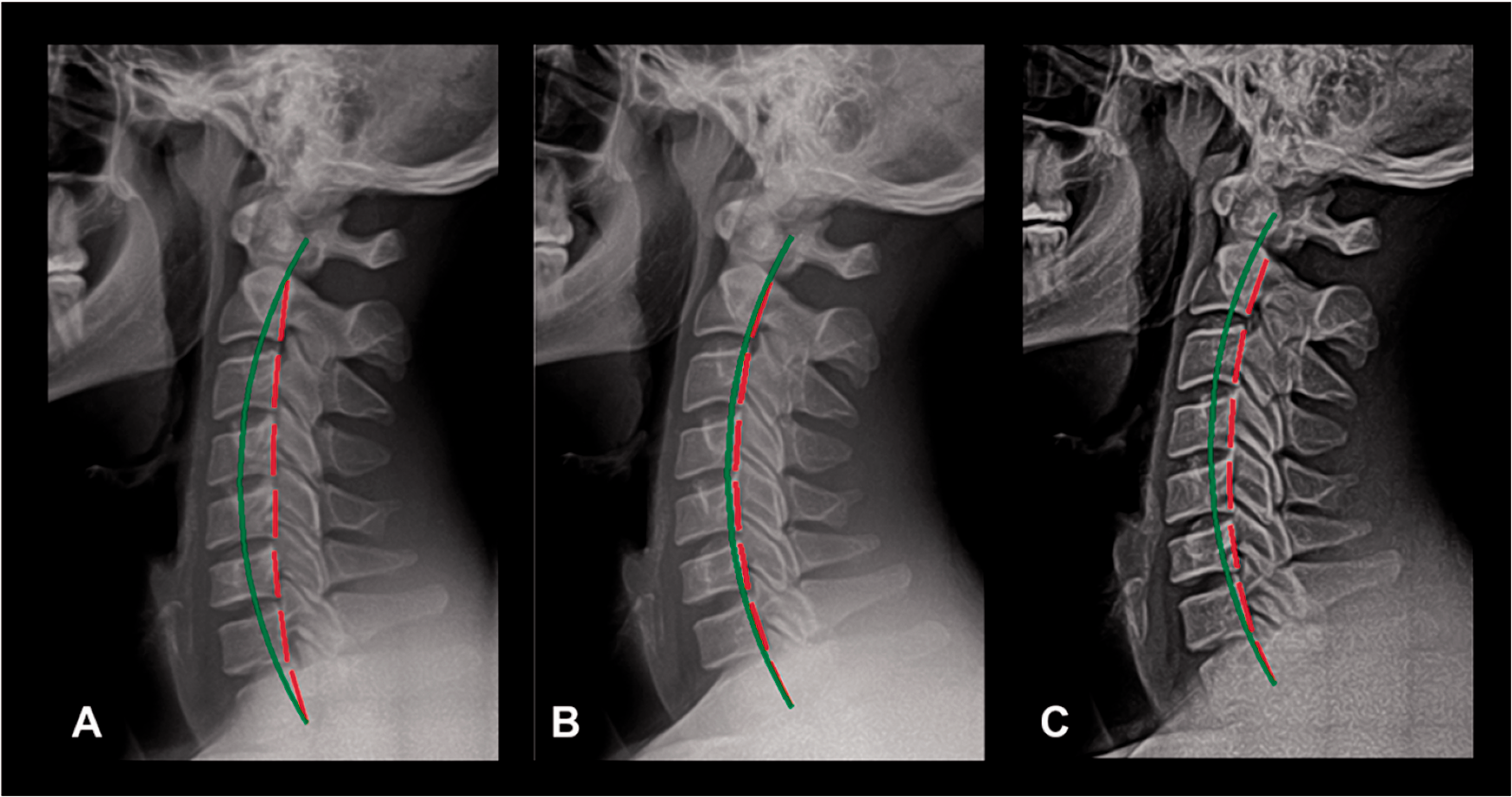


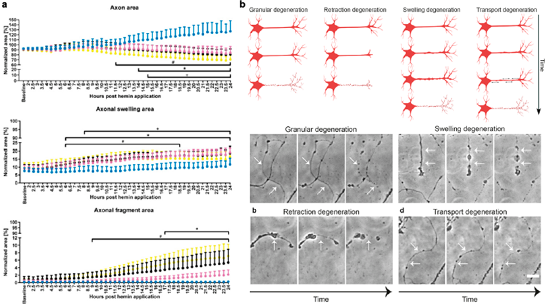
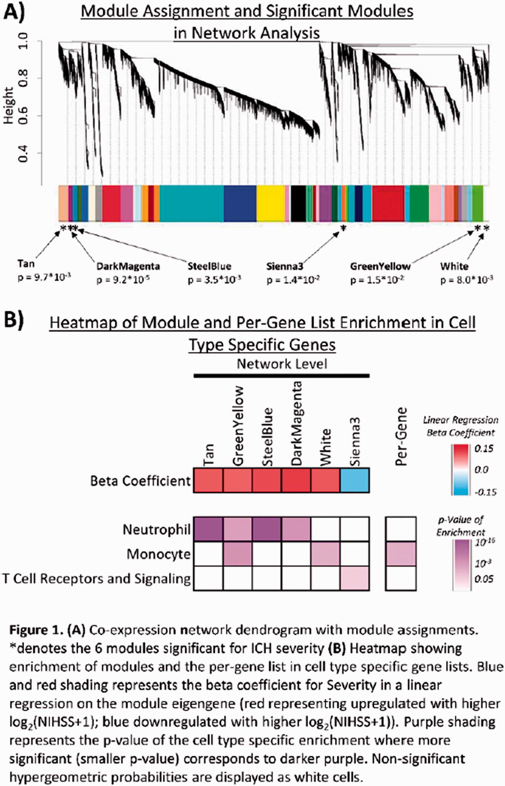
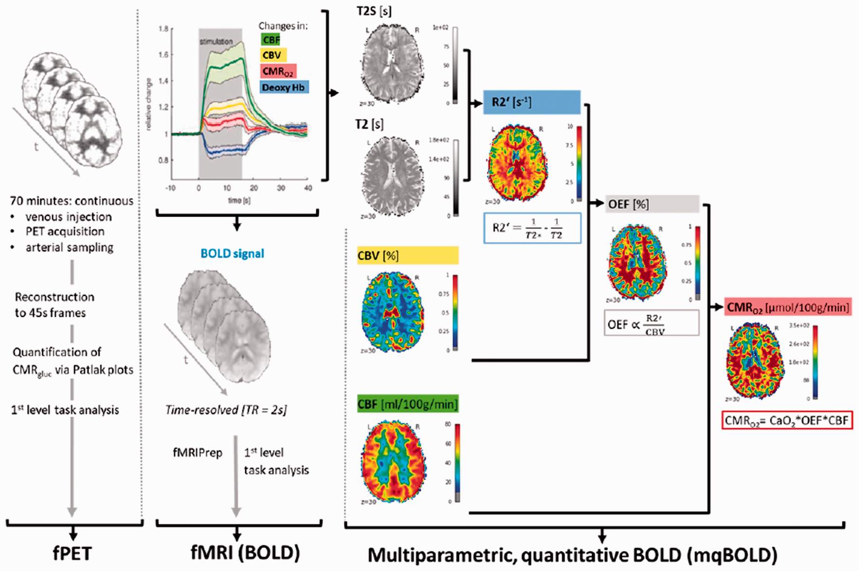
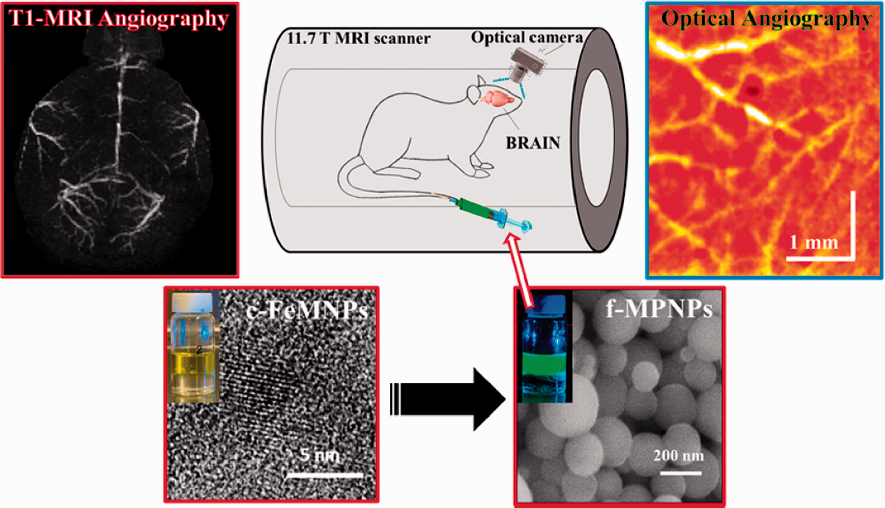
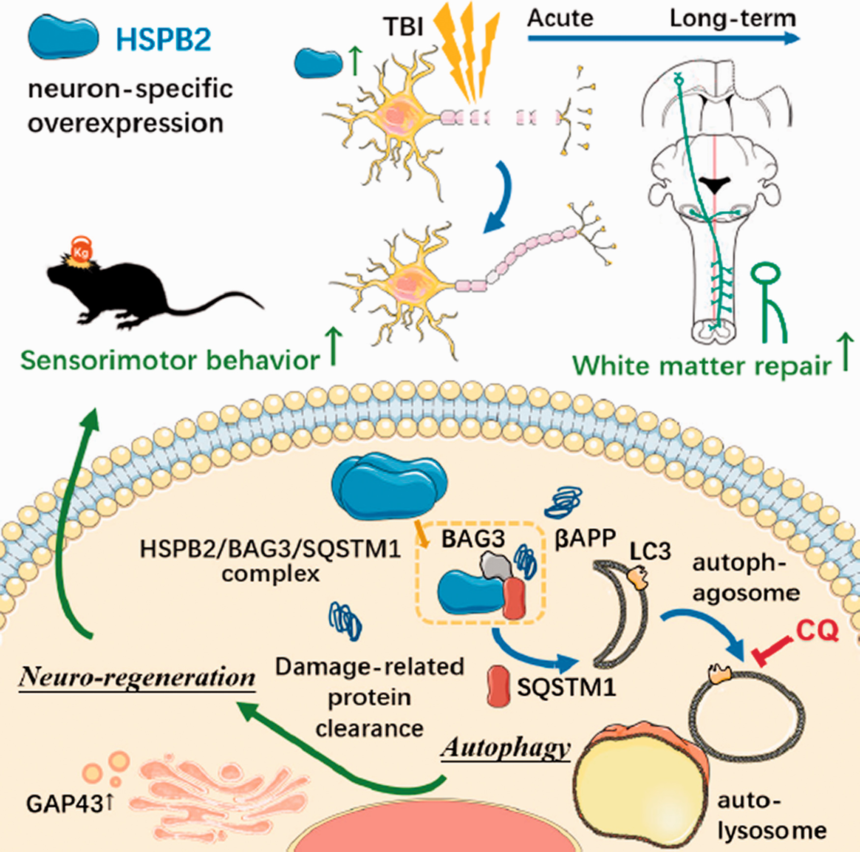
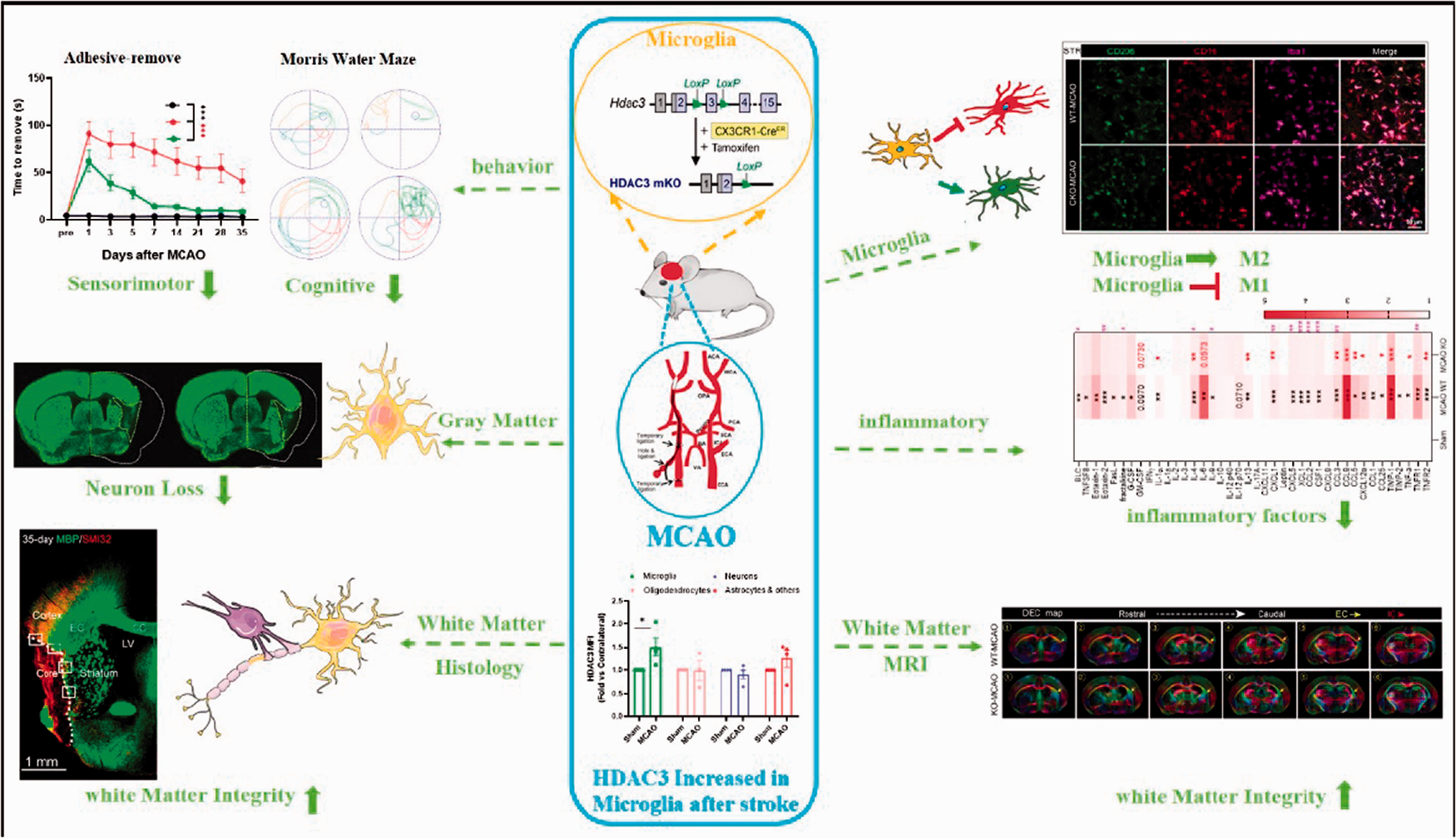
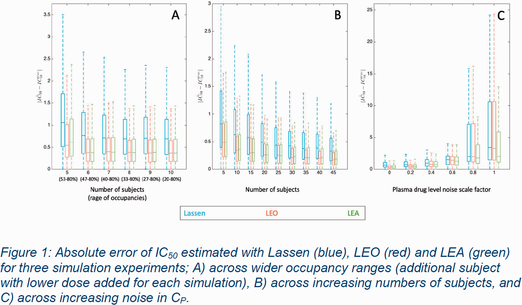


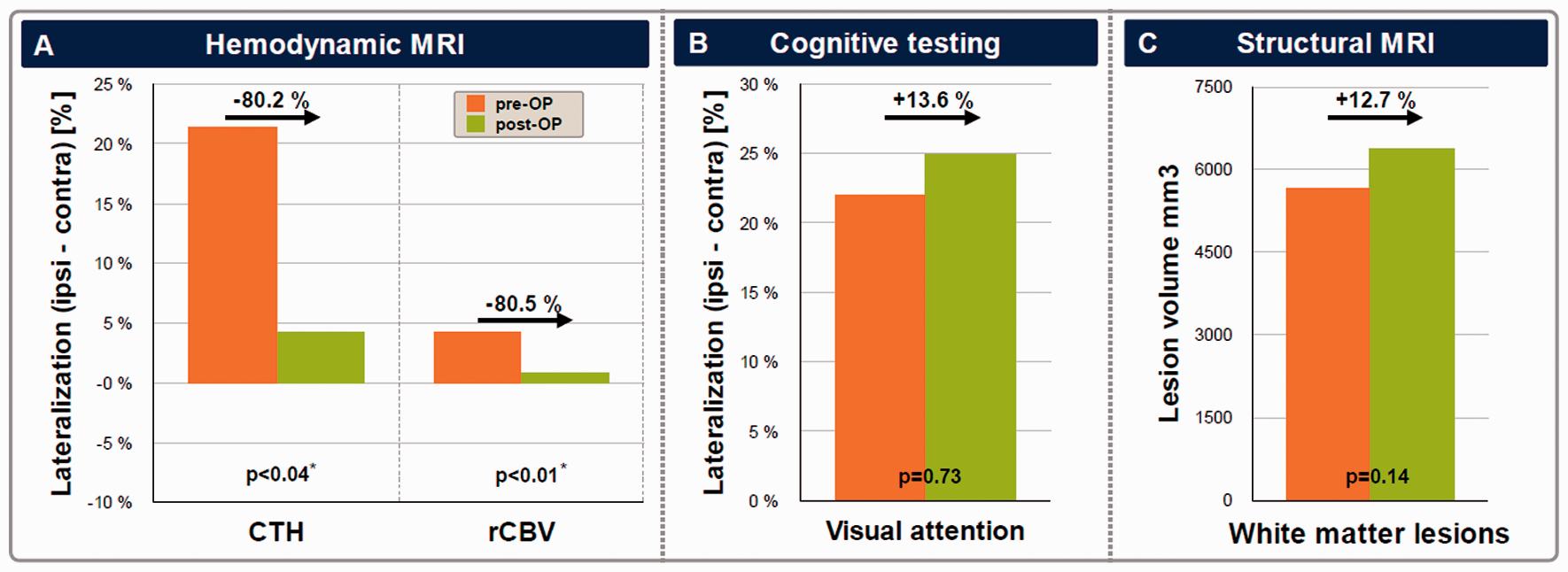

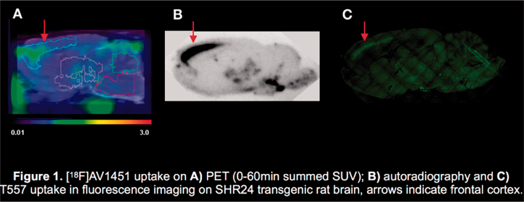


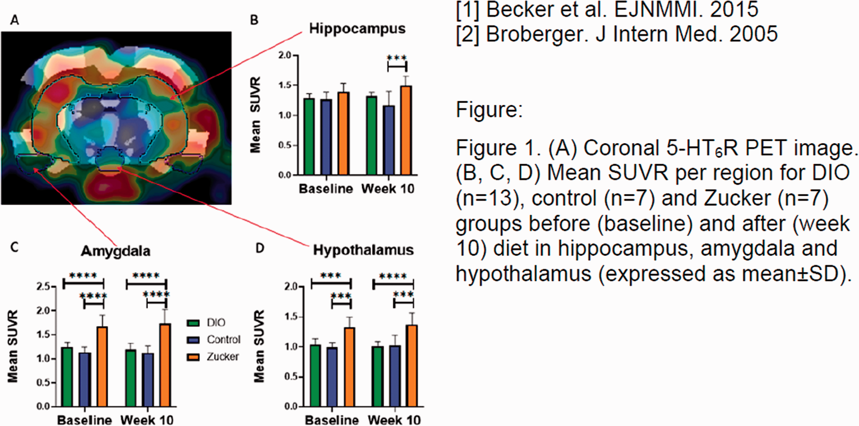
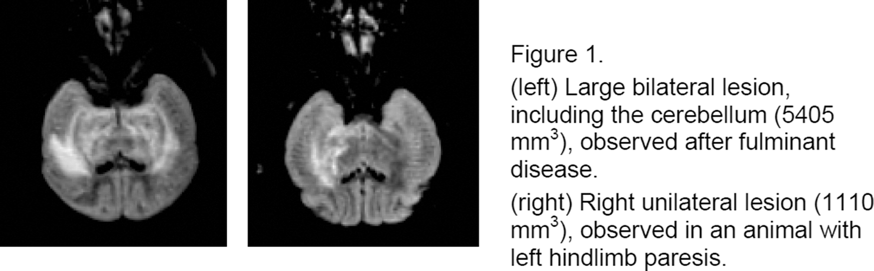
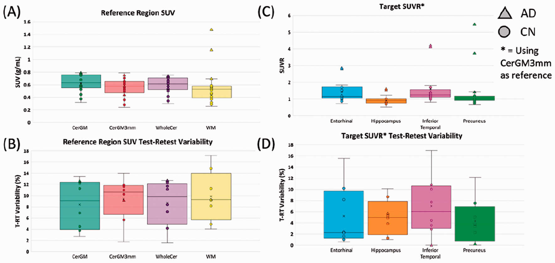

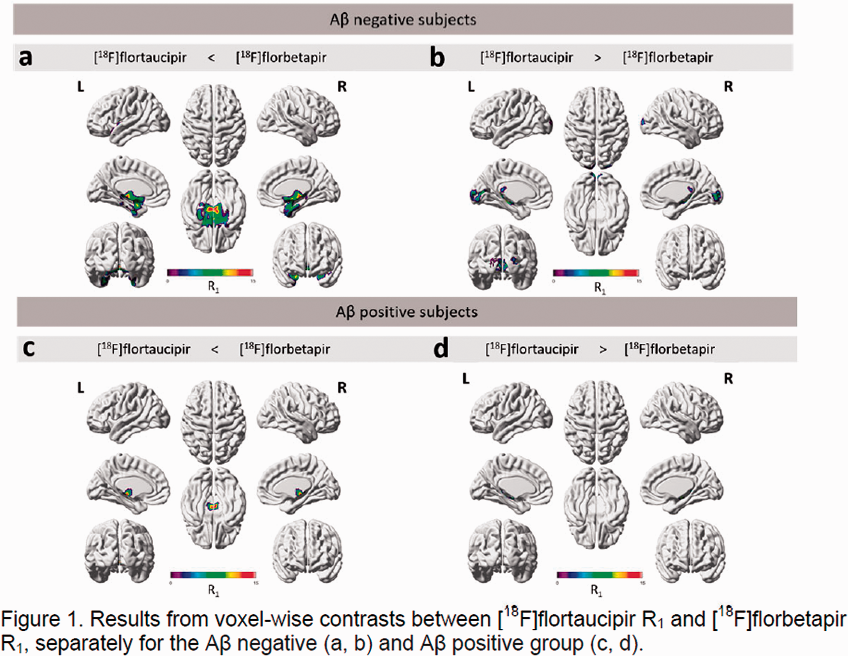
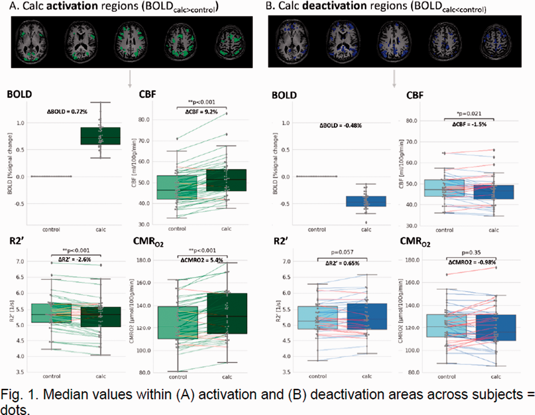

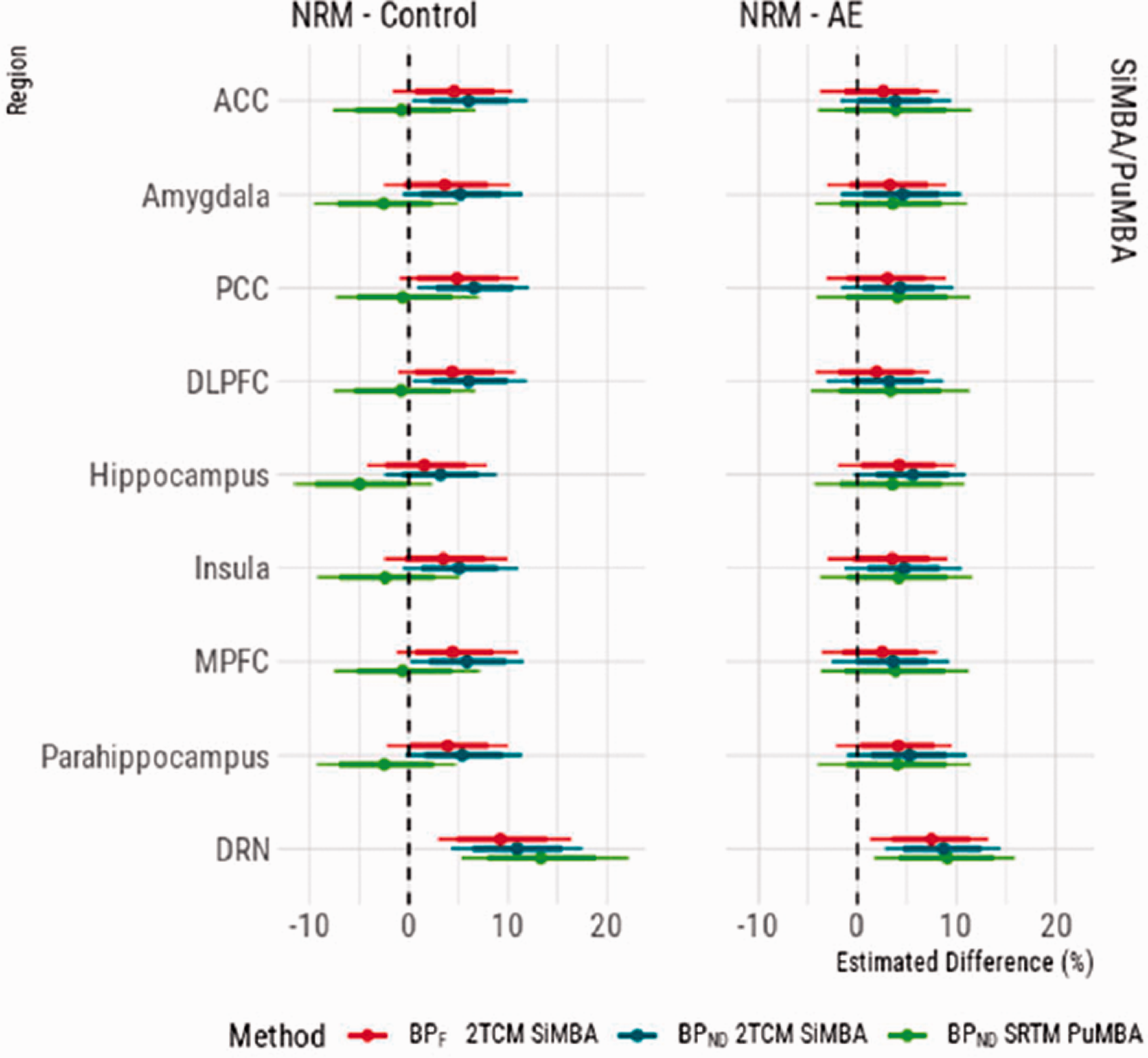
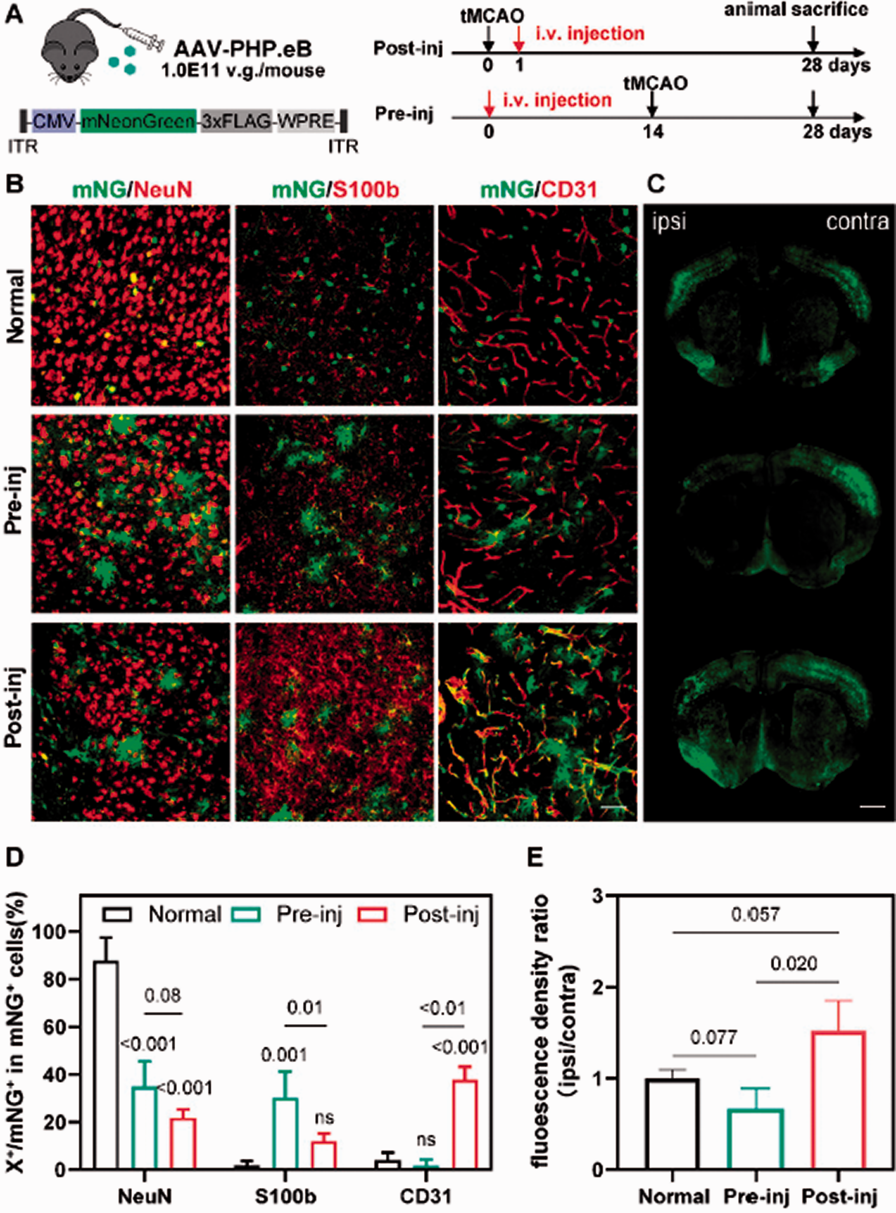
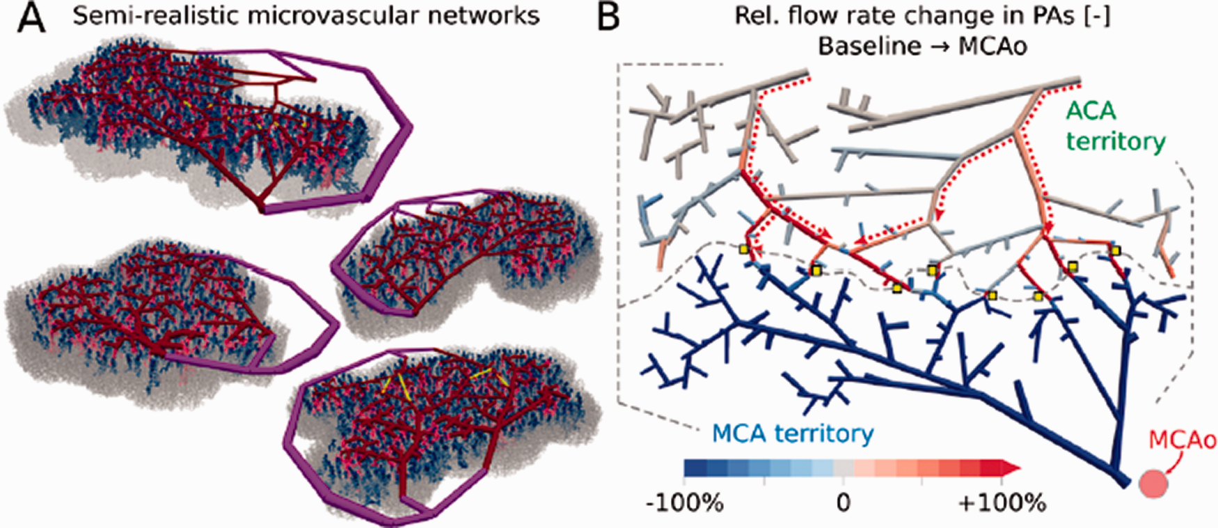


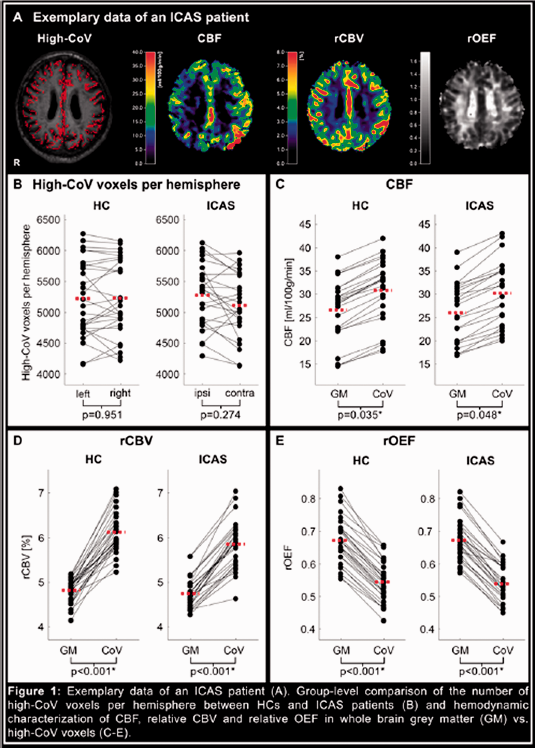
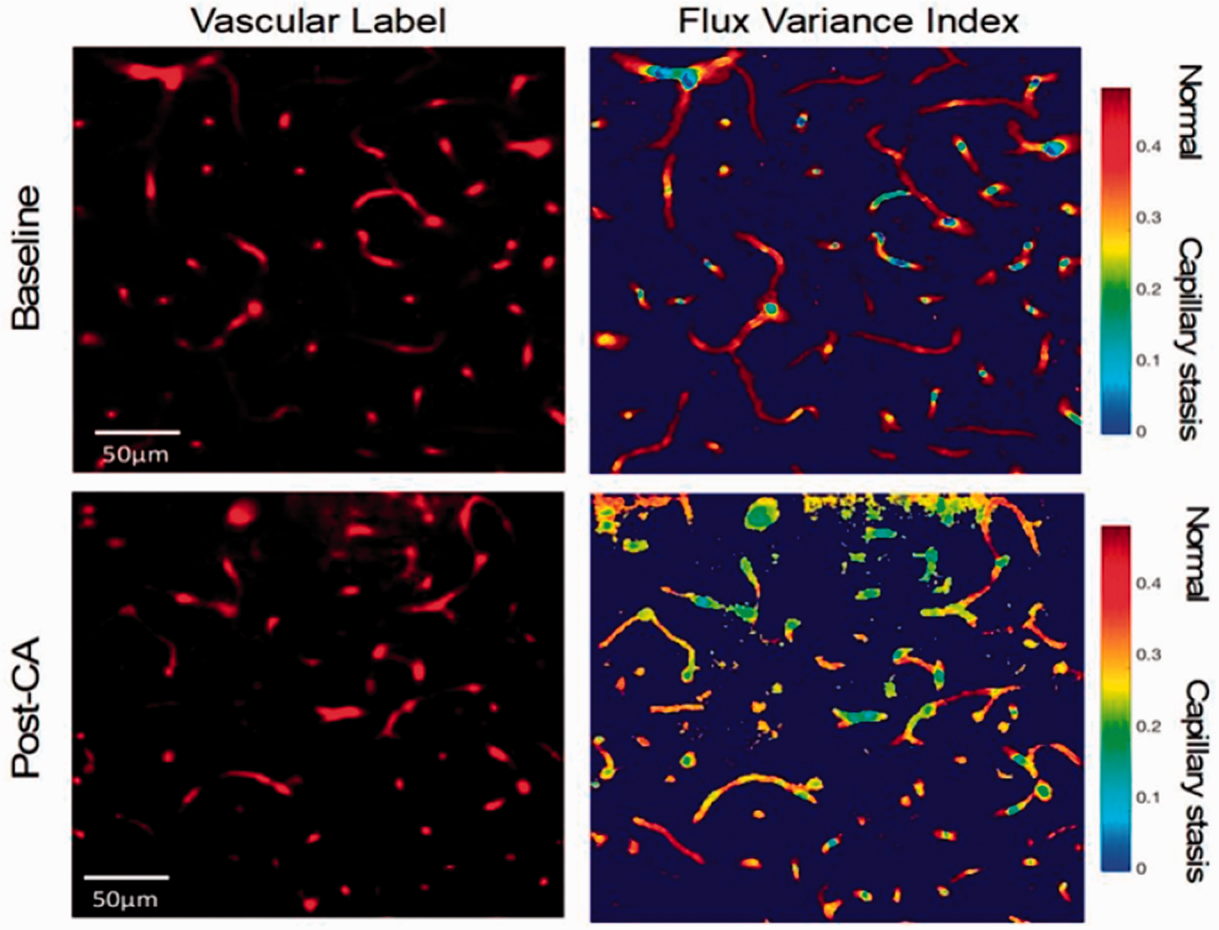

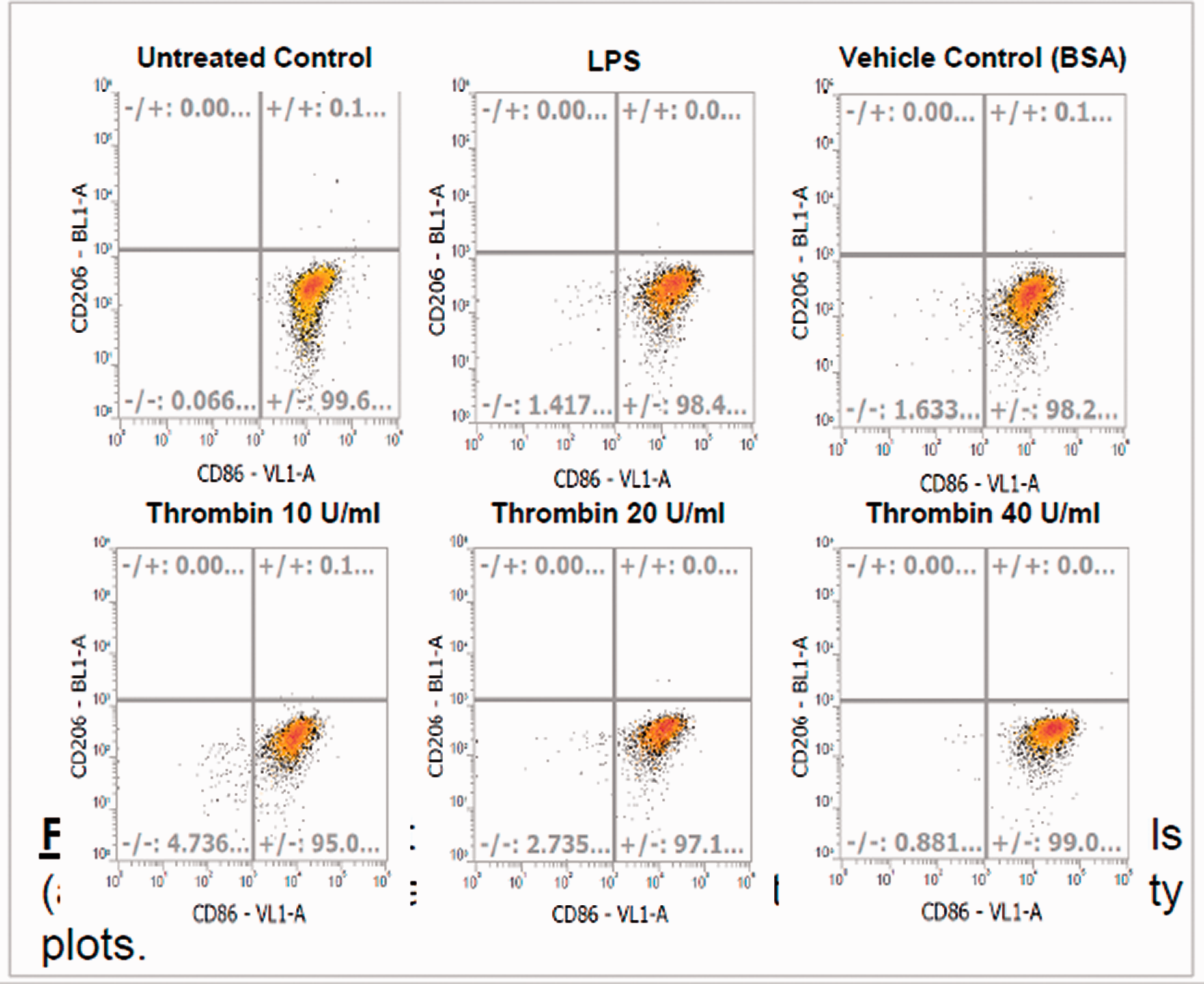
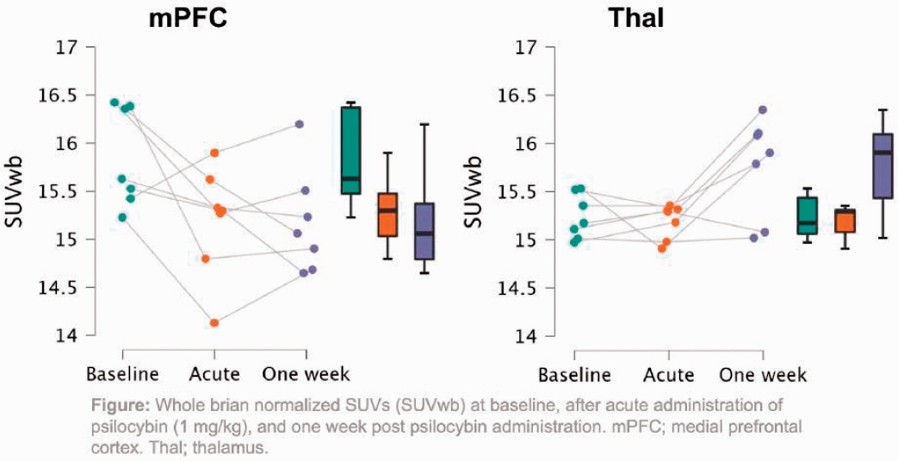
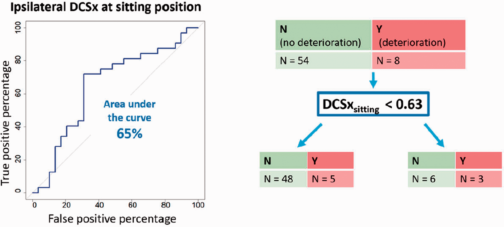
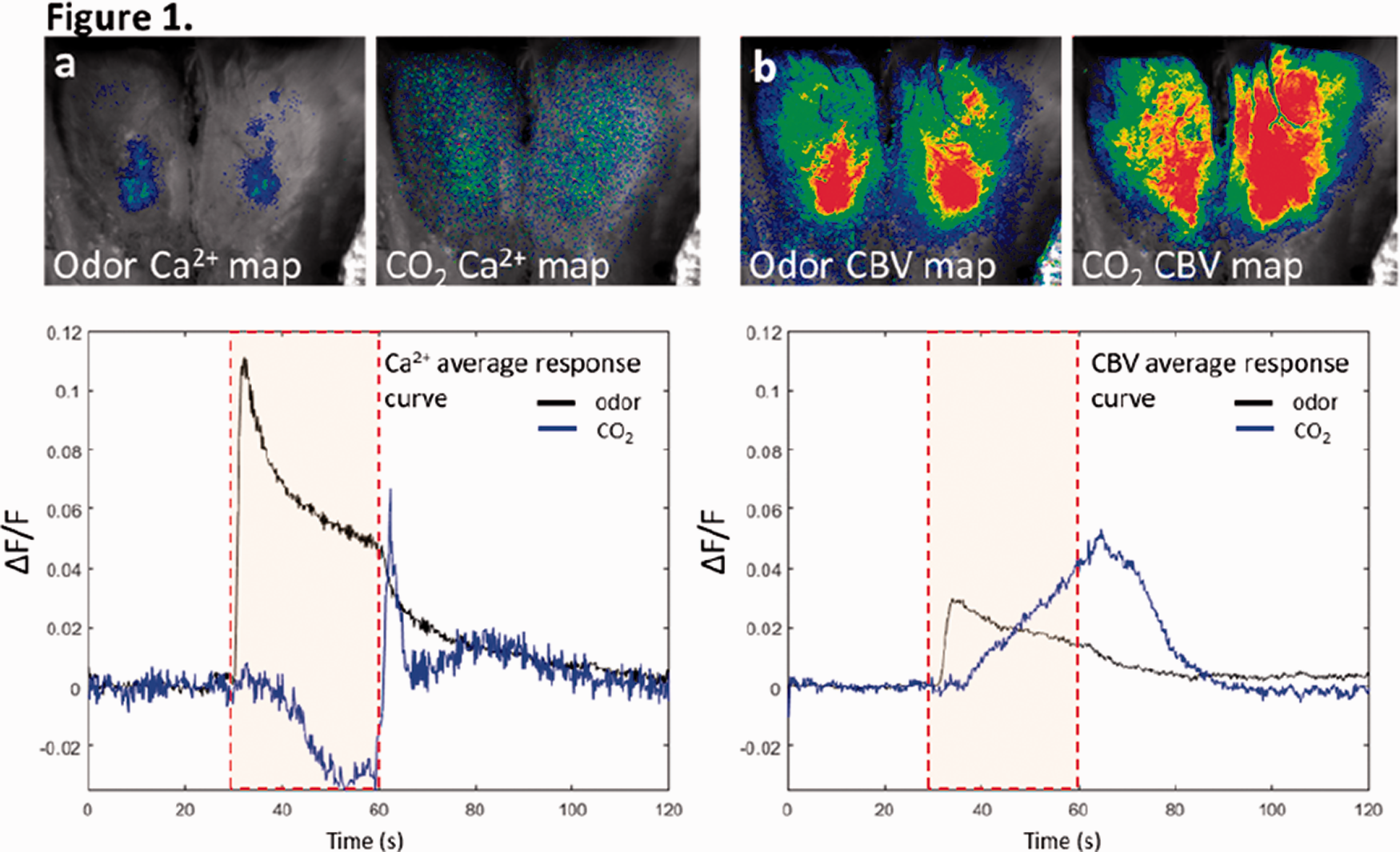
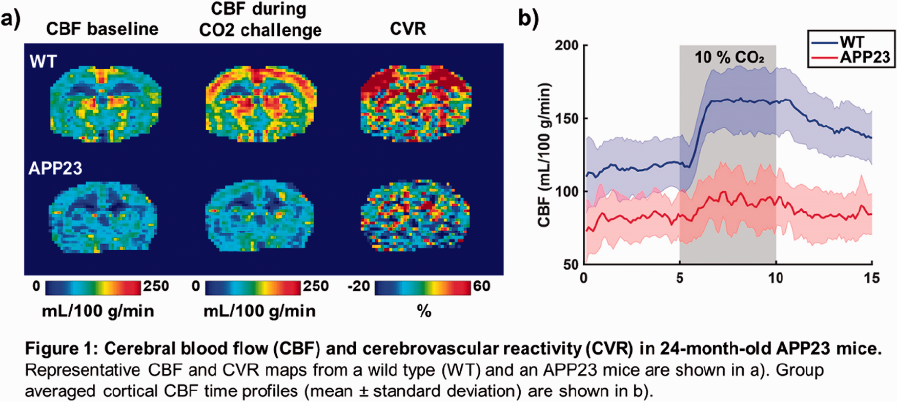
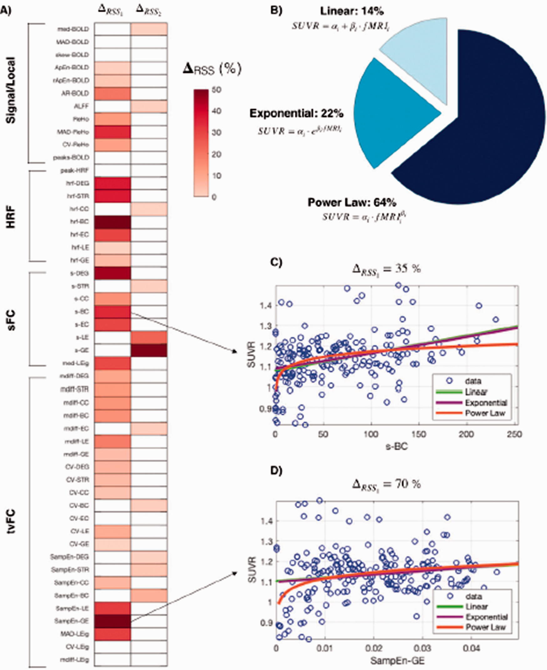
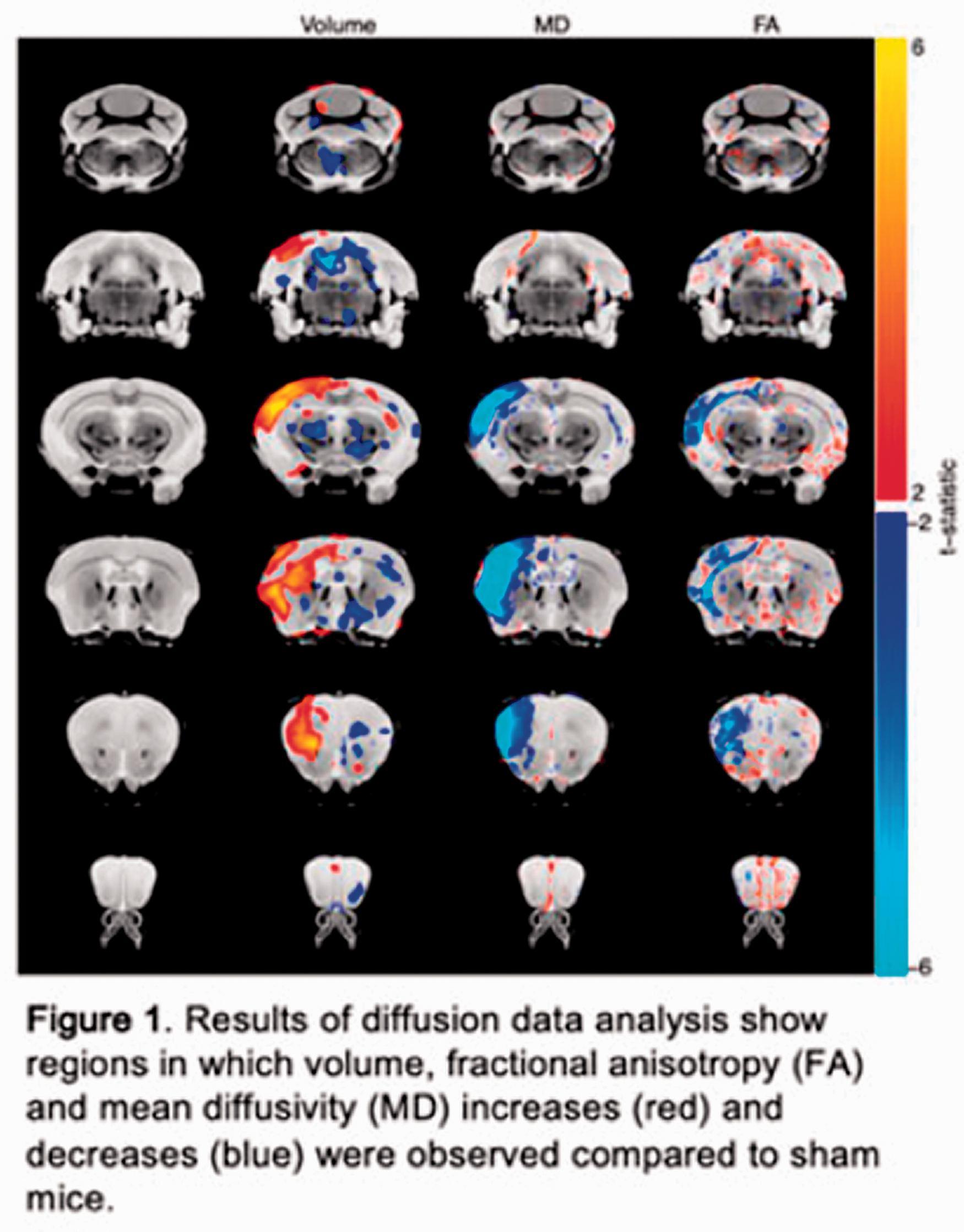

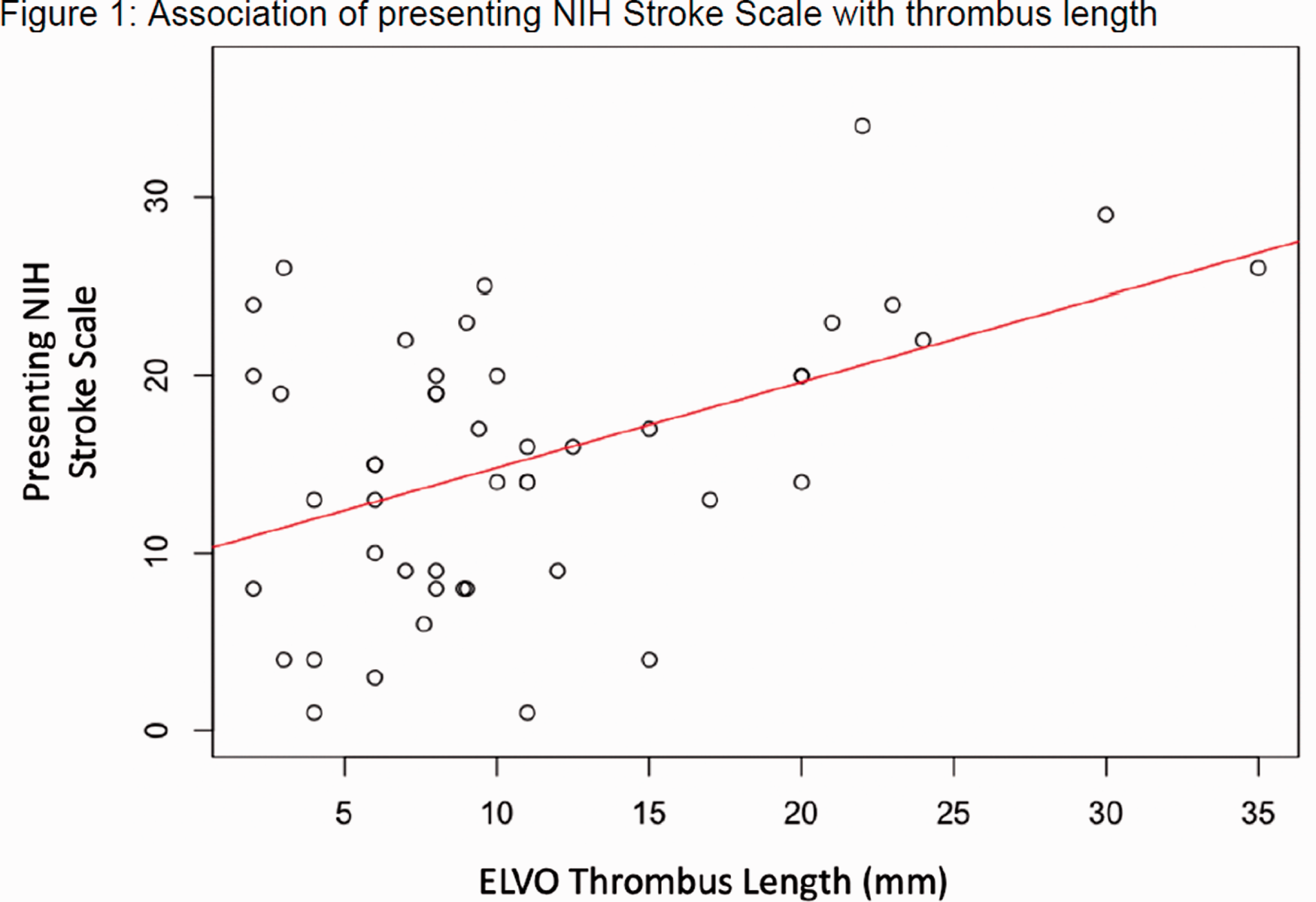


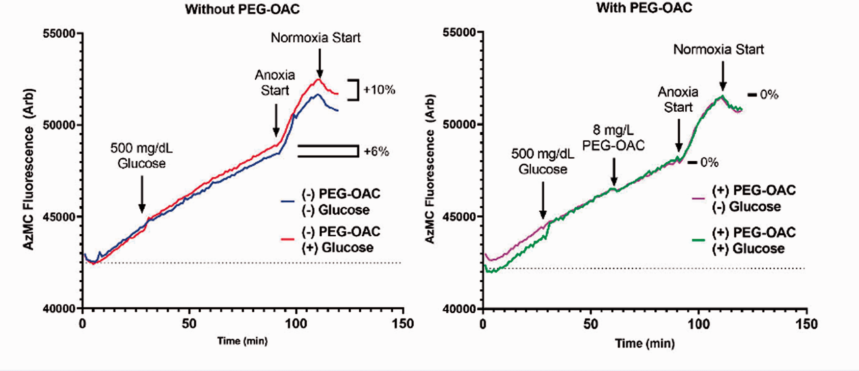

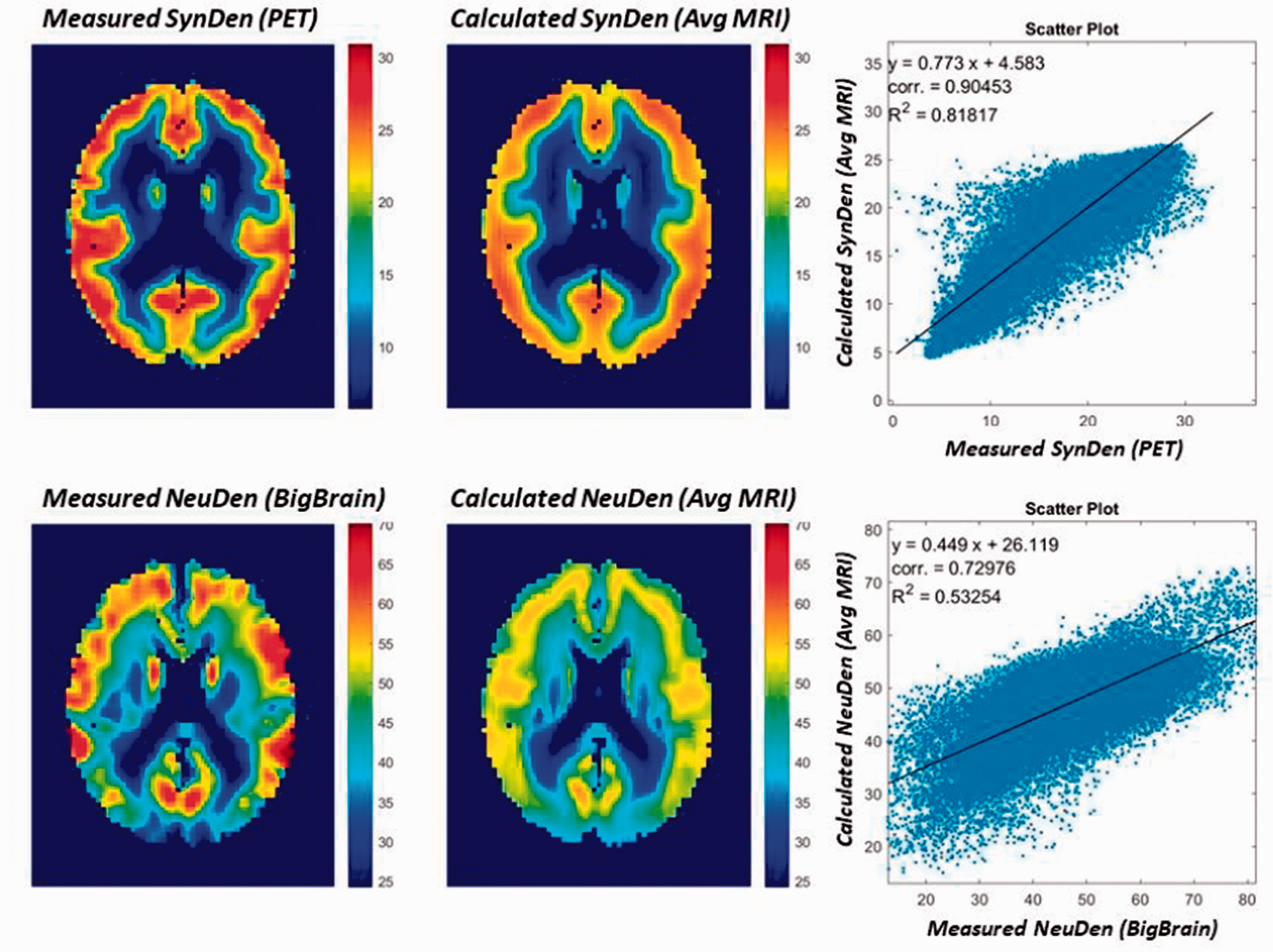
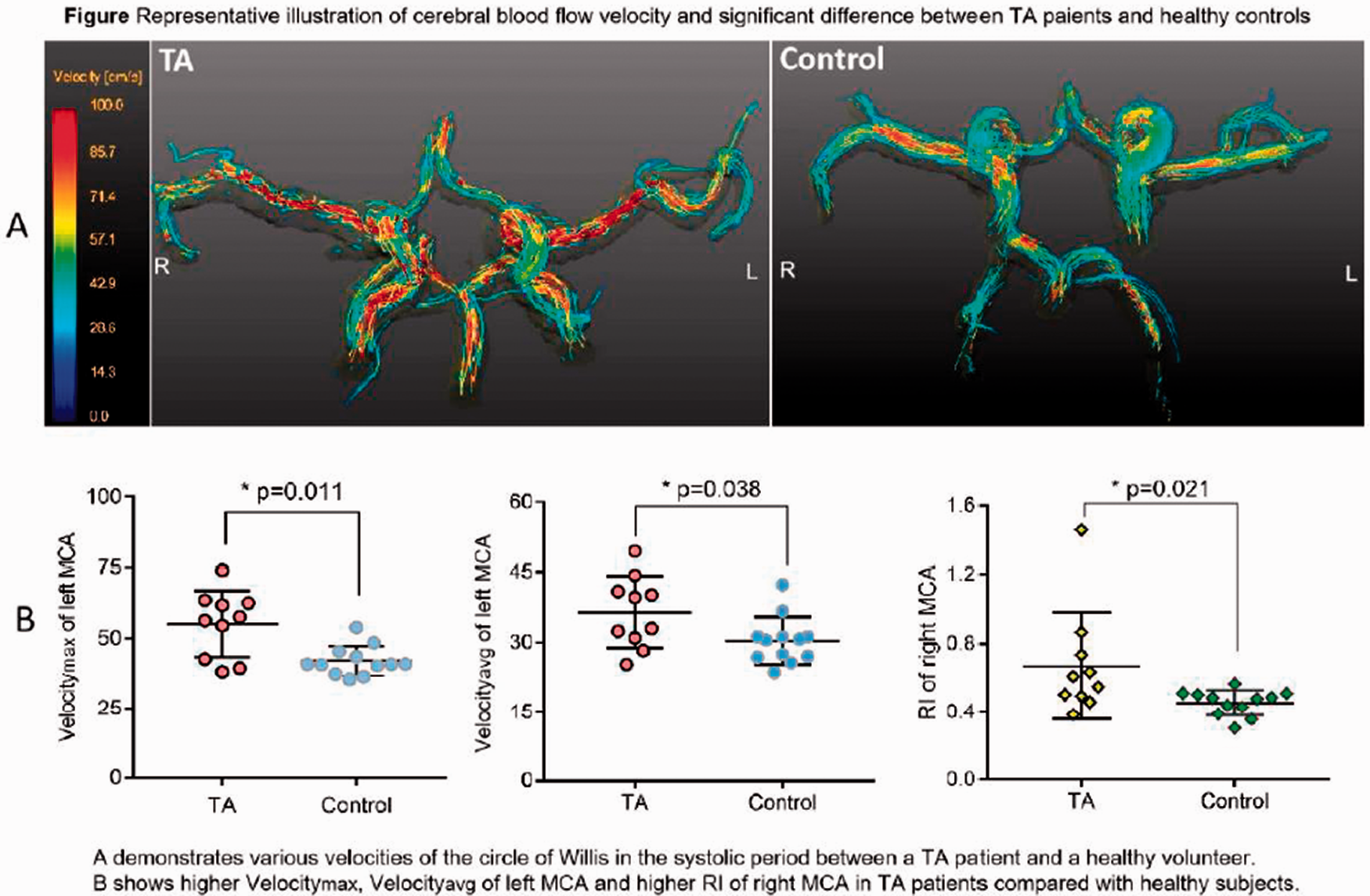

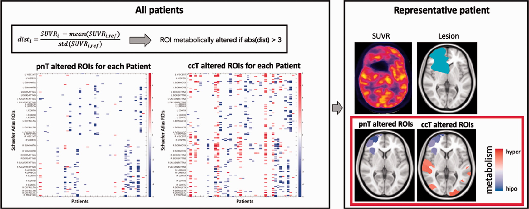
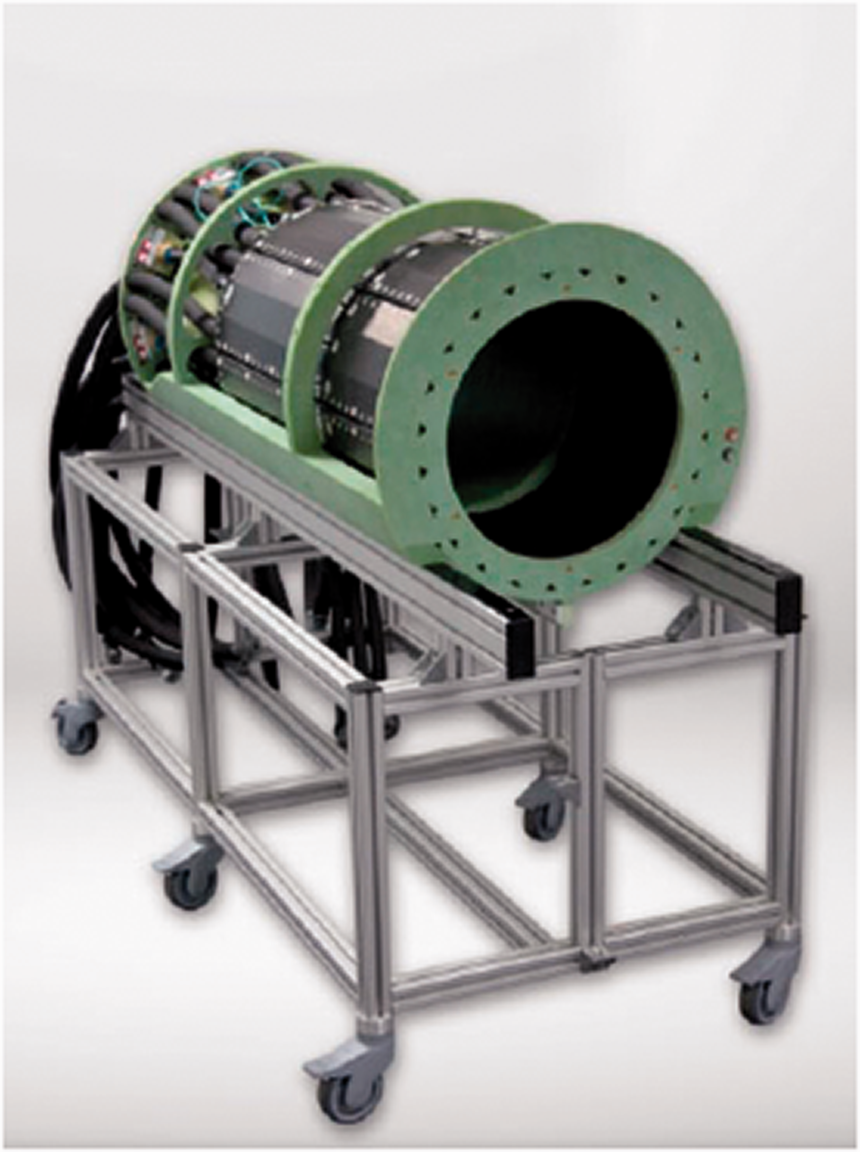
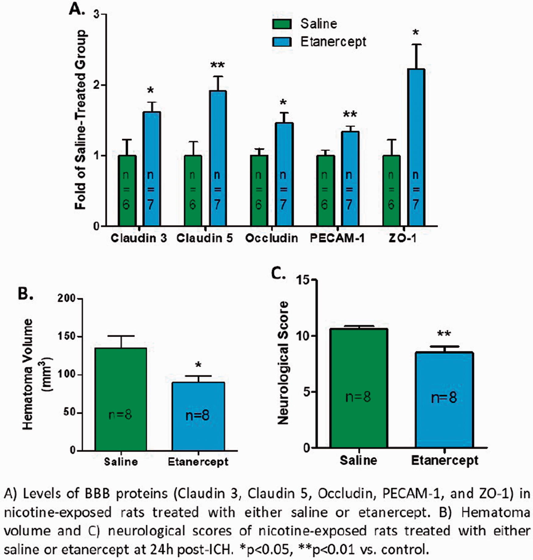 Figure 1. ▪.


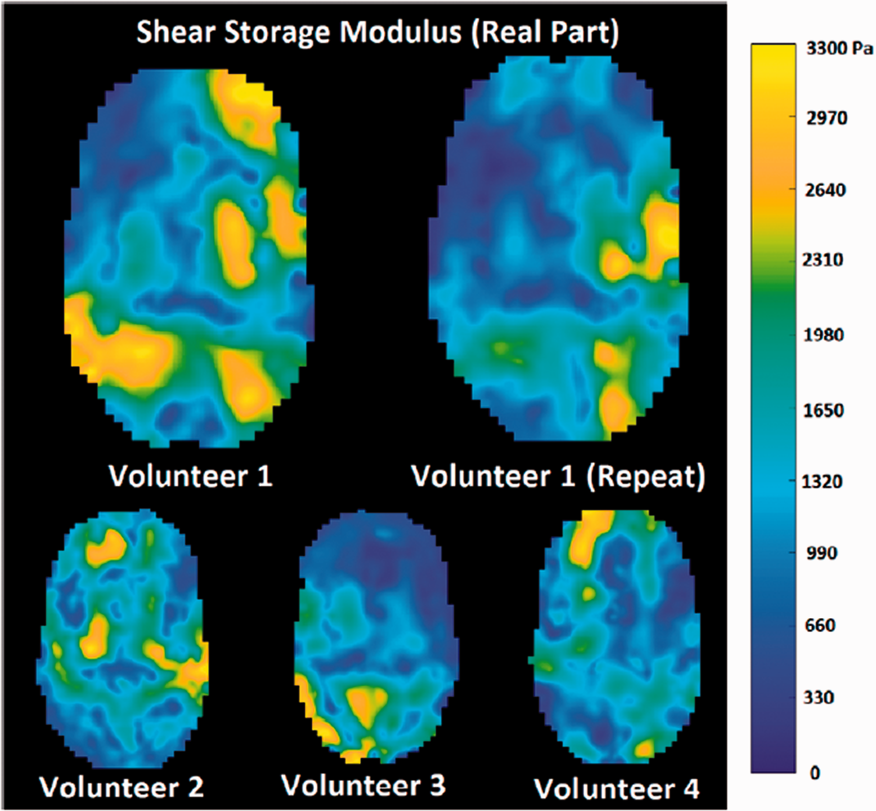
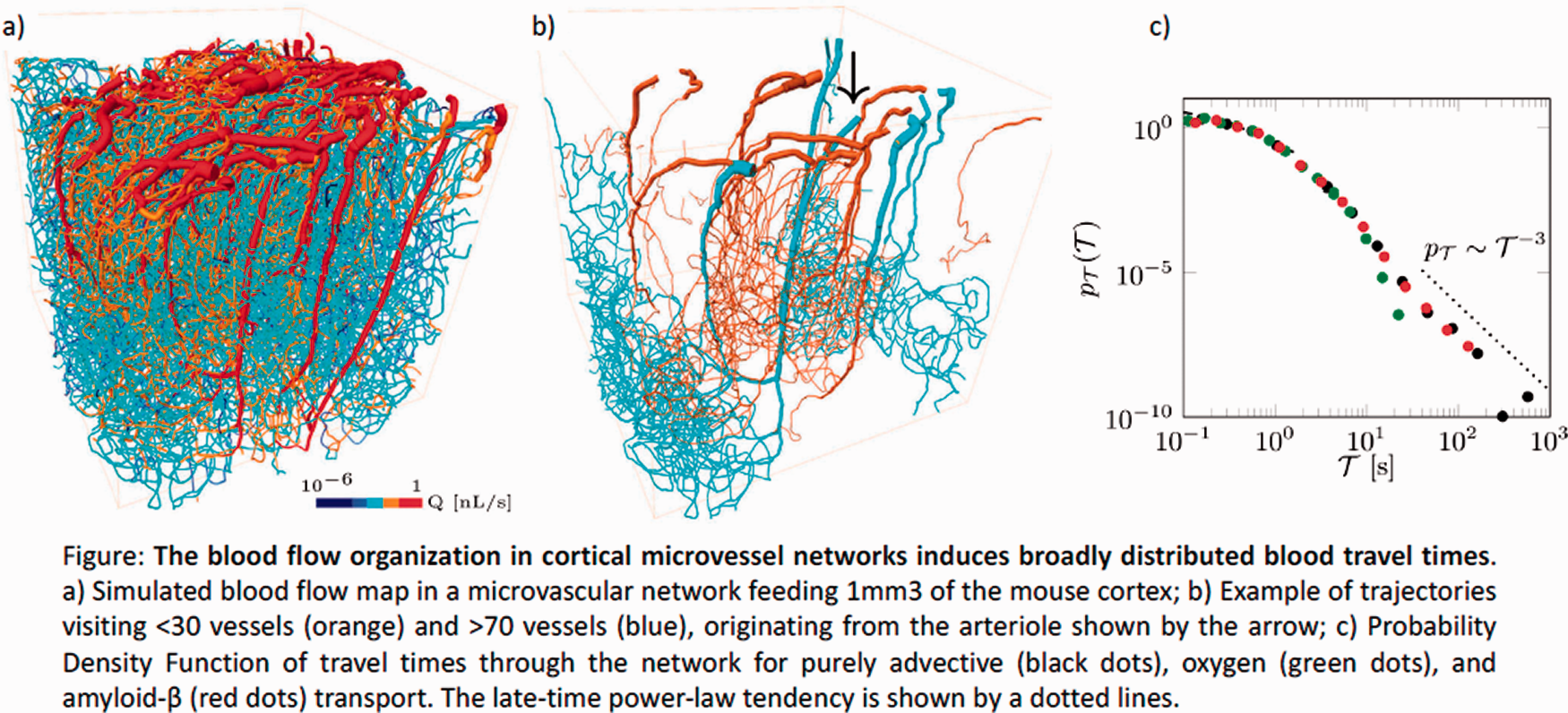
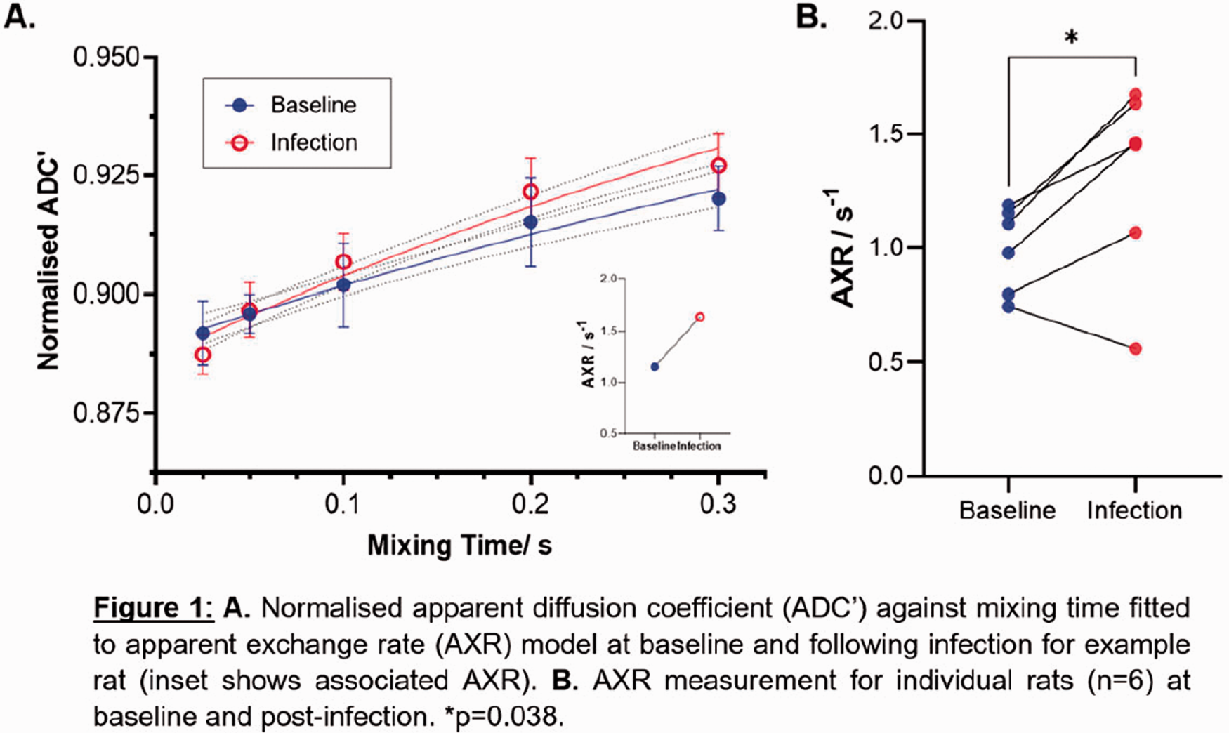


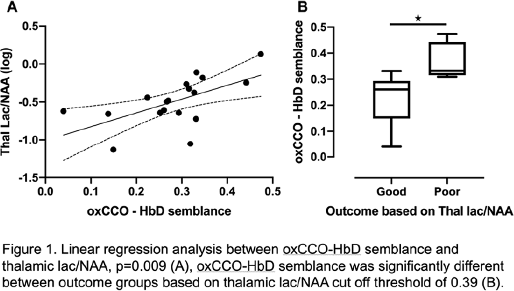


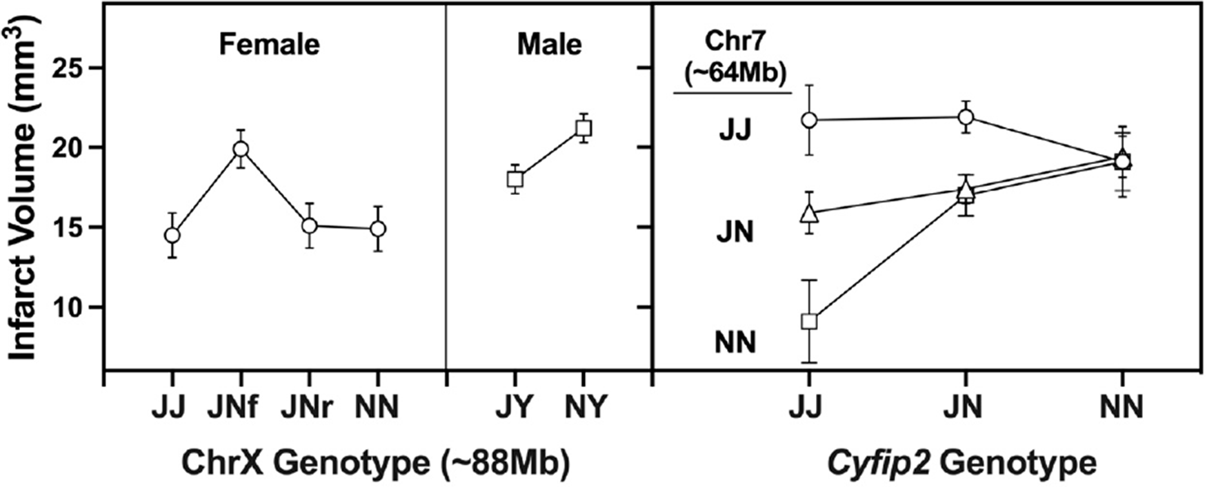
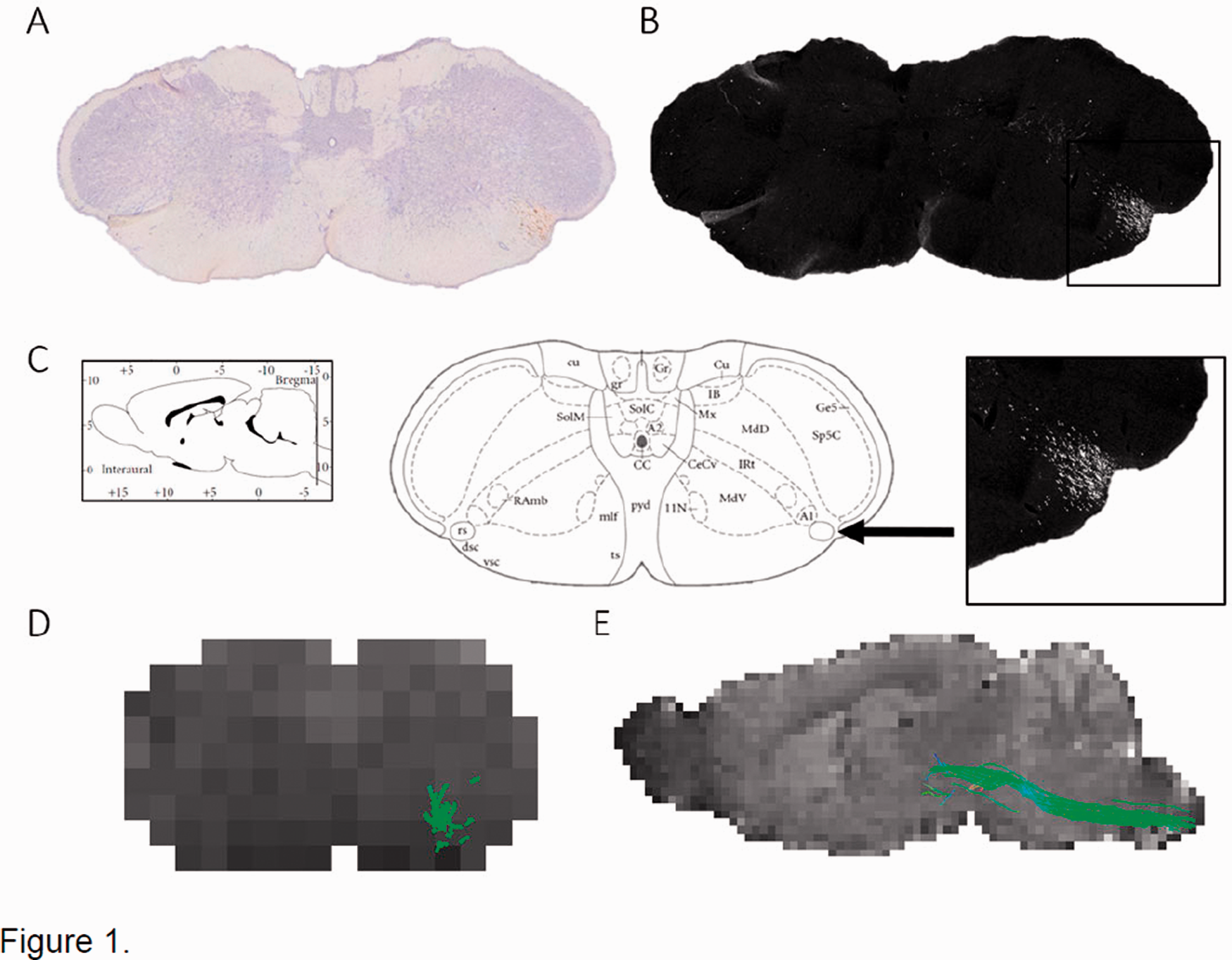 Figure 1. ▪.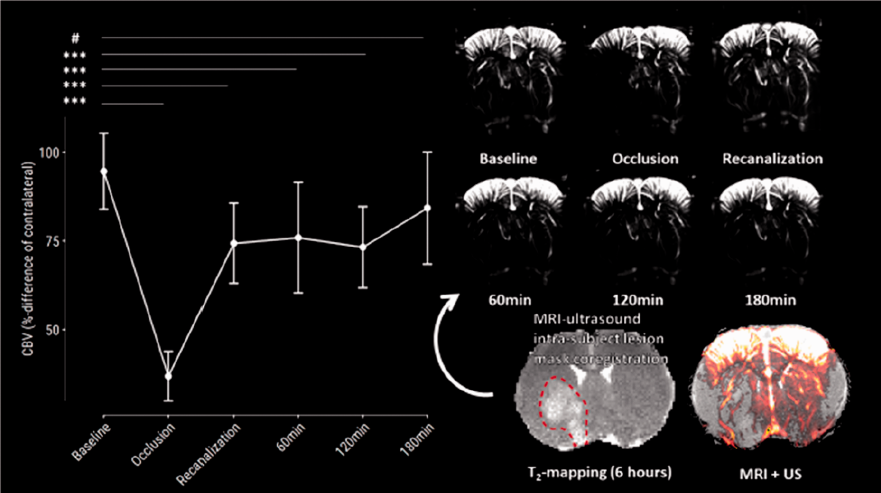

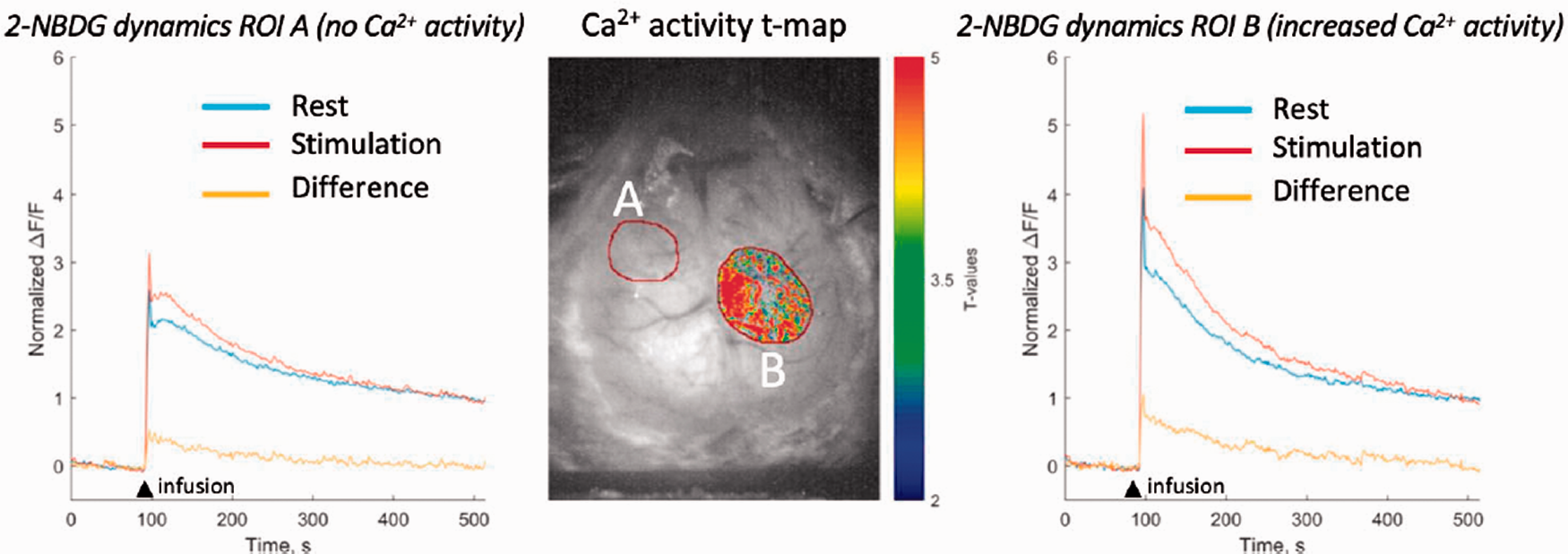
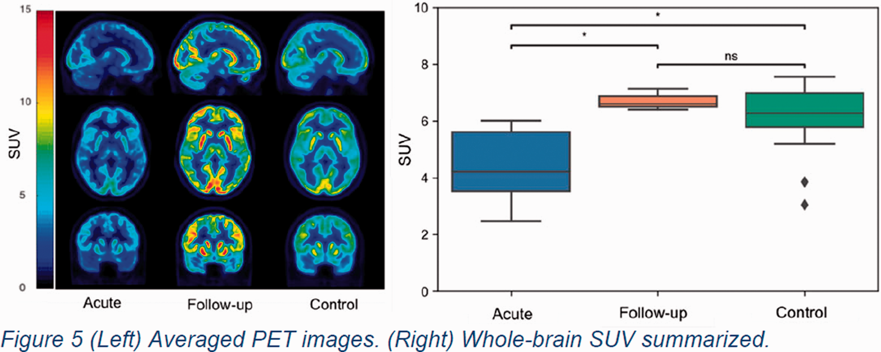
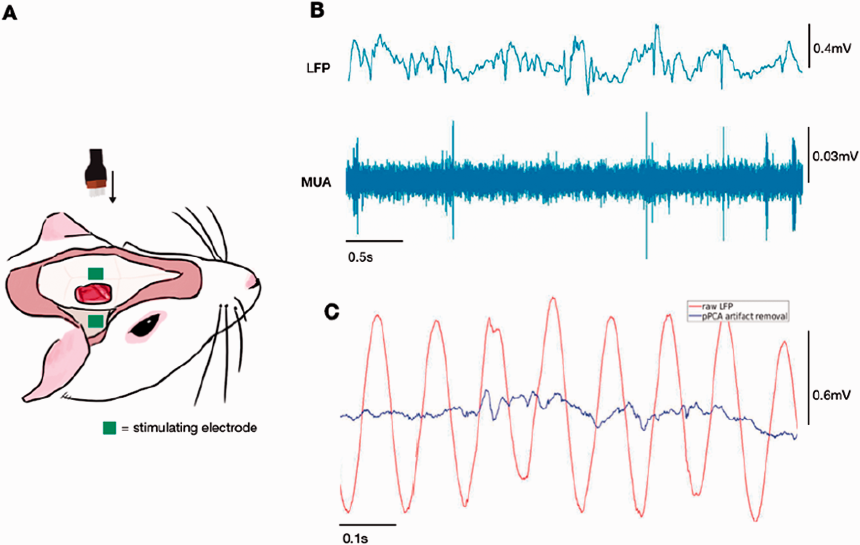
 Figure 1. ▪.