



Abstract

PA00-A01
Characteristics of the capillary bed – flow resistance, capillary dilation and red blood cell motion
1Institute of Pharmacology and Toxicology, University of Zurich, Switzerland
2Institute of Fluid Dynamics, ETH Zurich, Switzerland
Abstract
Objectives
The cortical vasculature consists of different vessel types, which differ topologically and which fulfil distinct functional tasks. Capillaries are the most frequent vessel type and highly relevant for oxygen supply. Besides their relevance, little is known about the topology of the capillary bed and their role in neurovascular coupling remains highly debated. Our goal is to improve our understanding of structural and functional properties of the capillary bed.
Methods
We use blood flow simulations in realistic microvascular networks (MVN) [1] to jointly study topology and perfusion of the capillary bed as well as the impact of diameter changes on the flow field. Additionally, we compute the flow resistance between two points of the capillary bed in the same MVNs [2]. The average flow resistance is compared for different hematocrit distributions and different pairs of points in the capillary bed.
Results
Our results reveal that the pressure drop in the capillary bed decreases with cortical depth and that red blood cells (RBCs) enter and leave the capillary bed at approximately the same cortical depth [1]. However, it remains unknown how these flow patterns are enforced by the cortical vasculature. To address this question we computed the flow resistance between two points of the capillary bed. The average flow resistance between descending arterioles (i.e. capillary start point) and ascending venules (i.e. capillary end point) reaches its minimum in cortical layer V. Comparison of the average flow resistance between randomly chosen points [2] and between start and end points of the capillary bed reveals that random point selection overestimates the flow resistance by a factor of ∼2.6. Interestingly, the average flow resistance is larger for a realistic hematocrit distribution than for a constant hematocrit of 0.3. Moreover, we show that a capillary dilation of 10% locally increases the flow rate by 23% per 100 µm dilation length and the number of RBCs by 20%.

Figure 1
(A) Realistic MVNs. Orange bands: outlines of barrels. (B) Average flow resistance of the capillary bed for different hematocrits (Htt). Encircled numbers indicate how the flow resistance was computed. Random: Two randomly chosen points. DA-AV: Outflow of descending arteriole (DA) and inflow of ascending venule (AV). DA-AV-paths: Equivalent to DA-AV, but only fluid dynamically connected points are considered. (C)Average flow resistance over depth for different hematocrits (DA-AV pairs). Error bars: median ± SEM.
Conclusions
The average flow resistance of the capillary bed is only twice as large as the resistance of descending arterioles. This is in contrast to previous results, where the resistances differed by a factor of 4 and where the capillary points were chosen randomly [2]. As the flow resistance decreases towards cortical layer IV, we conjecture that the capillary bed is designed to facilitate perfusion in the barrel layer. We demonstrate that capillary dilation is able to locally alter perfusion and provide indirect evidence for layer-specific regulation mechanisms.
References
PA00-A02
Acute phase of concussion in recreational rugby players is associated with changes in cerebral blood flow that are dependent on sex
1Department of Pharmacology, Physiology and Neuroscience, Rutgers, The State University of New Jersey, Newark, NJ USA
2War Related Illness and Injury Study Center, NJ VA Healthcare System, East Orange, NJ, USA
3MARCS Institute for Brain, Behaviour and Development, Western Sydney University, Sydney, Australia
Abstract
Objectives
Concussion is now recognized as a significant trauma with long term sequelae which has been found to affect women with greater severity. In fact, women experience worse symptoms, and take longer to recover (1). While the pathophysiology of concussion remains poorly understood, recent research suggests concussion may reduce cerebral blood flow (2). However, there is limited research regarding the acute effects of concussion on cerebral blood flow in women. The objective of our research study was to investigate sex differences in cerebral blood flow immediately post-concussion in recreational rugby players.
Methods
A total of 41 adult concussed players (25 men; 16 women) and 55 adult control players (39 men; 16 women) participated in our study. All participants had participated in a rugby match prior to enrolling. Concussed players had suffered a head injury during the match that resulted in their referral to the medical staff for evaluation. Players that did not need further treatment were offered the opportunity to participate in this study. Control participants were individuals who did not sustain a head injury during matches. Portable data collection tents were set up along the sidelines of ruby matches where a subset of participants underwent duplex ultrasonography of the internal carotid artery (ICA) in the seated and supine positions. Continuous blood pressure, ECG and end-tidal CO2 were obtained in addition to cerebral blood flow metrics.
Results
There was significantly lower ICA blood flow in the supine position in concussed male players (n = 14) compared to control male players (n = 25) (concussed =514 ± 85; control = 592 ± 149 ml/min; p = 0.043), however, there were no significant differences in the seated position between concussed (n = 11) and control (n = 21) male players (concussed = 622 ± 155; control =573 ± 122 ml/min; p = 0.327). In contrast, concussed female players (n = 9) demonstrated a trend towards greater ICA flow in the seated position in compared to control female players (n = 14) (concussed = 570 ± 110; control = 450 ± 153 ml/min; p = 0.055). There was no difference in supine ICA flow between concussed (n = 8) and control (n = 15) female rugby players (concussed =459 ± 92; control = 482 ± 183 ml/min; p = 0.751). There were no differences in end-tidal CO2 between concussed and control male players when supine (concussed =40.4 ± 6.5; control = 39.9 ± 5.5 mmHg; p = 0.790) or seated (concussed = 41.3 ± 4.3; control = 38.4 ±5.9 mmHg; p = 0.115) or female players when supine (concussed = 40.4 ± 6.5; control = 39.9 ± 5.5 mmHg; p =0.790) or seated (concussed = 41.3 ± 4.3; control =38.4 ±5.9 mmHg; p = 0.115).
Conclusions
Our data suggest that acute concussions are associated with reduced cerebral blood flow in adult male rugby players when supine. In contrast, concussed female players appear to be hyperperfused when seated. Cerebral blood flow changes could not be explained by end-tidal CO2, since there were no differences between groups. These data are the first to demonstrate that in the acute phase of concussion men and women demonstrate differing cerebral blood flow responses. However, this study involved a relatively small number of female players and a larger dataset is needed to determine confirm these findings. Overall our data supports the concept that concussive head injuries affect men and women differently and that further research is necessary to understand sex differences in cerebral blood flow immediately post-concussion.
References
PA00-A03
Microglia monitor and protect neuronal function via specialized somatic contact sites in an activity- and P2Y12R-dependent manner
1Laboratory of Neuroimmunology, Institute of Experimental Medicine, Hungarian Academy of Sciences, Budapest, Hungary
2MTA-SZTE Research Group for Cortical Microcircuits, Department of Physiology, Anatomy and Neuroscience, University of Szeged, Hungary
3Institute for Stroke and Dementia Research, Ludwig-Maximilians-University, Munich, Germany
4Laboratory of Molecular Neurobiology, Institute of Experimental Medicine, Hungarian Academy of Sciences, Budapest, Hungary
5Szentágothai János Doctoral School of Neuroscience, Semmelweis University, Budapest
Abstract
Objectives
Microglia are the main immunocompetent cells of the nervous system and their role in brain development and maintenance of proper neural function is now widely recognized.1 Microglia interact with most cell types via dynamic surveillance of the microenvironment by motile microglial processes.2 Since changes in microglial activity are linked with major human diseases including stroke, epilepsy and neurodegeneration, microglia-neuron interactions have been attracting a great deal of interest recently. To date, the majority of studies have focused on the interactions between microglial processes and synapses.3 However, there are several actions occurring at single-neuron level, which seem unlikely to be regulated merely by interactions between microglia and synaptic terminals.4 We hypothesized that specific sites on neuronal somata may also exist, allowing the dynamic monitoring and assistance of neuronal function by microglia.
Methods
We used in vivo two-photon imaging of CAG-TdTomato-electroporated CX3CR1-GFP mice, confocal laser-scanning-, STORM superresolution- and electron microscopy combined with electron tomography and advanced 3D-analysis to examine the communication interfaces between microglia and neurons in the mouse and the human brain. Chemogenetic neuronal activation was induced to study microglia-neuron interactions and NADH-fluorescence imaging was performed to measure mitochondrial activity. Functional connectivity and neuronal responses following experimental stroke were assessed by in vivo two-photon- and widefield calcium imaging.
Results
Here we identify a novel communication hot-spot between neuronal somata and microglial processes through which microglia sense and influence neuronal activity. We show that clustering of microglial P2Y12-receptors occurs in a contact-dependent manner at defined areas of neuronal soma identified by enrichment of Kv2.1 channels, mitochondria, mitochondria-associated membranes and increased expression of vesicular nucleotide transporter. This unique morpho-functional unit is present in most neurons in both mice and humans. Activity of neuronal mitochondria is linked with microglial contact formation and chemogenetic neuronal activation leads to increased microglial contact area. Blockade of P2Y12R-mediated actions prevents microglia-neuron interactions at somatic contact sites and alters neuronal network function. In turn, brain injury induced by cerebral ischemia dysregulates somatic communication hot-spots, while an absence of normal P2Y12R-signalling prevents microglial process recruitment and leads to increased brain injury.

Conclusions
Collectively, our results suggest that microglial processes recruited to these newly identified morpho-functional communication hot-spots are in ideal position to readily monitor and dynamically influence neuronal functions via mitochondrion- and P2Y12R-dependent signalling pathways. Bidirectional communication between microglia and neurons via this interface is likely to be important in the healthy brain as well as in a number of neurological conditions, even as a possible future therapeutic target.
References
PA00-A04
Characterizing microglial and macrophage-mediated repair of cerebral microbleeds in a mouse model of type 1 diabetes mellitus
1Division of Medical Sciences, University of Victoria, Victoria, Canada
2Deutsches Zentrum für Neurodegenerative Erkrankungen, Bonn, Germany
3University of Toronto, Toronto, Canada
Abstract
Microglia, the innate immune cells of the brain, rapidly respond to instances of cellular damage including ruptures in small blood vessels (4–7um), termed cerebral microbleeds (CMBs), and help to repair the damage. Type 1 diabetes mellitus is a highly prevalent metabolic disorder that is a risk factor for a host of vascular complications, including CMBs. Our previous work has shown that microglial responses are compromised in the diabetic brain, in terms of both the extent and the polarization of their responses. Since vascular repair is a process that evolves over days and weeks, it remains unclear how diabetes impacts this process over the long-term. Here we used 2-photon microscopy to image microglial responses and vascular repair in vivo in uncontrolled and insulin-treated type 1 diabetic male mice following induction of CMBs. These mice expressed green fluorescent protein (GFP) in microglia, via heterozygous replacement of a fractalkine (CX3CR1) allele, permitting the visualization of microglial motility and responses. We found that microglial aggregation around the CMB peaked 1–3 days after induction and declined thereafter (measured as area of GFP aggregation, % vessel coverage, # of cells), and did not differ significantly between groups. In contrast to nondiabetic mice where 100% of ablated vessels (43/43 vessels) regained blood flow, ∼20% of vessels from insulin-treated and untreated diabetic mice were pruned within 3 days.
Circulating leukocytes have been shown to infiltrate the brain following damage to larger penetrating arterioles (∼20um), and may impact vascular repair. To assess the relative contributions of microglia versus circulating leukocytes in vessel repair after CMB, we depleted each cell type using the PLX5622 diet to eliminate microglia or clodronate liposomes to deplete circulating phagocytic cells. By depleting microglia, we discovered that vessel pruning increased from 0 to 20% and 20 to 27% in nondiabetic and diabetic mice, respectively, suggesting that microglial responses contributed to vascular repair. By contrast, eliminating circulating phagocytic leukocytes reduced the rate of vessel pruning in diabetic animals from 20 to 7%, without compromising repair in nondiabetic animals (0/20 vessels eliminated). We are currently investigating aberrant phagocytic activity in microglia and macrophages after vascular injury, with particular emphasis on how diabetes perturbs these processes.
PA00-A05
Inhaled nitric oxide reduces adhesion molecule expression and blunts vascular inflammation after experimental stroke
1Laboratory of Experimental Stroke Research, Institute for Stroke and Dementia Research (ISD), University of Munich Medical Center
2Munich Cluster of Systems Neurology (Synergy), Munich, Germany
Abstract
Introduction
Inhaled nitric oxide (iNO) protects the brain from focal cerebral ischemia1. So far, the underlying mechanism was believed to be a selective dilatation of collaterals in the ischemic penumbra. An alternative explanation, however, could be that iNO has anti-adhesive effects and thus reduces leukocyte-endothelial interaction and vascular inflammation, processes well known to contribute to secondary damage after cerebral ischemia. Therefore, the aim of the current study was to investigate whether iNO has an effect on leukocyte-endothelial interaction (LEI) after experimental ischemic stroke.
Material and Methods
C57BL/6 mice (n = 8/group) were subjected to 60-min middle cerebral artery occlusion (MCAo) using an intraluminal filament or sham surgery and treated with 50 ppm iNO for five hours after reperfusion. Three hours later leukocytes and plasma were stained with Rodamine 6G and FITC-dextran, respectively, and rolling and adhesion of leukocytes to the vascular endothelium were visualized in vivoby 2-photon microscopy. Expression of adhesion molecules was investigated by qPCR and NO metabolites were measured using chemiluminescence.
Results
No rolling leukocytes were observed in sham operated mice. Cerebral ischemia resulted in significant rolling (11 +/− 7 cells/ROI) and sticking (12 +/− 9 cells/ROI) of leukocytes to venular and capillary endothelium. iNO significantly increased the amount of nitrite and nitrate in plasma and reduced the amount of adherent and rolling leukocytes by 75% (p < 0.05) and 98% (p < 0.00001), respectively, and the expression of ICAM1.
Summary and Conclusion
iNO increases plasma levels of nitrite and nitrate and almost completely blunts vascular inflammation after focal cerebral ischemia. Hence, reduction of neuroinflammation may represent a novel mechanism how iNO protects the brain after stroke. These findings further support the clinical evaluation of iNO as an acute therapeutic approach for ischemic stroke.
References
PA00-A06
The microRNA-7a-5p ameliorates ischemic brain damage by repressing alpha-synuclein
1Department of Neurological Surgery, University of Wisconsin – Madison, USA
2Cellular & Molecular Pathology Graduate Program, University of Wisconsin – Madison, USA
3Department of Medicine, University of Wisconsin – Madison, USA
4Williams S. Middleton Veterans Administration Hospital Madison, USA
Abstract
Objectives
Ischemic stroke, caused by a clot that blocks blood flow to the brain, can be severely disabling and sometimes fatal. We previously showed that transient focal ischemia in a rat model induces extensive temporal changes in the cerebral miRNAome, with a sustained decrease in the abundance of miR-7a-5p (miR-7)1, which represses the abundance of various proteins, including α-synuclein (α-Syn), that contribute to neurodegenerative disease.2 Here, we evaluated the therapeutic efficacy of a miR-7 mimic oligonucleotide after cerebral ischemia in rodents according to Stroke Treatment Academic Industry Roundtable (STAIR) criteria.
Methods
Rodents were subjected to intraluminal transient middle cerebral artery occlusion. Rodents were injected locally or systemically with miR-7 mimic either 2 hours before or 0.5–2 hours after transient middle cerebral artery occlusion. miR-7 and α-syn levels were measured with either qPCR or Western Blots. Post-ischemic functional recovery was evaluated with rotarod, beam walk, adhesive removal test as well as Morris water maze 1 to 30 days after ischemia, and brain damage was measured on cresyl violet stained brain sections. Cellular changes after ischemia were examined using immunofluorescence staining.
Results
Decreased miR-7 expression was observed in both young and aged rats of both sexes after cerebral ischemia. Pre- or post-ischemic treatment with miR-7 mimic decreased the lesion volume in both sexes and ages studied. Furthermore, systemic injection of miR-7 mimic into mice at 30 min (but not 2 hours) after cerebral ischemia significantly decreased the lesion volume and improved motor and cognitive functional recovery with minimal peripheral toxicity. The miR-7 mimic treatment significantly reduced the post-ischemic induction of α-Syn, which has been shown to induce mitochondrial fragmentation, oxidative stress, and autophagy that promote neuronal cell death. Genetic deletion of α-Syn abolished miR-7 mimic-dependent neuroprotection and functional recovery in young male mice. Further analysis confirmed that the transcript encoding α-Syn was bound and repressed by miR-7.
Conclusions
Our studies suggest that rapid treatment with the miR-7 mimic, or possibly preventive treatment in those at high risk, may therapeutically minimize stroke-induced brain damage and disability.
References
PA00-A07
Healthy adult blood treatment attenuates blood-brain barrier disruption and promotes re-myelination via FGF21/β-klotho signaling in a mouse model of middle cerebral artery occlusion
1School of Biomedical Engineering, Shanghai Jiao Tong University, China
2Shanghai Ruijin Hospital, School of Medicine, Shanghai Jiao Tong University, China
Abstract
Objectives
Exposure to young blood modulates aging brain, reverses cognitive deficit and promotes neuroplasticity.1,2 But whether it improves the prognosis of ischemic stroke remains unclear. In this study, we investigated the effect and mechanism of blood plasma on the functional recovery of ischemic brain injury in mice.
Methods
Adult female CD-1 mice (n = 48) were subjected to 90-minute middle cerebral artery occlusion. One hour after reperfusion,100 µl blood plasma from adult healthy female mice was intravenously injected via tail vein once daily for 8 consecutive days. Infarct volume, brain atrophic volume and neurobehavioral outcomes were performed to evaluate the effect of plasma treatment. Cell apoptosis, blood-brain barrier integrity, and white matter repair were examined following middle cerebral artery occlusion. Proteomics and transgenic mice were used to further characterize the underlying mechanism.
Results
Blood plasma treatment decreased infarct volume (p < 0.05) at 3 days after ischemia and brain atrophic volume (p < 0.01) at 14 days after ischemia compared to the control mice. In addition, blood plasma treatment showed better performance in Rotarod test, mNSS, corner test and smart cage after 14 days of ischemia. Immunostaining results demonstrated that the number of TUNEL+/NeuN+ and TUNEL+/PDGFR-α+ cells decreased in the blood plasma treated mice compared to the control mice. ZO-1, Occludin and Claudin-5 demonstrated blood plasma treatment increased tight junction expression (p < 0.05). Moreover, CD31 and myelin basic protein immunostaining indicated an increase of blood vessel density and remyelination (p < 0.05). Proteomics and Western blot showed that brain FGF21 increased and injecting blood plasma from FGF21 knockout mouse reduced its protective and restorative effects. Furthermore, FGF21 co-receptor?-klotho was increased in PDGFR-α+ oligodendrocyte progenitor cells, and injection of plasma into?-klotho knockout mice reduced re-myelination.

Conclusion
We demonstrated that healthy adult blood plasma treatment attenuated ischemia induced blood-brain barrier disruption, promoted remyelination and improved functional recovery via FGF21/?-klotho signaling, opening up a new avenue for ischemic stroke therapy.
References
PA00-A08
Patterns of intrinsic neural and hemodynamic activity recover uniquely following stroke
1Dept. of Radiology, Washington University in St. Louis, United States of America
2Dept. of Neuroscience, Washington University in St. Louis, United States of America
3Dept. of Physics, Washington University in St. Louis, United States of America
4Dept. of Biomedical Engineering, Washington University in St. Louis, United States of America
5Dept. of Neurology, Washington University in St. Louis, United States of America
Abstract
Stroke is the leading cause of serious long-term disability. Many patients partially recover following stroke, but the extent of functional recovery varies dramatically across patients. Functional recovery is associated with changes in resting-state functional connectivity (RS-FC) and remapping of injured brain regions.1 However, much of what is known about systems-level changes in brain function after injury comes from indirect measures of neural activity. It remains unclear how local neural circuitry reintegrates into the brain’s global functional architecture to support functional recovery. Understanding relationship between hemodynamic and neural activity would be valuable for interpreting human neuroimaging results. In this study we examine how functional recovery of local circuitry co-evolves with neural network dynamics following focal ischemia of right forepaw somatosensory cortex (S1fp).
Fourteen mice expressing GCaMP6f under a Thy1 promoter were used for experimentation. A cranial window was secured to each mouse as per our previous study.1 Stimulus-evoked and resting state hemodynamic and GCaMP6f (neural) activity were measured using a combination of optical intrinsic signal imaging (OIS) and planar fluorescence imaging, respectively (Fig.1A).2 For longitudinal observation, mice were imaged before, 1, 4, and 8 weeks after photothrombosis. Cortical GCaMPf fluorescence was corrected for hemoglobin based absorption changes.3 All data evaluated were filtered from 0.009 Hz to 0.5 Hz.
The spatiotemporal structure of intrinsic neural and hemodynamic activity was evaluated using seed-based RS-FC analysis (i.e. zero-lag correlation),1 as well as a lagged-correlation approach. Lagged correlation was evaluated by examining the time delay associated with maximum, pairwise correlation. All data were processed using custom written software in MATLAB.
Neural activity in response to forepaw stimulation shows that anterior portion of response largely disappears 1 week post ischemia (Fig.1B). Partial recovery of neural activation is observed 4 weeks post ischemia, while left forepaw activation was unaffected (Fig.1C). Zero-lag correlation (i.e., RS-FC analysis) and lagged correlation shows full recovery of hemodynamic connectivity to S1fp seed by week 8 (Fig.1D,E). While neural activity exhibited lag topography similar to baseline activity patterns, significant differences persisted at week 8 (Fig.1F).
Loss of S1fp function evaluated through changes in evoked response magnitude and RS-FC of hemodynamic measures show recovery by week 8. However, examination of neural circuit recovery reveals a possibly longer time required for damaged circuits to reintegrate into the brain’s functional architecture. Return of only the hemodynamic response in both zero-lag and cross-correlation of homotopic seeds could be explained by angiogenesis occurring before reformation of more complete neural structures.4 Increased lag time of more lateral somatosensory regions may be explained by ablation of direct neural connections and altered neurovascular coupling following ischemia.

References
PA00-A09
Remote cortical reorganization after experimental ischemic stroke
1Institute for Stroke and Dementia Research
2Cluster for Systems Neurology, University of Munich Medical Center, Munich, Germany
Abstract
Objective
Stroke induces neuronal deafferentation leading to remote anatomical and functional changes (diaschisis) via mechanisms not yet fully understood. The aim of the current study was to characterize the effect of a cortical infarct on contralesional cortical reorganization and dendritic dynamics and to correlate these changes to sensorimotor behavior and neurovascular function as a proxy for neuronal activity. For this purpose we retrogradely labelled neurons and their projections using adeno associated viruses and investigated dendritic spines turnover in the contralateral hemisphere by repetitive 2-photon in vivo imaging before and after damaging the projections of these cells by focal cerebral ischemia.
Methods
Male 6–8 weeks old C57BL/6N mice were implanted with a cranial window and neurons in layer II/III of the left sensorimotor cortex were retrogradely labeled by stereotaxic injections of AAV2-eGFP one-month prior to 1 h MCA occlusion or sham surgery. Over the following 14 days mice received optimized post-operative care to allow long-term survival.1 Dendritic spine turnover in the right/contralateral hemisphere was assessed by 2-photon microscopy before stroke induction and every two weeks thereafter for 8 weeks. In parallel global and focal neurological deficits were assessed by a modified Neurological Severity Score (mNSS). At the end of the observation period the contralesional neuro-vascular response to whisker stimulation and CO2 inhalation was quantified by laser speckle imaging and after sacrifice the number of excitatory and inhibitory neurons was quantified by immunofluorescence.
Results
Out of 35 investigated mice 80% survived MCAo for up to 2 months and showed sustained neurological deficits. In the contralesional cortex, cortical neurons deafferented by an ischemic infarct displayed dynamic dendritic spine turnover with more spine replacement. However their density decreased over the course of stroke recovery. This reorganization of dendritic spines in the contralesional cortex is accompanied by cortical thinning and a reduced increase of cerebral blood flow in response to whisker stimulation (8% vs. 12% in sham operated mice), while the CO2 response was not altered.
Conclusion
We developed an experimental paradigm which enabled us to study the dendritic reorganization of deafferentated neurons after stroke with high spatial and temporal resolution. Our data reveal that neurons projecting into the contralateral hemisphere and deafferentated by an ischemic infarct remodel their dendritic spines within the first two months after cerebral ischemia. This results in a decrease of evoked neuronal activity in the contralesional cortex and may thus contribute to sustained functional impairment in the chronic phase after ischemic stroke.
Support
Munich Cluster for Systems Neurology (SyNergy), Graduate School of Systemic Neurosciences (GSN), and ERANET Neuron (MISST).
Reference
PA00-A10
Antagonism of the prostaglandin F2a – FP receptor signaling pathway inhibits the evolution of injurious spreading depolarization in cerebral ischemia
1Department of Medical Physics and Informatics, Faculty of Medicine, Faculty of Science and Informatics, University of Szeged, Hungary
Abstract
Objectives
The prostaglandin F2a – FP receptor signaling pathway plays a significant role in Ca2+ mobilization in neurons and vascular smooth muscle cells. The inhibition of the FP receptor has recently been shown to limit secondary neurodegeneration in brain ischemia.1 Spontaneous, recurrent spreading depolarizations (SD) are increasingly more appreciated as the pathomechanism behind delayed ischemic brain injuries.2 Therefore, we set out to test the hypothesis that FP receptor blockade may achieve neuroprotection by the inhibition of SD.
Methods
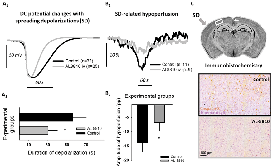
Global forebrain ischemia/reperfusion was induced in isoflurane-anesthetized, young, adult, male Sprague-Dawley rats (n=16) by the bilateral occlusion and later release of the common carotid arteries. Two open craniotomies on the right parietal bone served the continuous elicitation of SD with 1M KCl (caudal), and the acquisition of local field potential (rostral). The entire dorsal cranium was thinned to track regional cerebral blood flow (CBF) variations by laser speckle contrast imaging. The femoral artery was prepared for the monitoring of mean arterial pressure (MAP) and for blood sampling for blood gas analysis. The femoral vein was used for the infusion of an FP receptor antagonist (AL-8810; 1 mg/bwkg) or its vehicle (0.1% dimethyl sulfoxide, DMSO). Apoptosis was evaluated five hours following ischemia induction by cleaved-caspase 3 immunohistochemistry.
Results
Physiological parameters were similar in the two groups (e.g. MAP: 82.7 ± 8 vs. 84.5 ± 9.1 mmHg; AL-8810 vs. vehicle). However, AL-8810 markedly reduced the duration of evoked SDs (30 ± 10 vs. 56 ± 14 s; AL−8810 vs. vehicle). In addition, total depolarization time was reduced by 50% in the AL-8810 group (2642 vs. 5241 s, AL-8810 vs. vehicle). The CBF response to SD involved a more restricted cortical surface in the AL-8810-treated animals suggesting the involvement of a smaller tissue volume in SD. Both the number (12 vs. 9, AL-8810 vs. vehicle) and the amplitude of SD-related hypoperfusion as a result of inverse neurovascular coupling (–13.4 ± 3.2 vs. –6.8 ± 3 pp; AL-8810 vs. vehicle) were reduced in the AL-8810 group. Further, the amplitude of reactive hyperemia after reperfusion initiation was substantially greater (94.9 ± 20 vs. 79.7 ± 16%; AL-8810 vs. vehicle). The qualitative evaluation of the cleaved-caspase 3 immunolabeled brain sections revealed a decreased rate of Ca+-dependent neurodegeneration after AL-8810 treatment in the parietal cortex bearing SD.
Conclusion
In summary, the antagonism of FP receptors (located in the vascular wall or neurons) emerges as a promising approach to inhibit the evolution of injurious SDs in cerebral ischemia.
Funding
GINOP-2.3.2-15-2016-00006; NRDIO (NKFIH_K120358 and K111923); NTP-NFTÖ-18-B-0173; SzSA (No. 34232-3/2016/INTFIN).
References
PA00-A11
Circular RNA profiling of neutrophil transcriptome provides insights into asymptomatic moyamoya disease
1Institute of Cerebrovascular Disease Research and Department of Neurology, Xuanwu Hospital, the First Clinical Medical College of Capital Medical University, Beijing, China
2Beijing Geriatric Medical Research Center and Beijing Key Laboratory of Translational Medicine for Cerebrovascular Diseases, Beijing, China
Abstract
Objectives
Moyamoya disease (MMD), also known as spontaneous occlusion of the circle of Willis, is a rare cerebrovascular disease and prevalent in Eastern Asian countries, of which the cause is not well understood. Circular RNAs (circRNAs), a novel class of non-coding RNAs generated from protein-coding genes by backsplicing, are structurally stable, abundant in the brain and capable of crossing the blood-brain-barrier, which have been implicated as potential biomarkers in neurological disorders. We investigated the circRNAs expression profile of neutrophil transcriptome in asymptomatic MMD.
Methods
Subjects of asymptomatic MMD patients diagnosed by HRMRI and non-MMD matched healthy volunteers were enrolled. RNA was extracted from purified circulating neutrophil out of blood samples and circRNA expression profile was detected using circRNA microarrays and validated by real-time PCR. Transcription factor binding and miRNA binding sites of altered circRNAs were identified by bioinformatics analysis. Gene Ontology (GO) analysis and KEGG analysis was conducted to reveal the putative functions and involved pathways of their target genes.
Results
404 circRNAs were expressed differentially (Fold change > 1.5, P value < 0.5), mostly generated from exonic regions (343) of the genes in the circulating neutrophil of asymptomatic MMD patients. Of these, 260 were upregulated and 144 were downregulated compared to control. Hsa_circRNA_100146, hsa_circRNA_089761, hsa_circRNA_089762, hsa_circRNA_405463, hsa_circRNA_036592, hsa_circRNA_007011, and hsa_circRNA_044097 were differentially expressed with the highest fold change, which were detected by microarrays and confirmed by real-time PCR. Bioinformatics analysis revealed that these circRNAs altered in asymptomatic MMD might be regulated by a set of transcription factors and contain binding sites for a great amount of miRNAs. The major biological and molecular functions involved in altered circRNAs in asymptomatic MMD are cellular protein modification process, endodermal cell differentiation, cell adhesion, transforming growth factor beta receptor signaling pathway, cadherin binding involved in cell-cell adhesion, protein binding, ATP binding and protein serine/threonine kinase activity. The significant enriched pathways include focal adhesion, regulation of actin cytoskeleton, PI3K-Akt signaling pathway and thyroid hormone signaling pathway.
Conclusions
This is the first study that shows the profile of circRNAs of circulating neutrophil transcriptome in asymptomatic MMD. Our asymptomatic MMD-based study provided a set of aberrantly expressed circRNAs in asymptomatic MMD or at its early stage as potential diagnostic biomarkers. The focal adhesion pathway and thyroid hormone signaling pathway were found to be the critical regulatory pathways associated with the pathogenesis.
References
PA00-A12
Dose finding in preclinical research using adaptive designs
1Klinik fuer Neurologie mit Experimenteller Neurologie, Charite – Universitaetsmedizin Berlin, Corporate Member of Freie Universitaet Berlin, Humboldt-Universitaet zu Berlin, and Berlin Institute of Health
2Deutsches Zentrum für Herz-Kreislauf-Forschung (DZHK)
3Charite – Universitaetsmedizin Berlin, corporate member of Freie Universitaet Berlin, Humboldt-Universitaet zu Berlin, and Berlin Institute of Health, Department for Biostatistics and Clinical Epidemiology, Berlin, Germany
4Center for Stroke Research Berlin (CSB)
5German Center for Neurodegenerative Diseases (DZNE)
6Berlin Institute of Health (BIH), Berlin, Germany
7QUEST Center for Transforming Biomedical Research, Berlin Institute of Health (BIH), Berlin, Germany
8Department of Experimental Neurology and Center for Stroke Research Berlin, Charité – Universitaetsmedizin Berlin Corporate member of Freie Universitaet Berlin, Humboldt-Universitaet zu Berlin, and Berlin Institute of Health, Berlin, Germany
Abstract
Objective
To test whether an adaptive two-stage drop-the-loser design is applicable to preclinical biomedical research, using in silico simulation of data for a preclinical in vivo study.
Despite receiving increased attention in the arena of clinical trials, there are almost no reports of adaptive designs in preclinical animal research. The goal of an adaptive design is to reduce cost and development time by optimizing the information gained from each studied subject. Currently, in preclinical biomedical research, small sample sizes and high flexibility in hypothesis, experimental design and endpoint analyses are prevalent and pose serious threats to value and robustness of reported results. Adaptive designs could provide a more rigorous statistical framework to better account for the needs of preclinical research.
Material/Methods
We used the R software environment to simulate outcomes of adaptive designs in silico. We focused on a two-stage drop-the-loser design with three active and one reference group in the first stage, as this design most closely reflects dose finding and dose response experiments common in preclinical research. A maximum effect size of d = 1 was a priori assumed. The basic idea of adaptive testing goes back to Fishers’s p-value combination criterion.1 Here we generalize and modified it for the needs of the proposed experimental design.
Results
We were able to show that the number of animals allocated to a suboptimal treatment group was reduced by up to 15 % using an adaptive design compared to a standard block design assuming a large effect size of d = 1 commonly reported in preclinical biomedical research while maintaining levels for power and control for Type I error. Moreover, we showed that using an adaptive design reduced the number of animals spent in underpowered studies, when the a priori assumed effect size deviated from the observed effect size. In a next step, we demonstrated applicability and feasibility of an adaptive drop-the-loser design in the arena of preclinical stroke research in a study evaluating neuroprotective potential of antibodies against the N-Methyl-D-aspartate receptor in an established model of temporary middle cerebral artery occlusion
Conclusions
Adaptive designs are applicable to preclinical biomedical research. We demonstrated in silico that an adaptive two-stage drop-the-loser design can lead to a substantial reduction in animal numbers needed. By providing an appropriate statistical framework adaptive designs may better reflect the need of preclinical animal research for flexibility in study design while maintaining a high level of scientific rigour.
Reference
PA00-A13
Augmentation index is a predictor of cerebral blood flow across global grey matter in the elderly
1Department of Biomedical Sciences, Faculty of Medicine, Université de Montréal, Canada
2Centre de recherche de l’Institut Universitaire de Gériatrie de Montréal, Canada
3NeuroPoly Lab, Institute of Biomedical Engineering, Polytechnique Montréal, Canada
4Department of Neurosciences, Faculty of Medicine, Université de Montréal, Canada
5Institut de Recherches Cliniques de Montréal, Université de Montréal, Canada
6Montreal Heart Institute, Canada
7Department of Psychology, Faculty of Arts and Sciences, Université de Montréal, Canada
8Department of Pharmacology and Physiology, Faculty of Medicine, Université de Montréal, Canada
9Department of Medicine, Faculty of Medicine, Université de Montréal, Canada
10Physics Department, Concordia University, Canada
Abstract
Objective
Arterial stiffness and blood pressure (BP) are confirmed predictors of cardiovascular health and recognized contributors to cognitive decline and dementia.1,2 This cognitive decline is presumed to be the result of many biological alterations after years with a decline in cerebral blood flow (CBF) being one of the first manifestations.3,4 However, although many cardiovascular parameters have been associated with a decrease in global CBF,5,6 the best predictor of this decline among them has not yet been identified.
Our objectives are: 1) to determine whether the CBF in grey matter (GM) is associated with different measures of arterial stiffness and blood pressure. 2) to establish the best predictor of a lower CBF-GM amongst these cardiovascular parameters.
Methods
Fifty-three healthy participants between 65 and 75 years-old were evaluated. The measured parameters for arterial stiffness were the carotid-femoral pulse wave velocity (PWV) and the augmentation index (Aix), measured by applanation tonometry (SphygmoCor) following the Van Bortel L, et al. protocol.7 The mean systolic blood pressure (SBP) was monitored over 24-hours and analyzed following the guidelines from Hypertension Canada (2018). The coefficient of variation of 24-hours SBP was calculated by dividing the standard deviation by the mean. Resting CBF-GM was quantified from pseudocontinuous arterial spin labeling using Neurolens 2.0 (pcASL),8 and acquired on a 3T scanner (MAGNETOM Prisma Fit, Siemens) with TR/TE = 3850/8.2 ms, alpha = 90°, post-labeling delay = 1.55 s, label duration = 1.5 s, number of slices = 20, resolution = 5.0 mm isotropic.
The analysis consisted in a multiple linear regression model for each independent variable (cf-PWV, Aix, mean SBP in 24 hours and the coefficient of variation of 24-hours SBP) using age, sex, schooling and body mass index as covariates. To determine the best predictor amongst them, we created a multiple linear regression model including all four covariates in the first level and all four independent variables in a second level, and used stepwise selection to exclude variables not contributing to predict the outcome.
Results
Multiple linear regression models demonstrated that each independent variable could predict the level of CMF-GM: PWV (p = 0.010), Aix (p = 0.002), mean SBP in 24 hours (p = 0.015) and the coefficient of variation of 24-hours SBP (p = 0.015). The multiple linear regression model including all four independent variables, revealed that Aix was the best predictor of CBF-GM in our sample (p = 0.002).

Conclusions
In this cohort, we showed that while PWV, mean SBP in 24 hours and the coefficient of variation of 24-hours SBP are all predictors of CBF-GM levels, it is the augmentation index which has the highest predictive value. Based on these observations, Aix could be a useful tool to understand the interplay between lower CBF-GM and arterial stiffness. These results also indicate that Aix may be a good therapeutic target to preserve brain health and cognition.
References
PA00-A14
Effects of cerebral small vessels disease on brain perfusion in a memory clinic population
1Department of Pharmacology, National University of Singapore, Singapore
2Memory Aging & Cognition Centre, National University Health System, Singapore
3Department of Radiology, VU University Medical Center, Amsterdam, the Netherlands
4Department of Radiology, Brain Center Rudolf Magnus, University Medical Center Utrecht, the Netherlands
5Raffles Neuroscience Centre, Raffles Hospital, Singapore
6Departments of Epidemiology and Radiology and Nuclear Medicine, Erasmus University Medical Center, Rotterdam, the Netherlands
Abstract
Objective
Cerebral small vessel disease (SVD) typically manifests on MRI as white matter hyperintensities (WMH), lacunes, cerebral microbleeds (CMBs) and enlarged perivascular spaces (EPVs). It is suggested that SVD may alter cerebral blood flow (CBF) leading to brain changes and hence cognitive decline, functional impairment and dementia in elderly. CBF can be measured quantitatively by Arterial Spin Labelling (ASL). Recently, spatial coefficient of variation (CoV) has been proposed to quantify the presence of vascular artifacts and utilized as a proxy marker of vessel insufficiency.
We aim to investigate the association of location and severity of SVD with ASL-derived parameters in a memory clinic population from Singapore.
Methods
We included 375 subjects (mean age: 72.2 years, women = 53.1%) from an ongoing memory clinic based case-control study. All subjects underwent clinical assessment and 3T brain MRI. CMBs and lacune location were categorized into strictly lobar, strictly deep and mixed, and EPV location into centrum semiovale, basal ganglia, mesencephalon and hippocampus. WMH volume was automatically segmented. Total brain, gray matter and white matter CoV and CBF were analyzed with ExploreASL from 2D EPI pseudo-continuous ASL images. Linear regression models were constructed to analyze associations between SVD and ASL, and the mean difference (β) and 95% confidence intervals (CIs) were reported.
Results
41.6% of subjects had CMBs: 52.0 % strictly lobar, 19.2% strictly deep and 28.8 % mixed. 28.3% of subjects had lacunes (strictly lobar 30.2%, strictly deep 40.6% and mixed 29.2%). The mean (SD) number of EPVs was 17.9 (8.5), and mean (SD) WMH volume was 0.6 (0.5) ml.
In multivariate adjusted models increasing numbers of CMBs was associated with higher whole brain CoV [β = 0.02; 95% CI = 0.01, 0.02] and gray matter CoV [β = 0.02; 95% CI = 0.01, 0.02]. These associations were more specifically observed in subjects who had more than 4 CMBs. Location specific analysis showed mixed CMBs were associated with higher whole brain CoV [β = 0.02; 95% CI = 0.01, 0.02] and gray matter CoV [β = 0.02; 95% CI = 0.01, 0.02]. Similarly increasing numbers of lacunes was associated with increased whole brain CoV [β = 0.16; 95% CI = 0.05, 0.28] and gray matter COV [β = 0.17; 95% CI = 0.06, 0.29]. These associations were more specifically observed in subjects who had ≥2 lacunes. Region wise analysis showed mixed lacunes was associated with increased whole brain CoV [β = 0.22; 95% CI = 0.08, 0.35] and gray matter CoV [β = 0.21; 95% CI = 0.08, 0.35]. Increasing WMH volume was associated with whole brain CoV [β = 0.54; 95% CI = 0.32, 0.75], gray matter COV [β = 0.55; 95% CI = 0.34, 0.76] and white matter CoV [β = 0.41; 95% CI = 0.19, 0.63]. No association was observed between EPVs and ASL parameters. Furthermore, SVD markers were not associated with reduced CBF.
Conclusion
Increased CoV in ASL is associated with SVD, suggesting that SVD may induce brain changes via vascular insufficiency. Thus future research aimed to elucidate the pathophysiology of SVD and vascular insufficiency in elderly may help in developing prevention and treatment strategies.
PA00-A15
Rapamycin induces an eNOS dependent increase in brain collateral perfusion after acute experimental stroke in rats
1Radcliffe Department of Medicine, University of Oxford, UK
2Department of Neurological Sciences, University of Vermont, USA
Abstract
Objectives
Rapamycin (sirolimus) is a clinically approved mammalian target of rapamycin (mTOR) inhibitor shown to be neuroprotective in animal models of stroke. We hypothesized that one mechanism by which rapamycin is neuroprotective in stroke is through increasing collateral perfusion through leptomeningeal anastomosis (LMA), a strong predictor of outcome in experimental and clinical ischemic stroke. Interventions that can enhance this may be viable acute neuroprotective therapies. Thus, the aim of this study was to determine the effect of rapamycin on collateral perfusion during experimental stroke and if this is mediated by LMA dilation through endothelial nitric oxide synthase (eNOS) activation.
Methods
Proximal filament occlusion of the middle cerebral artery (MCAo) was induced for 90 min in male Wistar rats (300–350 g). Animals were randomized to receive 250μg/kg rapamycin or vehicle intravenously 30 min after onset of MCAo. Dual site laser Doppler flowmetry was used to continuously measure changes in LMA and MCA perfusion territories prior to and for 60 min after drug administration. Neuorbehavioral tests were performed 24 hours after MCAo. In a separate group of male Wistar rats (500 g), LMAs were studied isolated and pressurized in an arteriograph system. Diameter changes in isolated LMAs were studied after intraluminal pressure was increased stepwise from 40–80 mmHg. Dilation of LMAs to increasing concentrations of rapamycin (10−9 to 10−5M) was assessed in the absence and presence of a single concentration of the NOS inhibitor Nω-nitro-L-arginine methyl ester (L-NAME, 10−4M). Vessels were then washed with zero-calcium buffer with added diltiazem and papaverine to obtain fully relaxed diameters to calculate % tone and % reactivity to rapamycin.
Results
MCAo was confirmed by a 70% reduction in MCA perfusion. Rapamycin significantly increased LMA perfusion at 10 minutes after administration, during experimental stroke (Rapamycin (n = 8): 43.36 ± 17.45 vs. Vehicle (n = 6): –18.14 ± 8.55 % of pre-injection baseline, p = 0.0078, Figure 1A). Rapamycin also significantly increased average LMA perfusion during the 60 minutes post-drug administration (Rapamycin: 15.19 ± 10.02 vs. Vehicle: –15.51 ± 7.94 % of pre-injection baseline, p = 0.0425). Rapamycin significantly improved neuroscores (Rapamycin: median score of 2, range 1–3 vs. Veh: median score of 3.5, range 2–5 out of a max score of 6, p = 0.0353). Isolated LMA active inner diameters were 35 ± 5 µm (n = 4) and % tone was 15.2 ± 3.4%, (n = 4). Rapamycin caused dose-dependent vasodilation of LMAs (Figure 1B). Percent reactivity to 10–6M rapamycin was 79.7 ± 9.4 % without L-NAME that was only 7.3 ± 3.6% in the presence of L-NAME (p = 0.0024, Figure 1B).

Conclusions
Rapamycin significantly enhanced LMA perfusion during experimental stroke. Isolated LMA experiments demonstrated that that this increase in perfusion was potentially driven by rapamycin dilating LMAs. Rapamycin’s vasodilatory effect was largely inhibited by L-NAME, suggesting that rapamycin dilated LMAs through a mechanism involving eNOS. These results are in agreement with the eNOS and CBF enhancing effect of rapamycin previously observed in Alzheimer's disease mice and suggest that rapamycin may be an effective acute collateral enhancing therapy to maintain perfusion to potentially salvageable penumbra and improve stroke outcome.
PA00-A16
Pharmacological targeting of von Willebrand factor (VWF) strings provides improved stroke outcomes in aged mice
1Department of Neurology, McGovern Medical School, the University of Texas Health Science Center, USA
2Center for Translational Research on Inflammatory Diseases, Michael E. DeBakey Veterans Affairs Medical Center, USA
3Department of Medicine, Baylor College of Medicine, USA
4Department of Molecular Physiology and Biophysics, Baylor College of Medicine, USA
Abstract
Objectives
Von Willebrand Factor (VWF) strings are extruded from activated endothelial cells and can accumulate in the vascular lumen of cerebral arteries. Because VWF strings bind circulating platelets, we and others have proposed that these strings may contribute to the pathology of stroke. Our recent studies have demonstrated that VWF strings are anchored to the vascular lumen through a molecular interaction between the VWF A2 domain and endothelial extracellular vimentin. By targeting the VWF/vimentin interaction with a recombinant A2 domain protein (A2 protein), we were able to reduce string formation and improve reperfusion following stroke. We now seek to determine if A2 protein treatment can provide improved outcome following stroke in translationally-relevant aged mice.
Methods
Aged male mice (20–22 months) were subjected 60 min of MCAo and allowed to recover for three days. Mice were randomized to A2 protein (4 mg/kg i.p.) or vehicle treatment at 15 min before onset of reperfusion. Neurologic deficits were scored daily (NDS; 0–12). Infarct volume (corrected for brain swelling) was evaluated by TTC. The severity of hemorrhagic transformation (HT) was graded (0 to 5) for each section and expressed as a cumulative total HT score. Scoring was done blinded to treatment.
Results
Functional outcome (NDS) was significantly improved in the A2 protein treated mice for post-stroke days 1–3 (group difference, P = 0.003; n = 7–8 mice). In addition, A2 protein treatment demonstrated significant reduction in infarct volume (by 35%, P = 0.033) and profound reduction in HT score (by 58%, P = 0.009) at post-stroke day 3. There was no difference in post-stroke rectal temperature or body weight between groups. NDS at 60 min occlusion was not different between groups (12 vs. 12).
Conclusions
These data demonstrate the beneficial effect of targeting the VWF/vimentin interaction at the time of reperfusion. Using translationally-relevant aged mice, we demonstrated that a single dosing of A2 protein could reduce infarct volume and improve functional performance. Critically, HT was also significantly reduced with A2 protein treatment. These studies strongly implicate VWF strings in stroke pathology and provide a therapeutic rationale for targeting VWF string formation as an adjuvant treatment with recanalization therapies.
PA00-A17
Differentiation between recovered and deteriorated tissue after cerebral ischemia-reperfusion, based on MRI of blood-brain barrier leakage and cerebrovascular reactivity in a rodent stroke model
1Biomedical MR Imaging and Spectroscopy Group, Center for Image Sciences, University Medical Center Utrecht
Abstract
Objectives
Reperfusion therapy is currently the only approved treatment for acute ischemic stroke patients. However, despite its beneficial effects, reperfusion after ischemia may also trigger secondary tissue damage. Elucidation of tissue characteristics, associated with tissue recovery or secondary injury after reperfusion, could therefore aid in treatment monitoring. We investigated whether in vivo MRI-based assessment of blood-brain barrier (BBB) leakage and cerebrovascular reactivity can differentiate between recovered and secondarily injured tissue after reperfusion in a rodent model of stroke.
Methods
Cerebral ischemia was induced by intraluminal right middle cerebral artery occlusion in male adult Sprague Dawley rats (n = 6), followed after 90 min by induction of reperfusion.1 Serial MRI (at 9.4T) was conducted during occlusion and immediately after reperfusion (acute phase), and 4 days after stroke (subacute phase). During MRI, rats were anesthetized and mechanically ventilated with 2% isoflurane in air/O2 (2:1). Temperature was maintained at 37.0 ± 0.5°C. MRI included diffusion-weighted imaging (DWI, 150 × 150 × 600 μm voxels, b = 1454 s/m2) and T2-weighted imaging (150 × 150 × 600 μm voxels) for lesion detection. At the subacute phase we additionally performed T1 mapping (Look-Locker sequence, 300 × 300 × 1200 μm voxels) after i.v. Gd-DTPA injection, and blood oxygenation level-dependent (BOLD) MRI (3D spin-echo EPI: 1500 images, TR/TE = 70.5/40.0 ms, flip angle = 90°, 600 × 600 × 1200 μm voxels) combined with a 9% CO2 challenge. Semi-automatic thresholding was used to delineate the ischemic lesion in the acute (before and after reperfusion) and subacute phase. Recovered and secondarily injured (recruited) tissue areas were subsequently identified. The lesion core was defined as the acute ischemic lesion area before reperfusion subtracted by the recovered tissue area subacutely after reperfusion. Values derived from these regions of interest (ROIs) were normalized against values from identical contralateral areas. BBB leakage was determined by Gd-induced T1 shortening, while vascular reactivity was derived from the CO2-induced BOLD signal increase. One-way ANOVA and Bonferroni pot-hoc tests were applied for statistical analyses.
Results
The lesion core, rescued areas and recruited areas were successfully identified from apparent diffusion coefficient (ADC) (figure A) and T2 maps (figure B). The lesion core displayed highest level of T1 shortening (6.1 ± 1.7%) after gadolinium injection; significantly higher compared to recruited areas (1.6 ± 0.4%), while T1 shortening between core-rescued, and rescued-recruited areas did not differ (figure C). Further, CO2-induced BOLD signal increase showed an upward inflection in rescued compared to recruited and core areas (figure D).

Conclusion
Our study shows that post-stroke tissue areas that recover or deteriorate after reperfusion display mild to moderate BBB permeability, and cannot be straightforwardly differentiated based on contrast agent leakage. On the other hand, in contrast to post-reperfusion lesioned areas, recovered regions demonstrated substantial vascular reactivity, reflective of tissue convalescence.
Bart A.A. Franx,1 Geralda A.F. van Tilborg,1 Caroline L. Van Heijningen,1 Annette van der Toorn,1 Rick M. Dijkhuizen1
1Biomedical MR Imaging and Spectroscopy Group, Center for Image Sciences, University Medical Center Utrecht, Utrecht, Netherlands.
Funded by the Netherlands Cardiovascular Research Initiative through the Dutch Heart Foundation (CVON2015-01; CONTRAST) and the Brain Foundation Netherlands (HA2015.01.06).
Reference
PA00-A18
Dl-3-N-butylphthalide promotes angiogenesis and upregulates sonic hedgehog expression after cerebral ischemia in rats
1School of Biomedical Engineering, Shanghai Jiao Tong University, China
2Shanghai Ruijin Hospital, School of Medicine, Shanghai Jiao Tong University, China
Abstract
Objectives
Dl-3-N-butylphthalide (NBP), a small molecule drug used clinically in the acute phase of ischemic stroke, improves functional recovery and promotes angiogenesis and collateral vessel circulation after experimental cerebral ischemia.1 However, underlying molecular mechanism is unknown.1 In this study, we explored the potential molecular mechanism of angiogenesis induced by NBP after cerebral ischemia.
Methods
Ninety adult male Sprague Dawley rats were subjected to transient middle cerebral artery occlusion. NBP (80 mg/kg) was given by oral gavage once a day after occlusion. The effect of NBP was evaluated based on the changes in body weight, brain infarct volume, and neurobehavioral outcomes. The number of CD31+ microvessels and CD31+/BrdU+ proliferating endothelial cells were examined by immunostaining. The functional cerebral vascular density in living rats was assessed by synchrotron radiation angiography technique. Angiogenic growth factors was evaluated by Western blot. Sonic hedgehog expression was examined using immunostaining, Western blot and RT-PCR. Furthermore, the effect of silencing sonic hedgehog signaling on angiogenesis was investigated using cell culture experiment.
Results
NBP treatment attenuated body weight loss, reduced brain infarct volume, and improved neurobehavioral outcomes during focal ischemia compared to the control rats (p < 0.05). NBP increased the number of CD31+ microvessels, the number of CD31+/BrdU+ proliferating endothelial cells, and the functional vascular density (p < 0.05). Further study demonstrated that NBP also promoted the expression of vascular endothelial growth factor and angiopoietin-1 (p < 0.05), which was accompanied by up-regulated sonic hedgehog expression in astrocytes in vivo and in vitro. While Shh knockdown with siRNA in vitro, the expression of vascular endothelial growth factor and angiopoietin-1, the proliferation, migration and tube formation of HUVEC were inhibited after NBP treatment.

Conclusion
NBP treatment promoted the expression of vascular endothelial growth factor and angiopoietin-1, induced angiogenesis and improved neurobehavioral recovery. These effects were associated with increase of sonic hedgehog expression after NBP treatment. Our results broadened the clinical application of NBP to the later phase of ischemia.
Reference
PA00-A19
Cortical pericytes are more resistant to experimental stroke than neurons and start proliferating after reperfusion
1Laboratory of Experimental Stroke Research, Institute for Stroke and Dementia Research (ISD), University of Munich Medical Center
2Munich Cluster of Systems Neurology (Synergy), Munich, Germany
Abstract
Objectives
Neurons are very sensitive to cerebral ischemia, however, very little is known about the sensitivity of vascular cells to ischemic stroke, particularly pericytes. Therefore, the aim of the current study was to characterize the fate of pericytes in the acute phase after experimental stroke by profiling the extent and mechanism of cell death and by defining the transcriptomic profile of pericytes.
Methods
C57BL/6 or PDGFRb-eGFP/NG2-DsRed double reporter mice were not handled or subjected to 60 minutes occlusion of the middle cerebral artery (MCAo) by an intraluminal filament or sham surgery. One and three days after MCAo we utilized immunofluorescence techniques in combination with TUNEL, CollagenIV, and Ki67 staining to assess acute pericyte loss following ischemic stroke combined with analyses of PDGFRbEGFP and NG2DsRed reporter mice to investigate surviving pericyte density, proliferation, cell cycle entry, activation, and coverage. We performed FACS isolation of pericytes using the PDGFRb-eGFPxNG2-DsRed mice in combination with CD13 staining three days after MCAo to obtain the pathological transcriptome of pericytes and mural cells in the stroke core, ipsilateral, and contralateral hemisphere.
Results
Our results indicate that within the region where neurons die (as indicated by loss of NeuN staining) after 1 h of MCAo the majority of pericytes survives. Specifically, pericyte survival is about 50% adjacent to the MCA territory in striatal and lower cortical layers and increases significantly to 80% toward the anterior and the cortical surface of the brain. Loss of pericytes is coupled with a concomitant increase in TUNEL+ PDGFRb expressing pericytes (30%) in the infarct area. Further on, we report that surviving PDGFRb+ pericytes compensate widespread vascular disruption by significantly expanding their coverage within the basement membrane, activating and expressing cell cycle specific markers (Ki67) and proliferate in both the ischemic core and peri-infarct region. Finally, we employed a FACS isolation approach to determine the transcriptomic profile of pathophysiological pericytes in the sub-acute phase of experimental stroke. Here, we crossed mice expressing canonical mural cell specific reporters (NG2DsRed/PDGFRbEGFP) and CD13 staining to reveal a number of differentially regulated transcripts in pericytes within the ischemic core, ipsilateral and contralateral regions that shed-light on the pathophysiological function of pericytes in response to ischemia.
Conclusions
Taken together, our results demonstrate that pericytes are more resistant to transient cerebral ischemia than neurons and possess the ability to activate, enter the cell cycle and proliferate following reperfusion of the occluded artery in both the infarct core and peri-infarct region. Moreover, we report that pericyte loss occurs in a bregma dependent manner involving DNA fragmentation and that striatal pericytes and layer 4/5 cortical pericytes are most heavily affected by transient experimental stroke. These results demonstrate that depending on their location a large proportion of pericytes is resistant to extended periods of cerebral ischemia
PA00-A20
Endovascular model of ischemic stroke in swine
1Department of Neurosurgery, School of Medicine, Collegium Medicum, University of Warmia and Mazury, Olsztyn, Poland
2Central Clinical Hospital of Ministry of the Interior and Administration in Warsaw, Poland
3Department of Surgery and Roentgenology with the Clinic, Faculty of Veterinary Medicine, University of Warmia and Mazury, Olsztyn, Poland
4NeuroRepair Department, Mossakowski Medical Research Center, Polish Academy of Science, Warsaw, Poland
5Russell H. Morgan Department of Radiology and Radiological Science, Division of MR Research, The Johns Hopkins University School of Medicine, Baltimore, MD, USA
6Cellular Imaging Section and Vascular Biology Program, Institute for Cell Engineering, Johns Hopkins University, Baltimore, MD, USA
Abstract
Objectives
The main aim of this study was to use minimally invasive, catheter-based endovascular technique for catheter navigation under X-ray followed by MRI to monitor thrombin-mediated occlusion of cerebral vessels as a strategy to induce endovascular model of stroke.
Methods
The animal procedures were approved by local ethics committee and were performed according to ARRIVE guidelines. Juvenile, male, domestic pigs were anesthetized and 5 F Introducer (Prelude MeritMedical) was inserted into femoral artery after percutaneous puncture. Using this port, catheter was navigated under C-arm guidance to common carotid artery (CCA). Microcatheter (APOLLO™ Medtronic) was then introduced with its tip positioned in the ascending pharyngeal artery (APA) proximally to the rete mirabile. Animals with secured microcatheters were transported to 3T MRI scanner (Magnetom Trio, Siemens). Under real time MRI guidance with dynamic T2* weighted scans (temporal resolution 2–3s) thrombin solution (Biomed, Poland) mixed with gadovist (1:20) was injected. MRI protocol included dynamic GE-EPI for assessment of trans-catheter cerebral perfusion, GE-EPI for monitoring thrombin-mediated blood clotting as well as SWI, diffusion, T2w and T1w with contrast. MRI follow up was performed one day and one week post induction.
Results
Due to complexity of vascular anatomy direct blocking of cerebral vessels to induce ischemic stroke has not been possible. We circumvented this obstacle by intra-arterially injecting procoagulant thrombin with real-time MRI for instant assessment of cerebral vascular occlusion. Interventional MRI facilitated visualization of thrombus formation as evidenced by hypointense regions in SWI MRI (
Diffusion weighted MRI detected ischemic damage as hypointense region on ADC map with earliest evidence at 27 minutes and very robust effect at 90 minutes after thrombin injection (
Induction of stroke resulted in marked neurological deficits manifesting as severe hind limb paresis.

Conclusions
Our study has demonstrated feasibility of using endovascular route to induce ischemic stroke in pig. Most importantly, real-time MRI was instrumental to monitor and confirm formation of thrombin-induced clot and resulting blockade of cerebral perfusion with subsequent brain infarct. Our method produced cerebral ischemia in relatively large volume, which covers majority of MCA supply territory. Overall, we have established a new model of ischemic stroke in pigs without confounding effect of extensive stroke-independent morbidity occurring after surgical model. In this context, our model has improved clinical relevance.
Conflict of interests
Authors declare no conflict of interest.
Acknowledgements
Funding: NCBIR EXPLORE ME grant (STRATEGMED1/235773/19/NCBR/2016); NCN OPUS grant (UMO-2018/29/B/NZ5/00921)
PA00-A21
Extravasation of microspheres in a rat model of silent brain infarcts
1Biomedical Engineering and Physics, Amsterdam UMC, University of Amsterdam, Amsterdam, The Netherlands
2Department of Medical Biology, Electron Microscopy Center Amsterdam, Amsterdam UMC, University of Amsterdam, Amsterdam, The Netherlands
3Physiology, Amsterdam Cardiovascular Sciences, Amsterdam UMC, Vrije Universiteit Amsterdam, Amsterdam, The Netherlands
Abstract
Objectives
Silent brain infarcts (SBI) are small regions of ischemia caused by occlusion of microvessels, which occur without overt clinical symptoms. The lack of acute symptoms and their small size hamper investigations on the pathogenesis of SBI, which remains largely elusive. Here, we developed a rat model of SBI based on microsphere infusion and investigated their impact on perfusion and tissue damage. Secondly, we studied the extent and mechanisms of perfusion recovery.
Methods
At day (D)0, 15 µm fluorescent microspheres were injected into the right common carotid artery of F344 rats. At D1, D7 or D28, animals were i.v. injected with fluorescently-labeled lectin to stain the vasculature, flushed and fixed via transcardial perfusion. The brain was removed, cut in 100 µm cryosections and processed for immunofluorescent staining and analysis.
Results
Injection of microspheres caused mild and transient damage to the treated hemisphere, with a decrease in capillary volume fraction at D1, as compared to the untreated hemisphere. Apoptotic cells were rarely seen near the sites where microspheres had lodged. At D1 but not at D7 and D28, we observed IgG staining outside of the vessels, indicating vessel leakage. All microspheres were located inside the lumen of the vessels at D1, whereas the vast majority (∼80%) of the microspheres were extravascular at D7, and 100% at D28 (Figure). This was accompanied by restoration of perfused capillary volume.

Figure.
Examples of microspheres confined to the vessel lumen (D1) and extravasated microspheres (D7 and D28), as shown by lectin staining (red). Scale bar = 25 µm.
Conclusion
The effects of microspheres on cerebral arterioles are mild and, importantly, transient in nature. This transient effect is likely a result of effective extravasation mechanisms in the brain to clear micro-sized emboli from the vessels, which enables reestablishment of blood flow.
PA00-A22
Brain-wide ventricular-cerebral transport (vector) imaged by non-invasive dynamic MRI
1Centre for Advanced Biomedical Imaging, Division of Medicine, University College London, London, UK
Abstract
Objectives
A defining feature of the early stages of many neurological disorders is the accumulation of harmful material such as amyloid-beta in the CNS. Brain clearance mechanisms such as the glymphatic system involve exchange between cerebrospinal fluid (CSF) and interstitial fluid (ISF). Moreover, CSF-ISF exchange alterations have been implicated in the development of many common neuropathological disorders. Due to the mounting interest in this field, and associated methodological concerns, there is an urgent need to provide a physiologically intact view of CSF-ISF pathway function.
In this study we introduce a novel, non-invasive technique, for imaging whole-brain CSF-ISF exchange, named ‘ventricular-cerebral transport (VECTOR)’, using a single IV injection of gadolinium based contrast agent (GBCA) and MRI. This technique provides evidence for rapid transport from the circulation via the choroid plexus into the ventricles and subsequent brain-wide distribution in approximately 30 mins.
Methods
Mice (C57B6) were anaesthetized with 1.5–2% isoflurane. The dynamic contrast enhanced MRI platform comprised a 3d T1-weighted gradient echo MRI (9.4T) together with a single bolus (20 ml/kg) of contrast agent Gadodiamide (Omniscan) delivered over 5 minutes IV (n = 6). For autoradiography, 100 Mbq of 111In-DTPA was delivered IV. Mice were euthanized after 37 mins (n = 2). For perturbation of CSF-ISF exchange, 60 I.U./kg of [Arg8]-Vasopressin solution was delivered intraperitoneally 30 mins prior to GBCA injection (n = 6). For surgical tau injections, 50 ng of tau protein was injected into the left striatum. Vasopressin or vehicle was delivered 15 mins prior to tau injection (n = 5 per group). 15 mins post tau injection, CSF was collected from cisterna magna. Left and right striatum were also collected.
Results
Infiltration of GBCA into the brain parenchyma following a single IV injection was captured for the first time (Fig-1A). Rapid transfer of GBCA from the circulation into the CSF spaces (notably the ventricles) via the choroid plexus and subsequent ingress into periventricular brain tissue regions was monitored in real-time. Brain-wide signal hyper-intensity was visualized 37 mins post contrast injection on the MR images. Autoradiography with 111In-DTPA echoed these findings, where a similar pattern of signal hyper-intensity was observed (Fig-1B). Modulating brain fluid dynamics using vasopressin resulted in a significant contrast retention in the brain (Fig-1C). Increased retention of tau in the brain tissue together with a reduced clearance into the CSF, following invasive intra-striatal tau infusion, confirmed the retentive effect of vasopressin (Fig-1D).

Conclusion
In this study infiltration of systemic GBCA into the brain tissue, via a pathway named ‘ventricular-cerebral transport (VECTOR)’, was dynamically monitored. This novel platform is the first non-invasive imaging of CSF-ISF exchange and, as such, provides an unperturbed and physiologically intact view of exchange. Pharmacological modulation using vasopressin, demonstrated the utility of VECTOR in the study of the parenchymal accumulation of aberrant proteins that defines the most common neurodegenerative conditions.
PA00-A23
In vivo neurovascular response to focused photoactivation of channelrhodopsin-2
1Sunnybrook Research Institute
2Hospital of Sick Children
Abstract
Objectives
Optogenetic offers highly specific cellular targeting through genetic manipulations and precise spatio-temporal neuronal activation via parametrization of the optical stimulation. The difference between the most commonly used stimulation modalities, namely diffused (i.e. synchronous) and focused (i.e. asynchronous) stimulation has not been described. Furthermore, full realization of optogenetics’ potential is hindered by our incomplete understanding of the cellular and network level response to photoactivation.
Methods
Here we address these gaps by examining the neuronal and cerebrovascular responses to focused and diffuse photostimulation of channelrhodopsin in the Thy1-ChR2 mouse. We presented the responses of diffused photoactivation via 470-nm fiber optic illumination alongside focused 458-nm raster-scan stimulation of the barrel cortex. Local field potentials (LFP) assessment of intracerebral electrophysiology and two-photon fluorescence microscopy measurements of red blood cell (RBC) velocity (vRBC) in cortical penetrating vessels revealed ∼40% larger LFP responses (p = 0.05) and twice as large cerebrovascular responses (p = 0.002) under focused vs. diffuse photostimulation (focused: 1.64 ± 0.84 mV LFP amplitude and 75 ± 48% increase in vRBC; diffuse: 1.14 ± 0.75 mV LFP amplitude and 35 ± 23% increase in vRBC). Compared to diffuse photostimulation, focused photostimulation resulted in a ∼65% increase in the yield of cerebrovascular responses (73 ± 10% for focused and 42 ± 29% for diffuse photostimulation) and a doubling of the signal-to-noise ratio of the cerebrovascular response (20.9 ± 14.7 for focused and 10.4 ± 1.4 for diffuse photostimulation).

Conclusion
These data reveal important advantages of focused optogenetic photoactivation, which can be easily integrated into single- or two-photon fluorescence microscopy platforms, as a means of assessing neuronal excitability and cerebrovascular reactivity, thus paving the way for broader application of optogenetics in preclinical models of CNS diseases.
PA00-A24
Focal corpus callosum lesion disrupts resting state functional connectivity in mice: effects of Rho-kinase inhibitor Fasudil
1Neurovascular Research Laboratory, Department of Radiology, Harvard Medical School, Massachusetts General Hospital, Charlestown, MA, United States of America
2Department of Physical Medicine and Rehabilitation, Health University School of Medicine, Istanbul, Turkey
3Department of Physiology, Hacettepe University School of Medicine, Ankara, Turkey
4Stroke Service and Neuroscience Intensive Care Unit, Department of Neurology, Harvard Medical School, Massachusetts General Hospital, Boston, MA United States of America
5Department of Radiology, Massachusetts General Hospital and Harvard Medical School, MGH/HMS/MIT Athinoula A Martinos Center for Biomedical Imaging, Charlestown, MA, United States of America
6Department of Biomedical Engineering, Neurophotonics Center, Boston University, Boston, MA, United States of America
Abstract
Objectives
Small, lacunar-like, strokes involving subcortical white matter tracts contributes to the ∼25% of strokes in humans and lead to major disability. The corpus callosum (CC) is the largest white matter tract and critical for transhemispheric connectivity (1). Therefore, we investigated the effect of focal CC lesions on resting state functional connectivity (RSFC) in mice as a model of subcortical ischemic white matter injury.
Methods
We compared injection of 2 different doses of the vasoconstrictor L-NIO (0.4 uL and 2.4 uL of a 100 mM solution) with saline vehicle injection into the CC centered at two antero-posterior levels (0.5, and 1.5 mm anterior to bregma) in male C57bl/6 J mice. We assessed behavior a battery of sensory, motor and cognitive tests at multiple time points over 5 weeks. Fasudil (10 mg/kg, IP) or vehicle (saline 0.1 ml, IP) treatment started 24 hours after injection and continued twice a day for 2 weeks. We calculated various indices of RSFC using hemodynamic functional optical intrinsic signal imaging through intact, full thickness skull (2). In a separate set of longitudinal experiments we implanted a glass coverslip overlying unaltered skull for serial RSFC measurements. A seed-based connectivity index (SCI) was calculated by averaging the averaged positive correlations for each seed. Seeds were placed manually over: (a) secondary motor (M2), (b) the CC lesion based on histology (L), (c) whisker barrel (WB), and (d) occipital (OCC) cortex. A interhemispheric homotopic connectivity index (ICI) was calculated by averaging all the correlation coefficients between contralateral mirror pixels. We confirmed the size of the brain lesions with H&E staining.
Results
L-NIO reproducibly induced focal (1.65 ± 0.12 mm3) infarcts in CC on day 1, which remained limited to CC on day 35 (see Figure A for representative image). Mice injected with 2.4 but not 0.4 uL L-NIO developed pure-motor deficits in foot-fault test only, which persisted at least 2 weeks. Based on RSFC imaging, all contralesional seeds showed significant loss of connectivity to ipsilesional hemisphere after 2.4 but not 0.4 uL L-NIO. All, except occipital, ipsilesional seeds showed significant loss of connectivity to contralesional hemisphere as well. Ipsilesional motor cortex seed showed significant loss of connectivity to ipsilesional (i.e. same) hemisphere. The ICI decreased by ∼50% 1 day after a CC lesion (see Figure B for representative images). Fasudil significantly augmented the ICI on day 14 compared to vehicle treated mice (Figure C), without changing lesion volumes when quantified at 35 days (0.12 ± 0.1 mm3).

Conclusion
We show that RSFC imaging can be used to quantify the disruption and recovery of both interhemispheric and intrahemispheric long white matter tracts after a CC lesion, which can be more powerful than neurocognitive testing. Using the model, we show that fasudil improves RSFC two weeks after a CC lesion, without altering the lesion burden, suggesting enhanced plasticity.
References
PA00-A25
Middle cerebral artery occlusion induces remote white matter changes in the pontine reticulospinal tract in rats
1Biomedical MR Imaging and Spectroscopy group, Center for Image Sciences, University Medical Center Utrecht, The Netherlands
Abstract
Objectives
Recent studies have shown that damage to the corticospinal tract (CST) and alternate corticofugal motor fibers (aMF) are predictive of motor outcome in ischemic stroke patients.1 However, to what extent the CST and aMF are affected in relation to lesion size and location remains unclear.1 We have previously shown that direct and remote damage to the CST can be induced after middle cerebral artery occlusion (MCAo) in rats.2 The aim of the present study was to assess whether MCAo also induces damage in remote areas in the aMF. To this aim we compared measures of microstructural integrity in different areas of the CST and aMF, based on high-resolution post-mortem diffusion tensor imaging (DTI) of rat brain at 70 days post-stroke.
Methods
90-minute MCAo was induced with an intraluminal filament in male Sprague Dawley rats.2 Stroke animals were divided in 2 groups, based on lesion extent: cortical + subcortical lesion (StrokeLarge; n = 8) and subcortical lesion (StrokeMedium; n = 4). Control rats underwent sham surgery (n = 10). Animals were euthanized 70 days post-stroke, and high-resolution DTI (9.4T; 8-shot 3D-EPI; voxel resolution: 150x148x150μm3; b = 3842s/mm2; 60 diffusion-weighted directions) was performed on fixed post-mortem brain tissue.3 For analysis of post-mortem DTI data, a 3D-model of Paxinos’ stereotaxic rat brain atlas was registered to DTI images of individual rats. Regions-of-interest (ROIs) included the ipsi- and contralesional pontine reticular nucleus (pRetN), medial vestibular nucleus (mVeN), red nucleus (RN), and a subregion of the internal capsule containing CST fibers (ICCST; identified by MR diffusion tractography) (Figure A). Mean fractional anisotropy (FA) was calculated per ROI in both hemispheres, and statistically analyzed with ANOVA and post hoc t-testing.
Results
In the StrokeLarge group, the ICCST fell within the ischemic lesion, whereas the ICCST in StrokeMedium animals had <5% lesion overlap. The RN, pRetN, and mVeN were invariably outside the ischemic lesion territory in both groups. FA was considerably reduced in the ipsilesional ICCST in StrokeLarge rats compared to the contralesional hemisphere, and compared to controls (Figure B). Within the StrokeLarge group, FA was subtly but significantly reduced in the ipsilesional pRetN compared to the contralesional hemisphere (Figure C), which was not observed in the ipsilesional RN or mVeN. No significant FA changes were measured in the ROIs in the StrokeMedium group.

Figure
(A) Coronal (top) and sagittal FA maps with ipsi- (red) and contralesional (blue) ROIs. (B, C) FA (mean±SD) in ICCST and pRetN (**p < 0.001).
Conclusion
The present study shows that remote white matter degeneration, characterized by FA reduction, develops after transient MCAo. The altered structural integrity of aMF may contribute to post-stroke motor dysfunction and could provide a target for recovery-promoting strategies.
Funded by the Netherlands Organisation for Scientific Research (NWO) Graduate Programme.
References
PA00-A26
White matter fiber orientation effects of mq-BOLD derived oxygen extraction fraction
1Department of Neuroradiology, Technical University of Munich, Munich, Germany
2MRRC, Yale University, New Haven, CT, United States
3Clinic for Radiology, Technical University of Munich, Munich, Germany
4Philips Healthcare, Hamburg, Germany
5Clinic for Neurology, Technical University of Munich, Munich, Germany
Abstract
Objectives
Measurements of cerebral oxygenation supply in white matter (WM) have highest clinical relevance, as WM is especially vulnerable to haemodynamic changes.1,2 Relative oxygen extraction fraction (rOEF) can be measured by multiparametric quantitative-BOLD (mq-BOLD), which is based on T2*, T2 and relative cerebral blood volume (rCBV) MRI-measurements.3 However, mq-BOLD applicability in WM is questionable, as model assumptions4 are suspected to be violated within those highly oriented structures.1 WM anisotropy effects of T2*5,6 and rCBV are already known7and assumed to primarily arise from oriented myelin structures5,8 and partially oriented vessels.7,9 Additionally, T2 orientation effects are expected due to diffusion.10,11 The aims of this study were therefore, first, to characterize the anisotropy effect impact on mq-BOLD derived rOEF in healthy controls (HC) and second, to investigate whether haemodynamic changes2 in internal carotid artery stenosis (ICAS) affect those orientation dependencies.
Methods
Fifty-nine participants (29 asymptomatic, unilateral ICAS-patients, age = 70.1 ± 4.8y and 30 age-matched HC, age = 70.3 ± 7.3y) underwent MRI on a Philips 3T Ingenia with written informed consent. Separate acquisitions of T2*, T2 and dynamic susceptibility contrast (DSC) based rCBV12 were obtained to calculate rOEF by mq-BOLD.3 Fiber orientations were assessed by DTI, yielding polar angle θ-maps for each participant, which voxel-wisely describes the angle between main-fiber-orientations and the main-magnetic-field B0 (Fig.1A-C).5–7 On group level, orientation effects of T2*(θ), T2(θ), R2'(θ), rCBV(θ) and rOEF(θ) were evaluated by histogram analysis of median values in 5° bins within subject specific masks of oriented WM.13
Results
HC data show strong dependencies of T2*(θ), T2(θ) and thus also R2’(θ) (26.5%) on fiber orientation towards B0 (Fig.1D-F). Orientation dependencies of rCBV(θ) were even stronger with 37.1% (Fig.1G), while rOEF(θ) variations were lower with 15.0% (Fig.1H). Orientation dependencies of ICAS-patients are very similar to HC for all parameters, showing only slightly reduced orientation related variations.
Discussion
Strong dependencies of T2*(θ) and rCBV(θ) on tissue orientation towards B0 were measured in HC, in accordance with recent literature.5–7,10,11 Compared to strong R2’(θ) (≈27%) and rCBV(θ) (≈37%) orientation driven variations, rOEF(θ) variations are rather low (13.5%) due to partially counteracting effects of R2′(θ) and rCBV(θ).13 When considering the angle distribution (Fig.1I), average anisotropy related rOEF-deviations are only 3.8% in HC. Interestingly, orientation driven variations in ICAS were slightly reduced, pointing to pathologic effects on myelination and/or the vasculature. However, average anisotropy related rOEF variations in ICAS-patients were comparable to HC with 4.4%.

Conclusion
Our results demonstrate successful in-vivo measurements of mq-BOLD orientation effects by DTI. Measured T2*(θ), T2(θ) and rCBV(θ) dependencies for HC are in accordance with current literature and similar for ICAS-patients. Additionally, rOEF(θ) shows comparably low average orientation related variations for HC and ICAS-patients (<5%). Therefore, our results point to potentially meaningful evaluations of mq-BOLD based rOEF in WM, even in ICAS-patients. However, future validations are demanded, e.g. by MR-PET comparisons.
References
PA00-A27
Mapping functional capillary-weighted blood volume in the human brain using ultra-high field MRI
1CUBRIC Cardiff University Brain Research Imaging Centre, School of Psychology, Cardiff, UK
Abstract
Objectives
A simple non-invasive method is presented for mapping the functional cerebral capillary-weighted blood volume (CBVcap). MRI signal modelling is presented which demonstrates strong functional capillary weighting of the spin-echo hyperoxia BOLD signal at 7T. The method is demonstrated on a small pilot cohort (n = 5). The results from this pilot study indicate good agreement with expected physiology, with a mean grey matter CBVcap of 1.02 ± 0.13 %.
Methods
Spin-echo (TE 55 ms) MRI signals due to a hyperoxic susceptibility change were modelled at 7T for capillaries, venules, arterioles, and veins (5, 16, 16, and 200μm radii respectively). Extra-vascular signals were modelled using a Monte–Carlo simulation of water in a vascular network as per.1 Briefly, the ODIN framework 2 was used to produce numerical integrations of the Bloch equations along a large number of random walk paths for a range of baseline blood susceptibility values (0.1 to 0.9 ppm) and hyperoxic induced blood susceptibility changes (–0.05 to –0.1 ppm). The intravascular signal contributions were modelled considering expected changes in R2.3 The relative oxygen extraction of each vascular compartment was assigned as per.4
Experimental data was acquired from 5 volunteers (4 male, 31.4 ± 2.3 years). Acquisition: 7T research MRI scanner (Siemens, Erlangen). 10.5 minutes SE data (180 volumes), 64x64 matrix, 192 mm FOV, 28 slices, 3 mm slice thickness, 10% slice gap, GRAPPA factor 2, TE 55 ms. The gas paradigm consisted of three 1.5-minute blocks of 50% O2 interleaved with normoxia. A brief (15 seconds) period of 100% O2 was used at the beginning of each hyperoxic block to speed transition to a steady state. During all scanning sessions end-tidal CO2 and O2 were monitored. Structural images were segmented to create grey matter mask and applied to registered CBVcap estimates to calculate the mean grey matter blood volume for each subject.
Results
Modelling results demonstrated a linear relation between the hyperoxic SE BOLD signal change and the resting blood volume of each vascular compartment. However, the capillary compartment contributed more than two times the signal of the next most significant compartment (venule), see figure 1a. Variation in [Hb] only had a small influence on the modelling results, while the influence from baseline oxygen extraction fraction was more pronounced, see figure 1b. In-vivo estimates of CBVcap reported a group mean grey matter value of 1.02 ± 0.13 %, which demonstrates a small inter-subject variation and is close to the expected value of 0.8–1.0% reported in.3

Discussion
The proposed method of estimating CBVcap is simple to perform, non-invasive and initial evidence suggest it is robust and correlates well with expected physiological values. Future work to optimise the gas delivery and data acquisition is planned to reduce acquisition time and reduce the influence of field inhomogeneity in the inferior brain voxels.
References
PA00-A28
Impaired perfusion and capillary distribution of blood in mild cognitive impairment:Relation to oxygenation and amyloid load
1Center of Functionally Integrative Neuroscience, Aarhus University, Aarhus, Denmark
2Department of Nuclear Medicine and PET Centre, Aarhus University Hospital, Denmark
3Institute of Neuroradiology, Charité, Universitätsmedizin, Berlin, Germany
4Dementia Clinic, Department of Neurology, Aarhus University Hospital, Denmark
5Division of Neuroscience, Department of Medicine, Imperial College London, UK
6Division of Neuroscience, Newcastle University, Newcastle upon Tyne, UK
7Department of Neuroradiology, Aarhus University Hospital, Aarhus, Denmark
Abstract
Objectives
Alzheimer’s disease is associated with amyloid and tau proteinopathies, but also with hypoperfusion and small vessel pathology, including cerebral amyloid angiopathy. As local tissue oxygenation is maximized when blood is distributed homogeneously throughout the capillary network, vessel pathology that introduces heterogeneity in the capillary transit time distribution of blood essentially impede delivery of oxygen to the cells. In Alzheimer’s disease, hypoperfusion and increased capillary transit time heterogeneity (CTH) might be the result of mechanisms that adapts oxygen delivery according to a diminished oxygen metabolism caused by neurodegeneration. However, we hypothesized that both hypoperfusion and increased CTH would occur independent of neurodegeneration in subjects with mild cognitive impairment (MCI), and that the changes would be most prominent in MCI cases with a high β-amyloid load.
Methods
Using dynamic susceptibility contrast MRI, we compared mean cortical perfusion-indices of microvascular hemodynamics, CTH and of the resulting tissue oxygenation, in 35 subjects with MCI (11 females, mean age: 68.9 years, range: 50–85) with that of 21 healthy controls (13 females, mean age 69.4 years, range 59–79). Their hippocampal volumes and cortical thickness were determined from T1-weigthed MRI, while cortical amyloid-β levels in MCI subjects were measured with 11C-Pittsburgh compound B positron emission tomography. Subjects with a composite cortical:cerebellar 60–90’ uptake ratio above 1.5 were defined as amyloid-positive. Surface-based statistics with family wise error correction for multiple comparisons and adjustment for gender and age were applied to identify differences in cortical amyloid deposition, cortical thickness and perfusion-related measurements between subjects with MCI and healthy controls.
Results
22 subjects with MCI were amyloid-positive and 13 were amyloid-negative. The amyloid-positive MCI cases showed hypoperfusion, reduced blood volume, elevated CTH, and reductions in the resulting tissue oxygenation across their temporal, parietal and frontal cortices compared with controls, but only atrophy in the precuneus. The changes indicate an interaction between amyloid status and microvascular function; however, levels of 11C-Pitsburgh compound B only correlated positively with levels of capillary transit time heterogeneity and negatively with calculated tissue oxygenation within a subregion of the lateral temporal lobe.

Conclusions
Indeed cortical hypoperfusion, increased CTH and diminished cortical oxygenation were identified in amyloid-positive cases with MCI. The changes occurred in the absence of atrophy and extended beyond amyloid-β hotspots, indicating that hemodynamic changes in early Alzheimer’s disease may also develop independent of amyloid-β build-up. We speculate that both abnormal amyloid-β aggregation and declining tissue oxygenation caused by cortical hypoperfusion and dysfunctional capillaries are components of the early Alzheimer’s disease pathology.
PA00-A29
Major strain differences in a mouse model of intracranial aneurysm formation and rupture
1Neurovascular Research Laboratory, Department of Radiology, Massachusetts General Hospital, Harvard Medical School, USA
2Department of Neurosurgery, Massachusetts General Hospital, Harvard Medical School, USA
3Stroke Service and Neuroscience Intensive Care Unit, Department of Neurology, Massachusetts General Hospital, Harvard Medical School, USA
4Department of Neurosurgery, Jikei University School of Medicine, Japan
Withdrawn
PA00-A30
Mechanisms triggering spreading depolarizations in a mouse model of intracortical hemorrhage
1Neurovascular Research Laboratory, Department of Radiology, Massachusetts General Hospital, Harvard Medical School, Charlestown, Massachusetts, USA
2Charité – Universitätsmedizin Berlin, Berlin Institute of Health, Klinik und Hochschulambulanz für Neurologie und Centrum für Schlaganfallforschung Berlin (CSB), Berlin, Germany
3Stroke Service and Neuroscience Intensive Care Unit, Department of Neurology, Massachusetts General Hospital, Harvard Medical School, Boston, Massachusetts, USA
4Athinoula A. Martinos Center for Biomedical Imaging, Department of Radiology, Massachusetts General Hospital, Harvard Medical School, Charlestown, Massachusetts, USA
Abstract
Objectives
Cortical spreading depolarizations (CSDs) occur in all types of brain injury. Our understanding of CSDs in the setting of intracerebral hemorrhage (ICH) remains limited. We investigated the occurrence and causes of CSDs in hyperacute and acute stages of ICH in a mouse model.
Methods
ICH was induced in CD1 mice (n = 60) using intracortical injection of bacterial collagenase VIIs. Femoral artery was cannulated to monitor systemic physiology. Intrinsic optical signals (IOS), laser speckle flowmetry (LSF) and electrocorticography were recorded for 4 hours starting immediately, 8, 24 or 48 hours after collagenase injection. Brains were harvested at the end of the recording.
Results
Out of 41 animals studied between 0–4h, only 17 mice developed a total of 34 spontaneous CSDs (median 2 CSDs/animal), that often occurred in couplets. CSDs always originated from the hematoma and occurred 29 to 221 min after hemorrhage induction. Hematoma volumes at the time of CSD occurrence did not differ from hematoma volumes in animals not showing any CSDs (5.6 ± 2.7 vs. 5.0 ± 4.0 mm,3 respectively). Moreover, not a single CSD was detected at later time points of 8–12 h, 24–28 h, and 48–52 h after hemorrhage induction (n = 3 each), despite larger hematomas in these animals (8.8 ± 5.3 mm3). These data argued against blood constituents or breakdown products as CSD triggers after ICH. Cerebral blood flow in the vicinity of hematomas (monitored by LSF) at the time of CSD occurrence was significantly higher than minimum values recorded at any time point in animals not showing any CSDs (60 ± 15 vs. 49 ± 10% of baseline, respectively; p = 0.03). In a separate cohort (n = 9), normobaric hyperoxia (NBO), which is known to suppress CSD occurrence by 60% in focal ischemic brains,1 did not affect CSD occurrence in our ICH model. These data argued against perihematomal ischemia caused by growing ICH as a CSD trigger. Instead, we noted that spontaneous CSDs always occurred during periods of rapid hematoma growth (monitored using IOS). Hematoma growth rates were two-fold faster preceding a CSD compared to the peak growth rates in animals that did not develop a CSD (1.8 ± 1.2 vs. 0.9 ± 0.8 mm3/10 min, respectively; p = 0.01). In a separate cohort (n = 10), induced hypertension (phenylephrine) to accelerate hematoma growth (4.6 ± 3.0 vs. 1.6 ± 1.3 mm3/10 min, induced hypertension vs. normotensive controls, respectively; p < 0.0001) yielded four-fold increase in CSD occurrence (0.8 ± 1.2 vs. 0.2 ± 0.3 CSDs/h, respectively; p = 0.01).

Conclusion
These data suggest that spontaneous CSDs in ICH are triggered by the mechanical pressure effect of growing hematomas, while ischemia, blood constituents or breakdown products do not contribute to a significant extent. This is the first mechanistic insight to explain the origins of CSDs in cortical ICH. Besides ICH in general, the data have implications for the transient focal neurological deficits frequently observed in cerebral amyloid angiopathy.
Reference
PA00-A31
Assessment of territory-specific perfusion delay and its recovery after revascularization treatment in asymptomatic carotid stenosis patients
1Dept. of Neuroradiology, Klinikum rechts der Isar, Technical University Munich, Germany
2Dept. of Radiology, Klinikum rechts der Isar, Technical University Munich, Germany
3Dept.of Radiology & Biomedical Imaging, Magnetic Resonance Research Center, Yale University, New Haven, CT, USA
4Clinic for Neurology, Klinikum rechts der Isar, Technical University Munich, Germany
Abstract
Objectives
High-grade internal carotid artery stenosis (ICAS) is a major risk factor for stroke1 and cognitive decline.2,3 However, only some asymptomatic ICAS patients clearly benefit from revascularization procedures.4 Therefore, reliable quantification of hemodynamic effects of the stenosis is needed to better assess individual stroke and dementia risk. Dynamic susceptibility contrast (DSC)-based time-to-peak (TTP) is highly sensitive to perfusion delays and therefore widely used for detection of cerebral malperfusion.5 Thus, TTP might serve as a valuable biomarker to select asymptomatic ICAS patients for revascularization treatment and to monitor therapy success. To evaluate perfusion delays in patients with asymptomatic one-sided high-grade ICAS, we compared TTP increases between different vascular territories of the affected and unaffected side and assessed effects of revascularization.
Methods
Sixteen ICAS patients (73.0 ± 6.1years, five females) and 17 healthy elderly (72.3 ± 5.5years, eleven females) were examined twice with the same DSC-MRI protocol (TR = 1513 ms; Gd-DOTA bolus) on a 3T Philips Ingenia scanner. Patients had undergone successful carotid endarterectomy or carotid artery stenting between the scans; controls did not have any interventions (mean follow-up time: 14.6 ± 4.6months; at least three months after treatment). TTP maps were calculated,6 smoothed and normalized by the individual mean TTP of white matter in the unaffected/left hemisphere in patients/controls, respectively. Resulting rTTP-maps from patients were aligned with the stenosis on the same side and spatially normalized to MNI-space (Fig.1A). rTTP-maps were compared between groups using a voxel-wise two-sample t-test. Furthermore, mean rTTP-values were calculated in the vascular territories of the anterior-, middle- and posterior cerebral artery (ACA, MCA and PCA7; Fig.1B) and compared between both sides using two-sample t-tests.
Results
In the patients' scan 1 before treatment, rTTP was significantly increased on the side of the stenosis compared to controls (p < 0.05 FWE-corr., Fig.1C); in scan 2 after treatment, no significant differences were observed between groups. Mean rTTP in scan 1 was significantly increased in the affected vs. the unaffected MCA-territory in patients (p = 0.001; Fig.1D), whereas no significant side differences were observed in ACA- and PCA-territories. After revascularization treatment, rTTP in the MCA-territories equalized. In controls, no side differences of rTTP were observed in both scans.
Discussion
We found significantly prolonged rTTP in the MCA-territory of the affected carotid artery, whereas no significant increases were found in the remaining ipsilateral territories. Symmetrical TTP in ACA-territories can be explained by collateralization from the contralateral ICA via the anterior communicating artery (which was patent in 15/16 patients). The normalization of TTP after revascularization demonstrates reversibility of perfusion delay in the affected MCA-territory, being in line with according vascular territory shifts after treatment.8

Conclusion
We demonstrated perfusion delay in the MCA-territory of the stenosed carotid artery and its reversibility after revascularization treatment. Due to its robustness and ease of application, DSC-based TTP imaging might serve as a valuable biomarker to assess collateralization status before and after treatment of ICAS.
References
PA00-A32
TLR2 stimulation and neonatal stroke recruit myeloid cells through the choroid plexus in a Cx3Cr1-CCR2 manner
1Department of Neurology, University of California San Francisco, San Francisco, California 94158
Abstract
Objective
Both peripheral leukocytes and microglial cells play important modulatory roles in determining the extent of stroke injury in the developing brain. The Choroid Plexus (CP) has been shown to serve as a homing site for protective immune cells in several disease models in adult mice (1), yet less is known about the contribution of the CP as a gateway of immune cell traffic into the neonatal brain under physiologic conditions and after stroke. We previously demonstrated a distinct TLR2 response after acute neonatal stroke (2) as compared to that after adult stroke (3). We also demonstrated that the TLR2 activation leads to context-dependent effects after neonatal stroke, sterile inflammation and infection (2). To understand the role of leukocyte trafficking via the CP after neonatal stroke, we characterized the phenotypes of acutely CP-infiltrating myeloid cells after ligand TLR2 stimulation and after neonatal stroke.
Methods
We administered TLR1/2 ligand (Pam3CSK4, PAM, 5 mg/kg, 6 hr i.p.) to postnatal day 9/10 heterozygote Cx3Cr1GFP/+CCR2RFP/+ and Cx3Cr1GFP/GFP/CCR2RFP/RFP double knockout mice or subjected mice to a 3 h middle cerebral artery occlusion (tMCAO) followed by 3 h reperfusion. The presence of RFP+ and GFP+ cells in the CP and brain parenchyma was evaluated by immunofluorescence and the phenotypes of infiltrating CD11b+/CD45high, RFP+, GFP+, Ly6C+ and Ly6G+ myeloid cells characterized by flow cytometry.
Results
In Cx3Cr1GFP/+CCR2RFP/+ mice, compared to vehicle, PAM administration (n = 6) significantly increased the number of GFP+ cells in the CP (p = 0.024) and cortex (p = 0.036) as well as RFP+ cells in the CP (p = 0.024) and cortex (p = 0.024), whereas no increase was observed in Cx3Cr1GFP/GFP/CCR2RFP/RFP mice for either GFP+ or RFP+ cells (ns, n = 4-6/group). Flow cytometry revealed that PAM significantly increased accumulation of CD11b+/CD45high/Ly6C+ inflammatory monocytes and CD11b+/CD45high/Ly6G+ neutrophils in the CP (p = 0.002) and cortex (p = 0.002, n = 6) of heterozygote mice. tMCAO also induced accumulation of GFP+ cells in the ipsilateral CP (p = 0.0012, n = 7) and injured cortex (p = 0.032, n = 5) as well as RFP+ cells in the injured cortex (p = 0.016) in Cx3Cr1GFP/+CCR2RFP/+ mice as compared to sham-operated pups. The magnitude of acute accumulation of GFP+ and RFP+ cells in the CP was similar following PAM and tMCAO whereas in the cortex, the accumulation of GFP+ cells was significantly more robust following tMCAO. We are further defining the myeloid phenotypes at multiple time points and in relation to injury.
Conclusions
Neonatal stroke and TLR2 stimulation trigger robust Cx3Cr1-CCR2 dependent trafficking of myeloid cells via the CP, leading to context- and brain region-dependent responses. Understanding the migratory patterns and phenotypes of CP-infiltrating myeloid cells with intact and disrupted Cx3Cr1-CCR2 signaling would guide to novel therapeutic targets to limit injury after neonatal stroke and enhance neurological recovery.
Support
R21 NS098514, R01NS44025, R01 HL139685
References
PA00-A33
Regional assessment of aquaporin-4 polarisation dependent vascular water permeability in mouse brain using non-invasive multiple echo time arterial spin labelling (multi-TE ASL) MRI
1Centre for Advanced Biomedical Imaging, University College London, UK
2Office of the President, Karolinska Institutet, Sweden
3Institute of Basic Medical Sciences, University of Oslo, Norway
4Neuroradiological Academic Unit, University College London, UK
5Leonard Wolfson Experimental Neurology Centre, Univeristy College London, UK
Abstract
Objectives
Aquaporin-4 (AQP4) water channels play a central role in brain fluid movement and are highly polarised at the blood-brain interface (BBI). AQP4 also appears to have a key role in deleterious protein clearance, possibly via the glymphatic system. However, there is currently a lack of non-invasive tools available to assess AQP4 polarisation. We have recently developed an MRI technique (multi-TE ASL) that is able to measure the rates of vascular water flux across the BBI, and is sensitive to the removal of AQP4 in the mouse brain parenchyma.1 Here, we apply the technique to α-syntrophin-deficient (α-Syn -/-) mice, that have markedly reduced AQP4 channels in astrocyte endfeet facing blood vessels,2 to directly assess the effect of AQP4 polarisation (rather than global expression) on water flux across the BBI (Figure A). The technique is applied to cortical and cerebellar brain regions to assess regional variation in BBI water permeability, working towards better understanding of AQP4-polarisation-dependent water transport mechanisms across different brain regions.
Methods
Images were acquired in six α-Syn-/- mice and six C57/Bl6 WT mice using a 9.4T Bruker system. A multi-TE FAIR ASL sequence [1] was implemented at two slice positions with imaging parameters: TE = 8, 10, 12, 15, 18, 23, 30, 40, 50 ms; TI = 1500 ms; slice thickness = 2 mm; FOV = 25x25mm; data matrix = 64x64.
Multi-TE ASL data was evaluated using Matlab R2018a and GraphPad Prism 8, by assuming a two compartment biophysical model (intravascular (IV) and extravascular (EV) compartments): ΔM = ΔMIV.exp(- TE/T2IV) + ΔMEV.exp(- TE/T2EV). A kinetic perfusion model was used to measure the exchange time (Texw),3 which is the time for magnetically labelled vascular water to exchange into brain tissue, as a surrogate index of vascular water permeability.
Results
The multi-TE ASL technique is able to measure the exchange time (Texw) in both cortical and cerebellar regions of the brain as represented in Figures B-D. The ASL signal from each brain region is well described by the bi-exponential model (Figure D) despite the inherently limited SNR of ASL techniques. Mean Texw was 61% higher in the cerebellum in α-Syn-/- mice (502 ± 144 ms) compared to WT mice (311 ± 107 ms) (p = 0.026), Figure C. There is also a trend for increased exchange time in cortical brain region in α-Syn-/- mice (626 ± 251 ms) relative to WT mice (365 ± 161 ms), though this was not significant (p = 0.058). The subtle variation in water permeability in the two brain regions may be due to differences in AQP4 expression between regions.

Conclusion
Multi-TE ASL is able to detect changes in vascular water permeability with modulation of AQP4 polarisation. The technique is sensitive to changes in water permeability in the cerebellum which highlights the capability of the technique in targeting particular brain regions. Multi-TE ASL is a promising, non-invasive and clinically viable technique for better understanding the role of AQP4 polarisation in neurodegenerative diseases.
References
PA00-A34
Relationship between brain temperature during and after therapeutic hypothermia and neurodevelopmental outcome in neonates with hypoxic-ischemic encephalopathy
1Department of Pediatrics, Division of Neonatology, Children's Hospital Los Angeles, University of Southern California, USA
2Department of Radiology, Children's Hospital Los Angeles, University of Southern California, USA
3Department of Pediatrics, Division of Developmental-Behavioral Pediatrics, Children's Hospital Los Angeles, University of Southern California, USA
Abstract
Background
Brain temperature can rise above core body temperature following ischemic brain injury. By maintaining core temperature at 33.5°C, therapeutic hypothermia (TH) is moderately effective as a neuroprotective measure for neonatal hypoxic-ischemic encephalopathy (HIE). It is unclear if elevation of brain temperature despite TH confers a worse outcome.
Objective
To determine the relationship between regional brain temperature during and after therapeutic hypothermia and 18–24 month outcome in infants with hypoxic-ischemic encephalopathy (HIE)
Design/Methods
This is a prospective observational study. Neonates with HIE admitted for TH between April 2012 to Dec 2016 were enrolled. Neonates with culture-proven sepsis or congenital anomalies were excluded. Regional brain temperature in areas most susceptible to injury in HIE (basal ganglia-BG, thalamus-Thal, gray matter-GM, white matter-WM) were measured based on chemical shift differences on MR spectroscopy (MRS). Rectal temperature was recorded during each MRS acquisition. Poor outcome was defined by presence of the following: death, Bayley III MDI < 70, gross motor function classification system score 3–5, or bilateral cortical blindness or bilateral deafness at 18–24 month follow-up. Brain temperature data was normal in distribution and differences between good and poor outcome group were analyzed using t-test and two-way ANOVA.
Results
Fifty-six neonates were enrolled. Mean (±SD) gestation and birthweight were 38 ± 2weeks and 3240 ± 636grams, respectively. A total of 393 MR spectra were analyzed during (2 ± 0.6 days of age) and after TH (6 ± 2 days of age). Ten infants had poor outcome at follow-up. There was no significant difference in rectal temperature between the two groups during TH (33.4 ± 0.5°C vs. 33.3 ± 0.3°C, p = 0.9). During TH, mean brain temperature and brain-rectal temperature gradient were significantly higher in neonates with poor vs. good outcome (33.9 ± 0.9°C vs. 33.4 ± 0.8, p = 0.004 and 1.1 ± 0.8°C vs. 0.5 ± 0.5, p = 0.026),
Conclusions
Overall, brain temperature and brain-rectal temperature gradient were higher during TH in infants with poor neurological outcome. This transient phenomenon may be reflective of the timing of brain temperature measurement relative to brain injury. Non-invasive assessment of brain temperature by MR thermometry TH may uncover a subgroup of infants with elevated brain temperature despite TH. Understanding the mechanism behind brain and core temperature dissociation in neonatal HIE may aid in curtailing neuroprotective strategies.

PA00-A35
Non-invasive diffuse optical neuromonitoring predicts return of spontaneous circulation during CPR following asphyxial cardiac arrest in pediatric swine
1Division of Neurology, Children's Hospital of Philadelphia
2Department of Cardiothoracic Surgery, University of Pennsylvania Health System
3Department of Anesthesiology and Critical Care, Children's Hospital of Philadelphia
4Department of Physics and Astronomy, University of Pennsylvania
Abstract
Objectives
Pediatric cardiac arrest results in over 50% mortality and neurological morbidity. During cardiac arrest, high-quality cardiopulmonary resuscitation (CPR) is critical to restoration of cardiac function (i.e., return of spontaneous circulation, ROSC) and neurologically favorable survival. Lack of standardized neuromonitoring during CPR currently impedes neurological optimization of CPR quality. To address this clinical challenge, we explored the utility of non-invasive, quantitative frequency-domain diffuse optical spectroscopy (FD-DOS) to continuously measure cerebral tissue oxygenation (StO2, %) and total hemoglobin concentration (THC, µmol/L) during CPR in a high-fidelity pediatric swine model of asphyxial cardiac arrest. Our primary objective was to examine the association between cerebral hemodynamic measurements and subsequent achievement of ROSC.
Methods
FD-DOS neuromonitoring (Imagent; ISS Inc., Champaign, IL) of 1-month-old Yorkshire swine (n = 49) was conducted during three randomized controlled trials. Subjects were randomized to one of four CPR strategies: compression depth-directed American Heart Association guidelines (AHA, n = 14), AHA guidelines with supplemental inhaled nitric oxide (iNO, n = 10), blood pressure-directed with 100% fraction of inspired oxygen (FiO2; BP100, n = 16), and blood pressure-directed with 21% FiO2 (BP21, n = 9). All subjects underwent asphyxiation (via clamping of endotracheal tube) for 7 minutes followed by electrical induction of ventricular fibrillation (VF) to initiate cardiac arrest. Adhering to the assigned CPR strategy, CPR was performed for up to 20 minutes or until ROSC. In all subjects, compressions were paused every 2 minutes to determine cardiac rhythm with eligibility for external defibrillation after 10 minutes of CPR.
Neuromonitoring was continuously acquired at 10 Hz via a fiber optic probe secured to the forehead. Early ROSC association of absolute and relative StO2 and THC as a percentage of baseline, was assessed in the 5th minute of CPR by multiple logistic regression, controlling for CPR strategy. Identical analysis in the 10th minute of CPR, prior to first defibrillation, was also performed. Supplementary analysis of the predictive utility of significantly associated variables was assessed by receiver operating characteristic (ROC).
Results
ROSC was achieved in n = 38/49 subjects (77.6%), comprising n = 9/14 in the AHA group (64.3%), n = 10/10 in the iNO group (100%), n = 11/16 in the BP100 group (68.8%), and n = 8/9 in the BP21 group (88.9%). The median and interquartile range of continuous measurements, binned in 15 second intervals, during asphyxia and CPR are shown with respect to ROSC outcome (see Figure).
The relative THC (adjusted OR = 1.12, p = 0.024) value in the 5th minute of CPR and absolute StO2 (aOR = 1.12, p = 0.025), relative StO2 (aOR = 1.08, p = 0.012), and relative THC (aOR = 1.16, p = 0.004) values in the 10th minute of CPR were significantly associated with ROSC. Corresponding ROC AUC( ± SD) values of 0.76 ± 0.09, 0.78 ± 0.09, 0.81 ± 0.08, and 0.86 ± 0.07, respectively, also reveal significant predictive utility for ROSC. CPR strategy did not predict ROSC.

Conclusions
Our findings demonstrate the feasibility and predictive utility of non-invasive neuromonitoring during CPR. FD-DOS measurement of total hemoglobin concentration provided an early prognostic indicator of ROSC across all CPR strategies. While additional associations with neurological injury remain to be explored, FD-DOS neuromonitoring is a promising modality to facilitate real-time CPR optimization and improved survival and neurological outcomes following cardiac arrest.
PA00-A36
Pharmacological modulation of TSPO in a ME7 mouse model of prion disease
1Department of Neuroimaging, Institute of Psychiatry, Psychology & Neuroscience, King's College London, London, United Kingdom
2GlaxoSmithKline, Stevenage, London, United Kingdom
3Janssen Neuroscience External Innovation, Johnson & Johnson Innovation Centre, One Chapel Place, London, United Kingdom
4The Wellcome Trust Consortium for the Neuroimmunology of Mood Disorders and Alzheimer's Disease (NIMA)
5Biological Sciences, University of Southampton, Southampton General Hospital, Southampton, United Kingdom
Abstract
Neuroinflammation is an important component in most neurodegenerative diseases. Hence, there is a need to detect neuroinflammation in the human brain. Mitochondrial TSPO (18 kDa-Translocator protein) is one of the very few available biomarkers of neuroinflammation in humans which has a clinically available PET imaging agent. Increased TSPO expression is most commonly thought to be associated with microglial activation, however TSPO can also be expressed in other immune cell types. Microglial proliferation is a key component of the pathology of prion disease, and it is known that this process is driven by CSF1R activation (Gómez-Nicola et al., 2013). The selective CSF1R inhibitor JNJ-527 was demonstrated to significantly block microglial proliferation in ME7 mice a model of prion disease, and this has been detected by ex vivo autoradiography (Mancuso et al.,submitted), utilising the first generation TSPO ligand, [3H]PK11195. The aim of the study is to evaluate if autoradiography utilising [3H]PBR28, a second generation TSPO ligand, is sensitive to detect the neuroinflammatory process in the ME7 mice and its pharmacological attenuation by CSF1R inhibition. This study was ethically reviewed and conducted in accordance with Animals (Scientific Procedures) Act 1986. ME7 mice were treated from 12 weeks post injection of ME7, for 4 weeks with JNJ-527 (30 mg/kg) followed by termination. Normal brain homogenate (NBH) (n = 7), ME7 (n = 8) and ME7 + JNJ-527 (n = 8) coronal mouse brains slices were incubated for 30 min with 1 nM [3H]PBR28 and exposed to tritium-sensitive film (6 weeks). [3H]PBR28 binding in the hippocampus, cortex and thalamus was significantly increased in ME7 compared with NBH mice, consistent with previous data generated with [3H]PK11195. Importantly, the increase was significantly reduced in the hippocampus and cortex following treatment with JNJ-527. To corroborate these differences found with [3H]PBR28 binding, we performed immunohistochemistry of TSPO in adjacent coronal sections of the same animals to evaluate TSPO protein expression. We found increased TSPO protein expression in ME7 mice compared to NBH in CA1, dentate gyrus and CA3 areas of the hippocampus, cortex and thalamus. In addition, JNJ-527 reduced TSPO protein expression in some brain areas, with, the reduction greatest in the dentate gyrus. Taken together, these findings suggest that TSPO imaging is an important translational tool to detect neuroinflammation in neurodegenerative diseases. The in vitro study was part funded by GSK and a grant from the Wellcome Trust (Grant number:104025/Z/14/Z).

References
PA00-A37
Effects of a clinical course of excitatory and inhibitory theta burst stimulation on the dopaminergic system
1Department of Medicine/Neurology and Pacific Parkinson Research Center, University of British Columbia, Vancouver, Canada
2Department of Physical Therapy, Faculty of Medicine, University of British Columbia, Vancouver, Canada
3Faculty of Medicine, Graduate program of Rehabilitation Sciences, University of British Columbia, Vancouver, Canada
Abstract
Objective
Theta Burst Stimulation (TBS) is a patterned high-frequency form of transcranial magnetic stimulation and is currently used as an alternative treatment for depression. TBS can modulate neurotransmission, such as dopamine release, after a single session. However, it is still unknown whether the same effect is observed after a clinical course of TBS (multiple sessions)
Our objective was to investigate the effect of a single session and of a clinical course of continuous or intermittent TBS (cTBS and iTBS) on dopamine (DA) neurotransmission, using positron emission tomography (PET) and changes in 11C-Raclopride binding, a D2/3 antagonist, as a surrogate marker of DA release.
Methods
PET scans were acquired before and immediately after the first stimulation. A third scan was acquired 24 hrs after the completion of 12–15 sessions of TBS (delivered daily over 3 weeks, Monday to Friday in the awake animal) in a non-human primate (NHP) model to assess the effect of a clinical course on an intact brain.
Each condition was evaluated in a group of 8 NHP (males and females). 11C-Raclopride scans were acquired in anesthetized animals (isoflurane). Baseline and acute scans were acquired on the same day. NHP received either sham stimulation, cTBS or iTBS (both 600 pulses at 90% RMT) over left motor cortex (M1) using a human figure-8 shaped coil.
Logan’s reference tissue model was used to obtain the non-displaceable binding potential (BPND), using the cerebellum as the reference tissue, calculating the slope between 20 and 60 minutes.
Results
After acute cTBS, an increase in 11C-Raclopride binding was observed in left and right putamen. No significant change was observed in caudate. Acute iTBS and sham stimulation did not significantly modify BPND;
After chronic stimulation, there was no significant change in 11C-Raclopride binding for any cohort (cTBS, iTBS, and sham).

Conclusion
We showed that acute cTBS-induced modulation of M1 decreases DA release in the ipsilateral and contralateral striatum in NHP, but that acute changes disappear after chronic stimulation. The drop in DA release is possibly mediated through inhibition (due to cTBS) of local circuitry within M1 and/or of the glutamatergic cortico-striatal projections. The change in DA release observed in both sides is possibly due to modulation via interhemispheric connections.
The lack of significant difference in 11C-Raclopride binding after chronic stimulation has been previously reported in human studies. It may reflect an adaptive response of the DA terminals or receptors to repeated stimulations.
PA00-A38
Joint multimodal analysis revealed complementary spatial patterns of dopaminergic and serotonergic interactions related to levodopa response in Parkinson’s disease
1Department of Physics and Astronomy, University of British Columbia, Canada
2Djavad Mowafaghian Centre for Brain Health, Pacific Parkinson's Research Centre, University of British Columbia & Vancouver Coastal Health, Vancouver, BC, Canada
3Division of Neurology, Department of Medicine, University of British Columbia, Vancouver, BC, Canada
Abstract
Objectives
In Parkinson’s disease (PD), treatment is based on dopamine (DA) replacement therapy either with administration of levodopa (LD) or DA agonists. While generally effective in the early stages of the disease, treatment often induces aggravating complications within 5–10 years. It is hypothesized that processing of LD by serotonergic neurons may contribute to exacerbation of abnormal dopamine release following LD administration, contributing to treatment-induced motor complications.1 In this work, we used a novel joint multimodal analysis approach to explore complementary dopaminergic, serotonergic and DA release spatial patterns, and their relationships to motor responses to LD treatment.
Methods
15 early stable PD subjects (disease duration 5634 months) underwent positron emission tomography (PET) scans with: a) [11C]-DTBZ (a vesicular monoamine transporter type 2 (VMAT2) marker) and [11C]-MP (a dopamine transporter (DAT) marker) to assess dopaminergic integrity; b) [11C]-DASB to examine serotonergic integrity; c) [11C]-RAC (postsynaptic D2 receptor marker), at baseline and after LD administration to estimate percentage levodopa-induced dopamine release as (RAC baseline – RAC after LD)/RAC baseline. All subjects were scanned on the High Resolution Research Tomograph (HRRT) and underwent an additional anatomical MRI scan. Non-displaceable binding potential (BPND) values were calculated in 8 striatal ROIs (2 for caudate and 6 for putamen) using either Logan plot [2] for dopaminergic tracers or the simplified reference tissue model [3] for DASB. The reference regions were the occipital cortex for DTBZ and MP and cerebellum for RAC and DASB. Multiset canonical correlation analysis (MCCA) [4] was used to extract highly correlated subject scores along each canonical variate for different tracers. MCCA loadings (ROI weights) were used to visualize the spatial patterns for each tracer. Permutation tests and leave-one-out cross validation were used to examine the robustness of the obtained patterns.
Results
Subject scores for DA release along the third canonical variates (random permutation p = 0.004) correlated positively with DASB (p = 0.024), positively with change in Unified PD Rating Scale (UPDRS) after LD treatment (p = 0.045) and negatively with LD equivalent dose (p = 0.042) after correcting for age of onset and disease duration. The associated complementary spatial patterns comprised reduced dopaminergic function, increased serotonergic function and increased DA release in the putamen (Fig1A).

Conclusions
These preliminary results support the hypotheses that i) DA release induced by a standard dose of LD correlates positively with serotonergic innervation and negatively with dopaminergic function as observed DA transporter and VMAT2 density in Figure1A, and ii) early PD subjects with relatively preserved serotonergic innervation in the putamen respond better to LD therapy (i.e. need a lower LD equivalent dose) as shown in Figure1B. Full analysis leading to a robust interpretation of the data is still undergoing. In addition, follow-up clinical studies on these subjects are now being performed to examine the hypothesis that the preserved serotonergic function in early stages of disease may increase the risk of treatment-induced complications as disease progresses.
References
PA00-A39
Longitudinal quantification of mGluR1 using [11C]ITDM PET imaging in the q175dn mouse model of Huntington’s disease
1Molecular Imaging Center Antwerp (MICA), University of Antwerp, Belgium
2Department of Nuclear Medicine, Antwerp University Hospital, Belgium
3CHDI Foundation, Los Angeles, California, USA
Abstract
Objectives
Glutamate, the major excitatory neurotransmitter in the brain, plays an essential role in a variety of physiological processes and alterations in glutamate signaling have been linked to different neurodegenerative disorders, including Huntington’s disease (HD). Imaging of metabotropic glutamate receptors (mGluRs) with positron emission computed tomography (PET/CT) may represent a non-invasive tool to investigate pathological changes in glutamate signaling during pathology. As we previously characterized the temporal changes in mGluR5 in the Q175DN mouse model of HD,1 this study aims at investigating longitudinally mGluR1 using the PET radiotracer [11C]ITDM.2
Methods
Ninety-minutes dynamic microPET/CT imaging was performed in heterozygous (HET) Q175DN mice (n = 21) and wild-type (WT) littermates (n = 21) longitudinally at 6, 12, and 16 months of age. Total volume of distribution (VT) (Logan and 2TCM) was calculated noninvasively using an image-derived input function in striatum, motor cortex, thalamus, and cerebellum. Voxel-based statistical parametric mapping analysis of Logan VT images was performed to assess differences between genotype. Post-mortem autoradiography with [3H]ITDM at 16 months of age is underway to confirm in vivo quantification.
Results
Average parametric images are shown in Fig. 1A. At 6 months of age, HET Q175DN mice displayed a statistically significant increase in [11C]ITDM VT in motor cortex (+14.4%, p < 0.01) and cerebellum (+13.3%, p < 0.05) as compared to WT littermates (Fig.1B). At 12 months of age, HET Q175DN mice showed a further statistically significant increase in [11C]ITDM VT in cerebellum (+20.6%, p < 0.05), while VT values normalized in the remaining investigated regions (e.g. in motor cortex: +4.3%, p > 0.05) as compared to WT littermates. An age-related decline in [11C]ITDM binding was visible and statistically significant in both WT and HET Q175DN mice (e.g. cerebellum: –24%, p < 0.0001; –19%, p < 0.0001, respectively) (Fig.1B). Evaluation of radiotracer binding at 16 months of age and post-mortem autoradiography is underway.

Conclusions
Our initial findings suggest that mGluR1 levels in HD are characterized by an initial pathological increase, which seems to dynamically change during the progression of the disease.
Fig. 1 [11C]ITDM PET imaging in Q175DN mice. (A) Average microPET parametric images of [11C]ITDM in WT and heterozygous Q175DN mice at 6 and 12 months of age. Parametric microPET images are overlaid onto a MRI mouse brain template for anatomical localization. (B) [11C]ITDM VT (Logan) quantification in WT and HET Q175ND animals at 6 and 12 months of age. n = 21 per genotype. *p < 0.05, **p < 0.01, ***p < 0.001, ****p < 0.0001. Black stars indicate significance between genotypes, red stars within HET over time, and blue stars within WT over time. WT = wild-type, HET = heterozygous, M = months.
References
PA00-A40
Mild behavioral impairment is associated with β-amyloid and tau across the alzheimer’s disease spectrum
1McGill University Research Centre for Studies in Aging, Verdun, QC, Canada
2Translational Neuroimaging Laboratory, Verdun, QC, Canada
3Douglas Hospital Research Centre, Verdun, QC, Canada
4University of Calgary, Cumming School of Medicine, Calgary, AB, Canada
5Hotchkiss Brain Institute, University of Calgary, Calgary, AB, Canada
Abstract
Background
Mild behavioral impairment (MBI) is a neurobehavioral syndrome characterized by the emergence of sustained non-cognitive neuropsychiatric symptoms in elderly persons. MBI therefore represents a potential marker of preclinical Alzheimer’s disease. While studies have revealed correlations between certain neuropsychiatric symptoms and Alzheimer's disease (AD) biomarkers, little is known about the association of mild behavioral impairment with brain β-amyloid and tau pathologies in pre-dementia and dementia populations. Therefore, the objective of this study is to investigate whether mild behavioral impairment is associated with accumulation of β-amyloid and tau across the Alzheimer’s disease spectrum.
Methods
One hundred ten (N = 110) individuals aged from 56 to 88 years old were divided into cognitively healthy (N = 62), mild cognitive impairment (N = 16), and Alzheimer’s disease (N = 32) groups. All of these individuals underwent MRI, [18F]-AZD4694 β-amyloid PET, and [18F]-MK6240 tau PET. [18F]-MK6240 standardized uptake value ratios (SUVRs) used the cerebellum grey matter as the reference region and were calculated between 90 to 110 min post-injection, while [18F]-AZD4694 SUVRs were calculated 40 to 70 min post-injection. Mild behavioral impairment was assessed using the MBI Checklist (MBI-C) which is composed of 5 domains: decreased motivation, emotional dysregulation, impulse dyscontrol, social inappropriateness, and abnormal perception. The MBI-C was administered by a neuropsychologist to the subject’s informant. Pearson correlation was used to examine a relationship between global and striatal [18F]-AZD4694 SUVRs and total MBI-C scores. Voxel-based regression analysis was used to evaluate the relationship between [18F]-MK6240, adjusting for age, gender, education, APOE status, as well as diagnosis.
Results
Subjects across different groups did not differ significantly in age, gender, or education. A positive correlation was found between total MBI-C score and global [18F]-AZD4694 SUVRs (R = 0.46, p < 0.001) as well as striatal [18F]-AZD4694 SUVRs (R = 0.47, p < 0.001) (Figure 1). Results from voxel-based regressions showed a positive correlation between [18F]-MK6240 uptake and total MBI-C scores, particularly in the frontal pole, the posterior cingulate, the precuneus, as well as in the right temporal lobe (Figure 2).

Conclusions
This preliminary analysis is the first to reveal an association between brain deposition of β-amyloid and tau and mild behavioral impairment in pre-dementia and dementia populations. This supports a conceptual framework in which MBI constitutes an early clinical manifestation of Alzheimer’s disease pathophysiology and may be used as a target for clinical intervention.
PA00-A41
Functional connectivity investigation in alzheimer's disease using PET and fMRI imaging
1Translational Neuroimaging Laboratory, The McGill University Research Centre for Studies in Aging, Douglas Hospital, McGill University, Montreal, Canada
2Dept of Neurology and Neurosurgery, McGill University, Montreal, Canada
3Dept of Psychiatry, McGill University, Montreal, Canada
4Montreal Neuroimaging Institute, Montreal, Canada
Abstract
Introduction
The brain functional decline with the progression of Alzheimer's disease is one of the biggest Hallmark of Alzheimer’s disease (AD). Here, we examine the decline of functional connectivity associated with brain Tau and Amyloid protein aggregates in AD patients. We hypothesized that the protein aggregates deposition in the control normal should be minimum and gradually increase in the MCI and AD groups i.e., AD > MCI > CN and an exactly opposite pattern of decline in the functional connectivity i.e., CN > MCI > AD.
Method: Data
We studied CN (n = 90), mild cognitive impairment MCI (n = 22), and AD (n = 36) subjects of the translational biomarker aging and dementia cohort (TRIAD;McGill University), Canada. A global (whole brain gray matter voxels) correlation technique is used to measure the associations between protein deposition and functional connectivity across brain voxels. All the PET images from each study group were processed in the following steps. For T1 structural MRI images, we applied N4 bias correction, brain masking (extraction), tissue segmentation, and template registration. For PET images, we applied smoothing by c3d, T1 registration, template registration, and created SUVR images by ImageMath function of ANTs tool. Later these SUVR image volumes were concatenated in a time-series fashion to obtain the global connectivity maps. An AFNI program 3dTcorrMap was used for this purpose on PET images. It computes the voxel-wise correlation from each non-zero voxel as a seed to all the non-zero voxels in the brain and creates the connectivity maps. Multiband resting state fMRI data was processed using AFNI with non-linear structural MRI registration from Freesurfer software. An AFNI program, 3dNetCorr was used for generating the connectivity maps. Later these maps were z-transformed, then all these connectivity maps were averaged together to create a global average connectivity map. Finally, we created subtraction images from the final global correlation images to visualize the difference between each sub-group for the same protein aggregates and functional connectivity of the rs-fMRI.
Results
Our findings suggest that the increase of the Amyloid and Tau networks in PET images directly corresponds to the decrease in the resting state functional connectivity of the brain in rs-fMRI images. These findings are validated both at individual and on a group-level analysis.
Discussion
This preliminary analysis showed that the there is a direct link between the resting state functional degradation and protein deposition increment in the rs-fMRI scans and PET scans respectively.
PA00-A42
Longitudinal assessment of the novel tau tracer [18F]MK-6240
1McGill University, Dept. of Neurology
Abstract
Objective
To assess for the first time the longitudinal changes and the utility for clinical trials of the novel neurofibrillary tangles’ tracer [18F]MK-6240.
Methods
Nineteen individuals (10 cognitively unimpaired (CU) and 9 cognitively impaired (CI)) underwent positron emission tomography (PET) [18F]AZD4694 at baseline and [18F]MK-6240 at baseline and 1-year follow-up. [18F]AZD4694 and [18F]MK-6240 standardized uptake value ratios (SUVRs) used the cerebellum grey matter as the reference region and were calculated between 40 to 70 min and 90 to 110 min post-injection, respectively. Paired t-test assessed the differences between baseline and follow-up bindings. Sample size (using multiple regions-of-interest from the ICBM atlas) and voxel-wise (new method (1)) sample size calculations were performed.
Results
All CI and 1 CU were amyloid positive. In CI, there was no significant increase in [18F]MK-6240 uptake over 1 year. In CU, we found a significant increase in [18F]MK-6240 uptake in clusters in the posterior cingulate, parietal, and frontal cortices (Fig. 1A). We did not find significant correlations between baseline [18F]AZD4694 and [18F]MK-6240 changes. In CU, sample size calculation revealed that a clinical trial would require as few as 260 individuals per arm to test a 25% drug effect with 80% of power at 5% level on [18F]MK-6240 accumulation in the anterior cingulate cortex. Voxel-wise sample size calculation revealed that the clusters with the highest statistical power for testing disease-modifying interventions do not respect anatomical boundaries, requiring as few as 100 individuals per arm to test the same 25% drug effect (Fig. 1B).

Conclusions
In this preliminary analysis, our results highlight [18F]MK-6240 as a valuable tool for testing the effects of disease-modifying interventions on tau accumulation in presymptomatic individuals predominantly amyloid negative. [18F]MK-6240 accumulation in amyloid-negative individuals did not associate with baseline subthreshold amyloid load. The novel voxel-wise sample size calculation technique has the potential to significantly increase the statistical power of clinical trials designed to use voxel-based outcomes.
Reference
PA00-A43
A comparative study between 18F-FDG and 18F-DPA-714 PET for assessing neuroinflammation in multiple sclerosis
1Institut du Cerveau et de la Moelle épinière (ICM), Inserm U 1127, CNRS UMR 7225, Sorbonne Universités, UPMC Univ Paris 06 UMR S 1127
2Neurology Department, St Antoine Hospital, APHP, Paris, France
Abstract
Objectives
Microglial activation can be imaged in vivo using PET with TSPO radioligands. However, TSPO quantification is challenged by several factors, including the genetic polymorphism that affects the binding affinity of such tracers.1 It has been suggested that immune cells display accelerated glycolysis that could be captured by PET with 18F-FDG,2 which is extensively available and requires shorter acquisition times.
In multiple sclerosis (MS), 18F-FDG PET has been proposed as a tool for identifying active white matter lesions.3 However, its use in an animal model of MS, provided suboptimal results in the detection of inflammatory lesions in the brain.4
The aim of this study was to investigate the relationship between TSPO binding, measured with 18F-DPA-714 PET, and glucose metabolism, measured by 18F-FDG PET, in the same cohort of patients with MS.
Methods
Dynamic 18F-DPA-714 (90 min) and 18F-FDG (60 min) PET images were acquired on the same cohort of patients with MS (n = 28) and healthy controls (HC; n = 5). Parametric maps of distribution volume ratio (DVR) of 18F-DPA-714 were obtained using the Logan graphical analysis with a reference region extracted using a supervised clustering algorithm,5 while parametric maps of the net influx rate (Ki) of 18F-FDG were obtained with the Patlak graphical analysis using a population-based input function calibrated with 4 venous samples acquired from 30 to 60 min after 18F-FDG injection.
Mean values of DVR and Ki in gray matter (GM) and normal appearing white matter (NAWM) were compared between MS and HC controlling for age, gender and TSPO polymorphism for 18F-DPA-714 only. Partial correlations adjusted for age, gender and TSPO polymorphism between DVR and Ki in GM, NAWM and 668 manually segmented T2-w lesions were also calculated.
Results
Patients with MS were characterized by lower metabolism in both NAWM and GM compared to WM and GM in HC (Figure 1A-B, p = 0.041 and p < 0.001, respectively). On the contrary, NAWM in patients with MS showed higher 18F-DPA-714 binding (Figure 1C, p < 0.001) compared to HC. No significant difference between patients with MS and HC was found in DVR in GM (Figure 1D). Compared to NAWM, T2-w lesions had higher 18F-DPA-714 binding (p < 0.001) but no different 18F-FDG Ki. Partial correlation between 18F-DPA-714 DVR and 18F-FDG Ki was significant for T2-w lesions (Figure 1E, rho = 0.67; p < 0.001) but not for NAWM and GM.

Conclusions
We found a positive correlation between glucose uptake and 18F-DPA-714 binding in T2-w lesions, which confirmed that the glucose metabolism is modulated by activated microglia. However, the NAWM of patients with MS showed simultaneously higher inflammation but reduced glucose utilization compared to the WM of HC, suggesting that the global level of glucose metabolism mainly reflects a non-specific tissue damage. 18F-FDG net uptake value is thus affected by two opposite processes: tissue damage with consequent decrease of metabolism and inflammation with increase in glucose consumption. This limits the applicability of 18F-FDG to study neuroinflammation in MS.
References
PA00-A44
Non-invasive kinetic modelling using a constrained simultaneous estimation method: Further evaluation of proof of concept
1Martinos Center for Biomedical Imaging, Massachusetts General Hospital, United States
Abstract
Objective
Simultaneous estimation (SIME) is a data-driven method that estimates the arterial input function (AIF) by simultaneously modelling kinetic parameters from multiple regions-of-interest (ROIs). This method was initially applied to 18F-FDG studies [1] and later shown to provide robust volume of distribution (VT) estimates for PET radiotracers with radiometabolites.2 However, SIME does not fully remove the need for blood sampling as it requires one or more samples to scale the estimated AIF and ensure model identifiability. In recent work, SIME was modified (m-SIME) to include modelling of the parent fraction and use of an image-derived whole blood curve to estimate the AIF for a 5-HT4 PET radioligand.3 We aim to further evaluate and characterize m-SIME using different radioligands and subject groups with varying binding characteristics.
Methods
Motion-corrected dynamic 11C-PiB PET data (Siemens ECAT HR+, 90 minutes) were acquired in 5 amyloid(-) controls and 6 amyloid(+) Alzheimer’s disease (AD) and mild cognitive impairment patients. Thirty-five arterial samples were collected over the PET scan and the 11C-PiB parent fraction was measured.4 MR images were also acquired and used for carotid artery segmentation. Partial volume correction was performed on PET images using the segmented artery mask and image-derived whole blood curves were obtained. Time activity curves (TACs) for 13 regions (e.g. including cortical, sub-cortical and cerebellum) were extracted and k-means clustering was used to select 5 TACs with greatest kinetic variance. Each of these 5 TACs was modelled using a 2-TC model. SIME cost function included kinetic rate constants, parametric Hill function to represent parent fraction, and 2 additional parameters to account for the plasma:whole blood ratio. No blood or population-derived scaling was applied. For each ROI, kinetic rate constants and VTs were computed using both the estimated (m-SIME) and gold standard (measured) AIFs.
Results
Despite variability in the accuracy of the estimated parent fractions, there was a good correspondence between kinetic estimates determined using the estimated and measured AIFs (Figure 1). Across regions and subjects, the Wilcoxon signed-rank test showed no significant difference between the two methods for key kinetic macroparameters (VT:p = 0.41; K1/k2:p = 0.79; k3/k4:p = 0.19) and the Spearman test demonstrated a statistically significant correlation between these macroparameters (VT:rho = 0.80, K1/k2:rho = 0.81, k3/k4:rho = 0.81). On a regional basis, VT estimates were significantly correlated in 11 of 13 regions (p < 0.05), with poorer agreement for cerebellum and occipital cortices. The method performed similarly for controls and patients.

Conclusion
The m-SIME approach performed well in most cortical areas, with good correspondence between m-SIME and measured AIF derived macroparameters. A more comprehensive m-SIME framework is needed to yield better estimates of the parent fraction, to cover a wider range of in-vivo kinetics, and address other well-known challenges of non-invasive input function generation.5
References
PA00-A45
Occupancy of the kappa opioid receptor predicts reduction in drinking after naltrexone
1Department of Radiology, Yale School of Medicine, USA
2Department of Psychiatry, Yale University, USA
3Department of Biomedical Engineering, Yale University, USA
Abstract
Objectives
Naltrexone is a non-selective opioid receptor antagonist approved for the treatment of alcohol use disorder (AUD). However, the efficacy of naltrexone is modest. The ability to identify predictors of treatment response could improve clinical practice. We previously found that the efficacy of naltrexone depended on family history of alcoholism (FH) and speculated this could be due to differences in kappa opioid receptor (KOR) occupancy by naltrexone (Krishnan-Sarin, 2007). We used [11C]-LY2795050 positron emission tomography (PET) to investigate the level of occupancy that could be achieved by a week of 100 mg daily naltrexone. Participants also underwent an alcohol drinking paradigm (ADP) to assess the effect of naltrexone on drinking and craving. Effect of gender and FH on occupancy levels were examined, as were associations with drinking and craving during the ADPs.
Methods
Non-treatment seeking heavy drinkers meeting criteria for AUD underwent [11C]-LY2795050 PET before and after the week of naltrexone treatment. Occupancy was estimated using volume of distribution of [11C]-LY279505 before and after naltrexone using Lassen plots. Subjects also participated in ADP sessions before and after the week of naltrexone treatment. During each ADP, participants could drink up to 12 drinks (0.015 g/dl) following the consumption of a required priming drink (0.03 g/dl). The primary behavioral outcomes were reduction in number of consumed drinks from ADP1 to ADP2 (Ddrinks) and craving during each ADP (quantified via the Alcohol Urge Questionnaire (AUQ) and Yale Craving Scale (YCS)). Associations with KOR occupancy were assessed with mixed models for the reported craving levels, and with multivariable regression for the reduction in number of drinks (ΔDrinks). A logistic regression was performed to evaluate if associated variables could predict a reduction of >50% in drinking from ADP1 to ADP2 (ΔDrinks_50%).
Results
Forty-eight participants (16 F) drinking 47 ± 16 drinks per week participated. Participants were balanced in FH (41% negative, 59% positive) and smoking status (57% non-, 43% smokers). High occupancy (92 ± 1%) was achieved by the 100 mg naltrexone regimen. No effects of gender nor FH on occupancy were observed. Occupancy was associated with the number of years participants had been drinking (YOD) and this association was significantly different between FH positive and FH negative participants (p = 0.0003). ΔDrinks was associated with FH, YOD, and occupancy (p = 0.032). A logistic regression model including these 3 variables achieved 84% prediction accuracy for ΔDrinks_50% (Figure). Higher KOR occupancy by naltrexone was associated with higher craving levels during an ADP (measured by YCS (p = 0.03) or AUQ (p = 0.023)).

Conclusions
The relationship between occupancy and ΔDrinks differed by FH status. This difference could help to explain our previous finding that only in FH positive individuals was an increasing dose associated with a larger reduction in drinking (Krishnan-Sarin, 2007). Moreover, occupancy was found to be an important variable in predicting significant reductions in drinking. In conclusion, occupancy of KOR by naltrexone measured by PET combined with key demographics could provide valuable predictions of who will respond to naltrexone for the treatment of alcoholism.
PA00-A46
Obsessive compulsive disorder-like grooming in SAPAP3 knockout mice: a longitudinal evaluation with [11C]ABP688 PET targeting the metabotropic glutamate receptor 5
1Molecular Imaging Center Antwerp, University of Antwerp
2Department of Nuclear Medicine, Antwerp University Hospital
Abstract
Objective(s)
In the current study, we evaluate the potential of [3-(6-methyl-pyridin-2-ylethynyl)-cyclohex-2-enone-0-11C-methyloxime] ([11C]ABP688) small animal positron emission tomography (µPET) as a biomarker to visualize possible cross-sectional and longitudinal changes in metabotropic glutamate receptor 5 (mGluR5) availability in the brain of SAP90/PSD-95 associated protein 3 (Sapap3) knockout (ko) mice, showing obsessive compulsive disorder (OCD)-like behavior.
Methods
Following longitudinal video recording and assessment of grooming behavioral parameters, we performed [11C]ABP688 dynamic µPET/CT imaging in wild-type (wt; n = 10) and Sapap3 ko (n = 11) mice both at the age of 3 and 9 months (mo). Via kinetic modelling using the simplified reference tissue method (SRTM), the nondisplaceable binding potential (BPND) was calculated representing the availability of the metabotropic glutamate receptor 5 (mGluR5) in vivo. This parameter was calculated for multiple brain regions with the cerebellum as a reference region. Also, longitudinal voxel-based statistical parametric mapping (SPM) was performed on the resulting BPND images. Furthermore, we validated the obtained results using [11C]ABP688 in vivo autoradiography (20 slices/genotype) at 9 mo.
Results
A cross-sectional comparison at the age of 3 mo showed a significantly increased grooming initiation frequency in Sapap3 ko mice (resp. 15.2 ± 2.82 vs. 27.27 ±3.56; p < 0.01). In 9-month old mice, both grooming frequency (10.13 ± 1.93 vs. 48.00 ± 5.71; p < 0.0001) and % grooming duration (8.70% ± 2.86 vs. 35.50% ± 6.36%; p < 0.01) were significantly higher for the ko group. Also, a longitudinal aggravation of pathological grooming in ko mice was predominantly reflected by a significant increase in % grooming duration from 3 to 9 mo compared to wt (+9.64% ± 2.41% to + 35.50% ± 6.36%; p < 0.05).
[11C]ABP688 µPET imaging revealed significantly lower mGluR5 availability (BPND) in the brain of ko mice compared to wt counterparts (Figure 1), both at the age of 3 mo (cortex –16.73%, p < 0.0001; striatum –20.12%, p < 0.0001; hippocampus –18.02%, p < 0.0001; amygdala –11.10%, p < 0.001) and 9 months (cortex –25.63%, p < 0.01; striatum –27.31%, p < 0.01; hippocampus –25.88%, p < 0.01; amygdala –24.87%, p < 0.05), as also confirmed at this time point by [11C]ABP688 in vivo autoradiography. In contrast to wt animals, a significant longitudinal decline in [11C]ABP688 BPND was present for ko mice at 9 mo compared to 3 mo (Figure 1; cortex –17.14%, p < 0.01; striatum –19.82%, p < 0.01; hippocampus –15.53%, p < 0.05; amygdala–23.57%, p < 0.01). This significant longitudinal decline was confirmed by SPM (p < 0.01).

Conclusion
This study identified an in vivo temporal decline in mGluR5 availability in the brain of Sapap3 ko mice, parallel to aggravation of grooming behavior. Our results suggest the suitability of [11C]ABP688 PET to evaluate constitutive mGluR5 activation at the level of the cortex and the striatum, as present in the Sapap3 ko mouse model. Also, our findings demonstrate a potential role for[11C]ABP688 PET as a biomarker to visualize pathological progression and to further assess mGluR5 as a treatment target in OCD.
PA00-A47
[18F]FDG is associated with CSF neurofilament light chain in mild cognitive impairment
1Translational Neuroimaging laboratory – McGill Centre for Studying in Aging
2Brain Imaging Centre – Douglas Research Centre
3McConnell Brain Imaging Centre – McGill University
Abstract
Background
[18F]FDG PET is a well-established neurodegenerative biomarker to stage the progression of Alzheimer’s disease (AD). In recent years, cerebrospinal fluid (CSF) neurofilament light (NfL) has been suggested as a novel fluid neurodegenerative biomarker. However, the association between imaging and fluid biomarker needs further investigations. Here, we utilized the Alzheimer’s disease Neuroimaging Initiative (ADNI) database to investigate the link between CSF-NfL and [18F]FDG. We hypothesized that the increased CSF-NfL level is associated with the regional hypometabolism in Mild Cognitive Impairment (MCI) compared to Control (CN).
Methods
A total of 139 (45 CN and 94 MCI) participants underwent [18F]FDG PET and CSF collection. CSF-NfL concentration was measured using a commercially available enzyme-linked immunosorbent assay (NF-light; Uman Diagnostics) as described by the manufacturer. The [18F]FDG SUVR parametric images were generated from the linear and nonlinear transformations onto the ADNI template and normalized using pons as a reference region with final smoothing at 8 mm. Regions of interests (ROIs) include (orbital)frontal, cingulate, precuneus, temporal, occipitotemporal, entorhinal, parahippocampal, and hippocampus. The association between the CSF-NfL and [18F]FDG SUVR in each group was analyzed with the following model: CSF-NfL ∼ ROI SUVR + covariates (age, gender, APOE, and education). Furthermore, the voxel-wise analysis between CSF-NfL and [18F]FDG SUVR was performed using VoxelStats.
Results
CN did not show a significant association between CSF-NfL and [18F]FDG in either ROI or voxel-wise analysis. On the other hand, MCI showed a significant association between CSF-NfL and entorhinal cortex in the ROI analysis. Also, the voxel-wise analysis demonstrated a significant association between the CSF-NfL level and [18F]FDG in precuneus/PCC, entorhinal cortex, orbitofrontal cortex, and right hippocampus in MCI.

Conclusion
Our results demonstrate that the CSF-NfL level is associated with the hypometabolism in MCI but not in CN.
PA00-A48
SV2A synaptic density PET may be affected by neuronal activity
1PET Center, Department of Radiology and Biomedical Imaging, Yale University, USA
Abstract
Objectives
We have proposed synaptic density imaging with tracers targeting synaptic vesicle glycoprotein 2A (SV2A), a membrane protein on presynaptic vesicles. While the density of synapses clearly drives SV2A signal, the question remains as to whether synaptic activity or synaptic vesicle cycling affects the positron emission tomography (PET) signal. To address this question, we evaluated the effect of amphetamine activation and varying anesthesia levels (isoflurane; ISO) in nonhuman primates during equilibrium with 18F-SDM2, an 18F- labeled SV2A tracer.
Methods
To validate equilibrium imaging, one monkey underwent a 4-h baseline scan (2% ISO) with bolus plus infusion (B/I) administration of 18F-SDM2 (bolus fraction, Kbol = 75 min). In the second scan in this animal, amphetamine (0.4 mg/kg) was injected 60 min post-injection to stimulate neuronal activity in the dopaminergic system. Volume of distribution (VT) and binding potential (BPND) were calculated from the concentration ratio CT/CP for VT and CT/CND-1 for BPND, where CT, CND, and CP are regions of interest (ROIs) in gray matter (GM), centrum semiovale (CS; reference region), and plasma from arterial sampling with metabolite correction, respectively. Pre- and post-amphetamine VT and BPND were calculated from 45–60 and 95–120 min, respectively. Consecutively, four monkeys underwent 18F-SDM2 B/I PET with alterations in ISO level to vary neuronal activity for 1–3 time periods of duration of at least 45 min, beginning 90 min post-injection. VT and BPND were determined using 15 min of data at the end of each ISO period. For these ISO-varying scans (ISO: 0.75–2.5%), sequential periods were compared by calculating the percent difference in outcome measures in each ROI; there were 10 comparisons of low to high ISO.
Results
The baseline equilibrium scan showed flat time activity curves (TACs) from 60 min on (Fig1A). When amphetamine was administered, the tracer concentration in GM decreased, especially in caudate and putamen (Fig1B). The average VT decreased 2.1% in cortex (frontal, temporal, and occipital) and 6.6% in striatum (caudate and putamen). VT in CS increased by 5.6% so that BPND decreased by 21.1% in caudate and putamen.
When ISO level was altered, TACs clearly changed (Fig1C), with higher GM values at higher ISO levels. In GM, VT showed a nonsignificant increase of 2–4% with higher ISO, while VT in CS was significantly reduced by 7.6% with higher ISO. Thus, higher ISO significantly increased BPND in all GM ROIs by 11 ± 8%. Caudate showed the largest increase (13 ± 8%).

Conclusion
Equilibrium SV2A imaging with 18F-SDM2 PET showed that amphetamine and ISO alter the tracer concentration across the whole brain in a consistent manner. Specifically, periods of higher neuronal activity (lower ISO or post-amphetamine) had lower GM BPND than in the corresponding lower-activity periods (high ISO or pre-amphetamine). These changes could be caused by effects of synaptic vesicle recycling on SV2A availability or affinity. However, this interpretation is challenging due to the effects on CS uptake. Future studies in humans are needed to assess the significance of these effects in interpreting SV2A changes in neuropsychiatric diseases.
PA00-A49
In vivo PET determination of dose and strain-dependency for the delivery of CRISPR/Cas9 into the brain of rats using AAV-PHP.eb vectors
1Werner Siemens Imaging Center, Department of Preclinical Imaging and Radiopharmacy, Eberhard Karls University Tuebingen, Tuebingen, Germany
Abstract
Objectives
The overall aim of the project is to use the CRISPR/CAS9 system in adult rats to knockdown (KD) proteins involved in the pathogenesis of neurodegenerative disorders. CRISPR components (sgRNA, SaCAS9) can be delivered by adeno-associated viral (AAV) vectors, whereas tissue spread and stable transgene expression rely on the AAV serotype. The AAV-PHP.EB serotype has shown tremendous potential for focal vector delivery in the rat substantia nigra (SN), leading to significantly higher gene transfer compared to other serotypes.1 However, AAV-PHP.EB dose-response studies in adult rats are missing and the serotype has shown strain-limitations in mice.2 Therefore, we aim to use the CRISPR/CAS9 gene editing tool to KD the Slc18a2 gene encoding the vesicular monoamine transporter 2 (VMAT2), determine the AAV-PHP.EB lowest effective dose for gene transfer into the SN of adult rats and test the AAV-PHP.EB serotype strain-dependency in two rat strains. [11C]Methylphenidate (MP) and [11C]DTBZ PET scans will be used to quantify nerve terminal density as a measure of the AAV vector toxicity and VMAT2 expression changes as a measure of the CRISPR/CAS9 induced KD.
Methods
A sgRNA targeting the Slc18a2 was selected based on its in vitro KD efficiency in primary neurons (T7 assay, Immunofluorescence). Two AAV-PHP.EB vectors expressing the SaCAS9 and the selected sgRNA (SignaGen Lab., Rockville, USA) were used for in vivo stereotactic injections (1:1 ratio, 3 µL) into the right SN of naïve adult SD and LE rats.
The rats were divided into three groups (n = 5), receiving increasing AAV titers (∼2*109 to ∼2*1011 gc/mL) into the right SN and DPBS into the left SN. 4 weeks post-injection, AAV mediated toxicity and CRISPR/CAS9 induced VMAT2 KD were evaluated performing [11C]MP and [11C]DTBZ dynamic PET scans. The Simplified Reference Tissue Model was applied to calculate the binding potential (BPND) in the striatum.
Results
We observed no significant [11C]MP BPND differences between the right (AAV-injected) and left (DPBS-injected) striatum 4 weeks post- injection for AAV titers up to 1011 gc/mL in both SD and LE. [11C]DTBZ PET data indicated no appreciable VMAT2 KD with no significant differences between the right and left striatum BPND in both rat strains. In vivo PET scans 12 weeks post-injection are ongoing and data will be presented at the conference venue together with ex vivo IHC.
Conclusions
We report comparable AAV-PHP.EB vector behavior in two different rat strains. No toxicity for AAV titers up to 1011 gc/mL and no appreciable CRISPR/CAS9 induced VMAT2 KD were observed 4 weeks after viral vectors injection. Experiments at 12 weeks post-injection are ongoing to account for long-term effects. In addition, AAV titers up to 1013 gc/mL will be used to increase the expression of the SaCAS9 and sgRNA.