Brain temperature depends on degree of cerebral white matter damage in patients with subacute carbon monoxide poisoning
S. Fujiwara1, Y. Yoshioka2, T. Matsuda3, H. Nishimoto1, A. Ogawa1, K. Ogasawara1 and T. Beppu1,4
1Iwate Medical University, Department of Neurosurgery, Morioka, Japan
2Osaka University, WPI Immunology Frontier Research Center, Suita, Japan
3GE Healthcare Japan, MR Applications and Workflow Asia Pacific, Hino, Japan
4Iwate Medical University, Department of Hyperbaric Medicine, Morioka, Japan
Abstract
Objectives: Brain temperature (BT) elevation indicated misery perfusion that CBF decreased and CMRO2 was maintained. Carbon monoxide (CO) poisoning also caused misery perfusion1 and the patients with CO poisoning showed BT elevation in the acute phase2. On the other hand, in the subacute phase, the BT significantly decreased and it was comparable to the normal in patients with severe white matter (WM) damage2. BT may thus depend on the degree of the damage as the brain metabolism decreases. Here, we investigated whether BT correlated with WM damage in patients with the subacute CO poisoning.
Methods: In 16 patients with CO poisoning, proton magnetic resonance spectroscopy and diffusion tensor imaging (DTI) were performed on 3 Tesla MRI system in the subacute phase (mean, 15 days). BT at the centrum semiovale was estimated from the chemical shift difference from water (H2O) to N-acetylaspartate (NAA) signals with the following formula: T [°C] = 286.9–94 × Δ(H2O-NAA). We defined the mean + 1.96 standard deviation (38.3°C) of BTs from 15 healthy controls as the normal cut-off values. The WM damage was assessed by fractional anisotropy (FA) value of DTI. Correlation between BT and FA was examined by Spearman's rank correlation coefficient with p < 0.05.
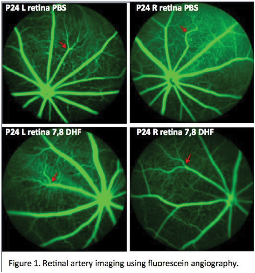
Results: All patients showed the abnormal high BT and the significant correlation was observed between BT and FA (rho = 0.542, p = 0.0302). Three patients who showed delayed neuropsychiatric sequelae after the subacute phase had already showed low BT relative to the others and severe WM damage at the subacute phase (black circle, Figure. 1).
Conclusions: BT could alter with the degree of WM damage in patients with the subacute CO-poisoning.
References:
1. De Reuck J, et al. Journal of neurology 1993.
2. Fujiwara S, et al. Neuroradiology 2015.
[Figure 1]
PS04-002
Poster Viewing Session IV
Immersive Medical Media - a platform for dynamic exploration of automatic, subject-specific atlases from standard medical images
G. Hartung1, G. Xu1 and A. Linninger1
1University of Illinois at Chicago, Chicago, United States
Abstract
Objectives: Unfortunately, the information embedded in medical images is currently limited to diagnostics and often merely archived. Currently no immersive visualization techniques allow subject-specific, real-time rendering of medical imaging data in a virtual space with interactive exploration. Immersive Medical Media will amplify the accessibility of cutting edge scientific and medical data within these images by incorporating automated feature recognition software to identify different structures and sub-regions of the brain into virtual reality hardware with immersive exploration tools.
Methods: We have utilized the graphics engine Unity3D where we created custom algorithms to parse medical imaging data, perform anatomical reconstructions, measure a variety of anatomical features from sub-structures of the brain, and independently modify the display properties and location of the reconstructed sub-structures. The images are visualized with our novel volume rendering technique allowing user modification of color and transparency for voxels belonging to individual sub-structures.
Results: We have achieved a level of performance that allows reconstructions to be performed within 10 minutes of the MRI, CT or PET scan using standard desktop hardware. We have also created a platform for immediate plug-and-play interfacing with all virtual/augmented reality devices as well as all hand and body gesturing tools.
Conclusions: This platform allows decreased time needed for patient image review, increased accessibility to anatomical measurements of patient-specific brain structures and enables the design of patient-specific medical devices such as 3D printed prosthesis and stents. This platform will allow large-scale epidemiological studies across multiple medical centers or research institutions nearly effortlessly to assist in generating quantitative standards by which to diagnose patients.A sample of this platform can be seen at https://www.youtube.com/watch?v=ALj_Hzv64AY.
PS04-003
Poster Viewing Session IV
Nonspreading depression followed by spreading depolarization in patients with cardiac arrest and analysis of platinum-iridium electrode interferences with oxygen and pH
J. Dreier1, S. Major1 and J. Hartings2
1Charité - University Medicine Berlin, Center for Stroke Research, Berlin, Germany
2University of Cincinnati, Department of Neurosurgery, Cincinnati, United States
Abstract
Spreading depolarizations (SDs) are propagating waves of neuronal and glial depolarization that either follow nonspreading depression or induce spreading depression of the spontaneous electrocorticographic (ECoG) activity. They are characterized by abrupt near-complete breakdown of ion homeostasis, acidification, efflux of excitatory aminoacids and loss in free energy. SDs occur abundantly in patients with stroke and traumatic brain injury (TBI). Here we monitored the electrophysiological changes in patients with either subarachnoid hemorrhage or TBI during cardiac arrest. Direct current (DC) and alternate current (AC)-ECoG was performed with either subdural electrode strips (n = 3) or depth electrodes (n = 4). In order to be able to analyze the ECoG, electrode interferences were investigated using a recording chamber with commercially available platinum-iridium (Pt/Ir) plate electrodes as used for the human recordings. In this setup we assessed the effects on the measured DC potential: (i) by electrode polarization when 5 min long, negative, square potential pulses were applied, (ii) by changes in partial pressure of oxygen, and (iii) pH. Pt/Ir electrodes were compared with a silver chloride electrode. We found that Pt/Ir-electrodes in contrast to a silver chloride electrode showed a positive shift with a median of 45.8 mV (1st quartile: 41.9 mV, third quartile: 49.8 mVmV, n = 6 Pt/Ir-electrodes in 3 experiments) during acidification by one unit of pH, which typically occurs in animals during SD including terminal SD. An increase of the partial pressure of oxygen from 0 to 136 mmHg caused a similar positive DC shift with a median of 44.0 mV (41.7 mV, 44.7 mV, n = 6/3). The initial DC response of Pt/Ir electrodes to a rectangular voltage stimulus of -44.6 mV was -39.6 mV (-39.4 mV, -39.8 mV, n = 6/2) with a rapid decay to -16.9 mV (-15.3 mV, 17.1 mV). Cardiac arrest in patients triggered nonspreading depression of spontaneous activity followed by terminal SD which was superimposed on giant DC shifts that likely result from changes in oxygen and pH.
PS04-004
Poster Viewing Session IV
Pharmacological and behavioral interventions for focus: The comparison of reversal learning under the influence of Lysergic Acid Diethylmide (LSD), Methylphenidate (MPH) and Mindfulness
and A. Zhuparris1
1Radboud University, Nijmegen, Netherlands
Abstract
Cognitive flexibility is the ability for one to adjust to unexpected and novel changes in the environment. Lysergic Acid Diethylamide (LSD), from anecdotal evidence, promotes divergent thinking, a more flexible, original and flowing thinking process (McGlothlin, 1963). Whilst Methylphenidate (MPH) promotes more convergent thinking, which tends to be more logical, rigid and less diverse (Tucha, 2011). Mindfulness has shown to both promote attention and cognitive flexibility (Moore, 2009). Hence, we've compared the effects of LSD, MPH and Mindfulness on cognitive flexibility. The Probabilistic Reversal Learning (PRL) task requires a subject to learn a rule in which there are three stimuli that are associated with the probabilistic feedback of either a reward (75 % of the time), punishment (75 %) or neutral responses (50 %). Midway through the experiment, this rule is reversed, so that subjects must now learn a new rule. From this it is possible to assess three aspects of cognitive functions: reward sensitivity (the tendency to repeat a response after a win, known as win-staying), punishment sensitivity (lose-shifting) and perseveration (tendency to remain on the same response, even after a punishment). Each subject's reaction time, perseveration error, and successful learning criterion were assessed. The data was analyzed using a mixed model as well as computational modelling to estimate how subjects integrate information on a trial-by-trial basis. Furthermore posterior predictive checks were used to assess whether the model captures observed effects from the raw data. Although there was no significance amongst the differences in the interventions, there was a trend in which LSD improved reversal learning and reduced perseveration, whilst MPH had the opposite effect. Mindfulness training lead to improved PRL scores, and PRL scores prior to the Mindfulness training were predictive of the outcome of the Mindfulness Skills scores.
PS04-005
Poster Viewing Session IV
Prediction of new cerebral ischemic events after endarterectomy for symptomatic unilateral internal carotid artery stenosis using crossed cerebellar hypoperfusion on preoperative brain perfusion
K. Oikawa1, K. Ogasawara1, H. Saito1, K. Yoshida1, H. Saura1, Y. Sato1, K. Terasaki2, T. Wada1 and Y. Kubo1
1Iwate Medical University, Department of Neurosurgery, Morioka, Japan
2Iwate Medical University, Cyclotron Research Center, Takizawa, Japan
Abstract
Objectives: Crossed cerebellar hypoperfusion (CCH) is defined as a reduction of blood flow in the cerebellar hemisphere contralateral to a supratentorial lesion1. By using the ratio of blood flow contralateral-to-affected asymmetry in the cerebellar hemisphere to blood flow affected-to-contralateral asymmetry in the middle cerebral artery (MCA) territory (ARcbl/ARMCA), brain perfusion can detect misery perfusion in the affected cerebral hemisphere in patients with unilateral occlusion of the middle cerebral artery or internal carotid artery (ICA). The aim of the present study was to determine whether ARcbl/ARMCA on preoperative brain perfusion images could identify patients at risk for new cerebral ischemic events after carotid endarterectomy (CEA) in patients with symptomatic unilateral ICA stenosis.
Methods: Brain perfusion was performed to assess brain blood flow in 101 patients with unilateral ICA stenosis. Using a 3-dimensional stereotaxic region-of-interest (ROI) template, ROI was automatically placed in the bilateral MCA territories and in the bilateral cerebellar hemispheres on the brain perfusion images after the standardization by SPM. Diffusion-weighted MRI was performed before and 24 hours after surgery. We defined the development of new postoperative ischemic lesions and neurological deficits before induction of general anesthesia and after recovery from general anesthesia as new cerebral ischemic events after CEA.
Results: In 12 of 101 patients (12 %), new cerebral ischemic events after CEA were observed. Multivariate analysis showed only ARcbl/ARMCA significantly was associated with the development of new postoperative cerebral ischemic events (P = 0.0070). The ARcbl/ARMCA provided 75 % sensitivity, 84 % specificity, 39 % positive predictive values, and 96 % negative predictive values in predicting development of new postoperative cerebral ischemic events.
Conclusions: The ARcbl/ARMCA on preoperative brain perfusion could identify patients at risk for new cerebral ischemic events after CEA for unilateral ICA stenosis.
Reference:
1. Komaba Y, et al. Stroke 2004.
PS04-006
Poster Viewing Session IV
Systemic inflammation impacts on central inflammatory changes and outcome after cerebral ischemia in stroke patients and experimental animals: a bench to bedside study
F.M. Vásárhelyi-Nagy1, N. Lénárt1, L. Csiba2, T. Hortobágyi3 and A. Denes1
1Laboratory of Neuroimmunology, Institute of Experimental Medicine, Hungarian Academy of Sciences, Molecular and Developmental Neurobiology, Budapest, Hungary
2University of Debrecen, Department of Neurology, Debrecen, Hungary
3University of Debrecen, Department of Neuropathology, Debrecen, Hungary
Abstract
Objectives: Systemic inflammation contributes to infavourable outcome in patients with cerebrovascular disease, but the mechanisms involved are poorly understood. It is also unclear whether systemic inflammation is associated with altered inflammatory responses in the brain of stroke patients and whether these changes are similar to those seen in experimental animals.
Methods: To investigate the potential cerebral effects of peripheral inflammation, patients with ischemic stroke and elevated systemic inflammatory burden (increased total white blood cell count, elevated erythrocyte sedimentation rate, C-reactive protein levels and/or evidence of infection at admission) were compared with stroke patients without an evidence of acute systemic inflammation and with low levels of inflammatory markers. Immunohistochemistry to detect neuronal injury and various markers of inflammation was performed on post mortem, paraffin embedded tissue samples. Data from clinical studies were compared with those from mice with or without systemic inflammation induced by lipopolysaccharide 2 h prior to experimental stroke.
Results: Systemic inflammation resulted in worse neurological outcome after experimental stroke, which was associated with microglial activation, increased granulocyte recruitment, and larger BBB injury in addition to elevated levels of proinflammatory cytokines in the brain. In stroke patients, white blood cell counts showed correlation with survival and correlations between blood parameters and microglial activation in the brain were also observed. Systemic inflammation was associated with increased recruitment of granulocytes into the area of the infarct. We also reveal changes in microglia / macrophages in response to systemic inflammation and stroke in patients and show that a population of these cells express markers of inflammasomes.
Conclusions: To our knowledge, this is the first study to systematically evaluate inflammatory changes in the human brain induced by systemic inflammation and stroke. Our data could support the translation of research findings into clinical benefit by facilitating the development of better diagnostic and therapeutic tools.
PS04-007
Poster Viewing Session IV
Detection of misery perfusion in patients with chronic unilateral major cerebral artery steno-occlusive disease using crossed cerebellar hypoperfusion on 123I-IMP single-photon emission computed tomography imaging
Y. Matsumoto1, K. Ogasawara1, H. Saito1, K. Terasaki2, Y. Takahashi1, Y. Ogasawara1, M. Kobayashi1, K. Yoshida1, T. Beppu1, Y. Kubo1, S. Fujiwara1, E. Tsushima3 and A. Ogawa1
1Iwate Medical University, Department of Neurosurgery, Morioka, Japan
2Iwate Medical University, Cyclotron Research Center, Morioka, Japan
3Hirosaki University, Graduate School of Health Sciences, Hirosaki, Japan
Abstract
Objectives: In patients with unilateral internal carotid or middle cerebral artery (ICA or MCA) occlusive disease, the degree of crossed cerebellar hypoperfusion (CCH) that is evident within a few months after the onset of stroke may reflect cerebral metabolic rate of oxygen in the affected cerebral hemisphere relative to that in the contralateral cerebral hemisphere1. The aim of the present study was to detect misery perfusion on 15O positron emission tomography (15O-PET) imaging in patients with chronic occlusive disease of unilateral ICA or MCA using CCH observed on the brain N-isopropyl-p-[123I] iodoamphetamine (123I-IMP) single-photon emission computed tomography (SPECT) imaging.
Methods:15O-PET and 123I-IMP SPECT were performed for assessment of oxygen extraction fraction (OEF) or cerebral and cerebellar blood flow. All images were anatomically standardized using SPM. A region of interest (ROI) was automatically placed in the bilateral MCA territories and in the bilateral cerebellar hemispheres using a three-dimensional stereotaxic ROI template. Then, affected-to-contralateral asymmetry ratio in the MCA territory (ARMCA) and contralateral-to-affected asymmetry ratio in the cerebellar hemisphere (ARcbl) were calculated on the images from each modality.
Results: A significant correlation was observed between ARMCA of PET-OEF and ARcbl/ARMCA on IMP-SPECT (r = 0.46, p = 0.0001). The correlation coefficient was higher when reanalyzed in a subgroup of 43 patients undergoing a PET study within 3 months after the last ischemic event (r = 0.61, p < 0.0001). ARcbl/ARMCA of IMP-SPECT in all patients provided 100 % sensitivity and 58 % specificity, with 43 % positive and 100 % negative predictive values to detect abnormally elevated ARMCA of PET-OEF.
Conclusions: The ratio of cerebellar blood flow asymmetry to cerebral blood flow asymmetry observed on 123I-IMP SPECT can detect misery perfusion observed on the affected cerebral hemisphere in patients with chronic occlusive disease of unilateral ICA or MCA.
References:
1. Komaba, et al. Stroke 2004.
PS04-008
Poster Viewing Session IV
Prediction of hyperperfusion after carotid artery stenting and carotid angioplasty using cerebral circulation time using syngo iFlow
K. Yamauchi1, Y. Enomoto1, Y. Egashira1, N. Nakayama1, S. Yoshimura2 and T. Iwama1
1Gifu University Graduate School of Medicine, Neurosurgery, Gifu, Japan
2Hyogo College of Medicine, Neurosurgery, Nishinomiya, Japan
Abstract
Background: Hyperperfusion syndrome (HPS) is one of the main complications after carotid artery stenting (CAS) and carotid angioplasty. The prediction of HPS is important for the prevention of HPS and the improvement of outcomes.
Objective: Syngo iFlow is the application software for digital subtraction angiography which provides the visualization of flow dynamics and enables us to evaluate the cerebral circulation. The aim of this study is to evaluate the usefulness of the cerebral circulation time (CCT) measured by syngo iFlow for the prediction of HPS after CAS.
Methods: 32 CAS / carotid angioplasty procedures in 29 patients between Feb 2014 and Dec 2015 were included. The CCT was defined as the time difference between the relative time to maximal intensity of arterial ROIs (proximal internal carotid artery) and venous ROIs (straight sinus or transverse sinus). Hyperperfusion phenomenon (HPP) was diagnosed with qualitative SPECT immediately after the procedure and defined as a CBF increase of more than 100% as compared with the normal side.
Results: The HPP was observed in 4 (12.5%) procedures. There was no HPS. The pre-procedural CCT in patients with HP was prolonged compared with patients without HP (9.7 ± 2.6 v.s 7.2 ± 1.4; mean (sec) ± SD; p = 0.045). The change of peri-procedural CCT (ΔCCT) was significantly greater in patients with HP (3.6 ± 1.0 v.s 0.5 ± 1.7;mean (sec) ± SD; p = 0.016).
Conclusions: Prolonged pre-procedural CCT and greater ΔCCT were associated with HP. The periprocedural evaluation of cerebral circulation time using syngo iFlow is useful for the prediction of HPP.
PS04-009
Poster Viewing Session IV
Increase of rCBF in low-perfusion area (L-pa) after extracranial-intracranial bypass is achieved regardless of targeting recipient artery (Ra) in non-moyamoya diseases (NMMD)
H. Katano1,2 and M. Mase1
1Nagoya City University Graduate School of Medical Sciences, Neurosurgery, Nagoya, Japan
2Nagoya City University Graduate School of Medical Sciences, Medical Informatics and Integrative Medicine, Nagoya, Japan
Abstract
Background: There are some differences in the methods and the perioperative assessment of superficial temporal artery - middle cerebral artery anastomoses (STA-MCA-A) among operators or centers.
Method: Sixty-four patients experienced STA-MCA-A (NMMD 34, 60.8 ± 11.8 y/o, ICA/MCA occlusion showing Stage II area; MMD 30, 35.3 ± 18.1 y/o) were investigated. Choice for single/double bypass and Ra was up to each operator. Group(Gr)I (operators A/B) preoperatively decided the target(t-)Ra with SPECT, while GrII (operators C/D) intraoperatively chose the appropriate Ra judging from its diameter, location and wall color. Results of the bypass were evaluated postoperatively by MRA, DSA (in NMMD, GrI only) and SPECT with normalization, compared with the results of MMD patients.
Result: In NMMD patients, single bypass was performed in 68.8 / 77.8 % (GrI/II). The patency of the bypass was confirmed by MRA in 93.8 / 100 %. Intraoperatively selected artery matched postoperative MRA in 80.0/77.8% and DSA in 84.6 % (cf. In MMD patients, 71.4 / 64.3 % and 70.0 / 55.6 %, respectively). MRA findings matched DSA in 76.9 %, while cases that intraoperatively selected artery did not match MRA but DSA was 38.5 %. Postoperative increased CBF areas matched with perfused areas of anastomosed arteries on MRA:DSA were found in 85.7/77.8 % : 86.9 %. As for 6 cases that mismatched between the intraoperatively selected artery and the MRA finding, increased CBF on the preoperative L-pa were observed in 5 (83.3 %). Any stroke or bypass occlusion occurred in perioperative period, 30 days and 6 months in 12.5/0, 18.8/0, 18.8/0 %. A patient in GrI showed stroke due to late allergy shock with contrast media.
Conclusion: Successful bypass to the directed Ra was confirmed by MRA around 80 % of the STA-MCA-A in both preoperatively aimed and non-aimed groups for NMMD patients. If preoperative accurate targeting to the Ra can hardly have serious meaning, DSA confirming anastomosis to the t-Ra precisely should be avoided to prevent dismal complications in NMMD.
PS04-010
Poster Viewing Session IV
Intracranial venous pulsatility is reduced after transverse sinus stenosis
A. Guenego1, A.C. Januel1, P. Tall1, N. Fabre2, Z. Czosnyka3, C. Cognard1 and E. Schmidt4
1University Hospital, Neuroradiology, Toulouse, France
2University Hospital, Neurology, Toulouse, France
3Brain Physic Lab, Neurosurgery, Cambridge, United Kingdom
4University Hospital, Neurosurgery, Toulouse, France
Abstract
Introduction: Transverse sinus stenosis is seen in the majority of patients with idiopathic intra-cranial hypertension (IIH). In case of a significant pressure gradient, transverse sinus is stented to reduce cerebral venous pressure, improve CSF resorption, reduce ICP and papilledema. However the pathogenesis of sinus stenosis and its effect on the cerebral venous system remain controversial. We hypothesize that transverse sinus stenosis stenting modifies the dynamic component of venous pressure.
[Table 1]
Methods: 10 IIH patients were prospectively enrolled. Under general anesthesia, a microcatheter was navigated into cerebral veins and sinuses. Intra sinus pressure was measured and recorded upstream and downstream the stenosis with a pressure transducer connected to the micro catheter. A stent was placed if a significant pressure gradient (>10 mmHg) was found across the stenosis. Finally pressure was measured upstream and downstream the stented stenosis. Off line mean venous pressure (VP) was calculated and waveform analyses were performed to extract fundamental harmonic A1 (heart-rate), second harmonic A2 (2*heart-rate), and respiratory component (Resp). Parameters are presented before and after stenting (mean ± SD) upstream and downstream the stenosis with p value (Kruskal-Wallis test).
Results: Table 1 displays venous pressure values of various indices upstream and downstream the stenosis, before and after stenting with percentage change and significance value. In our group, stenting significantly reduces mean venous pressure upstream the stenosis, which is expected. Stenting also significantly reduces fundamental harmonic A1 upstream with a trend in A1 decrease downstream the stenosis.
Conclusions: Transverse sinus stenosis stenting reduces mean venous pressure and venous pressure pulsatility. This might have indirect effect on CSF dynamics and ICP amplitude. Sinus stenting yields complex venous pressure profile changes and possibly in brain biomechanics.
PS04-011
Poster Viewing Session IV
Diagnosis of ventriculostomy-related infection: is cerebrospinal fluid lactate measurement a useful tool?
External ventricular drains (EVD) are devices commonly used in neurocritical care patients. Ventriculostomy related infection (VRI) is a serious complication of EVD. Its diagnosis is controversial due to: low sensitivity and specificity of CSF markers, negative CSF cultures because of antibiotic inhibition and lack of a standardized definition.
Objective: To evaluate the value of CSF lactate (LCSF) for the diagnosis of VRI, and compare it with other CSF markers.
Methods: Prospective study of neurocritical patients admitted to Maciel and Clinicas Hospital ICUs in which a EVD was inserted. In patients with clinical suspicion of VRI, a CSF sample was obtained through the EVD and submitted for CSF culture and markers analysis (glucose, protein, lactate and leukocytes). We defined VRI according to preset criteria as: fever, plus CSF alterations (glucose < 50 mg/dl or leukocytes > 500/ul), plus positive CSF culture. CSF markers were plotted in a receiver operating curve (ROC) to evaluate their diagnostic accuracy.
Results: 32 CSF samples were obtained: 12 corresponded to proven VRI and 20 to excluded VRI. Mean LCSF was 9.77 ± 5.24 mM for proven VRI and 3.16 ± 1.12 mM for excluded VRI (p = 0.0012). Both LCSF and CSF glucose showed a good diagnostic accuracy for VRI, with an area under the curve (AUC) of 0.873 and 0.933 respectively. We found the following diagnostic values for LCSF: sensitivity of 75 %, specificity of 89 %, PPV of 81 %, NPV of 85 %, cut-off value of 4.5 mM, positive likehood ratio of 6.8, negative likehood ratio of 0.28, Diagnostic Odds ratio of 25.6, accuracy test of 83 %, and Youden Index of 0.64.
Conclusions: Our current results show that LCSF represents a good marker for VRI, which could be a quick and specific test to identify the need for antimicrobial therapy in patients with clinical suspicion of VRI.
PS04-012
Poster Viewing Session IV
Incidence and types of arrhythmia in adult patients with uncorreected atrial septal defect
and M. Primasari1
1Faculty of Medicine Gadjah Mada University, Cardiology, Yogyakarta, Indonesia
Abstract
Background: There are high incidence of arrhythmia among adults patients with atrial septal defect (ASD). Arrhythmia, especially atrial filbrillation are well documented sequelae of ASD, associated with substantial morbidity, increasing risk of stroke and, occasionally, death. It is important to know the incidence and arrhythmia types in ASD patients, so that prevention or restoration of sinus rhythm in this defect is therefore desirable.
Objective: To know the incidence and arrhythmia types in uncorrected atrial septal defects (ASD) adult patients.
Method: A retrospective study was conducted using secondary data derived from registry of atrial septal defects patients at Cardiology Department of Sardjito Hospital, Yogyakarta, from July 2012 to December 2014.The inclusion criteria were all uncorrected ASD adults patients (18 - 60 years old) and having arrhythmia disorders. Patients with corrected ASD or had concomitant congenital heart disease were excluded.
Results: Ostium secundum accounts for 98 % of atrial septal defect types. In uncorrected atrial septal defects adult patients, the incidence of arrhythmia was 14.93 %, and female suffered from arrhythmia were more frequent than male (78.26 % vs 21.73 %). The most frequent arrhythmia types were atrial fibrillation (30.43%), followed by sinus arrhythmia (13.04 %) and AV block (13.04 %). The incidence of arrhythmia increases with age ; < 20 years of age (0 %), 20 - 40 years of age (5.84 %) and > 40 years of age (9.89 %).
Conclusions: The incidence of arrhythmia of adult atrial septal defect patients was 14.93 %, and female suffered from arrhythmia were more frequent than male. From the number patients with arrhythmia the most frequent arrhythmia types were atrial fibrillation, followed by sinus arrhythmia and AV block. The incidence of arrhythmia increases with age.
PS04-013
Poster Viewing Session IV
The low hSOD1 transgene copy number mice as an animal model dedicated to cell replacement strategies
M. Majchrzak1, L. Stanaszek1, P. Walczak2, M. Janowski1,2 and B. Lukomska1
1Mossakowski Medical Research Centre PAS, NeuroRepair Department, Warsaw, Poland
2Johns Hopkins University School of Medicine, The Russell H. Morgan Department of Radiology and Radiological Science, Baltimore, United States
Abstract
There is an increasing agreement that glial cells contribute to the premature death of motor neurons - a hallmark of ALS. Thus, global replacement of glial cells is a valid therapeutic strategy. The positive effects of stem cell therapies in mouse models of ALS have been observed by many groups, but no cure has been achieved. Actually, the time from human GRP transplantation to the myelin basic protein expression lasts 3–6 months. The life-span of most popular high copy number hSOD1 mice (130 days) might be too short to realize the full advantages of transplanted stem cells for the global replacement of potentially defective and toxic host glia. Thus, we focused on developing immunodeficient rag2-/-, long-living mice with a low copy number of the hSOD1 gene and a longer life-span. The obtained double-mutant hSOD1/rag2-/- mice have been characterized genetically and behaviorally. Quantitative PCR performed against the standards revealed that progeny in our colony had either 4 or 8 copies of hSOD1 gene. The death of long- and short-living hSOD1/rag2-/- mice is preceded by muscular weakness as measured by hind limb test as early as one month before the onset of the disease. Importantly, we were able to see the difference in magnetic resonance imaging in medulla, especially in motor related nucleus in terminal stage of the disease. To conclude, we developed long-living hSOD1/rag2-/- mice, which could be very attractive for testing therapeutic utility of human stem cells for glia replacement as the treatment of ALS. The correlation of copy number and the lifespan potentially allows for a high prediction of animal survival. Thus, the animals may serve as their own controls obviating the need for an additional control group reversely translating the concept of precision medicine to the preclinical research.
Supported by a NCR&D grant for STRATEGMED project: “GRP&ALS”
PS04-014
Poster Viewing Session IV
Effects of Na,K-ATPase alpha isoform deficiency on spreading depolarization studied in mice
C. Reiffurth1,2, M. Alam3, M. Zahedi-Khorasani4 and J.P. Dreier1,2,5
1Charité - University Medicine Berlin, Center for Stroke Research, Berlin, Germany
2Charité - University Medicine Berlin, Department of Experimental Neurology, Berlin, Germany
3Hannover Medical School, Department of Neurosurgery, Hannover, Germany
4University of Medical Sciences, Laboratory of Cerebrovascular Research & Physiological Research Center, Semnan, Iran, Islamic Republic of
5Charité - University Medicine Berlin, Department of Neurology, Berlin, Germany
Abstract
Objectives: The Na,K-ATPase plays a central role in modulating threshold and recovery of spreading depolarizations (SD) in the brain. Mutations in ATP1A2, the gene encoding the Na,K-ATPase alpha2 subunit, are associated with familial hemiplegic migraine type 2 (FHM2). It has been hypothesized that spreading depolarization might be facilitated by a loss of function of a single allele of the gene encoding the alpha2 subunit. To address the question whether a reduction of the alpha2 isoform affects the threshold for SD ignition we employed heterozygous knockout mice lacking one copy of the alpha2 subunit encoding allele (alpha2+/−) and provoked SD by various stimuli.
Methods: In acute brain slices, SD was triggered focally by droplet application of 1 M KCl solution, by electrical stimulation or by stepwise increasing the K+ concentration in the bathing solution. We recorded changes in extracellular K+ concentration, the accompanying slow extracellular potential shift, as well as changes in intrinsic optical signals to assess spatiotemporal patterns. To further investigate whether the observed effects were specific for a reduced amount of the alpha2 isoform, alpha1 and alpha3 heterozygous (alpha1+/− and alpha3+/−) mice were included in this study.
Results: In response to prolonged extracellular K+ exposure, we observed a significantly lowered (P < 0.001) threshold concentration necessary to trigger SD in alpha2+/− mice (13,03 ± 1,24 mmol/l, n = 18) compared to their wild-type littermates (14,92 ± 1,59 mmol/l, n = 23). This was reflected by a shortening of the wash-in time needed to induce SD. No statistically significant reduction in threshold concentration was found in alpha1+/− or alpha3+/− mice compared to their wild-type littermates indicating that the observed effect in the alpha2 group is specific for this isoform.
Conclusion: Different catalytic Na,K-ATPase alpha isoforms have distinct functional properties and functional haploinsuffiency may underlie increased susceptibility to SD in FHM2.
PS04-015
Poster Viewing Session IV
Anaplerotic triheptanoin preserves mitochondrial function and reduces oxidative stress in pilocarpine-induced status epilepticus
K.N. Tan1, C. Carrasco-Pozo1,2 and K. Borges1
1University of Queensland, School of Biomedical Sciences, Brisbane, Australia
2University of Chile, Department of Nutrition, Santiago, Chile
Abstract
Objectives: Growing evidence suggests that mitochondrial dysfunction contributes to the pathophysiology of epilepsy. Thus, preservation of mitochondrial function would be an ideal treatment strategy. Triheptanoin, a medium-chain triglyceride with three seven-carbon fatty acid molecules, was previously found to be anticonvulsant in several mouse seizure models and is currently undergoing clinical trials in patients with refractory epilepsy. The ability of triheptanoin to refill four-carbon intermediates of the TCA cycle (anaplerosis) is hypothesised to improve or preserve mitochondrial function. Here we investigated mitochondrial function and oxidative stress in mouse brains after treatment with triheptanoin using pilocarpine-induced status epilepticus (SE).
Methods: Mice were given control or 35 % (% calories) triheptanoin diet for ten days prior to pilocarpine injection (340 mg/kg; s.c.). Blood plasma and bilateral hippocampal formations were collected 24 h post seizure induction before significant neuronal death occurs. Mitochondrial function was assessed using an extracellular flux XFe96 analyzer based on oxygen consumption rate. Markers of oxidative stress were measured in the plasma and hippocampal formations based on antioxidant capacity of the plasma and lipid peroxidation respectively.
Results: Various mitochondrial function parameters including state 2, state 3 ADP, state 3 u respiration and respiration linked to ATP synthesis in the hippocampal formations of control-fed SE mice were reduced by 32 %, 26 %, 23 % and 28 % respectively compared to no SE mice (p < 0.05; n = 3–8 mice/group each). Antioxidant capacity in the plasma was also reduced by 13 % (p < 0.05) while lipid peroxidation was doubled (p < 0.0001; n = 3–8 mice/group each). Triheptanoin treatment abolished all the changes observed in control-fed SE mice and the protective effects were reproduced in another independent experiment (n = 5–8 mice/group each).
Conclusions: Triheptanoin preserves mitochondrial function and prevents oxidative stress in mouse hippocampal formations 24 h post SE. We are currently investigating whether triheptanoin is neuroprotective by assessing neuronal death three days post SE.
PS04-016
Poster Viewing Session IV
Amyloid-β- and tau-driven neurovascular dysfunction in a transgenic rat model of Alzheimer's disease
I. Joo1, A. Lai1, M. Koletar2, A. Dorr1, P. Bazzigaluppi1, M. Brown3, L. Thomason1, J. Sled4, J. Mclaurin3 and B. Stefanovic3
1Sunnybrook Research Institute, Toronto, Canada
2Sunnybrook Research Institute, Medical Imaging Research, Toronto, Canada
3Sunnybrook Health Sciences Centre, Toronto, Canada
4Hospital for Sick Children, Toronto, Canada
Abstract
Alzheimer's disease (AD), pathologically hallmarked by progressive amyloid-β (Aβ) peptide deposition, neurofibrillary tangle (NFT) formation, and neurodegeneration, is also associated with early-onset microvascular abnormalities. Despite their potential significance for early diagnostics and disease progression, much uncertainty still surrounds the mechanisms of cerebrovascular injury in AD. In the current study, we set out to investigate presymptomatic AD-associated structural alterations and functional impairment in cerebrocortical microvascular bed using TgF344-AD rat model that exhibits Aβ-peptide accumulation, NFT formation, and frank neuronal loss in addition to cognitive decline. Using in vivo linear array recordings and two-photon fluorescence microscopy, we examined in situ functioning of the neuronal and the cerebrovascular networks of the somatosensory cortex and related them to amyloidosis, tau hyperphosphorylation, and cerebrovascular wall morphology, as assessed on post mortem pathological analysis. The TgF344-AD rats exhibited significant amyloid deposition in the tissue and on the vasculature, significant hyperphosphorylation (doubled compared to nTg) and an impairment in cortical cross-frequency coupling, as evidenced by 60 % reduction in the modulation index of theta band phase on gamma band amplitudes (TgF344-AD: 18.1 ± 4.9 (mean ± SEM) vs. nTg: 45.2 ± 8.9; p = 0.029). Although mural cell and blood-brain barrier integrity were maintained, TgF344-AD rats exhibited 44 % reduction in arteriolar reactivity (TgF344-AD: 8.0 ± 1.8 % vs. nTg: 15 ± 2.7 %; p = 0.056) and 51 % reduction in venular reactivity (7.3 ± 2.4 % vs. 15 ± 2.8 %; p = 0.044) to CO2 challenge when compared to nTg rats. Moreover, cerebral blood flow increase to hypercapnia was abolished in TgF344-AD rats. In total, TgF344-AD rats show significant neurovascular network dysfunction in the presymptomatic stage of the disease progression. These findings underscore the significance of studying neurovascular network functioning in vivo, suggesting a new target for early diagnosis and preemptive treatment plans for AD.
PS04-017
Poster Viewing Session IV
Effects of arginine vasopressin on hippocampal network activity in a rat model of birth asphyxia
E. Prokic1, M. Summanen1, H. Hartung1, A. Alafuzoff1, J. Voipio1 and K. Kaila1
1Helsinki University, Department of Biosciences, Helsinki, Finland
Abstract
Activation of the stress axis during birth is critical to protect the fetus during delivery and for adaptation to extrauterine life. A surge of arginine vasopressin (AVP) takes place during delivery in fetal blood. This surge is exaggerated during birth asphyxia. Furthermore, birth asphyxia is associated with pathophysiological changes in the EEG, including either suppression of activity or manifestation of seizures, which are predictive of a poor neurodevelopmental outcome. The mechanisms underlying brain trauma following birth asphyxia remain unclear and it is unknown whether (i) the AVP surge takes place in vulnerable hippocampal-cortical structures and (ii) whether AVP has a protective role.
Here, we use a model where P6 rat pups are exposed for 45 minutes to asphyxic conditions (4–5% O2, 20% C02) followed by 2 hours of recovery in room air. We measured the levels of copeptin, the C-terminal fragment of prepro-AVP, with AlphaLISA. Copeptin is much more stable than AVP and therefore easier to measure. In addition, the impact of asphyxia and recovery on neuronal network activity was assessed in in vivo hippocampal local field potential recordings under urethane anaesthesia.
The highest plasma copeptin concentrations were observed at 15 minutes of asphyxia, after which copeptin concentrations decreased until a plateau was reached at 30 minutes post-asphyxia. There was no change in plasma copeptin in control pups. During asphyxia, field activity in the hippocampus was silenced. When switched to room air, network activity returned immediately, showing an overshoot from baseline in the frequency of events that waned over the recovery period. These effects were mimicked by intracerebral injection of AVP. Strikingly, intracerebral injection of SR49059, a specific V1aR antagonist, exacerbated the occurrence of epileptiform events at the start of asphyxia. The present data suggest a neuroprotective role for AVP during birth asphyxia.
PS04-018
Poster Viewing Session IV
Kinsbourne syndrome - opsoclonus myoclonus ataxia (clinical case report)
M. Azhermacheva1 and V. Alifirova1
1Siberian State Medical University, Department of Neurology and Neurosurgery, Tomsk, Russian Federation
Abstract
Opsoclonus myoclonus ataxia (also called Kinsbourne syndrome ) is an autoimmune disease of central nervous system. Kinsbourne syndrome is an extremely rare condition, affecting as few as 1 in 10,000,000 people per year. The present study reported the case of OMS in 29-years-old woman. The patient was examined and treated in neurological clinic of Siberian state medical university, Tomsk, Russia.
There have been provided a clinical examination, evaluation of neurological status, MRI of the brain, lumber puncture. The differential diagnosis was performed with hereditary spinocerebellar degeneration, neuroblastoma, infection diseases, multiple sclerosis, toxic encephalopathy, epileptic myoclonia.
The disease occurred after an acute respiratory infection (general weakness, fever up to 38°C, cough). At the beginning of the disease were observed autonomic disturbances: nausea, vomiting. Two weeks after were observed involuntary, rapid, multivectorial (horizontal and vertical) movement of the eyeballs, myoclonus of eyelids, chin and head. These forced movement intensified during looking at objects or medical examination. In neurological status: ataxic gait, walking only with support; seesaw motion in Romberg's position; intention tremor during coordination tests.
There were no changes in the cerebrospinal fluid, MRI brain.
For patient was been provided corticosteroid therapy and was been fond a positive response: nausea and vomiting disappeared, significantly reduced opsoklonus eyeballs and myoclonic hyperkinesia muscles of the face and tremor of the extremities,symptoms of ataxia went away and the patient began to walk without assistance.
Our observation were conform to all clinical symptoms of the disease, which we found in the literature:opsoclonus, polymyoclonia and cerebellar ataxia. This clinical case shows that Kinsbourne syndrome can develop not only in children but also in adult patients.
PS04-019
Poster Viewing Session IV
MRI imaging for sensitive tumour cell-tracking during development of early brain metastasis in a preclinical model
A. Corroyer-Dulmont1,2, S. Valable2, N. Falzone1, N.R. Sibson1, M. Bernaudin2 and K.A. Vallis1
1University of Oxford, CR-UK/MRC Oxford Institute for Radiation Oncology, Department of Oncology, Oxford, United Kingdom
Objectives: Breast cancer patients frequently develop brain metastases. Conventional treatment is palliative and often involves whole-brain radiotherapy. There is a pressing unmet therapeutic need to treat brain metastasis at an early stage, when relatively few tumor cells have invaded the brain parenchyma. However, to evaluate the efficacy of new therapeutic treatments, preclinical models of early stage brain metastases and tools to monitor response to treatment are needed. The aim of this study was to develop a model in which early brain metastases can be tracked by MRI using microparticles of iron oxide (MPIO).
Methods: MDA-231-BR, a human breast carcinoma cell line that preferentially metastasises to the brain, was used. For MPIO labelling, 5.105 cells were incubated for 24 h with a range of MPIO bead concentrations (0.5e109, 2.5e109 and 5e109). Internalisation of MPIO in cells and MDA-231-BR viability were assessed by fluorescence and flow cytometry. Four BALB/c nude mice received an intracardiac injection of MPIO-tagged cells using ultrasound-guidance. MRI, using Fast Imaging with Steady state Precession (FISP) and Rapid Acquisition with Relaxation Enhancement (RARE) (7T MRI, Pharmascan, Bruker, CYCERON platform), was performed to track labelled cells in the brain.
Results: Fluorescent and flow cytometry studies demonstrated maximal internalisation of MPIO beads in MDA-231-BR cells when the lowest tested concentration was used. Toxicity of beads was also observed across the concentration range. Given these results, the lowest concentration of MPIO was selected for in vivo studies. Using MRI (FISP sequence), metastatic cells were observed in the brain from 3 h after cardiac-injection. From Day 20 post injection (p.i.), T2w-sequence was used to visualize later phases of brain metastases (diameter > 420 µm).
Conclusions: In this study, we developed a sensitive, MRI-trackable model of early brain metastases which could be used to evaluate the treatment efficacy of novel therapy treatments.
PS04-020
Poster Viewing Session IV
A case of complicated migraine auras in temporal association with electrocorticographically recorded spreading depolarizations
S. Major1,2,3, D. Milakara1 and J.P. Dreier1,2,3
1Charité - Universitätsmedizin Berlin, Center for Stroke Research Berlin, Berlin, Germany
2Charité - Universitätsmedizin Berlin, Department of Experimental Neurology, Berlin, Germany
3Charité - Universitätsmedizin Berlin, Department of Neurology, Berlin, Germany
Abstract
In the last 15 years more than 500 patients with stroke and brain trauma underwent neuromonitoring of spreading depolarizations (SD) in neurocritical care within the framework of the Co-Operative Studies on Brain Injury Depolarizations (COSBID). Unequivocal electrophysiological evidence was found that SDs are involved in the pathophysiology of these human diseases in accordance with previous animal research. Preliminary clinical evidence in patients with subarachnoid hemorrhage (SAH) suggested moreover that clusters of SDs can be associated with waxing and waning and also permanent severe focal and global neurological deficits. However, Leão and Morison also proposed in 1945 that SD is the pathophysiological correlate of the migraine aura. For obvious reasons, it is not possible to implant the invasive recording tools for the measurement of SDs in patients before the occurrence of a migraine aura. Therefore, the electrophysiological proof whether or not SD is its pathophysiological correlate is still lacking. Yet, it is somewhat puzzling that not a single case report has described the patient percept of a migraine aura when SD was recorded in awake patients with cerebral injury. Here we present a 56-year old female who had suffered from SAH due to rupture of a left middle cerebral artery aneurysm. A subdural electrode array was implanted over the left frontal lobe after surgical clip ligation of the aneurysm. Eleven days after the initial hemorrhage the fully awake and oriented patient developed dysarthria followed first by facial paresis and then paresis of her right arm some minutes later in presence of a neurologist. Several minutes after symptom onset, SD was seen on the monitor propagating in caudal-rostral direction. Similar events recurred twice with full recovery in between and thereafter. To our knowledge, this is the first case report of complicated migraine auras in temporal association with electrocorticographically recorded SDs.
PS04-023
Poster Viewing Session IV
A web based meta-analysis of microdosing of psychedelic drugs as a form of nootropics
and A. ZhuParris1
1Radboud University, Nijmegen, Netherlands
Abstract
Objective: Testimonials dating back to the 1960s have described the microdosing of LSD to have nootropic properties such as cognitive enhancement, physical boost, increased creativity and mindfulness. Microdosing refers to ingesting subperceptual amounts of psychedelic drugs.
Research Design and Methods: This paper catalogued 600 reports from microdosing users from Reddit and Erowid. The drugs examined were Tryptamines (5-methoxy-N,N-dimethyltryptamine, Psilocybin),Ergolines, Phenethylamines (Mescaline, The 2C family, NBOMe derivative, 2,5-dimethoxy, 4-substituted amphetamines), Cannabinoids and Empathogens (Substituted methylenedioxy-phenthylamines). Self-reports of microdosing with prescribed nootropic drugs such as, psychostimulants (methylphenidate and amphetamine) and wakefulness-promoting agents (modafinil) were also collected. The data is composed of the methodology of the microdosing, which includes dosage, form of administration, tolerance and tolerance duration. Furthermore, the physiological and cognitive differs have been achieved, this encompasses sleep, side effects, appetite, motivation, concentration, memory, physical energy.
Results: 56 % of the users choose Ergolines for microdosing, 32 % with Tryptamines, and 12 % with Phenetylamines, Empathogens and Psychostimulants. Users experienced a variety of effects such as emotional clarity, enhanced senses, increased concentration and stamina. However there were several reports of reduced appetite, excessive sweating, reduced sleep, and digestive and warped time perception.
PS04-024
Poster Viewing Session IV
Reorganization patterns of cortical arm muscle representations: post-stroke longitudinal TMS study
L. Lazzouni1, A. Zumbansen1, H. Vogt1, P. Kramer1 and A. Thiel1
1McGill University/Jewish General Hospital, Montreal, Canada
Abstract
Patients suffering from stoke affecting motor cortex show different degrees of recovery over time. Transcranial Magnetic Stimulation (TMS) has been used to understand how motor recovery occurs after stroke. In the present study, we used TMS to map the cortical representation of upper limb muscles (first dorsal interosseous FDI, extensor carpi radialis ECR) in the affected and unaffected hemispheres in the acute phase post stroke and six months follow-up. We hypothesize that MEP mean amplitudes and surface areas of motor maps will show increase at follow-up as indicators of plastic changes during recovery.
Using TMS we mapped the FDI and ECR cortical representations in18 patients. We calculated the surface areas and the mean MEP amplitudes for all maps. We compared mean response amplitudes and surface areas, using repeated measures ANIOVAs with factors: Time, Side and Muscle and compared RMTs using paired t-tests. We used multiple linear regression to determine the variables predicting clinical scores at baseline, and change in clinical scores ( NIH and total Fugle Meyer) after six months.
In the absence of RMT change we found a significant increase of MEP amplitudes with Time in both hemispheres for the FDI, and in the affected hemisphere for the ECR. Surface areas increased significantly with Time for the FDI but not for the ECR. The ECR surface area at baseline was found to predict the change in clinical scores.
The increase in surface area and amplitude of the FDI in both hemispheres suggests a recruitment of ipsi- and contralesional neurons. The increase in map amplitude only for the ECR over time only in the affected hemisphere indicates a more ipsi-lesional reorganization. The size of the ECR surface area better predicts the change in clinical scores, those with larger muscle representation at baseline showing higher improvement six months after the stroke.
PS04-025
Poster Viewing Session IV
Longitudinal mapping of visual cortical network following partial optic nerve injury
M. Groleau1, M. Nazariahangarkolaee2, M.P. Vanni3, B.A. Sabel4, M.H. Mohajerani2 and E. Vaucher1
1Université de Montréal, Laboratoire de Neurobiologie de la Cognition Visuelle, École d'Optométrie, Montréal, Canada
2University of Lethbridge, Department of Neuroscience, Canadian Centre for Behavioural Neuroscience (CCBN), Lethbridge, Canada
3Brain Research Centre and Djavad Mowafaghian Centre for Brain Health, Department of Psychiatry, University of British Columbia, Vancouver, Canada
4Institute of Medical Psychology, Medical Faculty, Otto-v.-Guericke University of Magdeburg, Magdeburg, Germany
Abstract
Traumatic optic neuropathy damaging the optic nerve axons causes visual impairment and partial or complete loss of vision. Part of the retinal ganglions cells degenerate, but there is still a residual vision resulting from surviving retinal cells and cortical plasticity. The cortical network reorganization over time after an optic nerve injury is not known.
In the present study, we monitor the residual cortical function and its plasticity following a monocular partial Optic Nerve Crush (ONC) across time. The neuronal calcium response to a monocular flash illumination in each eye is measured using in vivo wide-field calcium imaging on awake mice (Thy1-GCaMP6s) at 1 hour, 1, 3, 7, 14 and 30 days after the injury in different areas of the visual cortical pathway. Long-range functional connections between areas of the visual cortex and high-order areas are mapped and the strength of the pathways during visual recovery is assessed.
The cortical activity of the visual cortex receiving input from the injured eye is not altered one hour and one day after the ONC compared to pre-ONC values, probably due to a progressive degeneration of the crushed axons. Changes in intra- and inter-hemispheric cortical responses are observed from the day 3 post-ONC. The visually-induced cortical response is weaker in the hemisphere receiving input from the injured eye compared to the pre-ONC values and to the opposite hemisphere. An inter-hemispheric shift of the visual cortical response is observed after stimulation of the injured eye.
In conclusion, our results show a reorganization of the connectivity between visual and associative cortical areas following a traumatic optic nerve injury which is indicative of visual cortical plasticity. Particularly, interhemispheric compensation is observed in the primary visual cortex. These results open an interesting avenue to manipulate visual recovery processes after a visual deficit.
PS04-026
Poster Viewing Session IV
Brain mechanisms of gait steering in young, normal subjects: a PET 18F-FDG PET study
J.-P. Soucy1,2, F. Starr3 and C. Paquette3
1McGill University, Neurology and Neurosurgery, Outremont, Canada
2Concordia University, PERFORM Centre, Montréal, Canada
3McGill University, Kinesiology and Physical Education, Montreal, Canada
Abstract
Directed locomotion requires implementation of gait steering and advanced sensorimotor integration, which is especially demanding in aged individuals with neurological diseases. It is unclear whether steering of gait and straight walking are supervised by different or similar neuronal networks. fMRI studies in subjects imagining they perform a steering task suggest involvement of the SMA, which would modulate more automatic brainstem structures controlling straight walking. Such an approach of necessity neglects sensory-motor integration. We adopted a more naturalistic approach to delineate neuronal networks controlling gait by contrasting brain activity when walking straight, standing upright and steering in young healthy subjects using 18F-FDG PET, hypothesizing that superior parietal and sensorimotor cortices would be activated during steering of gait while straight walking would be associated with activation of occipital and supraspinal structures.
Seven normal volunteers (mean age = 25) were studied while performing on different days 1 of 3 motor tasks for 40 minutes immediately following injection of 18F-FDG: 1) steering of gait, 2) straight walking and 3) upright standing. We subtracted 18F-FDG distribution maps for straight walking from those for steering of gait, and those for standing upright from those for straight walking. Regions of > 10 contiguous significant (p < .05) after correction for multiple comparisons were considered relevant.
Gait steering consistently co-activated the intraparietal sulcus, sensorimotor cortex and cerebellar vermis. Straight walking was associated with increased activity of the visual (occipital) areas, sensorimotor cortex and cerebellar vermis.
18F-FDG-PET is an excellent approach for measuring whole brain activation during sustained complex tasks. We were able to identify distinct networks for steering of gait and straight walking under natural conditions calling upon integration of sensory information with motor controls. Such knowledge should prove important for developing rehabilitation therapies in subjects with neurological deterioration and understanding the compensatory mechanisms of normal aging.
PS04-027
Poster Viewing Session IV
Wharton's Jelly-derived mesenchymal stem cells modulate autonomic activity and systemic inflammation in rats with sepsis
J. Cóndor1, C. Rodrigues1, R. Moreira1, I. Noronha1, F. Dos Santos1, C. Irigoyen1, S. Gomes1 and L. Andrade1
1University of Sao Paulo, School of Medicine, Sao Paulo, Brazil
Abstract
Objective: Sepsis may be associated with an impairment in heart rate variability suggesting that central autonomic regulatory dysfunction contributes to circulatory failure. It has been known that the administration of Wharton's jelly mesenchymal stem cells (WJ-MSCs) after sepsis induction improved survival; attenuated organs injury and reduced inflammation. The aim of this study was to evaluate the relationship among cardiovascular autonomic modulation, TLR4 and alpha 7 nicotinic acetylcholine receptor (α7nChR) during sepsis in a cecal ligation and puncture (CLP) model.
Methods: We used flow cytometry to evaluate WJ-MSCs phenotypes. We divided Wistar rats into groups: sham (sham-operated); CLP; and CLP + MSC (106 WJ-MSCs, i.p., 6 h after CLP). The arterial pressure and heart rate (HR) were recorded 24 h post-surgery. Overall variability for high- and low-frequency components (HF, and LF, ms2/Hz or mm Hg2/Hz) from heart rate (HR) and systolic (SBP) blood pressures spectra were obtained from 20-min recordings and baroreflex sensitivity (measure by bradycardic and tachycardic responses) were analyzed. At 24 h post-CLP, we also evaluated TLR4 and α7nChR protein expression.
Results: WJ-MSCs were negative for CD3, CD34, CD45 and HLA-DR, whereas they were positive for CD73, CD90 and CD105. At 24 h post-CLP, the CLP group showed a reduction in variance of overall variability and in high-frequency power of HR (heart parasympathetic activity); furthermore, a low-frequency reduction of SAP (blood vessels sympathetic activity). The treatment with WJ-MSCs improved the autonomic activity by increasing the high and low frequency power; and restore the baroreflex sensitive. Moreover, treatment decreased the protein expression of TLR4 and α7nChR in spleen and in heart.
Conclusions: WJ-MSCs attenuate the impairment of autonomic control of the heart and might play a neuroimunomodulation protective role in sepsis. (Supported by FAPESP).
Reference:
Cóndor, JM. et al. Stem Cells Transl Med. 2016.
PS04-028
Poster Viewing Session IV
An innovative regenerative medicine protocol to treat neurologic pathologies with examples of some clinical cases in non-provoked pathologies in dogs and horses
M. Polettini1, G. Zohar1 and C. Gabbiani1
1Thankstem, Udine, Italy
Abstract
Objective: To introduce into neurologic clinical practice, through a diverse paradigm, the use of regenerative medicine with a never before considered simple and safe therapeutic system.
Methods: Up to now regenerative medicine has considered one or another type of stem cell, mesenchymal, embryo, IPs, etc.. to obtain results on neurologic pathologies. Instead with this new method 3 stem cell types, hematopoietic, mesenchymal and pluripotent, are used contemporarily so as to form a network that can work on the different cell components involved in neurologic pathologies. From a blood sample cells from the white line are deprogrammed into stem cells through a minimal manipulation as previously described (Marfe et coll. J. Cell. Physiol. 227: 1250–1256, 2012) and, depending on the neurologic pathology, are injected systemically and/or locally. The stem cells used are autologous, blood withdrawal is non-invasive and preparation time is short.
Results: The clinical cases reported cover pathologies similar to Parkinson's, like equine PPID that is tied to the low level of dopamine in the brain, spinal lesions, reaction to vaccines with epileptiform crisis and loss of balance, heart pathologies with a strong neurologic component such as dilative cardiomyopathy, pathologies with ictus like symptoms. Through videos we show significant improvements and we confront results obtain with the stem cell network used and mesenchymal stem cells.
Conclusion: A new therapeutic system founded on the fact that a stem cell network can give a significant result on a neurologic pathology, that is in itself a complex network, introduces a new paradigm. The importance of being able to inter-react on neurologic pathologies is tied to the fact that the autonomous nervous system controls the reparative processes in every organ and plays a fundamental role in every kind of pathology.
PS04-029
Poster Viewing Session IV
MRI-navigated intra-arterial delivery of VLA-4 overexpressing mesenchymal stem cells in rat model of deep-brain lacunar infarct
A. Andrzejewska1, A. Nowakowski1, S. Koniusz1, M. Janowski1,2 and B. Lukomska1
1Mossakowski Medical Research Centre Polish Academy of Sciences, NeuroRepair Department, Warsaw, Poland
2The Johns Hopkins University School of Medicine, Russel H. Morgan Department of Radiology and Radiological Sciences, Division of MR Research, Baltimore, United States
Abstract
Introduction: Mesenchymal stem cell (MSC) transplantation is considered as a potential therapeutic strategy for central nervous system (CNS) diseases. Intra-arterial delivery has many advantages such as minimal invasiveness and precise delivery to the desired CNS territories, however it requires the effective extravasation of MSCs. Integrins play a pivotal role in diapedesis of leukocytes, therefore we aimed at taking over this feature through integrin overexpression in MSCs.
Aim: Aim of our study was to induce the expression of integrin ITGA-4 via clinically applicable mRNA-based method, and to test whether this overexpression leads to increased homing properties of MSCs after systemic transplantation.
Methods: The naive (non-transfected) and modified (mRNA-ITGA4 transfected) hBM-MSCs were labelled with iron nanoparticles coupled with Rhodamine-B (Molday, BioPAL) and transplanted into right internal carotid artery in rats with ouabain-induced stroke of right striatum performed 48 hours earlier. The infusion of hBM-MSCs was monitored with MRI directly after transplantation and 24, 48 and 72 h later.
Results: MRI study showed that both naive and VLA-4 overexpressing MSCs flowed into the right hemisphere of the brain after their infusion. Interestingly, directly after transplantation the strength of signal coming from VLA-4 overexpressing cells was higher in comparison to non-modified hBM-MSCs. The decay of signal observed 24 hours after transplantation in rats transplanted with modified hBM-MSCs was smaller compared to naive cell recipients. Immunohistochemistry analysis of rat brains revealed that both types of cells (modified and naive hBM-MSCs) remained inside the cerebral blood vessels 24 and 48 hours after transplantation. Three days after infusion hBM-MSCs showed the signs of diapedesis, but extensive parenchymal migration was not found.
Conclusion: Our results revealed that arterially transplanted hBM-MSCs are capable of extravasation within 3 days of delivery. The overexpression of VLA-4 seems to enhance homing of transplanted cells to the rat brain.
PS04-030
Poster Viewing Session IV
Induction of early gliogenesis in transplantation of hiPSCs-derived telenchepalon progenitors in rat focal brain ischemia
Y. Hermanto1,2, Y. Takagi1, J. Takahashi2 and S. Miyamoto1
1Kyoto University, Graduate School of Medicine, Department of Neurosurgery, Kyoto, Japan
2Kyoto University, Center for iPSCs Research and Application, Clinical Application, Kyoto, Japan
Abstract
Background: Stroke induce active gliogenesis form endogenous stem cells during early phase of stroke, thus contribute to glial scar formation. In this study, we sought to investigate whether the endogenous physiology also occurs in exogenous source of neural stem cells.
Methods: hiPSCs derived telenchepalon progenitors with SFEBq for 25 days were transplanted intracranially into ipsilesional cortex 7 days post MCAO (90 minutes) or intact rats. The transplanted rats received i.p. cyclosporine A 10 mg/kg bw every other day. After 4 weeks, the rats were sacrificed and subsequent proceed for histological examinations.
Result: Telenchepalon progenitor survived both in ischemic and normal brain (11,404 ± 3982 and 8358 ± 2601, respectively). Ischemic brain induced active lateral migration of telencephalon progenitor toward ischemic border (26.13 ± 1.52 vs 0.84 ± 0.14, p < 0.001). HuNuc+/GFAP+ was only obtained in MCAO group, this cells only observable in the migrating cells, meanwhile the astrocyte surrounding graft was host reactive gliosis. (21.75 ± 2.50 vs 0.00 ± 0.00, p < 0.001). Meanwhile, the profile of cortical maturation was not affected by the ischemia.
Discussion: Earlier gliogenesis of the transplanted cells was induced in the MCAO, this astrocyte was found in the ischemic border and contributed to astroglial scar formation. The subsequent migrating cells were Tuj1+ and directed toward ischemic border. The graft-derived astrocyte was never found in the graft core. Therefore, ischemic environment influence lineage commitment, perhaps in the early days of the transplantation. Nestin+ was rarely migrated and strongly indicated migration is part of maturation of cortical neuron, the modification of intermediate necessary for the maturation. Although, stroke contributes to long term increased of neurogenesis, there is no influence of the graft size and survival between normal and MCAO group, perhaps due to lack appearance activated microglia surrounding the graft as an oppose to ipsilesional SVZ.
PS04-031
Poster Viewing Session IV
Difference in sensitivity of immature and mature hESC derived neurons to OGD injury
Y. Liu1, A. Antonic2 and D. Howells3
1The University of Melbourne, Florey Institute of Neuroscience and Mental Health, Melbourne, Australia
2The University of Melbourne, Centre for Neural Engineering, Melbourne, Australia
3University of Tasmania and Florey Institute of Neuroscience, Neuroscience and Brain Plasticity, Hobart, Australia
Abstract
Objectives: We have used human embryonic stem cell (hESC) derived neurons to determine how structural and functional maturity determines sensitivity to oxidative injury. Here we describe the differential responses of neural progenitors (NPs) and structurally mature neurons to oxygen glucose deprivation (OGD).
Methods: Neural differentiation was achieved using Noggin induction [1] and the behaviour of 14DIV and 49DIV neurons are compared after exposure to OGD injury from 1 hour to 6 hours by measuring cell survival/death, gene expression by qPCR and morphology and protein expression by immunocytochemistry.
Results: 14DIV NPs exhibited a 3-fold increase in MAP2 expression. 49DIV neurons express even high level of MAP2 and also an abundance of synaptic markers: GAP43, DLG4 and SYN1 (3, 5 and 10-fold respectively), neurotransmitter specific genes, especially glutamatergic: NR1 (39 fold), GRM5 (41 fold), GRM2 (74 fold); and GABAergic: GAD1 (70 fold), GAD2 (99 fold) and GABAR1 (111 fold).
In NPs there was no significant increase in cell death within 6 hours. There was however a gradual reduction in MTT signal suggesting that these cells may have compromised mitochondrial activity.
The 49DIV mature neurons showed a 33% increase in cell death at 4 hours and 43% at 6 hours. MTT revealed more severe reduction in cell survival; 67 % reduction in the 1st hour and 75 % by 4 hours.
Conclusions: There are clear differences in injury responses to OGD between mature 49DIV neurons and NPs. In 49DIV neurons, mitochondrial activity measured by MTT was largely suppressed within the first 2 hours but without any significant cell death, suggesting that these cells may still have potential to be rescued. In NPs, mitochondrial activity was also suppressed, however, at a much slower rate and the window where salvage might be possible is longer.
References:
Dottori, M. M.F. Pera, Neural differentiation of human embryonic stem cells.
PS04-032
Poster Viewing Session IV
Enhancing the anti-inflammatory properties of mesenchymal stem cells
E. Redondo-Castro1, C. Cunningham1, J. Miller1, L. Martuscelli1, K. Kostarelos1, E. Pinteaux1 and S.M. Allan1
1University of Manchester, Faculty of Biology, Medicine and Health, Manchester, United Kingdom
Abstract
Objectives: Stroke is a major global health problem with only limited treatment options. Mesenchymal stem cells (MSCs) have great potential as a regenerative therapy although their mechanisms of action when transplanted have not been fully elucidated. MSCs exert immunomodulatory effects through secretion of cytokines such as interleukin-10 (IL-10) and pro-trophic effects through secretion of growth factors such as granulocyte-colony stimulating factor (G-CSF). Here, we investigated the effect of priming on the secretome of human bone marrow-derived MSCs.
Methods: To determine the anti-inflammatory effects, immortalised mouse microglia cells (BV2s) were stimulated with LPS and treated with conditioned medium from IL-1 primed hMSCs.
Results: Primed hMSCs showed over a 1000-fold increase in secretion of trophic factor G-CSF. Additionally, LPS-stimulated BV2s responded to hMSC conditioned medium treatment as demonstrated by decreased secretion of pro-inflammatory cytokines TNF-α and IL-6 (26 and 36% respectively).
Conclusions: Priming of hMSCs with the pro-inflammatory stimulus IL-1 modifies the secretome and drives hMSCs towards an anti-inflammatory and pro-trophic phenotype. Our future work will focus on investigating whether 3D culture as spheroids can further enhance this phenotype, and if these primed cells are beneficial in vivo in experimental stroke models.
PS04-033
Poster Viewing Session IV
Novel methods for intra-arterial injection of stem cells to the ischemic brain: A neurosurgical approach
F. Azeditehrani1, M.T. Joghataei1, K. Mousavizadeh2 and M. Mehrpur3
1Iran University of Medical Sciences, Neuroscience, Tehran, Iran, Islamic Republic of
2Iran University of Medical Sciences, Molecular Medicine, Tehran, Iran, Islamic Republic of
3Iran University of Medical Sciences, Neurology, Firoozgar Hospital, Stroke Unit, Tehran, Iran, Islamic Republic of
Abstract
Objectives: Intra-arterial (IA) injection represents an experimental avenue for minimally invasive delivery of stem cells to the injured brain. MCA occlusion (MCAO) has become the most commonly used model in the investigation of stroke in rodents. Stem cells or drugs can be injected into the internal carotid artery (ICA) for targeted delivery to the reperfused brain tissue. It has been reported that IA injection of stem cells carries the risk of reduction in cerebral blood flow (CBF) and microstrokes too. Therefore, it is desirable to develop methods when stem cells or drugs are delivered into the ICA.
Methods: To discuss additional aspects that may important to the Intra-Arterial Injection of Stem Cells to the Ischemic Brain with a neurosurgical approach. Moreover, advantages and limits and the potential of the animal models for the study of novel therapies are explored.
Results: The timing of injection is important. The longer recovery time after stroke can lead to a reduction in procedural mortality. MCA occlusion time resulting in a larger stroke size is considerable. Reduction of CBF caused by the obstructing indwelling catheter in the CCA together with cell-induced microembolic strokes explains the reported increased procedural mortality. Microcatheters do not obstruct intracranial vessels and it should be possible to safely inject cells that would reach the perfused penumbral area surrounding the core of the stroke.
Conclusions: Intra-arterial delivery is a promising clinically translatable and minimally invasive transplant paradigm for cell based stroke therapies. IA cell delivery with maintained blood flow can be used to successfully deliver cells into the cerebral vasculature.
[injection of stem cells to internal carotid]
References:
Ling Guo et al. A novel method for efficient delivery of stem cells to the ischemic brain.Stem Cell Research & Therapy 2013, 4:116. Glen C et al. Improving the translation of animal ischemic stroke studies to humans. Metab Brain Dis. DOI 10.1007/s11011-014-9499-2
PS04-034
Poster Viewing Session IV
Evaluating the neuroprotective effect of human dental pulp stem cells using a stroke-related neuronal survival assay
Y. Dillen1, P. Gervois1, J. Ratajczak1, P. Hilkens1, T. Vangansewinkel1, R. Driesen1, I. Lambrichts1, U. Himmelreich2, A. Bronckaers1 and E. Wolfs1
1Hasselt University, Morphology, Hasselt, Belgium
2KU Leuven - University of Leuven, Imaging and Pathology, Leuven, Belgium
Abstract
Ischaemic stroke is a severe condition which is defined by loss of brain function due to impaired blood flow to the brain. Current therapies are only available for a small subset of patients and are mostly unable to sufficiently improve the functional outcome following stroke. Cell-based therapy is considered a promising approach to minimize neurological damage and enhance functional recovery. The goal of this study is to evaluate the neuroprotective effect of human dental pulp stem cells (hDPSCs) in vitro and identify the key paracrine factors mediating this effect.
To assess the neuroprotective effect of hDPSCs in a context mimicking the stroke pathology, an oxygen-glucose deprivation (OGD) survival assay has been developed. Cortical neuronal cells are isolated from mouse embryo's (E17) and cultured in physiological conditions (4.6 % O2 and 37 °C). To simulate the pathological conditions of stroke, these cells are exposed to medium without glucose and 0.3 % O2 for 6 hours. Subsequently, cells are exposed to normal glucose levels and 2.3 % O2 to mimic reperfusion after a stroke insult. Metabolic activity as well as neuronal cell survival are measured by means of an AlamarBlue® viability assay and Propidium Iodide staining respectively. Twenty-four hours after reperfusion conditions, the metabolic activity of the neuronal cells dropped to about 20 % of the initial values and the amount of living cells decreased to approximately 65 %.
We will use this assay to evaluate the neuroprotective capacity of hDPSCs in comparison with human dermal fibroblasts and to identify the most significant neuroprotective factors present in their secretome. Our findings concerning the characteristics of the neuroprotective actions of hDPSCs will hopefully increase the current knowledge of stem cell therapy in stroke.
PS04-035
Poster Viewing Session IV
Cerebral hemodynamic changes after angioplasty of intracranial stenosis
M. Ghaffari1, C.-Y. Hsu1, A. Alaraj2 and A. Linninger1,2
1University of Illinois at Chicago, Department of Bioengineering, Chicago, United States
2University of Illinois at Chicago, Department of Neurosurgery, Chicgao, United States
Abstract
Introduction: Intracranial arterial stenosis secondary to atherosclerosis is a leading cause of stroke. The mechanism of stroke is related to decrease in distal cerebral perfusion once the stenosis is hemodynamically significant. Computational analysis of hemodynamic changes would help to stratify patients who have hemodynamic failure and thus are at higher risk of stroke and would benefit from interventional treatment. Currently benefits from computational analysis are limited because the analysis is performed on a short arterial segment.
Purpose: The objective of this study is to create a large-scale patient-specific arterial tree for patient with severe symptomatic intracranial stenosis. This will be used to generate hemodynamic simulation maps of cerebral blood flow at baseline and post angioplasty, thus demonstrating the hemodynamic response to the angioplasty.
Methods: Patient specific arterial trees were generated from a 3D digital rotational subtraction angiography from a patient with symptomatic severe (65 %) middle cerebral artery stenosis (MCA). Automatic parametric mesh generation was employed to develop high-quality mesh elements (Fig 1A). Large scale computational analysis was used to create a hemodynamic arterial maps at baseline and post angioplasty. Hemodynamic maps generated include the secondary flow, oscillatory shear index (OSI), time-averaged wall shear stress (TAWSS) and relative residual time (RRT). Post intervention data was compared to baseline.
Results: Post angioplasty, there was 36 % drop in the flow within the anterior cerebral artery (ACA) (Figure 1B), 76 % increase in flow within the MCA stenosis. At baseline, as a result of the stenosis, there was a high OSI region and low TAWSS proximal and distal to the stenosis resulting in higher RRT segments, which is considered a high risk for developing atherosclerosis. Post angioplasty there was normalization of the high RRT segments, both proximal and distal to the stenosis (Fig 1C).
PS04-036
Poster Viewing Session IV
Blood rheology in acute ischemic stroke and chronic microvascular ischemic disease
M. Azhermacheva1 and E. Stegmeier2
1Siberian State Medical University, Neurology and Neurosurgery, Tomsk, Russian Federation
2Social Impact Healthcare Fund, Aschau im Chiemgau, Germany
Abstract
Background and aims: The number of patients with cardiovascular diseases is increasing worldwide. Blood rheology has extremely important role in vascularisation of brain. Disturbances in hemorheological parameters provoke the development of zone of ischemia, local stasis, hypoxia.
The aim of study is measuring blood rheology in acute and chronic ischemia.
Methods: The study included 47 patients with acute ischemic stroke (AIS), age 62 [53; 69] years, 48 patients with chronic microvascular ischemic disease (CMID), age 60 [53;65] years, and 20 control patients, age 55 [54,59] years.
Hemorheological parameters assessed were: whole blood viscosity, plasma viscosity, hematocrit, red blood cell aggregation and deformability, plasma fibrinogen concentration.
Results: Patients with AIS had significant changes in hemorheological parameters: increased whole blood and plasma viscosity, increased plasma fibrinogen concentration, decreased red blood cell deformability and increased aggregation of erythrocytes.
Patients with CMID had increased blood viscosity at shear rates of 50–300 s-1, reduced deformability of red blood cells in shear rates 90–890 s-1, increased erythrocyte aggregation and plasma fibrinogen concentration.
Comparison of patients with AIS and CMID was been revealed, that acute ischemia had higher rates of blood viscosity in shear rate 3–10 s-1, increasing of plasma viscosity, hematocrit, fibrinogen and erythrocyte aggregation.
The blood viscosity in patients with AIS was higher by 16–30% compared to patients with CMID. Patients with chronic ischemia had significantly reduced erythrocyte deformability in shear rates 90–360 s-1, compared with patients with acute stroke.
Conclusions: Our results confirm the presence of high blood viscosity syndrome in acute and chronic ischemic cerebrovascular diseases. In AIS was been shown more pronounced changes in plasma and cellular hemorheological parameters compared with CMID. Increased blood viscosity, associated with disturbances of deformability in patients with microvascular ischemic disease is one of the possible pathogenic mechanisms leading to the development of acute cerebral ischemia.
PS04-037
Poster Viewing Session IV
Scanning electron microscopy identifies both active and mechanical interaction between stent retrievers and thrombus, which depends on surface characteristics
A.C.G.M. van Es1, A. Autar2, S. Ramlal2, B.J. Emmer1, B.W.F. der Kallen1,3, A. van der Lugt1, G. Lycklama à Nijeholt1,3, D. Dippel4 and H.M.M. van Beusekom2
1Erasmus MC, Radiology, Rotterdam, Netherlands
2Erasmus MC, Cardiology, Rotterdam, Netherlands
3MC Haaglanden, Radiology, The Hague, Netherlands
4Erasmus MC, Neurology, Rotterdam, Netherlands
Abstract
Background: Multiple randomized controlled trials have confirmed the positive effects of intra-arterial treatment (IAT) of stent thrombectomy on clinical outcome for patients suffering acute ischemic cerebral stroke (AIS). Although a pure mechanical interaction between clot and stent-retriever is suspected, no study has focused on the direct interaction between stent-retriever and clot.
Objectives: To study the interaction between stent and thrombus following thrombectomy for AIS.
Methods: In the MR CLEAN registry, seven stents were collected directly after IAT. The stent-retriever with thrombus was carefully rinsed in non-heparinized saline and fixed in buffered formaldehyde-glutaraldehyde in preparation for micro CT, scanning electron - and light microscopy (SEM, LM). All stent-clot interactions and clot surfaces were studied by SEM. In total 146 interactions were identified by two independent raters. Then, specimens were prepared for LM (methacrylate embedding), sectioned and stained using Hematoxylin-Eosin as an overview stain, Resorcin-Fuchsin as an elastin stain, and Okajima as a hemoglobin stain.
[Mechanical (L) and active (R) thrombus interaction]
Results: Stent-thrombus interaction consisted of either mechanical (wrapping around wires) interaction (Figure left) or active adhesion (Figure right). Mechanical interaction with the stent retriever (24 out of 35 interactions (69 %)) showed a predominantly loose fibrin-like thrombus surface, corresponding to old amorphous and fibrin rich parts of the thrombus. Active adhesion was associated with a more dense matrix (Figure left) composed of erythrocytes covered by a thin layer of fibrin (69 of 111 interactions (62 %), p = 0.001 (Chi-square)).
Conclusion: The interaction between stent retrievers and thrombus as revealed by SEM identifies both active and mechanical interaction which is associated with specific surface characteristics.
PS04-038
Poster Viewing Session IV
Relationships between coding variants in genes involved in vitamin B12 absorption, transport, and metabolism, response to supplemental B vitamins, and recurrent stroke
1University of Virginia, Center for Public Health Genomics, Charlottesville, United States
2Wake Forest School of Medicine, Biostatistical Sciences, Winston-Salem, United States
3East Carolina University, Biology, Greenville, United States
4University of Virginia, Neurology, Charlottesville, United States
Abstract
The Vitamin Intervention for Stroke Prevention (VISP) trial was designed to determine whether supplemental doses of B12, B6 and folate reduced recurrent stroke risk in the two years following an ischemic stroke. B12 measurements were available at study entry, 6 months, and 18 months' follow up. A VISP genome-wide association study identified coding variants in the cubilin (CUBN), transcobalamin 1 (TCN1) and fucosyltransferase 2 (FUT2) genes as associated with baseline B12 levels. We hypothesized that these variants modify response to B vitamin therapy. Using mixed effects and Cox regression models, we tested whether these variants contributed to change in B12 over the trial and incident recurrent stroke, respectively. The greatest change in B12 was seen in the high dose treatment arm, where mean levels rose from 362 pmol/L at entry to 598 pmol/L at 6 months and 680 pmol/L at 18 months. In the low dose arm, mean B12 levels were 360 pmol/L (entry), 407 pmol/L (6 months) and 428 pmol/L (18 months). The strongest influences on B12 change were baseline B12 (P < 0.001), treatment arm (P < 0.001), and the interaction between treatment and the follow-up visit (P = 0.002). TCN1 rs34324219 (D301Y) (P = 0.0025) and CUBN rs180122 (F253S) (P = 0.049) had significant effects on change in B12 over the trial, independent of treatment arm. The additive effect of TCN1 rs34324219 on B12 level differed by follow up visit (interaction P = 0.089). Marginal significance was detected between FUT2 rs601338 (W143X) (P = 0.10) and incident recurrent stroke. Our results suggest coding variants in genes involved in B12 absorption (CUBN) and transport (TCN1) influence response to supplemental B vitamins, and a well-characterized FUT2 functional variant affecting B12 metabolism may impact recurrent stroke risk in the setting of B vitamin therapy. Replication in independent cohorts is needed to verify these results.
PS04-039
Poster Viewing Session IV
Association of mitral annular calcification and/or aortic valve calcification and complex aortic atheroma in stroke patients
K. Ishizuka1, T. Hoshino1, K. Ashihara2, Y. Shirai1, S. Mizuno1, S. Toi1, K. Maruyama1, N. Hagiwara2 and K. Kitagawa1
1Tokyo Women's Medical University, Neurology, Tokyo, Japan
2Tokyo Women's Medical University, Cardiology, Tokyo, Japan
Abstract
Background and Aims: The aim of this study was to investigate the relation between mitral annular calcification (MAC) and/or aortic valve calcification (AVC) and complex aortic atheroma (CAA) using transesophageal Echocardiography (TEE) in patients with Embolic stroke of undetermined source (ESUS).
Methods: This hospital-based study included 130 (mean age 59.5 years; male 68.5%) consecutive patients with acute ischemic stroke and in whom TEE was undergone during hospitalization. ESUS was defined on the basis of the Cryptogenic Stroke/ESUS International Working Group criteria. MAC and AVC were defined as focal areas of high echogenicity of mitral valve annulus or aortic leaflet. Complex aortic atheroma was defined as ≥ 4 mm and/or ulceration and/or mobile plaques. Comparisons were made between CAA group and nonCAA group with regard to clinical findings and left-sided cardiac annular and/or valve calcification.
Results: Of the 130 patients, 26 (20.0 %) had CAA. The patients with CAA and nonCAA group showed significant differences in MAC (23.1 % vs. 2.9 %, P < 0.001), AAC (19.2% vs. 5.8 %, P = 0.022) and MAC or/and AAC (34.6 % vs. 7.7 %, P < 0.001). In ESUS patients, CAA group had significant difference in AVC (37.5 % vs. 6.8 %, P = 0.012) and MAC or/and AAC (34.6 % vs. 7.7 %, P = 0.030). No significant differences were shown between CAA group and nonCCA group with respect to prevalence of MAC (12.5 % vs. 2.3 %, P = 0.16).
Conclusions: The patients had AVC may be an important marker of CAA in acute ischemic stroke patients with ESUS.
PS04-040
Poster Viewing Session IV
Characterization of risk factors for stroke in a Hondura population of African descent aged between 35 - 85 years in the community of Ciriboya, Colon
F.E. Lobo Cerna1, L.J. Pinto1, J. Andrade1 and E. Soriano1
1Universidad Nacional Autónoma de Honduras, Tegucigalpa, Honduras
Abstract
Objectives: Stroke is characterized by a multifactorial etiology, risk factors are aften associated and enhanced one another. Aiming to characterize the risk factors for stroke in a population of African descents, taking modifiable and non-modifiable risk facts. The results will facilitate the creation of intervention programs of prevention and decrease risk factors in the population of Ciriboya and similar populations.
Methods: A transversal descriptive research was conducted with quatitative approach. The sample was 139 people with confidence level of 95 %. A structured survay was used as data collection instrument, which assigns scores to the risk factors according to their magnitude; house-to-house applied to household members in the age range studied.
Results: It was found that 67 % of the surveyed has risk; of women surveyed 64 % is at risk, while 71 % of men is at risk. The most prevalent risk factors are overweight (58 %), family history of hypertension (43 %), family history of diabetes mellitus (35%) and diagnosed hypertension (32 %).
Conclusions: In the studied population men have higher risk than women. Among the risk factors the most significant was overweight.
Reference:
1. Grau A.J., Buggle F., Heindl S., Steichenwiehn C., Banerjee T., Maiwald M., Rohlfs M., Suhr H., Fiehn W., Becher H., Hacke W. Recent infection as a risk factor for cerebrovascular ischemia. Stroke.1995;26(3):373–379.
2. Ankolekar S., Rewell S., Howells D.W., Bath. P.M.W. The influence of stroke risk factors and comorbidities on assessment of stroke therapies in humans and animals. Int. J. Stroke. 2012;7(5):386–397.
PS04-041
Poster Viewing Session IV
Observation of microvascular cerebral blood flow fluctuations due to undiagnosed breathing disorders in acute ischemic stroke
C. Gregori-Pla1, G. Cotta1, R. Delgado-Mederos2, M. Mayos2,3, A. Fortuna2, P. Camps-Renom2, J. Martí-Fàbregas2 and T. Durduran4,5
1ICFO-Institute of Photonic Sciences, Castelldefels, Spain
2Hospital de la Santa Creu i Sant Pau, Barcelona, Spain
4ICFO-Institut de Ciències Fotòniques, Castelldefels, Spain
5Institució Catalana de Reserca i Estudis Avançats (ICREA), Barcelona, Spain
Abstract
Objectives: During early hours after ischemic stroke onset it is crucial to maximize blood perfusion. Using diffuse optics, we have observed oscillatory decreases of cerebral blood flow (CBF) in acute ischemic stroke patients and have tested the hypothesis that these oscillations were due to breathing disorders such as sleep apnea and/or periodic breathing.
Emergence of breathing disorders has been reported in association with stroke [1], but their impact on cerebral hemodynamics has not been well described. Our study provides evidence to its hemodynamic effects.
Methods: For another protocol we have studied patients with a large anterior circulation acute ischemic stroke using hybrid diffuse optics [2] to monitor the microvascular blood flow and oxygenation in the frontal lobes. Heart rate, respiration rate, end-tidal CO2, and peripheral arterial saturation were measured by a capnograph (Capnostream20, Covidien, USA). In addition, abdominal and thoracic movement, air flow and temperature resistance were measured by polygraphy (Embletta, ResMed, USA) in one patient.
Results: Fourteen, ischemic stroke patients without a prior diagnosis of a breathing disorder (age 75 ± 14 y, 36 % male, median NIHSS = 12) were measured < 24 h after stroke. One was also measured with polygraphy. Four presented constant systemic and cerebral oscillations with a period of about a minute during the baseline, with amplitude changes of CBF of 10–50 %. To elucidate further, polygraphy was used in one patient confirming that the oscillations were due to periodic breathing.
Conclusions: Hybrid diffuse optics revealed bilateral oscillations in CBF and oxygenation in accordance with systemic parameters. More patients are now recruited to relate these results to patient outcome to explore their potential detrimental effects.
References
1-Hermann et al.. Stroke (2006)
2-Durduran et al..NeuroImage (2014)
Figure 1: Cerebral oscillations due to periodic breathing respiration.
PS04-042
Poster Viewing Session IV
miR-145 and proatherogenic low-density lipoprotein are associated with the pathogenesis of acute ischemic stroke
1China Medical University, Graduate Institute of Biomedical Sciences, Taichung, Taiwan, Republic of China
2China Medical University Hospital, Medical Research, Taichung, Taiwan, Republic of China
3China Medical University, Department of Biological Science and Technology, Taichung, Taiwan, Republic of China
4Texas Heart institute, Vascular and Medicinal Research, Huston, United States
Abstract
Objectives: Platelets are a significant source of Aβ. Proatherogenic low-density lipoprotein L5, as the most negatively charged subfraction of LDL, are elevated in stroke patients and increase Aβ production and release. Aβ is released and can stimulate platelet aggregation by activating glycoprotein (GP)IIb/IIIa via IKK2. miR-145 is a positive regulator of IKK2 through its actions on ubiquitin-specific peptidase 31 and ultimately IκBα. We examined whether miR-145, L5 and Aβ synergistically enhance platelets aggregation and thrombosis, contributing to ischemic stroke.
Methods: Human plasma LDL samples isolated from patients with stroke and healthy control individuals were chromatographically resolved into 5 subfractions (L1-L5) with increasing electronegativity. The expression of miR-145 in platelets was analyzed by stem-loop quantitative real time (qRT)-PCR. Using a mouse model of MCAO to examine the effects of L5 and miR-145 on the extent of ischemia- induced stroke and use the tail-vein bleeding time assay to determine the effects of miR-145 on L5-induced blood clotting.
Results: L5 but not L1 induced the upregulation of miR-145 in human platelet. L5 also enhanced Aβ-induced degradation of IκBα, thereby activating NF-κB and platelet aggregation. Aβ-induced NF-kB activation is inhibited by USP31, which is negatively modulated by miR-145. However, silencing miR-145 attenuates the synergistic effects of L5 and Aβ. In vivo studies, we injected L1, L5 or L5 + Aβ into C57/BL6 mice. The tail-bleeding times in L5 or L5 + Aβ-injected mice were shorter compared to L1-injected mice (n = 14, p < 0.01); this reduction was prevented by coadministering antagomiR-145 but not control antagomir (n = 14, p < 0.05). In MCAO study, L5 or L5 + Aβ mice significantly augment MCAO-induced brain injury (n = 8, p < 0.05) and this injury was prevented by antagomiR-145 but not control antagomir (n = 8, p < 0.05).
Conclusion: L5 and Aβ synergistically aggravate ischemia-induced stroke whereby L5 potentiates Aβ-induced platelet aggregation and this synergism can be prevented or attenuated by silencing miR-145.
PS04-043
Poster Viewing Session IV
The histone deacetylase inhibitor, sodium butyrate, exhibits neuroprotective effects for ischemic stroke in middle-aged female rats
M.J. Park1 and F. Sohrabji1
1Texas A&M University Health Science Center, Women's Health in Neuroscience Program, Department of Neuroscience and Experimental Therapeutics, College of Medicine, Bryan, United States
Abstract
Sodium butyrate (NaB) is a histone deacetylase (HDAC) inhibitor exhibiting anti-inflammatory and neuroprotective effects in a rat ischemic model of stroke. Although clinical evidence shows that older women are at higher risk for stroke occurrence and greater stroke severity, no studies have evaluated the effectiveness of NaB either in females or in older animals. To determine the effects of NaB on stroke in older females, acyclic middle-aged Sprague-Dawley female rats (9–11 months old) were subject to middle cerebral artery occlusion (MCAo). Rats were treated with NaB (300 mg/kg, i.p.) at 6 h and 30 h following ET-1 injection. NaB treatment reduced infarct volume and ameliorated sensory motor impairment in middle-aged female rats, when measured at 2 and 5 d post MCAo. At the early acute phase (2 d post stroke), NaB treatment decreased brain lipid peroxides, and reduced serum levels of GFAP, a surrogate marker of blood-brain barrier (BBB) permeability. NaB also reduced expression of the inflammatory cytokine IL-1beta in circulation and IL-18 in the ischemic hemisphere. At the late acute phase (5 d post stroke), NaB treatment further suppressed MCAo-induced increase of IL-1beta, IL-17A, and IL-18 in brain lysates (cortex and striatum) from the ischemic hemisphere, and decreased ischemia-induced upregulation of IL-1beta and IL-18 in circulation, indicating a potent anti-inflammatory effect of the HDAC inhibitor. Moreover, NaB treatment also increased expression of IGF-1, a known neuroprotectant, in peripheral tissues including serum, liver, and spleen at the late acute phase. Taken together, these data provide the first evidence that delayed (> 6 h) NaB treatment post-stroke is neuroprotective in older female rats. Additionally, these data also show that in addition to its well-known anti-inflammatory actions, NaB may exert a biphasic effect after stroke, operating initially to reduce BBB permeability and oxidative stress in the brain, and later, elevating IGF-1 expression in peripheral tissues.
PS04-044
Poster Viewing Session IV
High intracranial pressure triggers peri-infarct depolarizations and worsens outcome after focal cerebral ischemia
F. Oka1, H. Sadeghian2, A. Yaseen3, B. Fu3, S. Kura3, S. Sakadžić3, T. Qin2, M. Suzuki1 and C. Ayata2
1Yamaguchi University School of Medicine, Neurosurgery, Ube, Japan
2Massachusetts General Hospital, Harvard Medical School, Neurovascular Research Laboratory, Department of Radiology, Charlestown, United States
3Massachusetts General Hospital, Harvard Medical School, Optics Division, MGH/MIT/HMS Athinoula A Martinos Center for Biomedical Imaging, Department of Radiology, Charlestown, United States
Abstract
Objective: We have shown that supply-demand mismatch transients trigger peri-infarct depolarizations (PIDs) during focal cerebral ischemia. Here, we tested whether artificially-induced intracranial pressure (ICP) elevations influence PID occurrence and tissue outcome after middle cerebral artery occlusion (MCAO) in mice, and investigated the mechanisms.
Materials and Methods: Focal cerebral ischemia was induced by filament MCAO in male C57BL/6 mice. Arterial blood pressure was monitored via the femoral artery, and ICP was monitored and raised to 20, 25 or 30 mmHg for 30sec or 3 min every 10 min, via cisterna magna cannulation. PIDs were identified by laser speckle flowmetry (LSF). We also measured cortical oxy (oxyHb) and deoxyhemoglobin (deoxyHb) using multispectral reflectance imaging and extracellular K+ concentration ([K+]e) using K+-selective microelectrodes. Tissue outcome was assessed using TTC staining 24 h after fMCAO.
Results: In control animals (n = 5) (i.e., resting state ICP = 9.3 ± 2.6), PIDs spontaneously erupted at a rate of 2.6 ± 1.1/h, corresponding to an average spontaneous PID incidence of 13 % within any 3 min epoch. Transiently elevating the ICP to 20, 25 or 30 mmHg for 3 min (n = 6) markedly increased PID rate (67 %) in all three ICP groups. When the ICP transient was induced for only 30 sec, 30 mmHg still triggered a PID 67 % of the time, although PID rates during 20 and 25 mmHg ICP transients (17 %) did not differ from control (13 % as above). Infarct volume was 2-fold larger in high ICP groups. Surprisingly, ICP spikes did not significantly reduce peri-infarct CBF (9 ± 7 %) and oxyHb (2 ± 1 %) at the focus of PID origin. However, elevated ICP always caused an increase in tissue [K+]e.
Conclusions: During ischemic stroke, clinically common ICP spikes (20–30 mmHg) as brief as 30 seconds trigger PIDs and worsen the tissue outcome in mice. The underlying mechanisms relate to increase in tissue [K+]e.
PS04-045
Poster Viewing Session IV
Dynamic changes of phosphatidylcholine in rat hippocampal CA1 after transient global ischemia detected by imaging mass spectrometry
S. Miyawaki1, H. Imai1, T. Hayasaka2, N. Masaki3, H. Ono1, T. Ochi1, H. Nakatomi1, M. Setou3 and N. Saito1
1The University of Tokyo, Department of Neurosurgery, Bunkyo-ku, Japan
2Faculty of Health Sciences, Hokkaido University, Health Innovation & Technology Center, Sapporo, Japan
3Hamamatsu University School of Medicine, Department of Cell Biology and Anatomy, Hamamatsu, Japan
Abstract
Background and Purpose: The initial steps in the cascade leading to cell death are still unknown because of the limitations of the existing methodology, strategy, and modalities used.
Methods: Imaging mass spectrometry (IMS) was used to measure dynamic molecular changes of phosphatidylcholine (PC) species in the rat hippocampus after transient global ischemia (TGI) for 6 minutes. Fresh frozen sections were obtained after euthanizing the rats on Days 1, 2, 4, 7, 10, 14, and 21. Histopathology and IMS of adjacent sections compared morphological and molecular changes, respectively.
Results: Histopathological changes were absent immediately after TGI (at Day 1, superacute phase). At Days 2 to 21 after TGI (from subacute to chronic phases), histopathology revealed neuronal death associated with gliosis, inflammation, and accumulation of activated microglia in CA1. IMS detected significant molecular changes after TGI in the same CA1 domain: increase of PC (diacyl-16:0/22:6) in the superacute phase and increase of PC (diacyl-16:0/18:1) in the subacute to chronic phases.
Conclusions: Histopathology and IMS can provide comprehensive and complementary information on cell death mechanisms in the hippocampal CA1 after global ischemia. IMS provided novel data on molecular changes in phospholipids immediately after TGI. Increased level of PC (diacyl-16:0/22:6) in the pyramidal cell layer of hippocampal CA1 prior to the histopathological change may represent an early step in delayed neuronal death mechanisms.
PS04-046
Poster Viewing Session IV
Verapamil improves survival and dendritic branching in primary cortical neurons exposed to oxygen-glucose deprivation
A. Trout1, M. Maniskas2,3, J. Fraser2,3,4 and G. Bix2,3,4
1University of Kentucky, Sanders Brown Center for Aging, Lexington, United States
2University of Kentucky, Neurological Surgery, Lexington, United States
3University of Kentucky, Anatomy and Neurobiology, Lexington, United States
4University of Kentucky, Neurology, Lexington, United States
Abstract
Objectives: While t-PA and thrombectomy are mainstay therapies for occlusive stroke, there is no currently approved neuroprotective adjunct to further improve outcome. We previously demonstrated that verapamil, a calcium channel blocker, is significantly neuroprotective upon acute and selective intra-arterial administration after transient mouse middle cerebral artery occlusion. Although verapamil may be beneficial, in part, because of its vasodilatory effects, it may also have direct effects on neuronal tissue as it readily crosses the blood-brain barrier during ischemia. Therefore, we investigated the effects of verapamil administration on primary cortical neurons (PCN) exposed to oxygen glucose deprivation (OGD).
Methods: 7 days in vitro PCN cultures (prepared from embryonic day-18 C57/Bl6 mice) were exposed to 30 minutes of OGD, followed by a media change with vehicle (saline) or verapamil (250, 300, 500 ng/mL) for 24 hrs under normoxic and normoglycemic conditions. Percent cell survival (Hoechst) and neurite outgrowth (MAP2) were assessed from triplicate wells (≥ 5 images analyzed per well, n = 15) as compared to naïve PCN cultures.
Results: OGD was neurotoxic (70.0 ± 1.9 % survival in naïve cultures to 31.64 ± 3.3 % with OGD, p£0.001) and caused significant dendritic damage by reducing the branch length of primary dendrites by 48 % (64.4 ± 2.7 µm to 33.5 ± 2.9 µm, p < 0.001) and secondary dendrites by 58 % (47.7 ± 2.9 µm to 19.9 ± 2.1 µm, p < 0.001). Verapamil increased cell survival in a dose dependent manner (325 ng/mL - 49.7 ± 2.5 % and 500 ng/mL - 52.9 ± 2.8 %, p < 0.001), and significantly reduced the dendritic damage by decreasing the primary dendritic loss to 19.5 % (51.9 ± 2.0 µm, p = 0.01) and secondary to 25 % (35.7 ± 2.0 µm, p < 0.001).
Conclusions: Verapamil rescued PCNs from cell death and dendritic damage after OGD in vitro suggesting that verapamil could be directly neuroprotective.
PS04-047
Poster Viewing Session IV
The roles of Hax-1 in ischemic neuronal injury
X. Sui1, H. Yoshioka1, T. Yagi1, K. Kanemaru1 and H. Kinouchi1
1University of Yamanashi, Department of Neurosurgery, Chuou, Japan
Abstract
Objectives: HS-1 associated protein X-1 (Hax-1) was originally identified as a mitochondria protein that was suggested to be involved in B cell signal transduction. Subsequent studies have revealed that Hax-1 is expressed in various tissues and involved in the regulation of apoptosis. However, its role in ischemic neuronal injury remains obscure. In this study, we analyzed the expression change of Hax-1 after transient forebrain ischemia. In addition, we examined the effect of reactive oxygen species (ROS) on the expression of Hax-1 after ischemia, using knockout (KO) mice of NADPH oxidase (NOX), a major source of ROS.
Methods: Male NOX KO mice with a C57BL/6 background and their respective wild-type (WT) littermates were subjected to transient forebrain ischemia by bilateral common carotid artery occlusion for 22 minutes, and neuronal injury was assessed in the striatum. The brains were removed 3, 6, 24, and 72 hours after ischemia, and expression of Hax-1 was examined by western blot and immunohistochemical analysis.
Results: TUNEL-positive apoptotic cells appeared in the striatal neurons of the WT mice 24 hours after ischemia, and the number of these cells was significantly reduced in the NOX KO mice compared with the WT mice. Hax-1 was localized to the mitochondria of neurons under physiological conditions in both the WT and NOX KO mice. Western blot analysis revealed that the Hax-1 expression in the WT mice was gradually reduced 3 hours after ischemia, and significantly decreased 6–72 hours after ischemia. In the NOX KO mice, the down-regulation of Hax-1 after ischemia was significantly suppressed.
Conclusion: Since the down-regulation of Hax-1 after ischemia preceded appearance of apoptosis, there is a possibility that the decrease of Hax-1 would induce apoptosis after ischemia. Furthermore, ROS produced in NOX should be involved in the down-regulation of this anti-apoptotic protein.
PS04-048
Poster Viewing Session IV
Splenectomy failed to provide long-term protection to the ischemic brain
Y. Ran1, S. Huang1, W. Zhang2, J. Shen1, F. Li1, Z. Liu2 and X. Hu1
1China-America Institute of Neuroscience, Beijing Luhe Hospital, Capital Medical University, Beijing, China
2Central Laboratory, Beijing Luhe Hospital, Capital Medical University, Beijing, China
Abstract
Objects: Splenectomy before and immediately after stroke provided early brain protection, suggesting a detrimental role of spleen-initiated immune responses at acute stage of stroke. This study explored the effect of spleen on long-term neurological recovery after stroke.
Material and methods: Adult male rats are randomized into splenectomy or sham groups and then subjected to 90 min of MCAO. Spleen was removed right upon reperfusion or 3 d after MCAO. Infarct volume, sensorimotor and cognitive functions, and peripheral immune cell populations were assessed up to 28 d after stroke.
Results: Delayed removal of spleen at 3 d after MCAO did not reduce brain tissue loss at 5 d or 28 d after stroke. Such delayed-splenectomy showed no effect on sensorimotor function (corner test, rotarod test, cylinder test and adhesive removal test) or cognitive function (Morris water maze) up to 28 d post-MCAO. Spleen removal immediately upon reperfusion, although significantly reduced the infarct size 3 d after MCAO, also failed to reduce brain tissue loss at 28 d after MCAO. Flow cytometry analysis demonstrated that splenectomy immediate after tMCAO resulted in a prolonged decreases (up to 28 d) in the percentage of CD3(+)CD4(+) and CD3(+)CD8(+) T cells in total lymphocytes as compared to non-splenectomy MCAO rats. In contrast, the percentage of CD3(-) CD45RA(+) B cells on days 3, 7, 23, 28 post-MCAO was significantly elevated after splenectomy as compared to the spleen-intact group. As a result, the ratio of T/B cells was significantly reduced in stroke mice with splenectomy.
Conclusion: Delayed splenectomy failed to provide long-term protection to the ischemic brain or improve long-term functional recovery. The acute neuroprotective effect achieves by early splenectomy immediately after stroke cannot lead to long-term brain protection or recovery. The prolonged disturbance in the T cell and B cell ratio after spleen removal might be related to the loss of protection in long-term run.
PS04-049
Poster Viewing Session IV
Modeling the ischemic neuromuscular unit in a dish using a stem-cell based model
S. Page1, M. Cucciarre-Stuligross1, S. Faress1 and A. Al-Ahmad1
1Texas Tech University Health Sciences Center, Pharmaceutical Sciences, Amarillo, United States
Abstract
Stroke constitutes the 5th cause of death and leading cause of disability in industrialized countries. Despite the important effort made to identify therapeutical targets capable to reduce and improve stroke outcomes in patients, we are still failing to translate pre-clinical findings into clinical settings. We believe that current human in vitro models of the neurovascular unit are limited by the absence of one or several cell types (e.g. astrocytes, neurons, pericytes) and by their lack of barrier phenotype.
We have recently demonstrated the ability of induced pluripotent stem cell (iPSCs) derived brain endothelial cells (BMECs) to respond to oxygen-glucose deprivation (OGD) stress (an in vitro model of cerebral ischemia) better than hCMEC/D3 cells. Furthermore, we have recently documented our ability to differentiate BMECs, astrocytes and neurons from patient-derived iPSCs. In this study, we investigated the cellular response of these cells to OGD stress. Following such stress, we observed that such BMECs were capable to respond to OGD stress and capable to recover such barrier function following 24 h reoxygenation. Furthermore, we demonstrated that iPSC-derived neurons negatively responded to OGD stress by showing a decreased neurites and cell viability, whereas astrocytes showed a robust phenotype to such injury. We also demonstrated a differences in VEGF production between these cell types, as astrocytes showed the highest VEGF levels from all three cell types. We are currently further investigating the impact of hypoxia-inducible factor 1 (HIF-1) pathway and the interaction of such pathway with the canonical Wnt/beta catenin pathway.
PS04-050
Poster Viewing Session IV
The influences of PAR 1-agonists on brain cells of rat at ischemia in vitro and in vivo
E. Abramov1, T. Molchanova1, M. Sidorova2 and L. Gorbacheva1
1Lomonosov Moscow State University, Faculty of Biology, Moscow, Russian Federation
2Russian Cardiological Research-and-Production Complex Ministry of Health of the Russian Federation, Moscow, Russian Federation
Abstract
Ischemic damage of brain tissue is associated with impaired cerebral circulation. This process leads to activation of hemostasis proteases, such as APC and thrombin. These proteases activate protease activated receptors (PAR1) through the release of tethered ligand (TL). PARs can be activated by peptides with sequence homologous to TL. The aim of study was to investigate a role of PAR's agonists influence on the rat brain cells during ischemia.
Experiments were performed on model of ischemia in vitro by the incubation of astrocytes of rat cortex at the glucose deprivation and 5 % O2 and on model of ischemia in vivo by photothrombosis of cortex vessels on rat.
Ischemia led to an increase in the level of necrosis of cultured astrocytes on 19 % via 24 hours after damage. Thrombin (10 nM), APC and a new synthetic peptide-agonist PAR1 (pept9) reduced the necrosis level of astrocytes on 18 %, 10 % and 17 %, respectively. The ischemia resulted in a redistribution of actin from the membrane region to the nucleus region APC and pept9 prevent this effect of ischemia. Thrombin during ischemia caused the formation of stress fibrils of astrocytes. . At the model in vivo the intraventricular injections of APC and pept9 reduced the level of apoptosis caused by ischemia by 35 % and 40 %, respectively. Photothrombosis also led to activation of astrocytes in the sensorimotor cortex and GFAP+-cells rised to 44.5 %. The intraventricular injections of APC and pept9 decreased the GFAP+-cells in penumbra region to 25 % and 22 %.
Thus, in this study first demonstrated the protective effect of new synthetic peptide-agonist PAR1, which is similar to the APC's effect during ischemia. The low concentration of thrombin has a protective effect on rat astrocytes, which is consistent with the literature data on the dose-dependent effect of thrombin on neurons.
The work was support by RFFR16-04-01869A.
PS04-051
Poster Viewing Session IV
Proteinases of hemostasis are regulators of neurodegeneration
L. Gorbacheva1, E. Abramov1 and S. Strukova1
1Lomonosov Moscow State University, Moscow, Russian Federation
Abstract
Ischemic and hemorrhagic stroke, brain injury accompanied by glutamate (Glu)-induced toxicity, as well as the appearance in the brain tissue of thrombin and activated protein C (APC) as a result of damage to the vessel wall and the blood-brain barrier permeability disorders. The aim of this study was to investigate the effect of thrombin and the APC on the function of cultured neurons and astrocytes of rat brain in toxic conditions. We have shown that thrombin in high (50 nM) concentrations causes the death of more than 30 % of neurons (compared with control), comparable to the Glu-excitotoxicity. Pre-incubation of cells with APC (1–10 nM) abolished the neuronal death induced by thrombin as well as toxic effects of Glu. Moreover, the thrombin concentration of 1–10 nM prevented apoptosis induced by Glu. Analysis of the proliferation of cultured astrocytes revealed that increasing concentrations of thrombin activates cell proliferation dose-dependent manner. The pretreatment cell cultures with APC blocked this effect of thrombin. It is known that pro-inflammatory effect on endothelial cells is realized through translocation to the nucleus of the transcription factor NF-kB. We have shown that both Glu and thrombin at high toxic concentrations caused translocation of NF-kBp65 into the nucleus of neurons. APC protects neurons and reduces the level of NF-kBp65 in the nucleus. We have shown that the action of both thrombin and APC realizes via the receptor-activated proteases 1 (PAR1). Because the effect of these proteases is different, we believe that, despite the involvement of PAR1 in action both proteinases, the result of activation this receptor is probably due to the activation of various intracellular signaling pathways. This can be defined as the activation of different types of G proteins and different directions proteinase activity, splitting different peptide sequences of the receptor. The work was supported by RFBR.
PS04-052
Poster Viewing Session IV
Transthyretin provides trophic support via megalin by promoting neurite outgrowth and neuroprotection in cerebral ischemia
J.R. Gomes1,2, R. Nogueira1,2, M. Vieira1,2, S. Santos2,3, J. Ferraz-Nogueira2,3, J.B. Relvas2,3 and M.J. Saraiva1,2
1Institute for Molecular and Cell Biology - IBMC, Molecular Neurobiology Group, Porto, Portugal
2Instituto de Investigação e Inovação em Saúde (I3S), University of Porto, Porto, Portugal
3Institute for Molecular and Cell Biology - IBMC, Glial Cell Biology Group, Porto, Portugal
Abstract
Transthyretin (TTR) is a protein whose function has been associated to binding and distribution of thyroid hormones in body and brain. However little is known regarding the downstream signaling pathways triggered by wild type TTR in the CNS either in neuroprotection of cerebral ischemia or in physiological conditions. In this work we investigated how TTR affects hippocampal neurons in physiologic/pathologic conditions. Recombinant TTR significantly boosted neurite outgrowth in mice hippocampal neurons, both in number and length, independently of its ligands. This TTR neuritogenic activity is mediated by the Megalin receptor, and is lost in Megalin deficient neurons. We also found that TTR activates the mitogen-activated protein kinase (MAPK) pathways (ERK 1/2) and Akt through Src, leading to phosphorylation of transcription factor CREB. Additionally, TTR promoted a transient rise in intracellular calcium through NMDA receptors, in a Src/megalin dependent way. Moreover, under excitotoxic conditions, TTR stimulation rescued cell death and neurite loss in TTR KO hippocampal neurons, which are more sensitive to excitotoxic degeneration than WT neurons, in a megalin dependent way. CREB was also activated by TTR under excitotoxic conditions, contributing to changes in the balance between Bcl2 protein family members, towards anti-apoptotic proteins (Bcl2/BclXL vs Bax). Finally, we clarify that TTR KO mice subjected to pMCAO have larger infarcts than WT mice, due to TTR and megalin neuronal downregulation. Our results indicate that TTR might be regarded as a neurotrophic factor, since it stimulates neurite outgrowth under physiological conditions, and promotes neuroprotection in ischemic conditions.
PS04-053
Poster Viewing Session IV
Novel functional variant of nNOS in brain microvascular endothelial cells contributes to superoxide generation and affords protection against anoxic injury
V. Sure1, A. Chen1, N. Peterson1, N. Jain1, I. Merdzo1, G. Unis1, A. Gordon1, T. Baker1, I. Rutkai1, D. Busija1 and P. Katakam1
1Tulane University School of Medicine, New Orleans, United States
Abstract
Evidence from experimental stroke studies supported the paradoxically opposing roles of endothelial (eNOS) and neuronal (nNOS) nitric oxide synthase isoforms although they both catalyze NO generation. We have recently identified nNOS in the brain microvascular endothelial cells (MECs) and have shown that endothelial nNOS (enNOS) catalyzes the formation of superoxide. We hypothesize that enNOS is functionally distinct from nNOS of neuronal origin and eNOS and plays a central role in mediating anoxic injury to blood-brain barrier (BBB). We used rat brain microvessels, cultured primary rat cortical neurons, and primary MECs from humans (hMECs), rats (rMECs), and mouse (mMECs). We utilized electron spin resonance spectrometry to measure superoxide (1-Hydroxy-3-methoxycarbonyl-2,2,5,5-tetramethyl-pyrrolidine; CMH), and NO (colloid Fe(DETC)2). Pharmacological inhibition of nNOS (N-ω-Propyl-L-arginine and ARL-17477L) reduced superoxide levels but increased NO levels in rMECs compared to untreated cells. In contrast, eNOS inhibitor (L-N5-(1-Iminoethyl)ornithine) increased superoxide levels but reduced NO levels. Similarly, inhibition of nNOS in neurons increased ROS levels and decreased NO levels. Moreover, treatment of rMECs with angiotensin II (AngII) and high glucose levels increased superoxide levels. Superoxide generation in MECs at baseline and following the treatment with AngII was inhibited by gp-91-ds-tat peptide (NADPH oxidase inhibitor) or ARL-17477 with combined inhibition greater than agents alone. Transendothelial electrical resistance (TEER) measurements were made to quantitate the BBB integrity in confluent monolayers of hMECs grown in transwells and subjected to oxygen-glucose deprivation (OGD). nNOS inhibition increased TEER in MECs, increased sustained recovery of TEER following OGD compared to the eNOS inhibited and untreated cells. Thus, we identified a constitutively active nNOS variant in MECs that is distinct from the nNOS isoform of neurons and eNOS. enNOS makes significant contribution to superoxide generation at the baseline and following activation by AngII and high glucose. Lastly, nNOS inhibition enhances the BBB integrity and affords protection against anoxic-injury.
PS04-054
Poster Viewing Session IV
Using unbiased large scale 3 d microscopy deep phenotyping to investigate neuroprotective small molecule inhibitiors of pro-apoptotic Bax and Bak
P. Mergenthaler1,2,3, S. Hariharan3 and D.W. Andrews3
1Charité University Medicine Berlin, Dept. of Experimental Neurology, Dept. of Neurology, NeuroCure Clinical Research Center, Berlin, Germany
2Berlin Institute of Health, Berlin, Germany
3Sunnybrook Research Institute, Toronto, Canada
Abstract
The development of novel therapeutic approaches for diseases associated with acute or chronic neurodegeneration has reached a roadbloack. Although many of the underlying problems in preclinical neuroscience animal research have been identified, discovery research and basic mechanistic approaches are rarely considered.
With the spread of highly automated research equipment, including fast high-resolution microscopes, the generation of large multidimensional data sets, including 3-dimensional microscopy data, has become simple. However, unbiased analysis of such large data sets is generally computationally expensive and requires expert technical knowledge and is consequently rarely performed.
To address this problem, we have developed a novel scalable and versatile imaging-based approach for unbiase automatical analysis of high-content 3 d microscopy data and characterization of the functional impact of small molecule treatments as well as their effect on structural integrity or degradation of subcellular compartments.
We have developed the small molecule compound MSN-125, an inhibitor of the pro-apoptotic Bcl2-family members Bax and Bak, based on the well-defined molecular mechanisms of mitochondrial outer membrane permeabilization. We demonstrate on-target efficacy using biochemical, cell-free and cellular systems of mitochondrial apoptosis. Using live cell 3 d microscopy and our 3 d microscopy image analysis approach, we demonstrate that this small molecule inhibitor protects cultured primary neurons from injury after excitotoxic injury.
Indeed, by combining biochemical and biophysical approaches with unbiased large scale 3 d microscopy image analysis, we demonstrate feasibility of this approach in the development of small molecule inhibitors with neuroprotective properties. Furthermore, MSN-125 may serve as a tool compound for future drug development approaches and mechanistic studies.
PS04-055
Poster Viewing Session IV
Association between acute post traumatic epilepsy and type of cerebral ischaemia in neuroimaging
and D.P.C.K.A. Lal1
1National Hospital of Sri Lanka, Colombo, Sri Lanka
Abstract
Objective: Acute post traumatic epilepsy is a common occurrence following traumatic brain injury. Cerebral ischaemia secondary to primary brain injury is considered a pathogenic mechanism. Whether it has any relationship with the type and severity of brain ischaemia is yet to be determined. The Objective of this study is to describe the relationship between post traumatic epilepsy and type of cerebral ischaemia.
Methods: 76 adult patients with acute post traumatic convulsions were included in the study. Non contrast computerized tomographies of those patients were analyzed to find out the type of cerebral ischaemia associated with the traumatic cerebral pathology.
Conclusion: Acute post traumatic epilepsy is commonly associated with focal cerebral ischaemia. Convulsions in non-ischaemic brains are mostly due to cerebral oedema as it is the only traumatic cerebral pathology noted .
Recommendation: Patients with Computerized tomographic evidence of focal cerebral ischaemia following traumatic brain injury need more vigorous observation for convulsions.
[2] Gencer Genc et al. Post-Traumatic Late Onset Cerebral Ischemia. J Clin Anal Med 2015;6(4): 507–9.
[3] Shichuo LI et al. Cerebrovascular and posttraumatic epilepsy. Neurology Asia 2004; 9 (Supplement 1) : 12 - 13.
PS04-056
Poster Viewing Session IV
HQ22 treatment promote the neurogenesis and function recovery post stroke
and C. Xi1
1Shenzhen Bao'an People's Hospital, Shenzhen, China
Abstract
Stimulation endogenous neurogenesis has become a potential strategy for stroke treatment. Recently, studies demonstrated Radix Astragali exert neuroprotective effect against ischemia insult. However, its effect on neurogenesis and functional recovery post stroke has not been explored. HQ22 is one of active component from Radix Astragali. In present study, in order to explore active compound from Astragali contributing to neurogenesis, we investigated the effects of HQ22 on promoting proliferation and differentiation of neural stem cells (NSCs) in vivo and in vitro, the functional recovery were detected by behavioral tests as well. In this study, rats were underwent 1.5 hours middle cerebral artery occlusion (MCAO) and following reperfusion. HQ22 (2 mg/kg) was daily administrated by intravenous injection (i.v) for 7 days post ischemia and reperfusion. Two days before sacrifice, rats were injected with BrdU (10 mg/kg) to identify the proliferation of NSCs. In vitro study, MTT, neurosphere and BrdU assay were applied to study the proliferation of NSCs. Our results showed that HQ22 treatment promoted the rates of BrdU/Sox2 positive cells in dentate gyrus zone, subventricular zone and cortex in the ischemia brains in vivo. Consistently, in vitro studies revealed that HQ22 increased cell viability, the proliferation of NSCs and the size of neurospheres. Interestingly, HQ22 showed the EGF-like effect. Docking study revealed that HQ22 could directly bind to the EGFR binding region and western blot analysis showed that HQ22 up-regulated the expression of proteins involved into MAPK/ERK cascade. Furthermore, the effects of HQ22 were abolished by co-treated with the inhibition of EGF receptor and ERK. Behavior tests demonstrated that it can promote the functional recovery after stroke. Taken together, those studies suggest that HQ22 could promote the neurogenesis and stroke recovery. In conclusion, HQ22 is one of the active compounds contributing to stroke therapy.
PS04-057
Poster Viewing Session IV
Chronic photoperiod disruption does not increase sensitivity to transient focal cerebral ischaemia in spontaneously hypertensive rats
K.M. Ku Mohd Noor1, C. Wyse2, L. Roy1, S. Biello1, D. Dewar1 and C. McCabe1
1University of Glasgow, Institute of Neuroscience and Psychology, Glasgow, United Kingdom
2University of Glasgow, Institute of Biodiversity, Animal Health and Comparative Medicine, Glasgow, United Kingdom
Abstract
Objectives: Photoperiod disruption (PD), which occurs during shift work is associated with adverse health consequences1 that have the potential to affect stroke outcome2. We have shown that PD in young healthy rats did not alter sensitivity to permanent focal cerebral ischaemia3. In humans, stroke typically occurs as a result of the cumulative effects of pre-existing risk factors (i.e hypertension, diabetes) therefore we investigated the consequences of PD on sensitivity to experimental stroke in the presence of pre-existing hypertension.
Methods: Adult male spontaneously hypertensive (SHR) rats were housed singly under two different dark light cycle conditions (n = 20 each). Controls: standard 12:12 light dark cycle for 9 weeks, PD: 6 hour phase advance of light on time every 3 days for 9 weeks. Locomotor activity was monitored continuously by cage-top infrared movement sensors. At the end of the 9 week protocol all rats underwent transient middle cerebral artery occlusion (tMCAO; 30 min). Acute diffusion weighted imaging (DWI) was carried out at 25 min post-MCAO with reperfusion at 30 min. Day 7 infarct was evaluated by T2MRI. Data presented as mean ± SD.
Results: Initial lesion volume at 25 min post-MCAO was not statistically different between groups (288.4 ± 35.8 mm3 vs 280.6 ± 52.9 mm3) and similarly final infarct volume at day 7 was not different (191.0 ± 70.0 mm3 vs 172.3 ± 73.6 mm3). Reperfusion resulted in significant tissue salvage when assessing the reduction in lesion volume from 25 min to day 7 (33 ± 25 % vs 37 ± 27 %) however this was not significantly different between groups.
Conclusions: Photoperiod disruption in the presence of pre-existing hypertension, a known stroke co-morbidity, does not influence sensitivity to transient cerebral ischaemia or acute lesion growth.
PS04-058
Poster Viewing Session IV
Effects of yokukansan on NO, OH- metabolism during cerebral ischemia and reperfusion in mice
C. Kitabayashi1, I. Yasuo1, M. Yamazato2, N. Ryoji1, M. Hirayama1, T. Yamamoto1, K. Takahashi1 and N. Araki1
1Saitrama Medical University, Moroyama-machi Iruma-gun, Japan
2Higashimatsuyama Medical Assciation Hospital, Shimmeicho, Higashimatsuyama-shi, Japan
Abstract
Introduction: It is suggested that herbal medicine yokukansan may affect serotonergic and glutamatergic neuron. The purpose of this study is to investigate the effect of yokukansan on the nitric oxide production and hydroxyl radical metabolism during cerebral ischemia and reperfusion in mice.
Methods: C57BL/6 [n = 15] were used. Yokukansan 300 mg/ kg/ day was given in 7 mice for ten days, and eight mice were used as control. Both NO and hydroxyl radical metabolism were continuously monitored by in vivo microdialysis. Microdialysis probes were inserted into the bilateral striatum. The in vivo salicylate trapping method was applied for monitoring hydroxyl radical formation via 2,3-DHBA, and 2,5-DHBA. Forebrain cerebral ischemia was produced by occlusion of both common carotid arteries for 10 minutes. Levels of NO metabolites, nitrite (NO2-) and nitrate (NO3-), in the dialysate were determined using the Griess reaction.
Results: Blood pressure; There were no significant differences between the two groups. Cerebral blood flow (CBF): yokukansan group (50.6 ± 12.0 %; mean ± SD) showed significantly lower than that of the control group (64.9 ± 13.6) from 30 to 80 minutes after the start of reperfusion (p < 0.05). NO2-; There were no significant differences between the two groups. NO3-; yokukansan group (3.6 ± 0.8) showed significantly higher than that of the control group (2.6 ± 0.8) from 30 to 80 minutes and at 120 minutes after the start of reperfusion (p < 0.05). 2,3-DHBA; yokukansan group (92.2 ± 6.3) showed significantly lower than that of the control group (102.6 ± 11.5) during ischemia and at 120 minutes after the start of reperfusion (p < 0.05). 2,5-DHBA; yokukansan group (64.4 ± 32.5) showed significantly lower than that of the control group (103.3 ± 42.4 %) at 100 minutes after the start of reperfusion (p < 0.05).
Conclusion: These in vivo data suggest that yokukansan effect on hydroxyl radical metabolites in mice, and may have neuroprotective effect against cerebral ischemic injury.
PS04-059
Poster Viewing Session IV
A model of ischemic white matter injury in young and aged Fischer 344 rats
C. Cohan1, M. Youbi1, H. Stradecki-Cohan1 and M. Perez-Pinzon1
1University of Miami Miller School of Medicine, Miami, United States
Abstract
Introduction: Most ischemic injury research focuses on grey matter injury despite 30 % of strokes resulting in just subcortical white matter injury (WMI) (Sozmen et al., 2012). Additionally, 90 % of individuals older than 65 develop white matter hyperintensities, a marker of ischemic WMI (de Leeuw et al., 2001). Despite the human demographics, current animal models recapitulating WMI are primarily characterized in young animal models using drugs with off target effects on glia (such as endothelin-1).
Objective: the goal of this study was to characterize a model of WMI in young (3 months) and aged rats (12–18 months), using microinjections of the nitric oxide synthase inhibitor N5-(1-iminoethyl)-L-ornithine, dihydrochloride (L-NIO).
Methods: L-NIO microinjections in to the corpus callosum (CC) were completed using the neurostar automated stereotaxic system. 24 hour survival, immunofluorescence characterization of glial fibrillary acid protein (GFAP), apoptosis inducing factor (AIF), rotarod injury preinjury, and four days post injury were assessed.
Results: 24 hour survival following L-NIO injection was 100% in young rats and 80 % aged rats (n = 13/13, n = 8/10).
(A) AIF intensity increased 2.92 ± 0.18 fold in young animals and 1.78 ± 0.11 fold in aged animals (n = 3, p < 0.05, ANOVA).
(B) GFAP intensity was increased 1.31 + 0.06 fold in young animals (n = 3 p < 0.05, ANOVA) but was not significantly different in aged animals, likely due to increased baseline fluorescence.
(C) Rotarod performance decreased to 63.8 ± 5.92 % (n = 7, p < 0.05, t-test) in young animals, however the decrease in aged animals was not significant again likely due to low baseline performance.
Conclusions: L-NIO CC injections resulted in a consistent injury in young and aged animals that presented with differences in injury characterization and behavioral outcomes.
PS04-060
Poster Viewing Session IV
Effect of spreading depolarization on cerebral blood flow and development of infarction under experimental ischemia in anesthetized mice
M. Unekawa1, Y. Tomita1, H. Toriumi1, T. Osada1, K. Masamoto2,3, I. Kanno3 and N. Suzuki1
1Keio University School of Medicine, Neurology, Tokyo, Japan
2University of Electro-Communications, Brain Science Inspired Life Support Research Center, Chofu, Japan
3National Institute of Radiological Sciences, Department of Functional Brain Imaging Research, Chiba, Japan
Abstract
Background: Spreading depolarization (SD; a self-propagating wave of mass depolarization and breakdown of transmembrane ion gradients in neurons and astrocytes) sometimes occurs in patients with ischemic or hemorrhagic stroke.
Objectives: To evaluate spatiotemporal changes of cerebral blood flow (CBF) during middle cerebral artery (MCA) occlusion and to examine the relationship between SD occurrence and development of cerebral infarction.
Methods: CBF changes in male isoflurane-anesthetized C57BL/6 J mice were continuously recorded over the ipsilateral parietal bone by laser speckle flowgraphy during and after transient (45 min, n = 23) or permanent occlusion (n = 15) of MCA through a burr hole in the temporal bone. Brains were excised 24 hr after the experiment and infarct volume was evaluated by triphenyl tetrazolium chloride (TTC) staining.
Results: Upon MCA occlusion, CBF decreased by -56.7 ± 8.4 % in the core region and linearly recovered with increasing distance from the core (by approximately 20% for each additional 1 mm from the core). At 1–30 min after onset of occlusion, SD spontaneously occurred or was mechanically induced, and concentrically propagated from the core region in 88 % of mice. CBF markedly decreased in the core region, whereas a transient decrease and then increase, followed by long-lasting oligemia were seen in the intact zone (Figure A). SD spontaneously re-occurred and propagated around the ischemic area in 37 % of mice. Obvious decrease and marked increase of CBF were observed in the core and intact zones, respectively (Figure B). Infarction was not observed in mice with no SD. In mice with SD, the infarct area tended to be greater with increasing number of SD during MCAO (Figure C, D).
Conclusions: Occurrence of SD during ischemia might influence development of infarction. This model is useful to investigate development of infarction during and after experimental ischemia.
[CBF and infarction due to SD during MCAO]
PS04-061
Poster Viewing Session IV
Improved outcome utilizing a novel catalytic antioxidant carbon nanoparticle in hyperglycemic stroke in rats at clinically relevant recanalization times
T.A. Kent1, H.C. Rea1, W. Dalmeida1, R. Fabian1, J.M. Tour2, L.G. Nilewski2, W. Sikkema2 and P. Mandava1
1Baylor College of Medicine, Neurology, Houston, United States
2Rice University, Chemistry, Houston, United States
Abstract
Objectives: While oxidative stress contributes to stroke pathologies, antioxidant therapies have failed. Current antioxidants are limited by narrow range of radicals quenched, low capacity, or production of toxic intermediates. We discovered a novel high capacity catalytic antioxidant nanoparticle derived from graphene-rich nanotubes, PEG-ylated hydrophilic carbon clusters (PEG-HCCs; reference). These 40 nM particles generate oxygen 1:1 with quenched superoxide, are effective against -OH, recouple nitric oxide synthase and have shown no toxicity. Hyperglycemia occurs in 1/3 of stroke patients and worsens outcome in part by accelerating radical release during reperfusion, shortening the treatment window. Here, we studied PEG-HCCs under varying glucose levels and assessed their effect during recanalization at their baseline glucoses.
Methods: Transient MCAO was induced by suture insertion 2 days after IP streptozotocin in 30 rats. At 90 minutes, suture was removed and PBS or PEG-HCCs (4 mg/kg) was injected IV. At 3 days, corrected infarct size, hemorrhage and neurological function (Bederson Score, BS) was obtained. An outcome function was developed comparing baseline glucose in controls, and PEG-HCC outcomes superimposed at their glucose values.
Results: We found a strong sigmoidal relationship between infarct size and glucose level in control rats (Fig. r2 = .93; p = .0001; blue curve: control arm±95% confidence intervals). All PEG-HCC treated rats showed infarct size below the confidence interval (red stars; p < .003 vs control). Preliminary results for 120 min occlusion showed improved BS compared to control (3.5±2.4 vs 5.4 ± 1.8; p = .047). Reduction in hemorrhagic transformation will be presented.
[Glucose vs. Infarct Size]
Conclusions: This novel antioxidant improved outcome in a severe test of pre-clinical stroke at clinically relevant times. Ongoing studies are assessing the maximum treatment window and longer outcomes. (Supported by NIH R21NS084290).
Reference:
Samuel EL et al, Highly efficient conversion of superoxide to oxygen using hydrophilic carbon clusters.PNAS 2015;112:2343-8.
PS04-062
Poster Viewing Session IV
Chronic cerebral hypoperfusion accelerates Alzheimer's disease pathology with cerebrovascular remodeling in a novel mouse model
T. Yamashita1, Y. Zhai1, Y. Nakano1, R. Morihara1, J. Shang1 and K. Abe1
1Okayama University, Department of Neurology, Okayama, Japan
Abstract
Background: Recently, aging societies have been showing an increasingly strong relationship between Alzheimer's disease (AD) and chronic cerebral hypoperfusion (HP).
Methods: In the present study, we created a new mouse model for AD with HP, and investigated its clinical and pathological characteristics. Alzheimer's disease transgenic mice (APP23) were subjected to bilateral common carotid arteries stenosis with ameroid constrictors for slowly progressive cerebral HP.
Results: In contrast to simple APP23 mice, cerebral HP exacerbated motor and cognitive dysfunctions with white matter lesions and meningo-parenchymal amyloid-beta burdens. Strong cerebrovascular inflammation and severe amyloid angiopathy with cerebrovascular remodeling were also observed in APP23 + HP mouse brains. An acetylcholinesterase inhibitor galantamine improved such clinical dysfunctions, retrieved above neuropathological characteristics, and enhanced nicotinic acetylcholine receptor (nAChR)-binding activity.
Conclusion: The present study demonstrates that chronic cerebral HP enhanced cognitive/motor dysfunctions with parenchymal/cerebrovascular amyloid-beta accumulation and cerebrovascular remodeling. These neuropathological abnormalities were greatly ameliorated by galantamine treatment associated with nAChR-mediated neuroprotection by allosterically potentiating ligand action.
PS04-063
Poster Viewing Session IV
Long-term retinal vascular effects of TrkB agonist (7,8-dihydroxyflavone) therapy after neonatal hypoxic ischemic encephalopathy
O. Mezu-Ndubuisi1,2, D. Zafer1,3, T. Adams1, V. Chanana3, D. Hanalioglu1, A. Canturk1 and P. Cengiz1,3
1University of Wisconsin-Madison, Pediatrics, Madison, United States
2University of Wisconsin-Madison, Opthalmology, Madison, United States
3Waisman Center, Madison, United States
Abstract
Objective: Hypoxia ischemia (HI) related brain injury secondary to perinatal asphyxia leads to long-term neurodevelopmental disabilities. There are no clinically effective pharmacological treatments to decrease neurological damage post-HI. Neurotrophin activation has been shown to repair retinal neurovascular diseases. We hypothesize that highly selective tyrosine kinase B receptor (TrkB) agonist, 7,8-dihydroxyflavone (7,8-DHF), rescues retinal vascular injury after neonatal HI. This study investigates the long-term effects of 7,8-DHF post-HI on the retinal vascular recovery, cognitive and visual outcome.
Method: After exposing P9 mice to Vannucci's neonatal HI model, mice were randomly assigned to get phosphate buffered saline (PBS) or 7,8-DHF (5 mg/kg) i.p. daily starting from 10 mins for 7 days. In this model L retina/hemisphere is exposed to HI, while R retina/hemisphere is exposed to hypoxia only. Under anesthesia, retinal imaging is done with fluorescein angiography (FA) at early (P24-L, P28-R retina) and late (P38-L, P42-R-retina) time points post-HI. Retinal artery width (RAW) and retinal vein width (RVW) were measured using MATLAB program. Mice were tested for vision and cognition at P60 using flagged MWM and NOR. ANOVA and student t-test were used for analysis.
Results: At P24 L RAW were significantly higher in 7,8-DHF treated vs. PBS-treated mice (38.66 ± 4.54 µm vs. 28.40 ± 3.31 µm, p = 0.02) and unchanged compared to left RAW of P38 mice (p = 0.09). 7,8-DHF treatment resulted in higher RAW on the L compared to R retina (38.66 ± 4.54 µm vs 30.93 ± 0.79 µm, p = 0.013) (Fig.1). No differences were detected in the R RAW or in RVWs among the groups.
Conclusion: These observations suggest that L RAW in PBS treated mice were smaller compared to L RAW in 7,8 DHF treated mice 2 weeks and 4 weeks post-HI. This is likely due to facilitated recovery of retinal ischemia with 7,8 DHF treatment. Further studies are underway to correlate RAW with vision and cognitive function.
[Figure 1. Fluorescein angiography ]
PS04-065
Poster Viewing Session IV
Remote limb ischemic conditioning in murine focal cerebral ischemiaR
K. Kitagawa1, M. Saitoh1 and K. Ishizuka1
1Tokyo Women's Medical University, Department of Neurology, Tokyo, Japan
Abstract
Background & Aims: Remote ischemic conditioning (RIC) could induce brain protection in focal cerebral ischemia. The conditioning can be divided into pre-conditioning, per-conditioning and post-conditioning. The aim of this study is to clarify which RIC is the most effective in murine focal ischemia model.
Methods: Adult male C57BL/6 mice were used in this study. Transient focal cerebral ischemia was produced with nylon-suture by occluding middle cerebral artery (MCA) for 45 minutes. Cortical cerebral blood flow (rCBF) was continuously measured during ischemia. Twenty-four hours later, the animals were sacrificed and infarct volume was measured. Fifty mice were divided into five groups (each n = 10); sham RIC group, delayed preRIC group (RIC 24 hours before MCA occlusion: MCAO), early preRIC group (RIC 5 minutes before MCAO), perRIC group (RIC during MCAO), and postRIC group (RIC 5 minutes after MCAO). Reversible hind limb ischemia was induced for 5 minutes followed by recirculation, and four cycles were performed as RIC.
Results: Infarct volume were 59 ± 10 mm3 in sham RIC group, 55 ± 19 mm3in delayed preRIC group, 69 ± 11 mm3 in early preRIC group, 38 ± 22 mm3 in perRIC group, and 65 ± 14 mm3 in postRIC group. Infarct volume in perRIC group was significantly smaller than other groups (P < 0.01). After MCAO, rCBF reduced to 15 % in sham RIC group, and no difference was found among all groups. At the end of MCAO, rCBF compared to rCBF immediate after MCAO was 102 ± 21 % in sham RIC group, 112 ± 25 % in delayed preRIC, 98 ± 22 % in early pre RIC, 131 ± 3 3% in perRIC group and 105 ± 19 % post RIC groups. Relative rCBF change in per RIC group was significantly more than that in sham RIC group (P < 0.05).
Conclusions: Among four RIC procedures, only perRIC showed clear brain protection against transient MCAO. Enhancement of collateral circulation may play a role on brain protective effect of perRIC.
PS04-066
Poster Viewing Session IV
Severity of brain injury after global ischemia: Prognostic parameters of cardiac arrest survivors evaluated by brain autopsies
C. Endisch1, C. Leithner2, C. Storm2, C.J. Ploner2 and K.J. Streitberger2
1Charité - University Medicine Berlin, Department of Neurology with Experimental Neurology, Berlin, Germany
2Charité - University Medicine Berlin, Berlin, Germany
Abstract
Objectives: More than 50 % of initial cardiac arrest (CA) survivors die or remain in an unresponsive wakefulness syndrome due to severe hypoxic-ischemic encephalopathy (HIE). Early information on the severity of HIE is hence an important component in decisions on continuation of intensive care. Previous studies have established several prognostic investigations with high positive predictive value for severe HIE, e.g. bilateral absence of cortical somatosensory evoked potentials (SSEP), malignant EEG patterns, high serum NSE concentration or loss of gray-white matter discrimination in computed tomography (CT). By contrast, parameters that exclude severe HIE have received little attention so far.
Methods: We retrospectively identified CA patients treated on the intensive care units of the Charité University Hospital Berlin who received brain autopsies. The severity of HIE was histopathologically evaluated according to the selective eosinophilic neuronal death classification. Results of SSEP and EEG recordings, serum NSE concentration and brain CT obtained within the first week after CA were correlated with histopathological findings.
Results: Between 2007 and 2015, 148 patients with CA and initially successful resuscitation received brain autopsies. In 102 patients, NSE serum concentration was determined, 97 received a brain CT, 36 SSEP recordings and 23 EEG. NSE serum concentrations of up to 17 ng/ml obtained three days after CA was not found in patients with severe HIE. No patient was found with cortical SSEP amplitudes > 2.5 µV and severe HIE.
Conclusions: Our results indicate that lack of NSE elevation three days after cardiac arrest and high amplitudes of SSEP are promising early parameters which largely exclude severe hypoxic-ischemic encephalopathy. These parameters yield important information to early decisions on the extent of intensive care for cardiac arrest patients.
PS04-067
Poster Viewing Session IV
Characterization of cerebral hemodynamics following return of spontaneous circulation (ROSC) in a porcine model of pediatric asphyxial cardiac arrest and resuscitation
C. Mavroudis1, T. Ko2, T. Boorady3, K. Mensah-Brown3, R. Morgan4, A. Lautz4, M. Karlsson4, G. Bratinov4, R. Berg4, A. Yodh2, R. Sutton4, D. Licht3 and T. Kilbaugh4
1Children's Hospital of Philadelphia, Cardiothoracic Surgery, Philadelphia, United States
2University of Pennsylvania, Optical Physics, Philadelphia, United States
3Children's Hospital of Philadelphia, Neurology, Philadelphia, United States
4Children's Hospital of Philadelphia, Anesthesia and Critical Care, Philadelphia, United States
Abstract
Objectives: Pediatric cardiac arrest is associated with high mortality and severe neurological morbidity. Systemic hyperoxia post return of spontaneous circulation (ROSC) has been associated with increased risk of adverse outcomes, but there are few studies clarifying the role that cerebral-specific hyperoxia may play in secondary brain injury or the factors that may mitigate such cerebral hyperoxia due to lack of cerebral-specific monitoring during resuscitation. The purpose of this study is to define the changes in cerebral oxygenation that occur post ROSC in different CPR strategies in a porcine model of cardiac arrest.
Methods: One-month old female piglets (n = 7) underwent 7 minutes of asphyxia followed by shock-induced ventricular fibrillation. Subjects were randomized to three different resuscitation strategies: standardized American Heart Association (AHA) depth-guided compressions, compressions to reach a systolic blood pressure of 90 mmHg (BP90), or compressions to reach a systolic blood pressure of 110 mmHg (BP110). Cerebral monitoring included invasive measurements of oxygen content (PbtO2, mmHg; Licox, Integra LifeSciences, Plainsboro, NJ, USA), and non-invasive measurements of cerebral tissue oxygen saturation (StO2, %) and total hemoglobin concentration (THC, µmol/L) using multi-distance frequency-domain diffuse optical spectroscopy (FD-DOS; Imagent, ISS Inc., Champaign, IL).
Results: Four subjects (n = 0/3 AHA, n = 2/2 BP90, n = 2/2 BP110) were successfully resuscitated. In the post-ROSC period, median peak PbtO2 was 5.6 vs. 2.5 times that of baseline PbtO2 prior to asphyxiation achieved at a median time of 2.6 vs. 3.0 minutes post-ROSC in the BP90 vs. BP110 group, respectively. PbtO2 remained significantly elevated above baseline for a median time of 9.0 vs. 6.3 minutes post-ROSC.
Conclusions: Our results confirm the presence of cerebral hyperoxia post-ROSC in a model of pediatric cardiac arrest and resuscitation. Furthermore, CPR strategy may impact the extent and duration of post-ROSC hyperoxia. These findings support the use of neuromonitoring guidance during resuscitation to mitigate reperfusion injury.
PS04-068
Poster Viewing Session IV
Spatial and temporal heterogeneity of diffusion-weighted imaging findings in comatose cardiac arrest patients
O. Wu1, S. Winzeck1, W.A. Copen2, P. Schaefer2 and D.M. Greer3
1Athinoula A Martinos Center for Biomedical Imaging, Charlestown, United States
2Massachusetts General Hospital, Department of Radiology, Boston, United States
3Yale School of Medicine, Department of Neurology, New Haven, United States
Abstract
Objectives: Patients who survive cardiac arrest often suffer hypoxic-ischemic brain injury that leaves them initially comatose, and ultimately with long-term neurologic outcomes that vary widely. Diffusion-weighted MRI (DWI) detects this brain injury sensitively, but the optimal time for post-arrest imaging is unclear. We sought to characterize the spatial and temporal evolution of brain injury using automated methods.
Methods: Ninety DWI examinations from 80 comatose post-arrest patients were analyzed retrospectively. Lesions were defined as tissue with apparent diffusion coefficient values under 600 × 10−6 mm2/s. Lesion incidence maps were created and divided into groups according to time-to-MRI (Acute: Day 0–3; Late: Day > 3) and 6-month modified Rankin Scale (mRS) (good: mRS < =4; bad: mRS > 4) outcomes.
Results: No statistically significant differences in baseline Glasgow Coma Scale, age, or sex were observed between outcome groups. Sixty-six patients with poor outcomes underwent 52 Acute and 22 Late scans. Eight scans were acquired in the Acute stage and 8 in the Late stage for 14 patients with good outcomes. Acute incidence maps for the poor outcome group (see Figure) show cortical/subcortical gray matter are most frequently injured first with relative sparing of frontal regions. In the Late phase, for both groups, lesions involved subcortical white matter, with sparing of the genu.
[Incidence maps as a function of outcomes and time.]
Conclusion: Shifting anatomic patterns of hypoxic-ischemic brain injury following cardiac arrest suggest pathophysiologic changes that may have prognostic and/or therapeutic relevance. Patients with worse outcomes had early extensive injury compared to those with good outcomes, but this could be partly due to self-fulfilling prophecy bias. The delayed post-hypoxic leukoencephalopathy is also not well understood, with subjects with good outcomes also exhibiting late white matter injury, and should be investigated further. Given our small group sizes, our findings should be confirmed in studies with larger sample sizes.
PS04-069
Poster Viewing Session IV
In vivo assessment of neural and metabolic activity in cortex after pediatric cardiac arrest
M.D. Manole1,2, R.S.B. Clark1,2, P.M. Kochanek1,2, H. Alexander2, M. Fukuda3, L. Li2 and A.L. Vazquez3
1University of Pittsburgh, Pediatrics, Pittsburgh, United States
2Safar Center for Resuscitation Research, Pittsburgh, United States
3University of Pittsburgh, Radiology, Pittsburgh, United States
Abstract
Cortical perfusion and oxygenation are decreased after resuscitation from cardiac arrest (CA) from 60–150 min in a pediatric model. The purpose of the study was to characterize the neural and metabolic activity after pediatric asphyxial CA.
Postnatal 17 day old rats were equipped with a cranial window of 8 mm diameter over the right parietal cortex for in-vivo assessment of cortical metabolism and perfusion. Fentanyl and vecuronium were used for sedation/neuromuscular blockade. A carbon fiber microelectrode was used to record electrophysiological signals from the somatosensory cortex, and a laser Doppler flow probe was inserted to assess perfusion. Asphyxial CA of 9 min was induced. The rats were resuscitated with epinephrine, ventilation, and chest compressions. Evoked and spontaneous activity was recorded over the somatosensory cortex at baseline and serially after CA. The left forepaw was stimulated with 1.5 mA. To assess cortical metabolic activity, FAD signal changes were recorded.
Post CA, some spontaneous ongoing electrical activity was present; however it was markedly reduced in frequency and intensity vs. baseline. Local potential (LP) amplitude decreased by 70 % post-CA vs. baseline. Although the spontaneous LPs were severely depressed, the cortical areas responded to electrical stimulation immediately post CA from 15–60 min, followed by decreased response from 60–160 min.
[Cortical electrical and metabolic activity post-CA]
Stimulation of the forepaw produced somatosensory evoked potentials with a lag time of 11 ms and robust metabolic responses recorded by FAD (average amplitude of 0.4 %).
In conclusion, the electrophysiological activity progressively improved over the first 30–60 min after CA, but then decreased, similar to the blood flow and brain tissue oxygen changes previously observed. The evoked electrophysiological activity correlated with spontaneous FAD intensity. In future studies we will assess the impact of various clinical therapeutic modalities utilized post-CA on brain neural and metabolic activity.
PS04-070
Poster Viewing Session IV
The resting-state sensorimotor network is affected by stereotaxic injection
T. Hollyer1, L. Gallagher1, R.P. Stroemer2, K.W. Muir3, I.M. Macrae1 and J. Goense4
1University of Glasgow, Wellcome Surgical Institute, Glasgow, United Kingdom
2ReNeuron, Pencoed, United Kingdom
3University of Glasgow, Queen Elizabeth University Hospital, Glasgow, United Kingdom
4University of Glasgow, School of Psychology, Glasgow, United Kingdom
Abstract
Background: Injection, recording, and stimulation proceedures are commonly used in the neuroscience field, under the assumption that any damage caused by these procedures does not interfere with the property under study. We investigated this using resting-state fMRI, and determined the effects of intracerebral injection on the resting sensorimotor network in rats.
Methods: Four weeks after 90 min left tMCAO or sham surgery, adult, male, Sprague-Dawley rats underwent stereotaxic surgery where HypoThermasol was injected into the peri-infarct region of the caudate-putamen (n = 8 total) using a 50 µl Hamilton syringe (needle OD 0.72 mm). Resting-state fMRI was acquired under medetomidine (100 µg/kg/hr) and isoflurane anaesthesia (1%) using a single-shot EPI (TR = 1200 ms, TE = 16 ms, 500 repetitions, 10 min, 500 × 500 × 1000 µm3) and a T2-weighted RARE (TR = 5000 ms, TE = 55 ms, 100 × 100 × 1000 µm3) for anatomical reference.
The sensorimotor resting-state network was determined using seed-based connectivity analysis using the DBAPI toolbox for MATLAB with a seed region in the right S1FL. Group averages were generated and the mean networks were overlaid onto structural templates using MRICRO.
Results: 1 week prior to injection, rs-fMRI networks in vehicle-injected animals were bilateral with expected reduction in left hemisphere connectivity due to MCAO (see Figure). Six weeks after stereotaxic surgery, left sensorimotor cortex correlations previously seen were absent at the location of stereotaxic implantation in all experimental groups, irrespective of infarct location or volume (green arrows).
[Resting-state & anatomical indicators of damage]
Conclusion: The reduction of connectivity after the stereotaxic procedure is consistent with damage along the path of injection. Injection into the sub-cortical nuclei damages the corpus callosum, and affecting pial blood supply to the cortex. Here, we provide evidence of the sensitivity of rs-fMRI signals to minor physical damage. This phenomenon suggests caution is needed when evaluating functional connectivity after stereotactic injections or similar procedures.
PS04-071
Poster Viewing Session IV
Dynamical changes of cerebral vasculature in spontaneous hypertensive rats using synchrotron radiation angiography
L. Wang1,2, Z. Mu2,3, X. Lin2, J. Geng4, T. Xiao5, Z. Zhang2, Y. Wang2, Y. Guan6 and G.-Y. Yang1,2
1Ruijin Hospital, Department of Neurology, Shanghai, China
2Shanghai Jiao Tong University, Med-X Research Institute and School of Biomedical Engineering, Shanghai, China
3Kunming Medical University, Faculty of Basic Medical Sciences, Kunming, China
4Renji Hospital, Department of Neurology, Shanghai, China
5Shanghai Institute of Applied Physics, CAS, Shanghai Synchrotron Radiation Facility, Shanghai, China
6Ruijin Hospital, Department of Radiology, Shanghai, China
Abstract
Objectives: Hypertension has a profound influence on the structure and function of blood vessels1. However, dynamic changes of cerebral vascular morphology are largely unknown. In this study, we explored the dynamic changes of vascular morphology and function in hypertensive rats using novel synchrotron radiation (SR) angiography.
Methods: Spontaneous hypertensive rats (SHR, n = 24) underwent SR angiography. The dynamic changes of cerebral vessels in SHR were determined over a time period of 3 to 12-month. Ten SR images were obtained from each animal. The vascular diameter, curly small vessel counts and arterial elasticity were further analyzed by NIH Image J and Matlab software.
Results: In the early stage of hypertension, no vascular morphological differences between SHR and control rats were detected. The number of curly blood vessels increased at the 9-, and 12-month in the SHR compared to the control rats (p < 0.05) The vessel density of the cortex and the striatum in SHR was consistently higher than that in control rats at 3-, 9- and 12- month (p < 0.05). It was noted that the vascular elasticity of extracranial/intracranial internal carotid artery, middle cerebral artery, posterior cerebral artery and anterior cerebral artery decreased in the SHR compared to the control rats at 12-month (p < 0.01).
Conclusions: Our data directly demonstrated the cerebral vessel morphology and function deteriorated by hypertension with aging, providing useful information for the diagnosis and therapy of hypertension. SR angiography technique provided novel dynamic imaging data for the study of vascular morphology and function in rodents.
References:
1. Lin SZ, et al. Cerebral capillary bed structure of normotensive and chronically hypertensive rats. Microvasc Res. 1990;40:341–357.
PS04-072
Poster Viewing Session IV
Near-infrared, time-resolved optical tomography with a novel compact system
S. Tagliabue1, L. Di Sieno2, A. Farina3, E. Martinenghi2, S. Arridge4, F. Martelli5, T. Durduran1,6, A. Pifferi2,3, A. Torricelli2,3 and A. Dalla Mora2
1ICFO-Institut de Ciències Fotòniques, Castelldefels, Spain
2Politenico di Milano, Fisica, Milano, Italy
3Consiglio Nazionale delle Ricerche, Istituto di Fotonica e Nanotecnologie, Milano, Italy
4University College, London, United Kingdom
5Dipartimento di Fisica e Astronomia dell'Universitá degli Studi di Firenze, Florence, Italy
6Institució Catalana de Recerca i Estudis Avançats (ICREA), Barcelona, Spain
Abstract
Objectives: Near-infrared diffuse optical tomography (DOT) exploits the light 2–3 cm deep penetration in diffusive media to non-invasively rebuild their optical properties in three dimensions.In time domain, or time-resolved DOT, the information about depth is encoded in the photon time-of-flight and is richer than other approaches.Unluckily, it is slow, expensive and cumbersome.To overcome these drawbacks, we have introduced a novel, compact, low-cost, real-time system.
Methods: The device is composed by a compact pulsed laser diode, eight independent detection channels based on readily available silicon photomultiplier chips and a custom time-to-digital converter (TDC).The system takes advantage of an improved harvesting of the light, since the detectors are fiber-less and have a wide photosensitive area.The TDC allows us to obtain the digital arrival time of photons in real-time and to simultaneously handle eight channels, with no loss of information or waste of time.These characteristics make this device particularly suitable to follow brain's fast dynamic changes.
Results: We tested the system with simulations and measurements of an inhomogeneous solid switchable phantom.In both cases, we achieved satisfying tomographic reconstructions of the embedded inhomogeneity.We performed in-vivo measurements to detect the variations in blood components and, consequently, in the light intensity occurring during a sensorimotor functional stimulus.We successfully rebuilt the variations for the activations due to a finger tapping task for 2/3 subjects.The images show a good time and spatial localization (Figure 1): in time, in a specific position, the activation appears and then fades, up to restoring the initial condition.
Conclusions: The new portable 8-channel system dramatically reduces the footing of time-resolved tomographers, approaching continuous-wave imagers, yet with superior depth sectioning related to the photon traveling time.We will present the system, its characterization and in-vivo results.
PS04-073
Poster Viewing Session IV
Development of new pulsatile diffuse correlation spectroscopy (DCS) indices for evaluating infant hydrocephalus
J. Sutin1, B. Warf2,3, P.E. Grant1,4 and P.-Y. Lin4,5
1Boston Children's Hospital, Fetal-Neonatal Neuroimaging & Developmental Science Center, Boston, United States
2Boston Children's Hospital, Department of Neurosurgery, Boston, United States
3Harvard Medical School, Neurosurgery, Boston, United States
4Harvard Medical School, Department of Radiology, Boston, United States
5Massachusetts General Hospital, Athinoula A. Martinos Center for Biomedical Imaging, Boston, United States
Abstract
Objectives: In infant hydrocephalus, an excess accumulation of cerebral spinal fluid (CSF) in the brain can rapidly lead to severe brain injury and even death. While prompt treatment can prevent adverse outcomes, about 30 percent of treated infants do not respond to surgery. Increases in mean intracranial pressure and intraventricular CSF pulsatility during hydrocephalus are suspected of causing progressive ventriculomegaly in infants [1]. Investigations into the role of CSF pulsatility in treatment failure have been limited by the difficulty of measuring CSF dynamics. Cerebral blood flow (CBF) pulsatility is an alternative to CSF pulsatility which can be measured non-invasively. CBF pulsatility (PI) and resistive indices (RI) determined by Doppler ultrasound weakly correlate with hydrocephalus conditions, making them imperfect proxies for CSF pulsatility [2]. Here, we propose a new method to measure microvascular CBF pulsatility by diffuse correlation spectroscopy (DCS). We hypothesize microvascular PIDCS and RIDCS have different sensitivity to hydrocephalus progression than the corresponding Doppler ultrasound indices determined from blood flow in large cerebral arteries.
Methods: We have developed a custom correlator for fast DCS measurements of pulsatile CBF. We have shown our new method is sensitive to intracranial pressure changes in small animals [3]. We are extending our approach to measure PIDCS and RIDCS in hydrocephalus infants before and after treatment.
Results: We have demonstrated DCS measurements of mean CBF is feasible in hydrocephalus infants and work is ongoing to measure pulsatile CBF.
Conclusions: DCS measurement of pulsatile CBF is a new development and we are exploring its potential as a diagnostic for hydrocephalus.
References:
[1] Wagshul et al. Fluids and Barriers of the CNS 2011, 8:5
[2] Leliefeld et al. Ultrasound in Med. & Biol., 2009, 35:1601
[3] Sutin et al. 16th International Symposium on Intracranial Pressure and Neuromonitoring, 2016, O21
PS04-074
Poster Viewing Session IV
An improved pipeline for a computer-based stereological method of counting immuno-labelled cells in the rat brain tissue
C. Simmons1, A. Pellizzon1, M. Veronese1, S. Yildizoglu1, I. Rosenzweig1, C. Wild1, F. Turkheimer1 and D. Cash1
1King s College London, Institute of Psychiatry, Psychology and Neuroscience, Neuroimaging, London, United Kingdom
Abstract
A 'gold standard' method for unbiased stereological estimation of cell numbers in a volume, the Optical Fractionator (West 1991), has been widely used in neuroscience to quantify cells in brain tissue.
Since then various software has been developed to make use of this technique, but most are limited by the need for a microscope, making the systems and space requirements substantial and expensive.
Recently, a software tool (STEPanizer, Stepanizer.com) was developed to apply the principles of stereological assessments to the digitally captured high-resolution histological images, eliminating the need for microscopes and enabling the analysis to be conducted on any computer.
In order to use STEPanizer and comply with the principles of stereology, a series of images of regions of interest (ROIs) are required to have been acquired by systematic random sampling method - this is difficult to achieve with conventional imaging software. To overcome this challenge, we developed a pipeline that includes new software ('Brain Chopper') with which the images are sub-sampled according to a pre-determined grid within a manually drawn ROI. The resulting images are then processed by manual counting using STEPanizer.
We compared this new method with conventional microscope-based stereology (StereoInvestigator, SI) in two types of brain sections: 1) anti-GFAP stained astrocytes in a rat model of stroke, and 2) anti-Iba1 stained microglia in a mouse model of cuprizone-induced demyelination. Both involved thick sections (20–40 µm) from fixed tissue cut by freezing microtome and stained by free floating method, fulfilling requirements of true unbiased counting estimates.
We found the two methods to be comparable (GFAP, 14800 ± 1840 vs. 12570 ± 1256; Iba1, 9198 ± 1365 vs. 11678 ± 2043, SI vs. STEPaniser) and well matched according to Bland-Altman plots (between assay differences within 95 % limits of agreement). Our new method is simpler, faster and ideal to use in conjunction with digital microscopy images.
PS04-075
Poster Viewing Session IV
Bridging the gap between cellular and BOLD fMRI imaging
D. Boido1, R.L. Rungta1, B. Osmanski1, T. Tsurugizawa2, D. Le Bihan2, L. Ciobanu2 and S. Charpak1
1INSERM U1128, Laboratory of Neurophysiology and New Microscopy, Paris, France
2Commissariat à l'Energie Atomique et aux Energies Alternatives, NeuroSpin, Gif-sur-Yvette, France
Abstract
BOLD fMRI signals detect changes in the concentration of deoxyhemoglobin, and as such depends in a complex manner on functional hyperemia, oxygen consumption and blood volume. Consequently, the extent to which these signals report specific cellular activity and local vascular changes remains unknown. Here, we use the mouse olfactory bulb as a neurovascular model and three imaging techniques- two-photon imaging, ultrafast ultrasound imaging (fUS) and 17T BOLD fMRI to link microscopic neuronal and vascular activation to macroscopic vascular activation, i.e. changes in Power Doppler and BOLD signals, in response to odor stimulation. Transgenic mice expressing GCaMP6 under control of the Thy1 promoter were implemented with chronic windows compatible with the three imaging techniques and repetitively imaged under anesthesia. We first demonstrated that for each imaging technique, repetitive imaging sessions gave reproducible responses to odor stimulation. In each animal, we then imaged the same olfactory bulb region with the three imaging approaches. We report difference in the threshold and dynamic range of local changes in identified mitral cell activity, capillary red blood cell flow, Power Doppler and BOLD signals in response to odor. This work allows to discard some uncertainties regarding the relationship between local neuronal activation and both microscopic and macroscopic vascular responses, and characterizes some of the limits inherent to macroscopic imaging based on blood flow changes (fUS and BOLD fMRI).
PS04-076
Poster Viewing Session IV
Effect of background suppression and motion correction on pseudo-continuous arterial spin labeling CBF measurement
M. Ibaraki1, K. Nakamura1, H. Toyoshima1, K. Takahashi1, K. Matsubara1, J. Pfeuffer2, H. Kuribayashi3 and T. Kinoshita1
1Akita Research Institute of Brain and Blood Vessels, Akita, Japan
2Siemens Healthcare GmbH, Erlangen, Germany
3Siemens Healthcare KK, Tokyo, Japan
Abstract
Objectives: Arterial spin labeling (ASL) is a noninvasive MRI method for measuring CBF. The use of background suppression (BS) and motion correction (MoCo) is strongly recommended to improve the image quality. In this study, the effects of BS and MoCo in pseudo-continuous ASL (pCASL) were investigated for healthy subjects. We focused on left-right hemispheric asymmetry of normal CBF maps, which determines the degree to which ASL perfusion imaging can detect mild CBF reduction in an affected hemisphere of a patient with cerebrovascular disease.
Methods: ASL experiments for healthy volunteers (n = 13) were performed on a 3T scanner (MAGNETOM Verio Dot, Siemens) using a prototype sequence (pCASL; LD/PLD = 1,800/2,000 msec; number of repetition = 6; 3D GRASE with and without BS). MoCo processing was performed for raw images by an image registration algorithm (SPM8 “Realign” tool). Mean and temporal SNR (tSNR) of control-label subtraction images were calculated. CBFs were computed from the mean images. Hemispheric regions of interest were bilaterally defined on cortical MCA regions. Asymmetry index (AI) was calculated from right (R) and left (L) ROI measures of a CBF map: AI (%) = 100×|R - L| / ([R + L] / 2).
Results: CBF values were similar with and without MoCo, but significantly smaller, approximately 30 %, with BS compared without BS: 35.9 ± 6.0 vs. 49.6 ± 8.5 (mL/100 g/min). BS and MoCo improved tSNR with greater efficacy of BS compared to MoCo: 4.41 / 4.23 (with/without MoCo) and 1.71/1.57 (with/without MoCo) with and without BS, respectively. Average AIs were approximately 10 % without BS, but were improved with BS to 4.7 % / 6.2 % (with/without MoCo).
Conclusions: Acquisition with BS and MoCo processing are indispensable to obtain robust results in pCASL MRI.
PS04-077
Poster Viewing Session IV
Correction for non-rigid movement artefacts in calcium imaging using local-global optical flow and PCA-based templates
A. Brazhe1,2, J. Fordsmann2 and M. Lauritzen2
1Moscow State University, Biological Faculty, Biophysics Dept., Moscow, Russian Federation
2University of Copenhagen, Institute for Neuroscience and Pharmacology, Copenhagen, Denmark
Abstract
Objectives: Correction for lateral displacements of the imaged area is often a necessary first step of processing calcium imaging data, especially in awake animal studies. We address two problems: (1) image displacements (warps) can be poorly described by simple rigid-body translations or shifts and can be non-uniform across image; (2) due to fluorescence intensity changes single template image may not be optimal for a subset of the movie frames.
Methods: We address the first problem by using either a combined local/global algorithm of optical flow estimation or an original algorithm based on calculation of optical flow in image patches with global regularization. Both algorithms estimate smooth optical flow fields between a current image and a template image and allow for correction of large-scale displacements by employing a multiscale pyramidal approach. The second problem is solved by using a set of template images, obtained from clusters of image frames in low-dimensional PCA-based space. To allow for efficient storage of the estimated image warps, they can be represented as low-pass DCT coefficients or by other dictionary-based methods.
Conclusions: The proposed pipeline for motion correction of calcium timelapse imaging data is accurate, can represent non-rigid image distortions, robust to noisy data and allows for fast registration of large videos. The implementation is open-source and is programmed in Python, which provides for easy access and merging into downstream image processing workflows.
PS04-078
Poster Viewing Session IV
Localised structural brain differences between the BALB/cJ and BALB/cByJ mouse sub-strains and the relationship to aggression
T. Wood1, D. Cash1, C. Simmons1, A. Jager2, F. Mogavero2, M. Bernanos Mesquita1, S. Williams1 and J. Glennon2
1King's College London, Institute of Psychiatry, Psychology and Neuroscience, Neuroimaging, London, United Kingdom
2Donders Institute, Radboud University Medical Centre, Cognitive Neuroscience, Nijmegen, Netherlands
Abstract
BALB/cJ and BALB/cByJ are closely related inbred sub-strains of the common BALB/c mouse. Despite an almost identical genome with only 11 different CNVs, the two strains show a remarkable phenotypic difference in aggression, determined by the resident intruder test1 in which cJ's are significantly more aggressive than cByJ's. The specificity of these genetic changes for aggression (other behaviours do not appear to be affected) has prompted the search for possible identifiers of aggression in rodents, in terms of structural and functional differences between the cJ/cByJ substrains.
We used high resolution ex-vivo quantitative MRI to explore differences in brain volume and microstructure between the sub-strains. 12 cJ and 11 cByJ mice were killed by perfusion with paraformaldehyde and their heads stored in phosphate buffered saline2, then imaged with a 7T scanner (Agilent). The DESPOT1 & DESPOT2-FM methods were used to acquire quantitative T1&T2 maps. From these a high contrast structural image was synthesised for volumetric analysis.
We found that cByJ mice had a larger body weight (cByJ 33 ± 1, cJ 31 ± 1 g, p < 0.0004) but smaller brain volume (cByJ 392 ± 9, cJ 443 ± 11mm3, p < 0.0001). After correcting for the brain size, localised relative volume decreases were apparent in cByJs in the hippocampus and striatum (Fig1) amongst other regions. T1, a potential indicator of myelination, was lower in the hippocampus of the cByJ but higher in the brain stem, while T2 was higher in the amygdala. This corroborates the involvement of striatal and limbic systems in aggression and highlights the utility of whole brain imaging in rodent phenotyping.
References:
1. Velez L. et al (2009) Behav Genet 40, 201
2. Cahill L.S. et al (2012) NeuroImage 60, 933
[jacobians]
Fig 1. Relative volume changes in Balb/c mice: blue/red = decrease/increase in cByJ. Significant (p < 0.05) clusters are contoured.
PS04-079
Poster Viewing Session IV
Multimodal preclinical imaging concept with contrast agent MRI, diffusion-weighted MRI, fluorescence imaging and histology in a glioblastoma multiforme mouse model
H. Paland1,2, M. Grube1, S. Bien-Möller1,2, S. Hadlich3, S. Marx2, S. Langner3, J.-P. Kühn3, B.H. Rauch1 and H.W.S. Schröder2
1University Medicine Greifswald, Department of Pharmacology, Greifswald, Germany
2University Medicine Greifswald, Department of Neurosurgery, Greifswald, Germany
3University Medicine Greifswald, Institute for Diagnostic Radiology and Neuroradiology, Greifswald, Germany
Abstract
Objectives: Magnetic resonance imaging (MRI) is an essential tool for clinical diagnostics and management of brain tumors, in particular of glioblastoma multiforme (GBM). Volumetric MRI in preclinical GBM studies is also common, but a combination of advanced functional imaging modalities is currently not a standard procedure. Hence, we tested an approach to enhance imaging modalities in a GBM model in C57BL/6 mice by combining precontrast/postcontrast anatomical MRI, diffusion-weighted MRI, fluorescence based brain imaging via in vivo imaging system (IVIS), fluorescence based laser scanning microscopy (LSM) and hematoxylin-eosin (HE) microscopy.
Methods: GBM in C57BL/6 mice (n = 6) was induced by stereotactic intracranial injection of GFP tagged GL261 cells. After 12 and 26 days MRI scans were performed. Afterwards, the brain was dissected and fluorescence images were acquired using IVIS. Subsequently, frozen tissue slides were processed for fluorescence LSM and HE staining. The obtained data was assessed for tumor volume, fractional anisotropy (FA), Trace, diffusion pattern, tumor fluorescence, cell infiltration and tumor morphology.
Results: Tumor volume could be calculated with high accuracy via anatomical precontrast/postcontrast MRI. FA and Trace inside the tumor differed significantly from contralateral white matter. Correlation analyses between tumor volume, FA, Trace and tumor fluorescence revealed several significant associations, respectively, but this should be verified in a larger cohort. The diffusion pattern showed a unique appearance in GBM. Tumor cell infiltration into healthy brain could be demonstrated by LSM imaging. The morphological and cellular features of GBM described above could also be found in HE microscopy.
Conclusions: Classical MRI only shows altered blood brain barrier and not directly the GBM itself. Thus, the combination of modern imaging techniques allows a more critical evaluation of the GBM characteristics and the response to therapeutic agents by getting more detailed information about the morphological and cellular features inside the tumor and brain.
PS04-080
Poster Viewing Session IV
Progress on time-domain diffuse correlation spectroscopy
D. Tamborini1, B. Zimmerman1, D. Tyulmankov1, J. Sutin1, K.-C. Wu1, D. Boas1 and M.A. Franceschini1
1MGH/HST Martinos Center for Biomedical Imaging, Charlestown, United States
Abstract
Time-Domain Diffuse Correlation Spectroscopy (TD-DCS) is a new modality that merges two optical methods: diffuse correlation spectroscopy (DCS) and time-domain near-infrared spectroscopy (TD-NIRS). This method works by relaxing the narrow pulse requirement of TD-NIRS to pulse widths of 100–500 ps and by shortening the “long” coherence length of the DCS laser to a length within the laser's pulse width. These constraints allow us to operate DCS in the time domain and to time-tag each detected photon with two values: the time of flight from the source to the detector to obtain the temporal point spread function (TPSF), and the absolute arrival time to calculate the temporal autocorrelation function for DCS. By evaluating the correlation functions over different time gates of the TPSF, TD-DCS is able to differentiate between early and late arriving photons and evaluate the CBFi at different depths within the tissue. As a result, by estimating CBFi from gated long-path-length photons we achieve an 80% improvement in brain sensitivity with respect to CW-DCS. At the same time, tissue optical scattering and absorption coefficients are also determined from the TPSF, improving the reliability of absolute CBFi comparisons within and between subjects.
We have developed the theoretical model which describes the time-gated autocorrelation functions, we have built the first TD-DCS prototype and we have tested it in phantoms and animals [1]. Here, we will also present the progress made in moving the TD-DCS system to a clinical setting.
We believe that this is a very innovative method with a high likelihood of becoming an important neuro-physiological monitoring device to guide the clinical management of patients, for a wide-range of applications from the intensive care unit to the operating room, and even in the doctor's office.
Reference:
1. Sutin, J. et al. Time-domain diffuse correlation spectroscopy. OPTICA 3, 1006–1013 (2016).
PS04-081
Poster Viewing Session IV
The usefullness and problem of arterial spin labeling for moyamoya disease
Y. Takahashi1, E. Kudo1, S. Togashi1, K. Kokubun1, A. Okubo1, Y. Suda2 and H. Shimizu1
1Akita University Graduate School of Medicine, Department of Neurosurgery, Akita, Japan
2Yuri-Kumiai General Hospital, Department of Neurosurgery, Yuri-Honjo, Japan
Abstract
Background and Purpose: Arterial spin labeling (ASL) allows repeated measurement of estimated cerebral blood flow (CBF) without the need for exogenous contrast. Moyamoya disease is known to demonstrate dynamic CBF changes during perioperative periods. The aim of this study was to explore the value of ASL in the perioperative management of Moyamoya disease.
Methods: Thirteen patients underwent revascularization surgery and CBF was evaluated by using ASL and 123I- Iodoamphetamine single photon emission tomography (SPECT) pre- and post-operatively. Imaging included ASL using multiple post labeling delays (PLD, 1.5 and 2.5 sec.) to evaluate CBF ratio (operative to non-operative sides). Three-dimensional stereotaxic region of interest template was used to compare with CBF ratio of ASL and SPECT.
Results: Preoperatively, CBF ratios of ASL with PLD of 1.5 sec. did not correlate with those of SPECT. In 50 % of the patients the side of decreased CBF on SPECT was correctly indicated with ASL (match group), but in the other half of the patients the results was reversed (mismatch group). The reason for the mismatch was due to arterial transit artifacts (ATA) in all 5 patients. In mismatch group, the ivy signs were more prominent than in the match group. The localization of the ATAs and the ivy sign were well compatible. Preoperative ASL with PLD of 2.5 sec. failed to demonstrate CBF laterality which was evident in SPECT. In contrast, postoperative CBF ratios of ASL with PLD of 1.5 sec. tended to correlate with those of SPECT. Most of ATAs were diminished postoperatively.
Conclusions: Perioperative ASL with PLD of 1.5 sec was useful to evaluate CBF when the ivy sign is not evident. In patients with prominent ivy signs, ASL did not correctly demonstrate CBF due to ATA. Postoperative ASL tended to correlate with CBF on SPECT in association with diminished ATAs.
PS04-082
Poster Viewing Session IV
Quantitative T2'-mapping suggests increased oxygen extraction fraction in patients with unilateral high-grade ICA and MCA stenosis: What's behind?
A. Seiler1, R. Deichmann2, W. Pfeilschifter1, E. Hattingen3, O.C. Singer1 and M. Wagner4
1Goethe University Frankfurt, Neurology, Frankfurt, Germany
2Goethe University Frankfurt, Brain Imaging Center, Frankfurt, Germany
3University of Bonn, Neuroradiology, Bonn, Germany
4Goethe University Frankfurt, Neuroradiology, Frankfurt, Germany
Abstract
Background and Purpose: Quantitative T2'-mapping (qT2') detects changes in the relation of oxygenated and deoxygenated hemoglobin using their different signal characteristics in gradient echo imaging. T2'-values have been shown to decrease in regions with severe perfusion disturbances due to large-vessel stroke or significant stenoses of brain-supplying arteries, probably due to increased oxygen extraction fraction (OEF). However, elevated cerebral blood volume (CBV) as a consequence of autoregulatory vasodilation might have substantial influence on T2'-values. We evaluated the relationship between T2'-values and CBV within perfusion-restricted areas in the territory of the middle cerebral artery.
Methods: 16 patients (13 m, mean age 53 y, SD ± 12.5) with unilateral symptomatic or asymptomatic high-grade extracranial ICA stenosis/occlusion or MR-angiographic proven unilateral high-grade intracranial ICA or proximal MCA stenosis/occlusion were included. MRI comprised perfusion-weighted imaging and motion-corrected qT2'-imaging. Time-to-peak (TTP)- and mean-transit-time (MTT)-maps were thresholded for different degrees of perfusion delays in relation to the healthy hemisphere. T2'-values and CBV values in perfusion-restricted areas were compared to values in corresponding contralateral normoperfused areas.
Results: T2'-values were significantly lower (p ≤ 0.01) in all perfusion-restricted compared to corresponding contralateral areas. The qT2'-differences increased with the severity of the perfusion delay. No significant increases were detected for CBV in hypoperfused as compared to normoperfused areas. In areas with severe perfusion restrictions (MTT delay ≥ 6 sec) however, CBV was significantly reduced (p < 0.05) in comparison to contralateral areas.
Conclusions: T2'-mapping detects areas with increased OEF within perfusion-restricted tissue in patients with high-grade occlusive vessel disease. T2' decrease is independent from CBV and therefore not caused by CBV alterations. Although the present understanding of cerebral autoregulation in chronic cerebral hypoperfusion suggests autoregulatory vasodilation with consecutive increase in CBV as a compensatory mechanism, CBV decrease is more frequent than expected. T2'-imaging may add important additional information on the severity of the perfusion-disturbance.
PS04-083
Poster Viewing Session IV
Correlation of early diffusion-weighted magnetic resonance imaging findings with clinical outcome in patients with ischemic stroke in Kyrgyzstan
I. Lutsenko1,2,3, M. Ernisova1 and A. Shapovalova1
1Kyrgyz State Medical Academy, Neurology, Bishkek, Kyrgyzstan
2ESO-EAST, Kyrgyzstan, Bishkek, Kyrgyzstan
3SITS-registry Kyrgyzstan, National Coordinator, Bishkek, Kyrgyzstan
Abstract
Background and Purpose: Standard neurodiagnostic imaging techniques, i.e., computed tomography (CT) and T1-(T1WI) and T2-weighted (T2WI) magnetic resonance imaging (MRI), give high false negative rates during the first day after onset of ischemic symptoms, but diffusion-weighted imaging (DWI) detects changes in the self diffusion of water molecules that are associated with early cytotoxic edema in ischemic stroke.
Aim: To estimate neuroimaging of the brain using MRI characteristics with DWI regime and their correlation with specific pathogenic subtypes of ischemic stroke (IS) of different localization.
Materials and methods: We used TOAST criteria, NIHSS, Barthel Activities of Daily Living (ADL) Index. MRI was performed on the machine Hitachi Echelon OVAL, 1.5 TL in the T1 mode, T2, FLAIR and DWI.
Results: We studied 160 people (57.5% of males and 42.5% females) with median age 57,09 ± 16,32 years. In both genders according to TOAST predominates atherosclerosis of large arteries and cardiac embolism. According to ADL Index in the acute period of stroke severe functional dependence of patient correlated with a large ischemic focus on DWI (p = 0.001). We found a direct correlation between the average total score on the NIHSS scale in the acute period and the size of the ischemia on DWI (P = 0,0001). In our patients ischemia often occurred in the system of middle cerebral artery, mostly on the left (45.63%). Severe stroke according to NIHSS (p = 0.002), and ischemia with hemorrhagic impregnation (p = 0.01) on MRI was found only in cardioembolic stroke, in which edema was also expressed at the DWI and was absent in T2-mode.
Conclusions: In young patients with ischemic stroke extensive lesions on DWI were observed in cardiac embolism and thrombosis of the cerebral sinuses, while in elderly - in atherothrombotic stroke subtype (“triangle of the middle cerebral artery”).
PS04-084
Poster Viewing Session IV
Image-guided advances for radiologically silent transient ischaemic attacks: Ultra-sensitive molecular MRI discloses endothelial dysfunctions and improved MRI reveals perturbation of the glymphatic system
A. Quenault1, S. Martinez de Lizarrondo1, O. Etard2, M. Gauberti1, C. Orset1, H. Segal3, P. Rothwell3, D. Vivien1, E. Touzé1 and C. Ali1
1University of Caen Normandy, INSERM U919, Caen, France
2Caen Hospital, Caen, France
3John Radcliffe Hospital, Stroke Prevention Research Unit, Nuffield Department of Clinical Neurosciences, Oxford, United Kingdom
Abstract
About 20 % of patients with ischaemic stroke have a preceding transient ischaemic attack (TIA), which is a focal neurological symptoms of ischaemic origin resolving spontaneously. Failure to diagnose TIA is a wasted opportunity to prevent recurrent stroke. Unfortunately, diagnosis can be difficult, due to numerous mimics, and to the absence of specific test. New diagnostic tools are thus needed, in particular for radiologically silent cases (correspond to the recommended tissue-based definition of TIA).
Considering endothelial activation as a hallmark of cerebrovascular events, we postulated that it would be relevant to develop noninvasive in vivo imaging to detect this endothelial activation. Using transcriptional and immunohistological analyses for adhesion molecules in a mouse model, we identified brain endothelial P-selectin as a potential biomarker for TIA. We thus developed ultra-sensitive MRI using antibody-based microparticles of iron oxide targeting P-selectin. This highly sensitive imaging strategy unmasked activated endothelial cells after experimental transient ischaemic attack and allowed discriminating TIA from epilepsy and migraine, two important mimics.We provide preclinical evidence that combining conventional MRI with molecular MRI targeting P-selectin might aid for the diagnosis of TIA1.
In addition, specific MRI sequences also evidenced that TIA leads to déficits in the glymphatic system, which brings additional insights into our knowledge of post-TIA consequences.
References:
1- Quenault et al., Brain; DOI: 10.1093/brain/aww260
PS04-085
Poster Viewing Session IV
Functional diffuse correlation spectroscopy to investigate the perception of missing stimuli in a regular tactile sequence by preterm neonates
V. Dumont1, D. Zuba2, S. Lebargy3, M. Giovannella4, T. Durduran4, U. Weigel5, M. Zabalia1, B. Guillois6 and N. Roche-Labarbe2
1Normandie Univ. UNICAEN, MRSH, Caen, France
2Normandie Univ. UNICAEN, INSERM, COMETE, Caen, France
3Normandie Univ. UNICAEN, ENSICAEN, CNRS, GREYC, Caen, France
4ICFO - The Institute of Photonic Sciences, Barcelona, Spain
5Hemophotonics S.L., Barcelona, Spain
6Service de Néonatologie, CHU de Caen, Caen, France
Abstract
Identifying temporal structures in the environment and using them to form predictions is an essential function of the brain. This ability seems to appear very early, but its development is not known.
Our aim is to investigate the ability of preterm neonates to use interstimulus regularity to form sensory predictions in the tactile modality.
Preterm neonates (born 31 to 32 weeks of gestational age (wGA), tested at 32, 33 & 35 corrected wGA) were presented with a vibrotactile sequence on the palm of the hand, during sleep. The vibration (3 s) was interspersed with regular (5 s, Group 1) or irregular (3 to 7 s, Group 2) intervals. During the stimulation sequence, 10 stimuli were randomly omitted. Diffuse correlation spectroscopy, a non-invasive optical method based on light scattering by moving particles, was used to continuously monitor changes in blood flow due to the neurovascular coupling in the contralateral somatosensory cortex.
Results show a neurovascular response in the somatosensory cortex during omissions in the regular sequence: subjects form a sensory prediction on the basis of previous stimuli, activating their cortex in the absence of input.
Preterm infants have an increased vulnerability to neurodevelopmental disorders involving atypical tactile and temporal information processing such as Autism for which clinical signs could be explained by a predictive impairment. Characterizing tactile and temporal processing of preterm neonates is a necessary step to distinguish the impact of early sensory influences on their neurosensory development.
References:
Cascio, C. J. (2010). Somatosensory processing in neurodevelopmental disorders. Journal of Neurodevelopmental Disorders.
Durduran, T., & Yodh, A. G. (2013). Diffuse correlation spectroscopy for non-invasive, micro-vascular cerebral blood flow measurement. NeuroImage.
Sinha, P., Kjelgaard, M. M., Gandhi, T. K., Tsourides, K., Cardinaux, A. L., Pantazis, D., et al. (2014). Autism as a disorder of prediction. Proceedings of the National Academy of Sciences.
PS04-086
Poster Viewing Session IV
Concurrent diffuse optical measurement of cerebral haemodynamics and EEG during transcranial direct current stimulation (tDCS) in humans
M. Giovannella1, G. Mitjà2, C. Gregori-Pla1, M. Kacprzak1, D. Ibañez3, G. Ruffini2,3 and T. Durduran1,4
1ICFO - The Institute of Photonic Sciences, Castelldefels, Spain
2Neuroelectrics, Barcelona, Spain
3Starlab, Barcelona, Spain
4Institució Catalana de Recerca i Estudis Avançats (ICREA), Barcelona, Spain
Abstract
We present measurements of cerebral blood flow (CBF) and tissue oxygenation by a hybrid diffuse optical monitor during transcranial direct current stimulation (tDCS).
tDCS is a non-invasive form of brain stimulation that consists of applying weak direct currents to the brain through electrodes placed on the scalp. It is receiving increasing interest because effects have been demonstrated in a range of clinical conditions with a therapeutic potential in, e.g., stroke , depression , and epilepsy . There is a need to quantify the effects and, ultimately, the dosage of tDCS. CBF and cerebral tissue oxygenation, together with EEG, can thus be used as a measure of the effects stimulation.
A total of 20 subjects were measured in two sessions, before, during and after 10 minutes of anodal and cathodal stimulation of the left frontal cortex. Twelve subjects were also measured during sham stimulation.
Measurements were performed with a hybrid diffuse correlation spectroscopy (DCS) (HemoFloMo by HemoPhotonics) and time resolved near infrared spectroscopy (TR-NIRS) (TRS-20 by Hamamatsu Photonics) device. Stimulation was driven by Starstim 8 (Neuroelectrics) that also allows for EEG measurement. Two optical probes were placed bilaterally in the frontal lobes.
The CBF in the left frontal lobe was seen to increase during both anodal and cathodal stimulation and did not return to baseline, while in the sham stimulation we did not observe a significant increase (figure 1). Changes in tissue oxygenation due to stimulation displayed a far smaller contrast to noise ratio than blood flow. Stimulation was also seen to have an effect in EEG alpha and beta bandpower.
This study shows that DCS, TR-NIRS and EEG are suitable techniques for concurrent monitoring of cerebral activity during tDCS.
PS04-087
Poster Viewing Session IV
Spatiotemporal changes in cerebral blood flow induced by transcranial photostimulation to cortical neurons or astrocytes in the transgenic mice expressing channelrhodopsin-2
N. Hatakeyama1, K. Masamoto2,3, M. Unekawa4, H. Takuwa3, I. Kanno3, K. Matsui5, K.F. Tanaka6, Y. Tomita4 and N. Suzuki4
1University of Electro-Communications, Graduate School of Informatics and Engineering, Tokyo, Japan
2University of Electro-Communications, Brain Science Inspired Life Support Research Center, Chofu, Japan
3National Institute of Radiological Sciences, Department of Functional Brain Imaging Research, Chiba, Japan
4Keio University School of Medicine, Department of Neurology, Tokyo, Japan
5Tohoku University Graduate School of Medicine, Division of Interdisciplinary Medical Science, Miyagi, Japan
6Keio University School of Medicine, Department of Neuropsychiatry, Tokyo, Japan
Abstract
Background: Transcranial photostimulation to the cortical astrocytes that genetically expressed a photosensitive channel protein, channelrhodopsin-2 (ChR2), causes a widespread increase in cerebral blood flow (CBF) without inclusion of neural spiking activity. However, whether this widespread propagation of CBF responses from the irradiated spot is due to a scattering effect of the transcranially-delivered lights or intrinsic mechanisms of the evoked astrocytic communications remains unclear.
Objectives: The present study aimed to determine spatiotemporal dynamics of the photostimulation-induced CBF responses in the transgenic mice expressing ChR2 in either cortical neurons or astrocytes.
Methods: The animals expressing ChR2 (C128S) in either muscarinic acetylcholine receptor M4-expressing neurons (N = 7) or Mlc1-expressing astrocytes (N = 11) were used for the experiments. Under isoflurane or urethane anesthesia, a scalp over parietal region was removed, and the animal head was fixed on a metallic frame. The photostimulation consisting of 0.5-sec argon laser (a spot diameter of 0.5 mm) and subsequent 3-sec orange LED for open and close the ChR2, respectively, was transcranially delivered to the left somatosensory region, while spatiotemporal changes in local CBF over the optically-stimulated parietal region (a field of view: 4.9 mm × 4.7 mm) were evaluated with laser speckle flowgraphy. The measurements were conducted under either urethane anesthesia or awake conditions.
Results: Over ranges of stimulating laser power of 0.005–2.5 mW/mm2, the CBF signals monotonically increased with the laser power in both the ChR2-neuron and ChR2-astrocyte mice (4–111% and 3–44% relative to the pre-stimulus baseline, respectively) measured within the irradiation spot. The increase in CBF spread faster in the ChR2-astrocyte than ChR2-neuron mice under both the anesthesia and awake conditions.
Conclusions: These findings suggest that the astrocytic activation induce the intrinsic mechanisms of the vasoactive signals independent of the responses to neural activation.
PS04-088
Poster Viewing Session IV
Arterial stiffness due to carotid calcification disrupts cerebral blood flow regulation before cognitive deficits manifest
M.F. Iulita1, G. Muhire1, D. Vallerand1, J. Youwakim1, F.R. Petry2, M. Gratuze2, G. Ferland1, E. Planel2 and H. Girouard1
1Université de Montréal, Montreal, Canada
2Université Laval, Quebec, Canada
Abstract
Objectives: Arterial stiffness is a major risk factor for cognitive decline and dementia in the elderly and in hypertensive individuals. Arterial stiffness in large arteries refers to the reduced capability of these vessels to buffer pulsatile blood flow from ventricular ejection. Although the link between arterial stiffness and cognitive decline is increasingly recognized, the precise mechanisms by which this vascular parameter damages brain functions remain poorly understood. This is partly due to the lack of appropriate animal models. Using a new murine model of arterial stiffness, developed in our laboratory [1], the aim of this study was to evaluate the effects of arterial stiffness on cognitive and cerebrovascular functions as well as on β-amyloid (Aβ) levels and tau phosphorylation.
Methods: Arterial stiffness was induced by the application of 0.3 M CaCl2 on the right carotid artery (via a soaked sterile gauze, applied for 20 min) in C57BL/6 male mice aged 10–12 weeks. Control mice received a compress of saline in identical conditions. Animals were sacrificed 2–3 weeks after surgery.
Results: At 2 weeks post-calcification, arterial stiffness significantly attenuated resting cerebral blood flow (CBF) as determined by autoradiography (P < 0.05; n = 7–8 mice/group). Similarly, it diminished CBF responses to whisker stimulation (neurovascular coupling) and to the topical application of the endothelium-dependent vasodilator acetylcholine, monitored by laser-Doppler flowmetry (P < 0.05; n = 2–3/group). At 3 weeks, mice with carotid calcification exhibited a slower learning acquisition and memory deficits in the Morris water maze task (P < 0.05; n = 10–13 mice/group). Arterial stiffness also led to a small but significant increase in the Aβ40/Aβ42 ratio in the frontal cortex (P < 0.01; n = 6–7/group), without affecting tau phosphorylation.
Conclusion: Treatments that correct arterial stiffness could offer a novel paradigm to protect CBF and cognitive functions.
References:
[1] Sadekova N et al., J Am Heart Assoc. 2013;2(3):e000224.
PS04-089
Poster Viewing Session IV
Neurovascular coupling independent of astrocyte Ca2+ transients
A. Institoris1, D.R. Rosenegger1 and G.R. Gordon1
1University of Calgary, Hotchkiss Brain Institute, Department of Physiology and Pharmacology, Calgary, Canada
Abstract
Using 2-photon fluorescent Ca2+ imaging in acute rat brain slices we have previously shown that below a threshold of synaptic activation, neurovascular coupling on penetrating arterioles occurs without an increase of astrocyte endfoot Ca2+. This is in correspondence with observations in anesthetized rodents during functional hyperemia.
Here we extend these findings and demonstrated that this form of neurovascular coupling occurred unabated when intracellular Ca2+ was clamped in the perivascular astrocyte network with patch-loaded BAPTA (Panel A,B). Moreover, when several proposed astrocyte-specific pathways were inhibited (phospholipase A2, phospholipase D, BK channels, Kir channels), synaptic activation-induced neuronal Ca2+ responses and vasodilation again remained intact. Next, we found that blocking AMPA receptors but not NMDA or metabotropic glutamate receptors reduced both stimulation-evoked neuronal Ca2+ transients and vasodilation (Panel C). Topical AMPA application elicited robust vasodilation and Ca2+ elevation in neurons but not in astrocytes in the presence of tetrodotoxin and the NMDA receptor blocker APV (Panel D). General nitric oxide synthase (NOS) inhibition with L-NAME reduced the evoked vasodilation while neuronal NOS inhibition with NPLA did not, suggesting a role for endothelial NOS in this coupling.
Next, 2-photon fluorescent Ca2+ imaging was performed through a closed cranial window over the barrel cortex of head-fixed mice freely moving on an air-supported ball. Air puff to the contralateral whiskers dilated penetrating arterioles without a preceding endfoot Ca2+ increase. Dilation was absent in response to ipsilateral body puff (Panel E). In summary, we confirm that neurovascular coupling onto penetrating arterioles occurs in slice and in vivo during sensory stimulation independent of astrocyte endfoot Ca2+ elevation. AMPA receptor activation in neurons induces the release of a yet unclarified vasoactive substance that recruits endothelial NO.
PS04-090
Poster Viewing Session IV
Individual neural activity and capillary diameter responses measured in a three-dimensional spatial domain of awake mouse somatosensory cortex
H. Takeda1, T. Sugashi2, H. Takuwa3, B. Ji3, N. Sahara3, T. Suhara3, M. Higuchi3, I. Kanno3 and K. Masamoto1,3
1University of Electro-Communications, Brain Science Inspired Life Support Research Center, Chofu, Japan
2University of Electro-Communications, Graduate School of Informatics and Engineering, Tokyo, Japan
3National Institute of Radiological Sciences, Department of Functional Brain Imaging Research, Chiba, Japan
Abstract
Background: Neurovascular coupling has been quantitatively determined in a relatively large scale of population activity in the rodent brains.
Objectives: Here we aimed to determine the quantitative relationships between a capillary level of temporal fluctuations in the vascular response and a single-cell level of neural response to air-puff whisker stimulation in the awake mouse cortex.
Methods: We used transgenic mice (Cre-CaMKII/GCaMP3, N = 6) in which the neurons genetically express fluorescence calcium indicator GCaMP3, and their calcium response was captured with two-photon microscopy. Sulforhodamine 101 was intraperitoneally injected to label blood plasma and volume imaging over depths of 50 to 320 µm from the cortical surface was conducted at the activated somatosensory cortex with a fine spatial resolution (0.25 µm/pixel and 0.8 µm/slice) and subsequently with a low spatial resolution (1.0 µm/pixel and 1.6 µm/slice) but a fast volume acquisition mode (< 20 sec) for activation experiments. The activation experiments consisting of three stimulation paradigms, no stimulation, 1 Hz and 4 Hz whisker stimulation for 20 sec were repeated with an interval of 20 sec for 60 trials. In each volume image, capillary diameters were measured at 5,000–7,000 points, and the number of the activated neurons was counted.
Results: We observed that the capillaries (< 8 µm in diameter) showing a lower fluctuation (< 1SD) at rest had a relatively large diameter (5.2 ± 1.0 µm) and little changes to the stimulation, whereas those showing a high fluctuation (> 1SD) at rest had a smaller diameter (4.3 ± 0.9 µm) but a greater response (2.9% and 4.7%) to the stimuli (1 Hz and 4 Hz, respectively). The number of neurons activated during 1 Hz and 4 Hz stimuli was 1.7 and 2.6 fold higher, respectively, than those at the rest.
Conclusions: These findings suggest that small population of the capillaries responds to the nearby increase in neural activity.
PS04-091
Poster Viewing Session IV
Neurovascular uncoupling predicts cognitive decline and gait abnormalities in a clinically relevant mouse model of whole brain irradiation
A. Yabluchanskiy1, S. Tarantini1, P. Hertelendy1, M.N. Valcarcel-Ares1, T. Gautam1, E. Farkas1, W.E. Sonntag1, A. Csiszar1 and Z. Ungvari1
1University of Oklahoma Health Sciences Center, Reynolds Oklahoma Center on Aging Research, Oklahoma City, United States
Abstract
Whole brain irradiation (WBI) is known to promote accelerated brain senescence and leads to progressive cognitive dysfunction in up to 50 % of tumor patients surviving long-term after treatment, due to, at least in part, gamma-irradiation induced cerebromicrovascular injury. Moment-to-moment adjustment of cerebral blood flow (CBF) via neurovascular coupling (NVC) has a critical role in maintenance of healthy cognitive function. To determine whether cognitive decline induced by WBI associate with impaired NVC, C56BL/6 mice (3 mo) subjected to a clinically relevant protocol of fractionated WBI (5 Gy twice weekly for 4 weeks) and control mice were compared. Mice were tested for spatial memory performance (radial arm water maze), sensorimotor coordination (computerized gait analysis, CatWalk) and NVC (laser Doppler flowmetry) at 3 and 6 months post-irradiation. We found that mice with WBI exhibited impaired NVC at 3 months post-irradiation, which was associated with impaired performance in the radial arm water maze, while gait parameters remained unaltered. At 6 months post-irradiation persisting neurovascular dysfunction and cognitive impairment were evident, which were associated with significant impairment in gait coordination (including changes in the regularity index and phase dispersion). Collectively, our findings provide evidence for early and persisting neurovascular un-coupling after a clinically relevant protocol of fractionated WBI, which predict early manifestations of cognitive impairment.
PS04-092
Poster Viewing Session IV
Correspondence between fMRI and electrophysiology during visual motion processing in human MT+
A. Gaglianese1,2, M.J. Vansteensel1, B.M. Harvey3, S.O. Dumoulin3, N.F. Ramsey1 and N. Petridou2
1University Medical Center Utrecht, Department of Neurosurgery and Neurology, Brain Center Rudolf Magnus, Utrecht, Netherlands
2University Medical Center Utrecht, Department of Radiology, Utrecht, Netherlands
3Utrecht University, Experimental Psychology, Helmholtz Institute, Utrecht, Netherlands
Abstract
Objectives: Analysis of fMRI responses assumes a linear relationship between stimulus intensity, neuronal activity, and BOLD signals. However, a recent study that combined 7T fMRI and intra-cranial electrocorticography (ECoG) in human motor cortex showed that neuronal activity, as reflected in the ECoG high frequency broadband (HFB) response can predict BOLD responses more accurately than a standard linear model based on stimulus intensity, in the case of increasing frequency of movement (Siero et al., 2013). This supports a linear relationship between neuronal activity and BOLD signals, but not with stimulus intensity. However, it is unclear whether this phenomenon extends to others brain areas. Here, we investigate the coupling between the shape and amplitude of neuronal (ECoG) and BOLD (3T fMRI) responses in the human Middle Temporal complex (hMT+), a higher order brain area involved in visual motion processing (Dumoulin et al., 2000).
Methods: During fMRI and ECoG measurements, subjects viewed a visual stimulus moving at different speeds, that consisted of a high-contrast black-and-white dartboard with spatial frequency of 3deg/cycle and temporal frequencies of 1Hz, 3Hz and 5 Hz. We used the HFB ECoG responses to predict the corresponding BOLD responses (ECoG-BOLDHFB) and compared them with measured BOLD responses at the same cortical locations, per subject (Fig.1).
Results: We show a close correspondence between predicted and measured BOLD (Fig.1, A-D). ECoG-BOLDHFB amplitudes and area under the curve (AUC) were found to be significantly correlated with measured-BOLD amplitudes and AUC (Fig.1, E-F).
Conclusion: Our results demonstrate a linear relationship of the BOLD responses in hMT+ to both the strength and the shape of the underlying neuronal activity, extending previous findings from primary motor cortex to higher order visual cortex.
Impact of chronic cerebral hypoperfusion on neurovascular coupling and cognition: role of NADPH oxidase
M. Li1, J. Duncombe1, J. Beverley1 and K. Horsburgh1
1University of Edinburgh, Centre for Neuroregeneration, Edinburgh, United Kingdom
Abstract
Chronic cerebral hypoperfusion is suggested to be a key factor leading to degenerative processes and cognitive decline in Alzheimer's disease. The mechanisms may involve dysfunction of the neurovascular unit and oxidative stress. This study sought to investigate whether 1) hypoperfusion precipitates vascular lesions, impairs neurovascular coupling and cognitive impairment in TgSwDI mice (a model of amyloidosis relevant to Alzheimer's disease) and whether these impairments may be rescued by NOX inhibition (apocynin).
Male 9-month old TgSwDI mice underwent chronic cerebral hypoperfusion induced by bilateral common carotid stenosis or a sham procedure. Apocynin (30 mg/kg/day) was administered in drinking water and compared to vehicle. Baseline cerebral blood flow (CBF) was measured by laser speckle contrast imaging and arterial spin labelling; CBF responses to whisker stimulation was used to measure neurovascular coupling. Spatial learning and memory was assessed using a Barnes maze. At 3 months histological evidence of vascular lesions was assessed.
Cortical CBF was significantly reduced at 24 hours, 1 month and 3 months in vehicle treated hypoperfused mice with improved CBF at 3 months following apocynin treatment. Hypoperfusion caused ischaemic lesions in the cortex but this burden was reduced with apocynin treatment. Neurovascular coupling was profoundly impaired with hypoperfusion which was restored with apocynin treatment. There was significantly poorer spatial learning and memory in the hypoperfused group but apocynin had no effect.
Chronic hypoperfusion contributes to degenerative processes, impaired neurovascular coupling and cognitive impairment in the TgSwDI model. These effects can be partially restored by NOX inhibition and suggest that whilst NOX may be an important target in vascular disease additional targets may need to be sought.
PS04-094
Poster Viewing Session IV
Hypercapnia and NMDA elicit layer-specific local field potential changes in the cerebral cortex associated with increases in blood flow in newborn pigs
G. Remzső1, J. Németh1, V. Tóth-Szűki1, V. Varga1, V. Kovács1 and F. Domoki1
1University of Szeged, Department of Physiology, Szeged, Hungary
Abstract
Introduction: Hypercapnia and NMDA-induced pial arteriolar vasodilation were used to demonstrate neurovascular dysfunction elicited by cerebral hypoxia/ischemia and to assess the efficacy of putative neuroprotective interventions in this translational model of neonatal hypoxic/ischemic encephalopathy. However, neuronal responses as well as parenchymal changes in perfusion to these stimuli remained essentially uncharted. Therefore, our major aim was to characterize the hypercapnia and NMDA-induced responses by recording local field potentials (LFPs) using multi-channel laminar electrodes and cortical blood flow (CoBF) with Laser Speckle Contrast Imaging (LSCI).
Results: 5 % CO2 increased PSD in all except the delta frequency bands, dominantly in the upper cortical layers (< 500 µm, maximal θ change:132 ± 15 %*; mean ± SEM, *p < 0.05 vs. baseline RM-ANOVA). In contrast, 10 % CO2 depressed PSD in all frequency ranges starting in the deepest layers (maximal θ change:-40 ± 8 %*), gradually shifting upwards. NMDA triggered significant increases in the lower frequencies ( < 500 µm, maximal δ change:210 ± 29 %*) and severe depression in all higher frequencies. These changes coincided well with increases in CoBF by LSCI.
Conclusion: We could demonstrate the concentration-dependent and layer-specific effects of CO2 and also of NMDA on neuronal activity associated with CoBF changes. Our results may help to identify mechanisms of neurovascular dysfunction after experimental asphyxia in this model.
PS04-095
Poster Viewing Session IV
Alterations in neurovascular coupling in the diabetic brain: A pilot study
A.R. Nippert1, J. Kur1, K.R. Biesecker1, P.-P. Chiang1 and E.A. Newman1
1University of Minnesota, Neuroscience, Minneapolis, United States
Abstract
Objectives: Cognitive decline is a serious complication of diabetes. Vascular abnormalities are believed to contribute to this decline. Decreases in both basal blood flow and activity-dependent increases in blood flow, called neurovascular coupling, have been reported in patients with diabetes. To date, no study has characterized neurovascular coupling in the diabetic brain while simultaneously monitoring neuronal activity. This is necessary to determine whether reductions in blood flow responses are due to reduced neurovascular coupling or simply reflect reduced neural activity. The goal of the proposed study is to test, with proper controls, the hypothesis that neurovascular coupling is reduced in a pharmacological induced rat model of type 1 diabetes.
Methods: The hind paw of anesthetized rats was stimulated electrically while resulting changes in blood flow were measured with laser Doppler flowmetry in the somatosensory cortex through a cranial window. Neuronal activity was monitored at the same site by recording local field potentials. The relationship between blood flow changes and neuronal activity, a measure of neurovascular coupling, was characterized using a series of graded stimuli in diabetic and control animals.
Results: Both neuronal activity and blood flow in the somatosensory cortex increased with increasing hind paw stimulus intensity. The relation between blood flow increases and neuronal activity, a measure of neurovascular coupling, was approximately linear. Experiments are currently being conducted to determine whether the relation between blood flow increases and neuronal activity differs in control and diabetic animals.
Conclusion: By determining if neurovascular coupling is changed in diabetes in the brain, we provide a possible mechanism for damage. This may contribute to the cognitive decline and incidence of neurodegeneration seen in patients with diabetes. Investigating neurovascular coupling may provide new therapeutic targets for intervention in diabetes-related cognitive decline.
PS04-096
Poster Viewing Session IV
The effects of capillary transit time heterogeneity (CTH) on the BOLD signal
H. Angleys1, L. Østergaard1,2 and S. Jespersen1,3
1Aarhus University, Center of Functionnaly Integrative Neuroscience and MindLab, Aarhus, Denmark
2Aarhus University Hospital, Department of Radiology, Aarhus, Denmark
3Aarhus University, Department of Physics and Astronomy, Aarhus, Denmark
Abstract
Objectives: In the brain, the MR signal increases with neural activity, owing to changes in blood oxygenation. This blood oxygenation level dependant (BOLD) signal is the basis of functional MRI studies done to map patterns of activation in the working human brain. Depending on the physiological condition, this signal shows interesting and robust transients, so-called initial dip, overshoot and post-stimulus undershoot, which suggest an interesting interplay of physiological parameters regulating blood oxygenation.
Capillary transit time heterogeneity (CTH) has been shown to have an important effect on blood oxygenation [1,2], but the influence of this parameter on the BOLD signal has not been assessed yet.
Here, we examine whether dynamic changes in CTH can better predict BOLD response and explain signal transients, which after 25 years of research remain poorly understood.
Methods: We develop a three compartment (hemoglobin, plasma, tissue) model to predict dynamic changes in blood oxygenation and in the BOLD signal. The model incorporates the effects of dynamic changes in CTH.
Results: Figure 1 shows predictions of the BOLD signal during brain activation under different conditions, such as hypercapnia, hyperoxia, and caffeine uptake, where the physiology, and hence the BOLD response is altered.
Our model predictions show good agreement with experimental measurements of BOLD signal under these conditions. In particular, signal transients are consistently reproduced only when CTH is included in the model.
Conclusion: Including dynamic changes in CTH provides a way to accurately predict the BOLD signal under a wide range of physiological conditions, based on realistic physiological mechanisms.
References:
[1] S.N. Jespersen, L. Østergaard, J. Cereb. Blood Flow Metab. 32 (2012) 264–277.
[2] H. Angleys, L. Østergaard, S.N. Jespersen, J. Cereb. Blood Flow Metab. 35 (2015) 806–817.
PS04-097
Poster Viewing Session IV
RCBF - rCMRO2 interrelation of neonatal premature brain
M. Nourhashemi1, G. Kongolo1, M. Mahmoudzadeh1, S. Godjil1 and F. Wallois1
1University of Picardie, INSERM U1105, Amiens, France
Abstract
Introduction: Under resting physiologic conditions, identifying a causal relationship between CBF and CMRO2 in neonatal brain helps to infer the principles of cortical hemodynamic during development. While the linear relationship between CBF and CMRO2 changes was known for resting-state in adult , the nonlinear relationship has not been studied in preterms.
Methods: We simultaneously recorded CBF and CMRO2 by two separate devices relying on different approaches; the NIRS and the DCS, respectively. Continuous measurements were performed in healthy (n = 9) and Intra-Ventricular Hemorrhage (IVH grade II) (n = 1) preterm neonates (28–35 weeks GA). To determine the coupling between rCBF an rCMRO2 and the dominance of this coupling, the transfer entropy (TE) and the Mutual Information (MI) were calculated. We applied the surrogate method to determine the statistical significance.
Results: To elucidate whether there is a dominant influence of CBF on CMRO2 or inverse (CMRO2 on CBF), the directionality index (D) between rCBF and rCMRO2 was calculated. Statistical confidence for MI and TE were calculated using surrogate method determining the limits at 95th percentile. The statistical evaluation of TE values robustly detected the correct direction of CMRO2 on CBF for 10 subjects. In premature infant at rest during sleep, the consumption of oxygen (CMRO2) had a predominant driving influence on CBF, as indicated by the negative values of the index D with appropriate statistics.
Conclusions: This study focusing in the nonlinear CBF-CMRO2 coupling in the resting state of neonatal brain conclude that even if their interactions are bilateral, the dynamics of metabolic rate as assessed by rCMRO2 has a dominating influence over blood flow rate (rCBF). These results help to infer the principles of cortical hemodynamic during development in neonatal brain.
PS04-098
Poster Viewing Session IV
Could ATP produced in neurotransmitter glutamate metabolism meet the energy demands of glutamatergic neurotransmission?
and P.K. Maciejewski1
1Weill Cornell Medical College, Department of Radiology, New York, United States
Abstract
Objectives: To estimate ATP demands of synaptic function relative to potential ATP supplies from neurotransmitter glutamate metabolism for hippocampal CA3 and Calyx of Held synapses.
Methods: Marx, Billups and Billups (2015) provided information on morphology and glutamate trafficking capacity of CA3 and Calyx of Held terminals. Physiology Web (www.physiologyweb.com) provided an estimate of number of Na+ ions transferred into a presynaptic terminal per unit terminal surface area during a typical action potential. Maciejewski and Rothman (2008) provided estimates of ATP produced and consumed for metabolic and transport processes associated with a proposed glutamate-(glutamine-malate) cycle (GGMC) for neurotransmitter glutamate trafficking. This information was combined to estimate ATP produced in neurotransmitter glutamate metabolism, ATP required to sustain synaptic function, and distribution of ATP use within CA3 and Calyx of Held synapses.
Results: For CA3 synapses, ATP produced within the GGMC exceeds total ATP required for synaptic function; 40 % of ATP use is in restoring terminal membrane potential, 33 % is in packing glutamate into vesicles, and 20 % is in astrocytes. For Caylx of Held synapses, ATP produced within the GGMC provides 36 % of total ATP required for synaptic function; 83 % of ATP use is in restoring terminal membrane potential, 9 % is in packing glutamate into vesicles, and 5 % is in astrocytes.
Conclusions: Large, high capacity, terminals such as the Calyx of Held require sources of ATP beyond cycles for neurotransmitter glutamate metabolism. ATP requirements of CA3 synapses could be met by the operation of a cycle such as the GGMC that integrates metabolism and synaptic trafficking of neurotransmitter glutamate with glucose oxidation.
References:
Marx M-C, Billups D, Billups B. Maintaining the Presynaptic Glutamate Supply for Excitatory Neurotransmission. J Neurosci Res 2015; 93:1031–1044
Maciejewski PK, Rothman DL. Proposed cycles for functional glutamate trafficking in synaptic neurotransmission. Neurochem Int 2008; 52:809–825
PS04-099
Poster Viewing Session IV
The role of lactate shuttle for synaptic transmission and seizure propagation in neocortical slices from patients with temporal lobe epilepsy
E.A. Angamo1,2, U. Heinemann2 and R. Kovács1
1Charité - Universitätsmedizin Berlin, Institute of Neurophysiology, Berlin, Germany
2Charité - Universitätsmedizin Berlin, Neuroscience Research Center, Berlin, Germany
Abstract
Regional glucose hypometabolism has been observed in temporal lobe epilepsy (TLE) patients using (18)F]fluoro-2-deoxyglucose (FDG)-PET studies and is usually confined to the lesion and perilesional area. Molecular studies have also shown decreased activity of electron transport chain complexes and mitochondrial dehydrogenase resulting in impaired neurometabolic coupling in chronic epileptic tissue. Even though neurons mainly depend on glucose metabolism, lactate from astrocytic glycolysis or ketone bodies can also serve as energy substrate during state of high activity. Indeed astrocytic neuronal lactate shuttle has been shown to support synaptic transmission, ionic homeostasis, memory formation and synaptic plasticity. Although tissue lactate levels are elevated during seizures, very little is known about its role in chronic epileptic tissue.
Hence, we investigated the effect of monocarboxylate transporter blocker, a-cyano-4-hydroxycinnamate (4-CIN), on synaptic transmission and ionic homeostasis in human neocortex from TLE patients. We evaluated field potential responses and associated changes in extracellular potassium concentration, oxygen tension (ΔpO2) and pH in neocortical layer V/VI while electrically stimulating the white matter. Electrical stimulation resulted in extracellular potassium accumulation and decrease in tissue pO2, resembling enhanced oxidative energy metabolism.
Blocking lactate uptake by 4-CIN (200 µm), led to tissue acidosis due to extracellular lactate accumulation and induced a decrease in ΔpO2 suggesting that lactate is being used for oxidative metabolism in human neocortical slices. However, 4-CIN did not affect field potential responses or the amplitude and decay kinetics of extracellular potassium. This observation is different from what we found in acute rat hippocampal slices, where lactate shuttle supports synaptic transmission and ionic homeostasis. Interspecies differences in lactate metabolism or pathologically decreased MCT expression in human neocortex from TLE patients could explain this phenomenon. Remarkably, 4-CIN was able to block 4-aminopyridine (4-AP) induced seizures in these slices which allows targeting lactate shuttle for antiepileptic drug development.
PS04-100
Poster Viewing Session IV
The price of auditory sharpness: model-based astimate of metabolic demands of the octopus-cells
O. Zhukov1 and A. Brazhe1
1Moscow State University, Faculty of Biology, Biophysics Department, Moscow, Russian Federation
Abstract
Octopus cells (OC) of the mammalian auditory brainstem cochlear nucleus (CN) are among the fastest and most temporally precise neurons in the brain, being able to generate action potentials at rates up to 1000 Hz [1]. This is underpinned by unusually low membrane resistance and a range of specific ion conductances [2]. Evaluating energy expenditures of such fast spiking neurons is invaluable for better understanding of fine-grained brain metabolic demands in pathology and physiology.
Objectives: Our goal is to estimate resting and spiking OCs' energy demands (EDs).
Methods: We employ conductance-based mathematical models. We use both point and distributed models [3,4] and put energy demands of OCs in the context of other CN neurons as well as of estimates for various CNS neurons from literature [5].
Results:
1. OCs need about 2 × 10⁹ ATPs/s at rest, based on both point and distributed model estimates, which is several times as much as for other CNS neurons and up to 2 orders of a magnitude higher than in other CN neurons.
2. Activity-related EDs are increased more than twofold in comparison to rest.
3. Both point and distributed models showed over 99% domination of resting ion currents in shaping the resting EDs.
Conclusions:
1. OCs energy demands are extremely high.
2. Major determinants of OCs' energy demands are specific ion currents that provide for their speed and precision.
References:
[1] D. Oertel, R. Bal, S.M. Gardner, P.H. Smith, P.X. Joris. Proc. Natl. Acad. Sci. U. S. A., 97 (2000) 11773–11779.
[2] N. L. Golding, D. Oertel. J. Physiol. 590 (2012) 5563–5569.
[3] J.S. Rothman, P.B. Manis. J. Neurophysiol., 89 (2003) 3097–3113.