
Abstract

5
BRAIN-0960
Brain Course
MODELING COMMON STROKE MORBIDITIES AND THEIR IMPACT ON STROKE OUTCOME
1Laboratory of Neuroimmunology, Institute of Experimental Medicine, Budapest, Hungary
Abstract
Common risk factors for stroke such as old age, atherosclerosis, hypertension, diabetes, obesity or infection promote the occurrence of acute cerebrovascular events and impact negatively on outcome. Although the development of vascular diseases usually precedes stroke by several years or decades, mechanisms how these conditions contribute to brain injury are improperly understood. Recent data indicate that stroke comorbidities commonly involve systemic inflammation, which is associated with the development of inflammatory alterations in the brain prior to any acute cerebrovascular event takes place. Experimental data show that immune cells and inflammatory mediators contribute to brain injury after stroke. Dysregulation of systemic inflammatory processes is observed in common risk factors for stroke and elevated systemic inflammatory burden before or after stroke is associated with worse outcome in both patients and experimental models. Recent preclinical studies show that therapeutic blockade of key inflammatory pathways reduces brain injury and improves outcome in comorbid animal models of stroke. One major challenge is to establish appropriate animal models that allow the understanding of how inflammatory mechanisms and vascular risk factors contribute to stroke outcome and translate these results to clinical benefit. In spite of the good representation of disease mechanisms by several animal models of comorbidities, their limitations must be understood. Development of new imaging technologies, transgenic models, and appropriate biomarkers will greatly improve the translational value of preclinical models in brain diseases.
6
BRAIN-0979
Brain Course
DESIGN AND INTERPRETATION OF APPROPRIATE BEHAVIORAL ANALYSIS
BEHAVIOURAL OUTCOME MEASURES IN PRECLINICAL MODELS OF STROKE-PITFALLS IN EXPERIMENTAL DESIGN AND DATA INTERPRETATION
1Cellular & Molecular Medicine, University of Ottawa, Ottawa, Canada
Abstract
While animal models of stroke will never perfectly capture all the elements of the human condition it is essential that key clinical features be incorporated wherever possible. For example, animal models should reflect the most common brain regions and relative injury sizes that are observed clinically. Knowledge about the fundamental characteristics of human stroke (e.g. specific functional deficits, chronicity of impairments, etc.) should be part of new investigators' training prior to designing experiments. For these reasons, this workshop will emphasize a “bedside to bench” approach instead of the universal “bench to bedside” line of thinking that characterizes and undoubtedly hampers current preclinical stroke research.
The first question to be addressed is: What is one trying to model? When using rodent stroke models it seems obvious that the model should be relevant to the clinical problem being investigated but this is often not the case. Accordingly, a brief but critical review of commonly used rat and mouse stroke models will be presented highlighting the similarity and differences to stroke in humans.
While histological outcome measures (e.g. infarct volumes and cell counts) provide valuable information, behaviour is the most important clinical outcome measure. Consequently, investigators need to select a battery of tests that are not simply convenient or easy, but instead appropriate for the specific type of stroke being modeled. The time course of injury progression is an important variable that can affect interpretation of both neuroprotective or neurorecovery interventions. The choice of behavioural tests can ultimately determine the “apparent” level of neuroprotection or functional recovery so a number of widely used sensory-motor and cognitive tests will be discussed and critically evaluated with regard to their sensitivity and relevance to human stroke. Differences in the timing and repetition of behavioural testing can also markedly affect outcome and illustrative examples will be provided. Special consideration will be given to use of appropriate control groups used to assess new treatments. Another issue of considerable importance is the extent to which improvement in post-stroke performance reflects true recovery or instead is partly or entirely the result of compensatory strategies.
At the conclusion of the workshop attendees will have acquired new knowledge that will help them approach and design more thoughtful and valid experiments that better mimic the clinical condition of interest. In so doing, there is a greater likelihood that results from the laboratory will be successfully translated to the clinic.
11
BRAIN-0983
Brain Course
NEUROGENIC AND ASTROGLIOGENIC PERTURBATION OF LOCAL CBF
1Brain Science Inspired Life Support Research Center, Univ. of Electro-Communications, Tokyo, Japan
2Molecular Imaging Center, National Institute of Radiological Sciences, Chiba, Japan
Abstract
Cerebral blood flow (CBF) is maintained globally at constant, whereas local blood velocity and volume in cerebral microcirculation was found to fluctuate continuously. The cerebral microcirculation is maintained by regional interactions between vascular cells (i.e., vascular endothelial cells, pericytes, and smooth muscle cells) and brain cells (i.e., neurons and glial cells). In this talk, we will focus on the role of the neurons and astrocytes on a perturbation of the cerebral microcirculation. First, we study the anatomical features of the cellular communications between the vascular and brain cells. Because the neurons and astrocytes reside with variable densities across the cortical layers and areas, this causes a region-dependent variation in the vascular actions. Secondly, we look at the mode of transmission from the neurons/astrocytes to the vascular cells, including a diffusion process of transmitters and mediators. Those signaling mode confines a spatial extent of the blood velocity/volume changes and their propagations. Finally, we examine the vascular targets via neurogenic and astrogliogenic perturbations, and discuss the importance of the dual control of local CBF in view of the pathogenesis of neurodegenerative disorders.
12
BRAIN-0975
Brain Course
QUANTITATIVE OPTOGENETIC INTERACTION
1Radiology, University of Pittsburgh, Pittsburgh, USA
Abstract
Optogenetics has emerged as a powerful tool to precisely modulate neuronal activity. This course will describe technical and physiological considerations for the investigation and quantification of neuro-vascular (and neuro-metabolic) interactions using optogenetic approaches. The advantages and disadvantages of several different optogenetic models that target Channelrhodopsin expression in cortical neurons will be discussed. Quantification of spatial and temporal neuro-vascular relationships will be discussed as well. Finally, a summary of important findings and frontiers will be overviewed.
13
BRAIN-0966
Brain Course
METABOLIC BALANCE OF NEURONS, GLIA, AND VESSELS
1Department of Neurology, Keio University School of Medicine, Tokyo, Japan
Abstract
Astroglia play a pivotal role in the brain metabolism as well as in the regulation of cerebral blood flow. In particular, the astroglial metabolic compartment exerts supportive roles in making neurons dedicated to generating action potentials and protects them against oxidative stress associated with high energy consumption. Thus, the metabolic responses of the astroglia in the normal physiological state would possibly be neuro-protective. Under ischemia, numerous metabolic derangements occur, resulting in irreversible neuronal damage. The neurovascular unit (NVU) is a conceptual framework used to better understand the pathophysiology of cerebral ischemia. The major components of the NVU are neurons, microvessels and the astroglia that are interposed between the neuronal synapses and the microvasculature. Therefore, the metabolic responses of the astroglia in the early stage of ischemia could be either protective or deleterious. In this review, I focus on three major metabolic compartments: the 1) glucose, 2) fatty acid and 3) amino acid, especially D-/L-serine, compartments. Both the beneficial and detrimental roles of the metabolic responses induced in the astroglia will be discussed. A better understanding of the astroglial metabolic response in normal physiological state may be expected to lead to the development of a novel strategy in stroke therapy.
References:
16
BRAIN-0953
BrainPET Course
FUNDAMENTALS OF THE PET SIGNAL: SCANNERS, SIGNAL DETECTION & RECONSTRUCTION
1Radiology & Nuclear Medicine, VU University Medical Centre, Amsterdam, Netherlands
Abstract
PET is a molecular imaging technique allowing to quantitatively measure the distribution of a molecule (radiotracer) labelled with a positron emitting isotope, such as 18F, 11C or 15O. By using different radiotracers information on various molecular or functional processes can be obtained, such as (changes in) receptor density, metabolism or perfusion.
PET is based on the simultaneous detection of 2 annihilation photons that are generated upon emission of a positron (from the radiotracer). The positron travels a short distance (a few mm) in tissue and, when its kinetic energy is sufficiently reduced, annihilates with an electron thereby generating two 511 keV photons that are emitted in (near) opposite directions. A PET system consists of thousands small detectors allowing to detect these photons. When these 2 photons are detected simultaneously, i.e. typically within 4 to 6 ns, the PET systems registers a coincidence detection. The line connecting these 2 detectors is called a line of response (LOR). However, some of the emitted photons may be deflected from its original direction due to (Compton) scatter resulting in a dislocation of the LOR. In addition, 2 photons from uncorrelated positron emissions at different locations may be detected at the same time as well. This so-called accidental or random coincidence detection is an invalid LOR and should be corrected for. Finally, the patient itself attenuates the emitted photons. As a consequence only a few percent of all positron emissions are being detected. In order to quantitatively measure the distribution of the radiotracer in vivo accurately, corrections for randoms, scatter and attenuation need to be applied.
Upon data collection all coincidences are stored in list mode or sinograms and represent the projection data. The projection data are then reconstructed into an image representing the activity (radiotracer) distribution in the patient. Corrections for randoms, scatter and attenuation are applied during this process. Various different reconstruction methods are available such as filtered back projection and various iterative methods. In addition, during image reconstruction time of flight information and the PET system resolution characteristics can be included to enhance the quality of the reconstructed PET image.
During this presentation the principles of positron emission tomography (PET) data acquisition and image reconstruction will be addressed and the impact of acquisition and reconstruction settings on image quality will be discussed.
17
BRAIN-0958
BrainPET Course
FUNDAMENTALS OF PET RADIOCHEMISTRY: RADIONUCLIDES, RADIOLABELING TECHNIQUES, AND RADIOLIGAND SYNTHESIS
1Department of Clinical Neuroscience, Karolinska Institute, Stockholm, Sweden
Abstract
PET provides a new way to image the function of a target in a translational way from mouse to man, and by elevating the mass, to pharmacologically modify the function of the target. The main applications of radioligands in brain research concern human neuropsychopharmacology and the discovery and development of novel drugs to be used in the therapy of psychiatric and neurological disorders. A basic problem in the discovery and development of novel drugs is the absence of relevant in vitro or in vivo animal models that can yield results to be extrapolated to man. Unlike MRI or CT which mainly provide details about the anatomy, PET can measure chemical changes that occur before macroscopic anatomical indications of a disease may be observed. A basic problem in PET brain receptor studies is the lack of useful radioligands with ideal binding characteristics. Prerequisite criteria,such as high affinity and selectivity etc, need to be satisfied for a radioligand to reveal target binding sites in vivo. Molecular biological techniques have now revealed the existence of hundreds of novel targets for which little or no prior pharmacological or functional data existed. The development of synthetic strategies for the synthesis of novel positron-emitting molecules is, however, not trivial. This talk review key aspects of radionuclides, radiolabeling strategies, and radioligand synthesis of PET radioligands with short-lived positron emitting radionuclides such as carbon-11 and fluorine-18 with emphasis both on classical but also the most recent labeling strategies.
19
BRAIN-0987
BrainPET Course
QUANTIFICATION OF PET SIGNAL: BIOLOGICAL PARAMETERS DERIVED FROM THE PHYSICAL SIGNAL
1Yale University PET Center, Yale University PET Center, New Haven CT, USA
Abstract
This presentation will introduce basic concepts underlying quantitative analysis of PET data. It will explain the standard study endpoints for receptor-ligand imaging and compare the standard study designs that produce those endpoints. The endpoints are physiological parameters of interest. Unavoidable ambiguities in interpretation of the common endpoints will be discussed. The presentation will develop modeling and analysis concepts in qualitative but intuitive ways. It will conclude with designs and results of different study types drawing examples from ongoing receptor-imaging studies of addiction and cancer at the Yale PET Center.
PET is a powerful high resolution in vivo imaging technique with unparalleled molecular specificity. The high resolution owes much to the physics of PET and the specificity derives from PET chemistry. But the full power of PET would remain unrealized were it not for proper kinetic modeling of the emission images to yield “images of physiology”.
The talk is appropriate for anyone who has struggled to read a PET paper, contemplated acquiring some PET data of their own … or seeks greater insight into the data they already have.
22
BRAIN-0959
Brain Course
OPTOGENETIC AGENTS INTRODUCTION AND APPLICATION FOR STUDIES OF SLEEP
1Dept of Neurology, University of bern, Bern, Switzerland
Abstract
While the functions of sleep are still a matter of debate and may include memory consolidation and plasticity, the neural substrates of sleep and wake states are the subject of intense study. Successive sleep-wake cycles rely on an appropriate balance between sleep-promoting nuclei of the brain located in the anterior hypothalamus and, arousal-promoting nuclei from the posterior hypothalamus and the brainstem including the dopamine, norepinephrine, serotonin systems. We have investigated some of the neural substrates of arousal and REM sleep using a combination of electrophysiology and optogenetics in freely-moving mice. We recently demonstrated that a subset of hypothalamic cells expressing the peptide melanin-concentrating hormone (MCH) are both sufficient for the induction of rapid-eye movement (REM) sleep (or paradoxical sleep), and required for theta rhythm stability during REM sleep. We further identified a subset of inhibitory cells from the same hypothalamus area that directly control the activity of the reticular thalamus nuclei and induce arousal through feed-forward disinhibition of thalami-cortical loop during NREM, but not REM sleep. This lecture will present these recent work and proposed an integrated model of hypothalamic regulation of sleep-wake states, as well as consciousness.
23
BRAIN-0972
Brain Course
RECOMBINANT SENSOR DEVELOPMENT FOR THE NEUROVASCULAR UNIT, SECOND MESSENGER AND CALCIUM DEPENDENT REPORTERS
1Physiology, University of California Los Angeles, Los Angeles, USA
Abstract
Intracellular Ca2+ signaling is considered important for multiple astrocyte functions in neural circuits. However, resolving role(s) of astrocyte calcium signaling in the CNS will require a more complete morphological and functional description of astrocyte signals in brain slices and in vivo. To address this issue, we developed a set of sensitive tools that can report calcium signals with high spatial and temporal precision. Our tools include: (1) newly engineered adeno-associated viruses (AAVs) expressing the latest genetically encoded calcium indicator (GECI), GCaMP6f under control of an astrocyte-specific promoter, GfaABC1D, (2) a semi-automated image analysis program, GECIquant, that can demarcate and quantify a large number of calcium signals with subcellular resolution and (3) confocal and 2-photon imaging methods to study astrocyte function in live brain slices and in vivo.
We employed our tools and techniques to address an unresolved question in the field. Mice devoid of inositol triphosphate type 2 receptors (IP3R2) reportedly lack all astrocyte Ca2+ signals, but display no behavioral, neuronal or neurovascular deficits, implying that astrocyte Ca2+ signals play no role(s) in these processes. An invariable assumption has been that loss of somatic Ca2+ signals also reflects similar loss in astrocyte territories. We tested this assumption using our newly developed tools and found diverse types of Ca2+ signals within astrocytes, with most signals occurring within processes rather than in somata. These signals were preserved in IP3R2-/- mice in brain slices and in vivo, were increased by G-protein-coupled receptor activation and by startle-induced neuromodulatory responses. In addition, we identified in astrocyte territories a delayed Ca2+ response to in vivo startle that was independent of both IP3R2- and GPCR-mediated pathways. Our data revealed enduring novel Ca2+ signals within astrocyte territories and highlight limitations of studies that used IP3R2-/- mice to evaluate astrocyte contributions to neural circuit function and plasticity.
We have recently expanded our tools to include knock in mice with a loxP flanked STOP cassette that will drive GCaMP6f or membrane targeted Lck-GCaMP6f in an inducible manner when the mice are crossed with appropriate mouse lines expressing cre recombinase. Double transgenic mice derived from crosses of knock in and Cre/ERT2 mouse lines enabled us to study and directly compare astrocyte calcium signals from multiple brain regions.
Funding: Supported by NINDS and NIMH
28
BRAIN-0971
Brain Course
MIRNA AND ISCHEMIC NEUROPROTECTION
ALTERING microRNA LEVELS AS A WAY TO PROTECT FROM STROKE AND FOREBRAIN ISCHEMIA
1Anesthesiology, Stanford University, Palo Alto, USA
2Neurosurgery, Stanford University, Palo Alto, USA
Abstract
Objectives: Stroke is a major cause of death and long term neurological disability worldwide. Despite many years of research, the development of effective clinical treatments other than thrombolysis has been elusive. Similarly, brain injury induced by cardiac arrest also has few treatment options. miRNA are now appreciated to play a role in ischemic brain injury, as well as in normal brain function and the immune response. One important aspect of the regulation of gene expression by miRNAs is that a single miRNA may target multiple mRNAs and thus reduce expression of multiple proteins. Further, in the progression of stroke and recovery, it is likely that different miRNAs may be relevant at different times. We altered levels of miRNAs and tested whether altering levels of individual miRNAs could alter the outcome from stroke or forebrain ischemia using rodent models.
Methods: The mouse model of transient middle cerebral artery occlusion (MCAO) was used (1), and outcome evaluated by assessment of infarct volume using TTC or cresyl violet staining, and neuroscore. For animals allowed to survive to 1 month, rotarod was used to assess outcome (2). For forebrain ischemia a rat model of transient bilateral carotid occlusion and hypotension was used (3). Immunohistological staining was performed to evaluate expression of relevant proteins and identify cells. Alteration of miRNA levels was achieved using commercially available mimic, inhibitor and antagomir, administered intracerebroventricularly or intravenously. RT-qPCR wa used to evaluate levels of miRNA and mRNA.
Results: We investigated the effects of several different miRNAs. miR-181 contributes to injury, and reducing levels of miR-181 were protective, when lowered prior to MCAO or prior to forebrain ischemia. Pretreatment resulted in reduced infarct size and improved neuroscore. We also tested the effect of reducing miR-181a levels after focal ischemia, and found this could still reduce infarct volume at 24 hr reperfusion, and this treatment resulted in long lasting improvement in rotarod performance out to 1 month. There are multiple potential targets of miR-181 that might be relevant to ischemic outcome. We validated Bcl-2, XIAP with post-treatment, and GRP78 and Bcl-2 with pretreatment. Astrocytes are known to play a central role in forebrain ischemia, and increasing miR29, a miRNA known to be highly expressed in astrocytes, provided protection from forebrain ischemia (4).
Conclusions: It is possible to both increase and decrease miRNA levels in brain. Depending on the target of the miRNA, appropriate changes can lead to protection, which can be long lasting. Due to their ability to target multiple mRNAs the prospect for developing new treatments for stroke that may be effects, both early and late, is an exciting possibility.
References:
29
BRAIN-0951
Brain Course
LNC RNA
1Neurological Surgery, University of Wisconsin School of Medicine and Public Health, Madison, USA
Abstract
By definition, a non-coding RNA (ncRNA) is a functional RNA molecule that is not translated into a protein. In mammals, >98% of the RNAs transcribed are ncRNAs that belong to many functional classes. The ncRNAs can be small (∼20 nucleotides; e.g. microRNAs) to very big (∼10,000 nucleotides; e.g. long noncoding RNAs). The precise functional significance of various classes of ncRNAs is still being evaluated, but many of them are thought to regulate chromatin function, transcription and translation to maintain genome fitness and function. CNS is known to have a very high level of ncRNA expression that might be essential for maintaining the complex brain functions. Many recent studies show that chronic and acute insults to CNS alter the expression profiles of ncRNAs with functional impact on the disease outcome. Emerging evidence also indicate that modulation of ncRNAs can be therapeutically beneficial. The long non-coding RNAs (lncRNAs; lincRNAs) have recently emerged as one of the epigenetic modulators of cells under physiological and pathological conditions. These gigantic RNAs (typically ∼1-10 kB) are expressed through development and adulthood in a stage and cell-specific manner. Several recent studies have suggested that lncRNAs may provide a missing link between DNA, histones and chromatin modifying proteins (CMPs), either acting as structural scaffolding or directly recruiting epigenetic factors to chromatin. LncRNAs have been shown to interact with a litany of epigenetic factors, including the polycomb repressive complex 2 (PRC2), heterogeneous nuclear ribonucleoprotein K (hnRNP-K), lysine (K)-specific demethylase and euchromatic histone-lysine N-methyltransferase-2 and thus regulate the epigenetic processes such as chromatin remodeling and chromosome inactivation. The basis of lncRNA action, however, remains poorly understood at the molecular level. Perturbations in lncRNA expression have been implicated in a variety of human diseases including Alzheimer’s disease, myocardial infarction and multiple forms of cancer. However, very little is known about the dynamics of lncRNAs following acute insults to the CNS. Recent studies showed that the expression profiles of cerebral lncRNAs are altered temporally after stroke. In this lecture, I will discuss the functional significance and mechanism of action of certain lncRNAs in post-stroke pathophysiology. I will then give examples and modalities of how to use them to design and test lncRNA-based therapies for minimizing post-stroke brain damage.
31
BRAIN-0952
Brain Course
TREATMENT OF STROKE AND NEURAL INJURY WITH EXOSOMES AND MIRNA CARGO
1Neurology, Henry Ford Health System, Detroit, USA
Abstract
Traditionally, treatment of neural injury (e.g. stroke, traumatic brain injury neurodegenerative disease) has focused on reduction of the lesion, with, as of now, little translational benefit to humans. A more effective and viable translational approach for the effective treatment of neural injury may reside in stimulating and amplifying endogenous restorative mechanism. Thus, we redirect the focus of therapy from the lesion (i.e. neuroprotection) to the intact central nervous system (CNS) (neurorestoration), to remodel the CNS. In this presentation, data will be presented illustrating robust post neural injury plasticity, and coupling of neurovascular restorative processes. I will describe ways by which we may amplify these processes by both cellular and pharmacological means to promote neurological recovery. Molecular underpinnings of these restorative events will be described, e.g., where the developmental morphogen, sonic hedgehog (Shh) is activated by effective neurorestorative treatments of neural injury. Delving deeper into the molecular targets of recovery, I discuss how restorative therapies, such as cell-based therapies, which amplify neurological recovery and stimulate remodeling of tissues, communicate with and alter their environment. I will describe the essential roles of microRNAs, master molecular switches-that regulate gene translation and subsequently many biological processes, in promoting neurological recovery. I will demonstrate that stem-like cells act as “factories” to produce tiny lipid particles, exosomes (∼40-100nm). Exosomes encapsulate proteins, mRNAs, and miRNAs within. Stem-like cells, and many others, generate these exosomes, and thereby transfer key genetic regulatory instructions to tissue adjacent to and remote from the administered stem cells. These exosomes may be employed without their mother-cells as a monotherapy for the treatment of stroke and neural injury. In addition, I will describe how the cargo of the cell generated exosomes may be tailored to contain specific miRNA to promote neurite outgrowth. This exosome/miRNA communication network underlies a vast arena of biological processes and may be employed to promote recovery post stroke and neural injury.
36
BRAIN-0963
BrainPET Course
POSITRON EMISSION TOMOGRAPHY OF HUMAN BRAIN CAN MONITOR NEUROINFLAMMATION AND CAMP SIGNALING: APPLICATIONS TO ALZHEIMER'S DISEASE AND DEPRESSION
1Chief Molecular Imaging Branch NIMH, NIH, Bathesda, USA
Abstract
I will overview studies in my laboratory on two targets: 1) translocator protein (TSPO), a putative marker of neuroinflammation and 2) phosphodiesterase4 (PDE4), the major enzyme in brain to metabolize the second messenger cAMP.
48
BRAIN-0968
Special Symposium
BRIGHT AND DARK SIDES OF INNATE IMMUNITY IN STROKE AND DEMENTIA
1Brain and Mind Research Institute, Weill Cornell Medical College, New York, USA
Abstract
Owing to the blood-brain barrier,the brain was traditionally considered an “immune privileged” organ, nearly impenetrableto immune cells. However, a growing body of evidence indicates that cells ofthe immune system traffic in and out of the brain, and, while preserving brainhealth in the normal state, they can also cause brain damage in disease. Thislecture will highlight these double-edged roles of the immune system in twomajor brain diseases: stroke and dementia. Whereas innate and adaptive immunitycontribute to the acute phase of the tissue damage associated with experimentalcerebral ischemia, immune cells protect the brain from impending damage inmodels of preconditioning, and contribute to tissue repair in the late phasesof ischemic injury. In addition, immune cells play a critical role in conditionsassociated with cognitive impairment, such as Alzheimer’s disease and vasculardementia. Thus, innate immunity cells and their receptors contribute to the neurovasculardysfunction associated with amyloid-beta, a key pathogenic factor inAlzheimer’s disease, or with hypertension, a major risk factor for vascularcognitive impairment. The realization that the immune system is critically involvedin the pathobiology of major brain diseases, provides the opportunity tomodulate immune function to reset the balance between its detrimental andbeneficial effects on the brain, and to develop new approaches for thetreatment of stroke and dementia.
51
BRAIN-0973
Symposium
ROLE OF ASTROCYTE IN CEREBRAL AUTOREGULATION
1Physiology, Georgia Regents University, Augusta, USA
2Brain and Behavior Discovery Institute, Georgia Regents University, Augusta, USA
3Internal Medicine, University of Cincinnati, Cincinnati, USA
Abstract
Basal and activity-dependent cerebral blood flow changes are tightly coordinated by processes including cerebral autoregulation, endothelial-mediated signaling and neurovascular coupling. The cellular mechanisms underlying these events are incompletely understood.
References:
52
BRAIN-0012
Symposium
CEREBRAL BLOOD FLOW MODULATION CAN OCCUR INDEPENDENTLY OF LARGE CYTOSOLIC CA2+ SIGNALING IN ASTROCYTES
1Brain Science Institute, RIKEN, Wakoshi, Japan
Abstract
Activation of the nucleus basalis of Meynert (NBM), the primary source of cholinergic projection to the cerebral cortex, has been reported to cause significant changes in cerebral cortical blood flow (CBF) in rodents. The NBM-driven increase of CBF has been described to be dependent in part on muscarinic acetylcholine receptors (mAChRs). Lately, several groups reported that astrocytes modulate local CBF via intracellular Ca2+ signaling. Considering that cortical astrocytes express mAChRs and in vivo activation of NBM leads to mAChR-dependent Ca2+ surges in astrocytes, cholinergic modulation of CBF via astrocytic Ca2+ surges is conceivable. We find that a brief stimulation of the NBM induces a biphasic CBF response as measured by laser Doppler flowmetry in the somatosensory cortex of C57BL/6J mice. This response consists of a rapid increase, followed by an overshooting slower decrease that goes back to baseline within a minute. The stNBM-induced CBF response was sensitive to the mAChR antagonist atropine. Surprisingly, we find that IP3R2 knockout mice, which lack cytosolic Ca2+ surges in astrocytes, show a similar CBF response to stNBM. Moreover, whisker stimulation resulted in similar degrees of CBF increase in IP3R2 knockout mice and the background strain C57BL/6J. Our results show that neural activity-driven CBF modulation can occur without large cytosolic Ca2+ increases in astrocytes. We are currently investigating cerebral vessel diameter change in response to optical activation of G-protein coupled receptors in astrocytes.
53
BRAIN-0989
Symposium
THE BASIS OF VASCULAR REACTIONS DURING SPREADING DEPRESSION AND ASTROGLIAL CALCIUM WAVES
1Neuroscience and Pharmacology, Panum University of Copenhagen, Copenhagen, Denmark
Abstract
Cortical spreading depression (CSD) is a transient depolarization wave that pervades cerebral grey matter. CSD is associated with large scale changes of cerebral blood flow (CBF) and oxygen consumption (CMRO2) and followed by prolonged reduction in CBF accompanied by persistent rise in CMRO2. The overall decrease of vascular reactivity after CSD explains in part the accompanying impairment of neurovascular coupling. Numerous factors contribute to the rise in CBF during the initial 1-2 minutes of CSD and it is unclear whether a single substance plays a key role. After CSD, the vasoconstrictor 20-HETE accumulates and blockade of 20-HETE synthesis ameliorates the hypoperfusion after CSD, but not the reduced vascular reactivity. In comparison, L-arginine, the substrate for NO synthesis and blockade of the mitochondrial permeability transition pore ameliorates both the reduced CBF and the reduced vascular reactivity. Astroglial calcium waves (AGCW) enable astroglial cells to communicate, but our knowledge of AGCW in disease models is incomplete. AGCWs propagate 20-50 µm from the initiation site and are rarely observed in young healthy mice, but AGCWs may accompany CSD. The incidence is increased in aging rodents, in hypoxia and in Alzheimer’s disease. AGCWs are distinct from CSD by being restricted in the extent of propagation and by being strictly astroglial. AGCWs are accompanied by slight rises in oxygen use and in a low proportion of cases AGCW induce capillary constriction, but not vasodilation. Both CSD and AGCW may contribute to the changes of CBF and CMRO2 in patients with migraine or acute brain injury.
55
BRAIN-0670
Symposium
MACROPHAGES PREVENT HEMORRHAGIC TRANSFORMATION FOLLOWING STROKE
1Neurology, Heinrich-Heine-University, Düsseldorf, Germany
Abstract
Objectives
Macrophages (MP) and their circulating monocyte (MO) precursors are key players in the innate immune response to ischemic stroke. However, their functional role was so far poorly understood. Basic immunological work has identified distinct subpopulations of inflammatory and non-inflammatory MO/MP. Goal of our study is to delineate the temporal pattern, mechanism and function of hematogenous MO/MP recruitment in stroke-induced neuroinflammation.
Methods
We addressed the role of bone-marrow-derived MO/MP in mouse models of permanent and transient focal brain ischemia using a combined cell-specific depletion, chemokine receptor knock-out, bone marrow chimeric, and pharmacological approach.
Results
Inflammatory Ly6chi/CCR2+ monocytes infiltrated into the infarct borderzone within 24 h of stroke onset and subsequently differentiated into mature Ly6clo/CX3CR1+ phagocytes within the CNS compartment. Functional studies revealed a critical role of the hematogenous MO/MP response for infarct demarcation and neovessel stabilization via a transforming growth factor (TGF)-β1-dependent mechanism. Accordingly, early depletion of circulating MO/MP precursors caused delayed clinical deterioration and hemorrhagic conversion of the infarctions. MO/MP recruitment and subsequent differentiation towards a non-inflammatory phenotype proved also essential in order to prevent secondary intracerebral hemorrhage (sICH) after stroke in orally anticoagulated mice. Clinically relevant risk factors such as diabetes interfered with appropriate MO/MP differentiation, impaired their repair function, and increased the rate of sICH. Further studies revealed a role of MO/MP-specific PPARγ activation for neurovascular repair after ischemic stroke.
Conclusions
Our study elucidated an essential repair function of hematogenous MO/MP and identified targets for novel therapies preventing secondary infarct bleeding after ischemic stroke.
References
57
BRAIN-0965
Symposium
REGULATORY B CELLS IN EXPERIMENTAL STROKE
1Neurology and Anesthesiology and Perioperative Medicine, Oregon Health & Science University, Portland, USA
Abstract
It is now increasingly clear that human stroke creates not just a single organ insult, but a complex interaction between two physiological systems: the CNS and the peripheral immune system. Until recently, the events behind how stroke induces pathology in distant immune organs (e.g. spleen and thymus) has been relatively unstudied. Furthermore, the significance of, and mechanisms underlying, cerebral ischemia-induced immune dysfunction remain poorly understood. However, using animal and cell models, we have observed that systemic immunopathology evolves in tandem with the maturing central cerebral infarct. I will present evidence from our work, characterizing the systemic immune response after experimental stroke, the cell players, and their interactions with the injured brain.
The implications of brain-spleen injury cycling for future immunotherapy will be presented. MCAO triggers early signaling from the ischemic brain to spleen, resulting in a massive production of inflammatory factors and transmigration of splenocytes to the circulation and brain. Whereas inflammatory cells from the periphery have now been shown to contribute to CNS damage and cell death, other regulatory immune cells can reduce inflammation and limit damage within the brain. A major conundrum in the immunology of stroke is how to enhance the early immunoregulation that limits CNS inflammation while preventing excessive systemic suppression. We evaluated the ability of regulatory B-cells from the peripheral immune system to exert immunosuppressive effects, diminish stroke lesion size and protect from neurological damage. Recent evidence demonstrated that absence of B-cells led to larger infarct volumes and CNS damage after middle cerebral artery occlusion (MCAO) that could be prevented by transfer of IL-10+ B-cells. Our study demonstrated beneficial immunoregulatory effects on MCAO of the IL-10+ B-cell subpopulation in the B-cell-sufficient mice. CNS inflammation and infarct volumes were evaluated in male C57BL/6J mice that received either RPMI or IL-10+ B-cells and underwent 60 min of middle cerebral artery occlusion followed by 96 hours of reperfusion. Transfer of IL-10+ B-cells markedly reduced infarct volume in WT recipient mice when given 24 hours prior to or 4 and 24 hours after MCAO. B-cell protected MCAO mice had increased regulatory subpopulations in the periphery, reduced numbers of activated, inflammatory T-cells, decreased infiltration of T-cells and a less inflammatory milieu in the ischemic hemispheres of the IL-10+ B-cell-treated group. Moreover, transfer of IL-10+ B-cells 24 hours before MCAO led to a significant preservation of regulatory immune subsets in the IL-10+ B-cell protected group presumably indicating their role in immunomodulatory mechanisms, post-stroke. These novel observations demonstrated the previously unrecognized activity of B-cells to limit infarct volume and functional neurological deficits as well as to inhibit activation and recruitment of inflammatory T-cells, macrophages and microglia into the growing CNS infarct after experimental stroke in mice. A key function of B-cells is their secretion of IL-10, an anti-inflammatory cytokine that has been studied extensively in stroke. Taken together, these findings suggest that local secretion of IL-10 by circulating or CNS-infiltrating B-cells may be preferable to systemic delivery.
This work was supported by NIH/NINDS 1RO1NS075887.
58
BRAIN-0906
Symposium
INFLAMMATION AFFECTS CEREBRAL PERFUSION IN ISCHEMIC STROKE
1Neurology, Affiliated Drum Tower hospital of Nanjing University medical school, Nanjing, China
Abstract
61
BRAIN-0837
BrainPET
FROM MOLECULE TO MAN: DEVELOPMENT OF NOVEL AGONIST PET RADIOTRACERS FOR IMAGING THE KAPPA OPIOID RECEPTOR IN VIVO
1PET Center Department of Diagnostic Radiology, Yale University, New Haven, USA
Abstract

References:
62
BRAIN-0303
BrainPET
VERMIS – TO EXCLUDE OR NOT EXCLUDE? DEFINING A PROPER REFERENCE REGION FOR PET MODELLING OF THE SEROTONIN SYSTEM
1Neurobiology Research Unit and Center for Integrated Molecular Brain Imaging, Rigshospitalet, Copenhagen, Denmark
Abstract
Reference-Tissue modelling (RTM) necessitates a proper reference region and for that purpose cerebellum is most commonly used. The cerebellum can, however, be subdivided into sub-regions that may vary in terms of their suitability as reference tissue. We investigate regional differences in binding within the grey matter of the cerebellar hemispheres (CH) and the cerebellar vermis (CV) for PET radioligands targeting the serotonin system.
The following PET scans were available for analysis: 5-HT1AR ([11C]CUMI, n=8), 5-HT1BR ([11C]AZ10419369, n=12), 5-HT2AR ([18F]Altanserin, n=7) and ([11C]Cimbi-36, n=29), 5-HT4R ([11C]SB207145, n=51), and 5-HTT ([11C]DASB, n=63). All subjects were scanned on a HRRT scanner and corresponding T1-weighted MRI scans were also available. We employed the software packages SUIT [1] to segment CV and FreeSurfer [2] to delineate CH. We used mean standardized uptake values (SUV) weighted by frame-length as a measure of brain uptake. SUV is defined as the time-activity-curve in the region-of-interests divided by injected dose per kg bodyweight. Statistical difference was assessed with paired two-tailed t-tests.
Figure 1 shows the ratio (CV/CH) in mean SUV between CV and CH. A significant (p<0.001) difference between CH and CV was found for [11C]CUMI, [11C]AZ10419369 and [11C]SB207145. No significant difference was found between CH and CV with the antagonist Altanserin, but uptake was significantly lower (p<0.001) in CV with the agonist [11C]Cimbi-36. We found no significant difference between CH and CV for [11C]DASB. All these data are consistent with the available literature on human post-mortem autoradiography [3][4][5][6], with the exception of 5-HT4R, where Hall et al [7] reported no evidence for specific binding to 5-HT4R in cerebellum.
We demonstrate radioligand specific regional differences in cerebellar uptake, of relevance for its use as a reference region in PET imaging. These differences may be ascribed to differences in concentration of the receptor or transporter in question in CV vs. CH, could reflect off-target binding of the radioligands or – less likely – differences in the non-displaceable binding in the two tissue types. There is evidence from post-mortem autoradiography of the presence of 5-HT1ARs in CV [3] and we observe a significantly higher [11C]CUMI uptake in the CV compared to CH. We also found significantly higher uptake of [11C]AZ10419369 in CH compared to CV, which is consistent with an autoradiographic study showing presence of 5-HT1BRs in CH [6]. Additionally, we observed a CH-CV difference between the 5-HT2AR agonist [11C]Cimbi-36 and the 5-HT2AR antagonist [18F]Altanserin. Our data highlight the importance of validating each radioligand carefully with regard to the suitability of including or excluding CV in the reference region definition. Additionally, we recommend the use of automatic segmentation software based on individual structural MRIs to accurately delineate cerebellum subregions.

References:
63
BRAIN-0637
BrainPET
TRACER KINETIC ANALYSIS OF [18F](S)-THK5117 AS A PET TRACER FOR TAU PATHOLOGY
1Nuclear medicine & PET, Uppsala University, Uppsala, Sweden
2Translational Alzheimer Neurobiology, Karolinska Institutet, Stockholm, Sweden
3Pharmacology, Tohoku University School of Medicine, Sendai, Japan
4Pre-clinical PET platform, Uppsala University, Uppsala, Sweden
Abstract

References:
64
BRAIN-0791
BrainPET
REALISING THE BINDING POTENTIAL DIRECTLY AND INDIRECTLY
1Modelling and Analysis, Imanova Centre for Imaging Sciences, London, United Kingdom
Abstract
Objectives
The two tissue compartment model (2TCM) is often identified as the optimal model for quantifying PET radioligand binding. 2TCM quantification produces estimates of rate constants as well as the equilibrium partition coefficient VT. When a reference region exists, the binding potential BPND can be calculated directly (from k3 and k4) or indirectly (via VT data). It has previously been observed [1] that the estimation of microparameters (k2, k3, k4) is often poor, whilst the macroparameters (VT, K1) are robustly estimated. Despite this, direct calculation of BPND as k3/k4 continues to appear in the literature.
The objective of the work presented here is to investigate the performance of direct and indirect 2TCM methods to estimate BPND from human PET data.
Three methods to estimate BPND were investigated:
The Indirect method is considered the gold standard, but requires a reference region. Estimates of BPND from Direct I & II were compared with the Indirect method for 15 PET scans (7 [11C]-IMA107; 8 [11C]-PHNO) in 7 brain regions.
BPND values calculated using Direct I & II are plotted against the Indirect Method in Figure 1. For [11C]-IMA107 and [11C]-PHNO, Direct I produces very poor correspondence to the Indirect method. For Direct II the correlation is better, but a significant bias still remains. The quality of model fits to the time-activity curves was markedly poorer for Direct II as compared to the Indirect Method, (AIC: Indirect: -49, Direct II: -1).
Abstract
Direct methods to quantify specific binding in the absence of a reference region suffer from significant bias and variance when evaluated on measured PET data. Caution should be used when validating methods to estimate the binding potential using only idealised 2TCM simulated data that does not reflect the real world situation.
Whilst the 2TCM structure suggests a direct relationship between the partitioning of kinetic components of the PET signal and the underlying biology in terms of the specific and non-displaceable compartments, in practice this is not the case. There are many more complexities to the data including tissue heterogeneity, non-equilibrium of non-specific binding, imperfections in scatter, attenuation, subject motion as well as partial volume and finally noise that compromise the interpretation of the 2TCM derived microparameters. Thus, the ratio k3 to k4 is not a good unbiased and low variance estimator of the binding potential. When no reference region is available it may be more appropriate to limit one's endpoints to the outcome measure VT and derivatives thereof.
BPND estimated via Direct I & II plotted against Method 1.
References
65
BRAIN-0731
BrainPET
PHARMACOKINETIC EVALUATION OF THE TAU RADIOTRACER [18F]T807 WITH ARTERIAL KINETIC ANALYSIS
1CAMIS/Radiology, Massachusetts General Hospital and Harvard Medical School, Boston, USA
2Physical Medicine and Rehabilitation, Spaulding Rehabilitation Hospital, Boston, USA
3Radiology, Massachusetts General Hospital and Harvard Medical School, Boston, USA
Abstract
[18F]T807 arterial kinetics. (Top) %PPF and (Bottom) [18F]T807 arterial time course. (Bottom) Logan regression plot and (Top) compartmental model fits to measured PET data (cerebellum, occipital lobe, temporal lobe, frontal lobe, parietal lobe). Logan DV images in a subject with TBI and a subject with MCI.


66
BRAIN-0457
BrainPET
THE USE OF PET DISPLACEMENT STUDIES AS AN INDIRECT MEASURE OF BRAIN PENETRATION OF ANTIEPILEPTIC DRUGS
1Medicinal Chemistry, UCB Pharma, Braine l'Alleud, Belgium
2Yale PET Center, Yale University, New Haven, USA
3Non Clinical Development, UCB Pharma, Braine l'Alleud, Belgium
4Clinical Pharmacology, UCB Pharma, Smyrna, USA
5Global Exploratory Development, UCB Pharma, Braine l'Alleud, Belgium
Abstract
OBJECTIVES: Levetiracetam (LEV) is an antiepileptic drug that binds to synaptic vesicle glycoprotein 2A (SV2A). Preclinical data suggest that brivaracetam (BRV) – an SV2A antiepileptic drug in Phase 3 development – has higher brain permeability than LEV.1 Here we evaluated the use of PET displacement studies in nonhuman primates to measure the brain entry rates of these two drugs.
METHODS: Using the SV2A PET tracer [11C]UCB-J,2,3 four blocking studies and four displacement studies were performed in rhesus monkeys after approval by the Institutional Animal Care and Use Committee. Care/handling of animals was in accordance with NIH guidelines. In the blocking studies, BRV (4 mg/kg) or LEV (30 mg/kg) was given intravenously (IV) 15 min before tracer injection. Receptor occupancy was determined using occupancy plots. In the displacement studies, BRV (5 mg/kg) or LEV (30 mg/kg) was given IV 45 min after tracer administration. Displacement halftime was defined as the time it takes from drug administration until 50% of maximal tracer displacement is achieved. Displacement halftimes were estimated using average standardized uptake values (SUV) from baseline and displacement experiments. The difference in SUV (ΔSUV) between conditions over time was plotted and fitted to the following model:

Abstract
RESULTS: The estimated displacement halftimes were 10 min for BRV and 30 min for LEV. The tracer k2 was 0.1 min−1, which gives a tracer clearance halftime of ∼7 min. Thus, in a theoretical displacement study in which all specific binding sites were occupied instantaneously, the displacement halftime would be 7 min. Therefore, the entry speed of BRV is very fast, since the tracer clears almost as fast as its theoretical maximum (10 vs. 7 min). If we subtract the tracer clearance halftime from the displacement halftimes, the drug entry halftimes would be 3 min for BRV and 23 min for LEV, respectively. Preliminary data indicate that the [11C]UCB-J clearance halftime in humans is ∼13 min, suggesting that a similar study would be feasible in humans.
CONCLUSIONS: PET data in rhesus monkeys confirmed the faster brain entry of BRV vs. LEV, which is consistent with rodent efficacy data. It also shows that using a tracer with fast kinetics, a PET displacement study can be used to estimate a drug’s speed of entry into the brain.
REFERENCES:
69
BRAIN-0146
Brain Oral Communication
ARTERIOLE DILATION TO SYNAPTIC ACTIVATION THAT IS SUB-THRESHOLD TO ASTROCYTE ENDFOOT CA2+ TRANSIENTS
1Department of Physiology and Pharmacology, Hotchkiss Brain Institute, Calgary, Canada
Abstract
Ca2+ dependent pathways in neurons and astrocyte endfeet can initiate arteriole diameter changes to control local brain blood flow. Discrepancies between the clear involvement of Ca2+ transients in astrocyte endfeet in brain slices versus controversial endfeet signals in vivo during functional hyperemia, prompted us to determine whether there exists an activity threshold in which neurons control arteriole diameter independent of astrocyte endfeet. We used two-photon fluorescence imaging of acute brain slices of the sensory-motor cortex from Sprague-Dawley rats to examine both synthetic (Rhod-2/AM) and genetically encoded Ca2+ indicator signals. We systematically examined different imaging planes and used various rates of data acquisition (1Hz and 30Hz) during a range of electrical evoked vasodilations.
Ramping stimulation (0.9-1.8V, 50Hz, 1s) at a particular intensity triggered a time-locked, uniformly spreading neuropil (relative fluorescence change: ΔF/F=21±7%) and neuron somata Ca2+ signals (ΔF/F=252±87%)(Fig.1D) encompassing the arteriole prior to vasodilation (4.0±0.5%)(Fig.1A-C) with no appearance of astrocyte somata (ΔF/F=3±2%) or endfeet (ΔF/F=1±1%) Ca2+ transients (n=6; * labels spontaneous Ca2+ signals). When imaging in additional image planes around the vessel, the same stimulation failed to trigger endfeet Ca2+ transients despite clear neuropil and neuron somata Ca2+ transients (Fig.1E). Blocking voltage-gated sodium channels with tetrodotoxin (500nM) eliminated arteriole dilation (0.3±0.4%), neuropil (ΔF/F=2±1%) and neuronal somata (ΔF/F=2±1%) Ca2+ signals (n=6)(Fig.1C-D). To potentially detect a fast astrocyte Ca2+ signal, we next imaged neuropil and endfeet apposed to the region of peak vasodilation at 30Hz. The neuropil Ca2+ signal was again reliably evoked (ΔF/F=43±10%), but without endfoot Ca2+ transients (n=6, ΔF/F=7±2%)(Fig.1E). When a different pattern of afferent fiber activity was evoked (20Hz, 5s stimulation), it still failed to trigger reliable astrocyte somata (ΔF/F 3±2%) or endfeet Ca2+ transients (ΔF/F=0±1%) preceding arteriole dilation (5.7±0.6%; n=6)(Fig.1F). Since ambient O2 concentration can influence vasodilation and vasoconstriction pathways, we confirmed that bubbling with 30% O2 evoked larger vasodilation (7.5±0.9%, n=7, P<0.05)(Fig.1F) than standard, 95% O2 in response to 50Hz 1s stimulation, with a clear neuropil Ca2+ signal (ΔF/F=24±4%) and a lack of endfoot activity (ΔF/F=4±1%).
Abstract
In contrast, higher stimulation voltages crossed a threshold for astrocyte activation (supra-threshold), where we detected consistent Ca2+ transients in astrocyte somata (ΔF/F=117±44%) and endfeet (ΔF/F=42±19%), which preceded vasodilation that was of similar magnitude to respective sub-threshold dilation (sub=4.5±0.8% vs. supra=5.0±0.4%, n=7)(Fig.2A-B). Next, we employed a GFAP-controlled genetic Ca2+ indicator to enable astrocyte-specific Ca2+ measurements using AAV2/5-gfaABC1D-cyto-GCaMP3 (Fig.2C-D). We could elicit vasodilation (6.5±1.1%, n=6) in the absence of Ca2+ transients in astrocyte processes (ΔF/F=5±3%, n=6) and in endfeet in multiple imaging planes (central: ΔF/F=4±2%, superficial: ΔF/F=5±0.1%, n=3; deeper: ΔF/F 5±2%, n=3)(Fig.2E). 30Hz image acquisition using GCaMP3 did also not detect a reliable endfoot Ca2+ transient (ΔF/F=6.7±5%)(Fig.2F). Notably, supra-threshold stimulation activated astrocyte processes (ΔF/F=125±46%) and endfeet (ΔF/F=102±57%) as well as caused vasodilation (3.6±0.5%, n=4)(Fig.2E).
Abstract
These data demonstrate that neuronal control of hyperemia can be functionally separated from astrocytes, and below a certain threshold of neuronal activity, endfoot Ca2+ transients are not an essential initiation signal for activity-dependent vasodilation in acute brain slices.


70
BRAIN-0709
Brain Oral Communication
SPONTANEOUS CALCIUM TRANSIENTS PRECEDE HEMODYNAMIC ACTIVITY AND SHOW HOMOTOPIC FUNCTIONAL CONNECTIVITY PATTERNS
1Biomedical Engineering, Washington University in Saint Louis, Saint Louis, USA
2Radiology, Washington University in Saint Louis, Saint Louis, USA
Abstract
References:
71
BRAIN-0681
Brain Oral Communication
ODOR-EVOKED FMRI MAPS ARE COUPLED TO CALCIUM-SENSITIVE DYE IMAGING PATTERNS OF INPUT ACTIVITY IN THE OLFACTORY BULB
1Diagnostic Radiology, Yale School of Medicine, New Haven, USA
2The John B. Pierce Laboratory, Yale School of Medicine, New Haven, USA
3Neurobiology, Yale School of Medicine, New Haven, USA
Abstract
ACKNOWLEDGEMENTS: Supported by NIH (R01 DC-011286, P30 NS-52519).

REFERENCES:
72
BRAIN-0306
Brain Oral Communication
LONGITUDINAL TWO-PHOTON IMAGING OF CORTICAL MICROVESSELS AND NEURAL ACTIVATION IN AWAKE MARMOSET MONKEYS
1National Institute of Neurological Disorders and Stroke, National Institutes of Health, Bethesda, USA
2National Institute of Mental Health, National Institutes of Health, Bethesda, USA
Abstract


73
BRAIN-0765
Brain Oral Communication
ROLE OF INHIBITORY NEURON ACTIVITY ON VASCULAR REGULATION AND HEMODYNAMIC RESPONSES
1Radiology, University of Pittsburgh, Pittsburgh, USA
Abstract
Introduction
The role of inhibitory neuron activity on vascular regulation have been difficult to determine because it is difficult to stimulate and isolate inhibitory neuron activity in vivo, particularly in cortex. Recent advances in optogenetics allow for the selective stimulation of cortical inhibitory neurons. The goal of this work is to use this optogenetic model to investigate the contributions of inhibitory neuron activity, including g-aminobutyric acid (GABA) neurotransmission, on vascular regulation and hemodynamic signals.
Methods
Transgenic mice expressing Channelrhodopsin-2 (ChR2) under the control of the vesicular GABA transporter (VGAT) promoter were obtained from the Jackson Laboratory (Bar Harbour, ME) for experimentation. Mice were induced using ketamine and xylazine and placed in a stereo-taxic frame. An acrylic well was placed over the somato-sensory cortex and a craniotomy was performed. A fiber optic (125µm), electrode (to measure LFP and MUA) and laser Doppler flowmetry (LDF) probe (sensitive to CBF) were placed in the forelimb area. Different photo-stimulation parameters were tested using a 473nm laser light source. Photo-stimuli delivered for 4-sec at a frequency of 5-Hz were repeated every 60-sec for a total of 10 times. Experiments were performed under three conditions: control, glutamate receptor blockade (GRB) and GABA-and-glutamate receptor blockade (GGRB). The GRB condition was established by intra-cortical administration of ionotropic glutamate receptor antagonists APV (50mM) and NBQX (5mM) to block excitatory input to excitatory and inhibitory neurons while sparing GABA neurotransmission. Recent studies have found that inhibitory neuron activity can not only dampen excitatory activity, but also increase excitatory activity via inhibitory-to-inhibitory connections that disinhibit excitatory neurons. The GGRB condition was established by the administration of APV (50mM), NBQX (5mM) and BMI (0.5mM; a GABAA receptor antagonist) to isolate pre-synaptic inhibitory activity. Forelimb stimulation experiments were performed for comparison (1mA, 0.5ms pulses).
Results and Discussion
Photo-stimulation of inhibitory neurons under control conditions generated LFP and MUA responses that were effectively modulated by the photo-stimulus duration (Figure 1). More importantly, the evoked hemodynamic responses were much larger than those evoked by forelimb stimulation and they were also slower (longer time-to-peak; Figure 2). Inspection of the evoked LFP activity shows prolonged features similar to that of excitatory activity generated by forelimb stimulation. Preliminary experiments using two-photon calcium imaging were performed to visualize neuronal activity, inhibitory and excitatory. YFP-positive inhibitory neurons as well as many other cells, showed increases in neuronal activity (data not shown). These preliminary experiments suggest that the activity of inhibitory and excitatory neurons increases with inhibitory photo-stimulation under control conditions. Experiments were performed under GRB conditions show robust but slightly reduced MUA and hemodynamic responses relative to control (Figure 3). This condition is not sufficient to determine if GABA release plays a significant role in vascular regulation since post-synaptic inhibitory neuron activity is not blocked. Experiments were also performed under the GGRB condition. Electrophysiology experiments showed temporal narrowing of the LFP and significantly reduced hemodynamic responses (Figure 3). In summary, increasing inhibitory activity has a profound impact on vascular regulation and the hemodynamic response.


74
BRAIN-0716
Brain Oral Communication
ASTROCYTIC DEPOLARIZATION INDUCED RAPID AND BROAD INCREASE IN CBF IN IN VIVO MOUSE CORTEX
1Brain Science Inspired Life Support Research Center, Univ. of Electro-Communications, Tokyo, Japan
2Department of Neurology, Keio University School of Medicine, Tokyo, Japan
3Faculty of Informatics and Engineering, University of Electro-Communications, Tokyo, Japan
4Molecular Imaging Center, National Institute of Radiological Sciences, Chiba, Japan
5Division of Interdisciplinary Medical Science, Tohoku University Graduate School of Medicine, Sendai, Japan
6Department of Neuropsychiatry, Keio University School of Medicine, Tokyo, Japan
Abstract
[Objectives] To examine the astrocytic role on the neurovascular coupling, specific manipulation of astrocytic activity is required. We introduced a recently developed mouse with genetically targeted expression of a light-gated cation channel, channelrhodopsin-2 (ChR2), in astrocytes. In this model, photo-stimulation to the ChR2-expressing astrocyte was shown to induce a transient depolarization of the astrocytic membrane potential depending on an irradiated light power and duration [1,2]. Here, we tested whether photo-stimulation to the ChR2-expressing astrocytes causes a local change in cerebral blood flow (CBF) in in vivo mouse cortex.
[Methods] A total of twenty-one Mlc1-tTA::tetO-ChR2(C128S)-EYFP double transgenic mice (10-30 weeks) were used for the experiments. ChR2(C128S) is opened by a blue light, and it requires an yellow/orange light to close. Thus, photo-stimulation was delivered with a 488-nm argon laser followed by a brief irradiation with 595-nm LED light. The irradiation spot evoked with the blue laser was a center of the parietal bone (0.5 mm in diameter), and the irradiation power (0.1-3.3 mW) and duration (0.5-3.0 sec) were varied to test the irradiation-dependences of the CBF. A skull over the somatomotor cortex of both hemispheres was exposed under urethane anesthesia, and a spatiotemporal dynamic CBF response to photo-stimulation was measured using laser speckle flowgraphy. Additionally, a pharmacological test was performed after the skull and dura were removed and topical application of drugs were conducted. The CBF responses to photo-stimulation were further compared between pre- and post-treatment in the same hemisphere.
[Results] A brief photo-stimulation induced a fast and transient increase in CBF (20-60% relative to the baseline), and the response magnitude relied on the irradiated laser power and duration. The evoked response was reproducible for repeated photo-stimulation. An onset time of the CBF was 0.6 sec at the response foci where the blue laser was irradiated, while this CBF response spread widely (3.4 ± 2.2 mm2) from the irradiation spot. Pharmacological manipulation further showed that a topical administration of either BaCl2 (0.5 mM) or 4-aminopyridine (1 mM), known as potent inhibitors of K+ channel activities, significantly reduced the CBF response to the ChR2-activation. However, topical administration of indomethacin (a non-selective cyclooxygenase inhibitor), tetrodotoxin (a sodium channel inhibitor), and MPEP (metabotropic glutamate receptor antagonist) had negligible effects on the CBF response to the ChR2-activation. These findings therefore indicate that the ChR2-evoked depolarization of astrocyte may induce K+ signaling to the vascular smooth muscle cells.
[Conclusion] The ChR2-evoked depolarization of astrocytes may directly induce K+ signaling to the vascular smooth muscle cells. Thus, the optogenetic mouse model with ChR2-expressing astrocytes is a powerful tool for exploring a role of the cortical astrocytes in the neurovascular coupling.
References:
77
BRAIN-0740
Brain Oral Communication
THE NEURONAL ENDOPLASMIC RETICULUM (ER) UNDERGOES RAPID AND REVERSIBLE FISSION IN VIVO. THE ER FISSION-FUSION CORRELATES WITH CHANGES IN NEURONAL ACTIVITY FOLOWING CORTICAL SPREADING DEPRESSION.
Abstract
1 Department of Healthy Ageing, University of Copenhagen, Copenhagen, Denmark
78
BRAIN-0719
Brain Oral Communication
NOVEL DANGER SIGNALING MOLECULE PEROXIREDOXIN-1 INDUCES NEUROTOXIC MICROGLIAL ACTIVATION AFTER EXPERIMENTAL CARDIAC ARREST
1Anesthesiology & Perioperative Medicine, Oregon Health & Science University, Portland, USA
Abstract
Objectives: Ischemic heart disease frequently manifests as cardiac arrest (CA). While recent advances in cardiopulmonary resuscitation (CPR) and critical care have improved survival after CA, brain injury that leads to long-term cognitive dysfunction remains a common problem among survivors. CA causes wide spread inflammation and activation of microglia, the brain resident immune cells, followed by neuronal death in ischemia-sensitive brain regions [1]. We hypothesized that injured neurons release a danger signaling molecule after CA, which activates microglia to a neurotoxic phenotype, and that neurotoxic microglia exacerbate neuronal death and functional deficit after CA. We tested whether the antioxidant protein Peroxiredoxin-1 (Prx1) acts as a danger signaling molecule after experimental CA.
Methods: In vivo: CA was induced in anesthetized and intubated male adult C57BL/6 mice by injection of potassium chloride. CPR was initiated after 10 min of CA. Hippocampal tissue was harvested and cerebrospinal fluid (CSF) collected 1 or 3 days after CA/CPR for quantification of Prx1 by immunoblot (tissue) or ELISA (CSF). Recombinant Prx1 was injected into the hippocampus of additional mice and microglial activation assessed by immunohistochemistry using Iba1 antibody 1 day later. In vitro: Primary cultured mouse neurons were exposed to oxygen-glucose deprivation (OGD) to simulate ischemia, and cell death assessed 1 day later. Neurotoxicity of primary mouse microglia was assessed by measuring neuronal death in microglia-neuronal co-cultures after OGD. Microglial release of cytokines was quantified by ELISA. Group differences were evaluated using ANOVA or Student’s t-test, as appropriate. Results are mean±SEM.
Results: Prx1 protein was upregulated in mouse hippocampus within 2 hrs after CA/CPR (Fig. 1A) and released into the CSF. Prx1 was absent in CSF of control mice, but readily detectable by ELISA on the first day after CA/CPR (Fig. 1B). Similarly, cultured neurons released Prx1 into the medium after OGD. This neuron-conditioned medium (NCM) induced a pro-inflammatory phenotype in cultured microglia, characterized by release of TNF-α (104±31 pg/105 cells vs 0.8±0.4 pg/105 untreated cells) and IL-β (11.4±6.5 vs 0±0 untreated). Similar release of pro-inflammatory cytokines was induced when microglia were treated with recombinant Prx1 (TNFα 1482±798, IL-1β 8.0±5), while depletion of Prx1 from neuron-conditioned medium by immunoprecipitation abolished the cytokine release. Microglia activated by NCM or recombinant Prx1 significantly increased neuronal cell death after OGD, compared to untreated microglia (NCM 37±6% death, Prx1 34±3%, untreated 25±5%, P<0.05). Finally, injection of recombinant Prx1 into the hippocampus caused morphologic activation of microglia that mimicked activation after cardiac arrest.
Conclusions: We identified Prx1 as a novel danger signaling molecule that is released by ischemia-injured neurons and activates microglia to a pro-inflammatory and neurotoxic phenotype, which exacerbates neuronal death. As Prx1 release after CA/CPR precedes microglial activation, it provides a promising new target for interventions aimed at reducing brain injury and subsequent dysfunction after CA/CPR by blocking inflammation and microglial neurotoxicity.

Reference:
79
BRAIN-0212
Brain Oral Communication
MIRNA PROFILING AND MODULATION FOLLOWING ISCHAEMIC STROKE
1Institute of Cardiovascular and Medical Sciences, University of Glasgow, Glasgow, United Kingdom
2Institute of Neuroscience & Psychology, University of Glasgow, Glasgow, United Kingdom
Abstract
80
BRAIN-0512
Brain Oral Communication
AMNIOTIC MESENCHYMAL STROMAL CELL SECRETOME INDUCES PROTECTION AFTER BRAIN ISCHEMIA
1Neuroscience, IRCCS - Mario Negri Institute for Pharmacological Research, Milano, Italy
2Centro di Ricerca E. Menni, Fondazione Poliambulanza, Brescia, Italy
3Environmental Health Sciences, IRCCS - Mario Negri Institute for Pharmacological Research, Milano, Italy
Abstract
Objectives. Mesenchymal stromal cell (MSC) paracrine action now stands as the main driver of protection after acute brain injury, suggesting the potential for a cell-free therapeutic strategy. It is still debated, whether MSC neuroprotective effectors are secreted in response to an ischemic environment or also in unchallenged conditions. In this work we tested the effects of human amniotic-derived MSC (hAMSC) versus their secretome in an in vitro model of brain ischemia.
In order to characterize the nature of the protective bioactive components contained in the hAMSC secretome, we first heat-treated CM (80℃, 30 min) and tested its efficacy on organotypic slices exposed to OGD at 48h. Heat-treated CM reduced PI incorporation after OGD with similar effects compared to unheated CM (CTRL:0.84±0.35%, OGD:100±39.48%, OGD+CM:24.44±8.19%, OGD+CM heat-treated:26.92±9.17%) suggesting that the protective effectors are not proteins with secondary structures. We then fractionated CM with vivaspin-column with a 2kDa cut-off. CM fraction <2kDa, but not >2kDa, induced a protective effect on organotypic slices 48h after OGD (CTRL:1.52±0.80%, OGD:100±59.34%, OGD+CM:42.52±28.13%, OGD+CM<2kDa:44.67±0.27%, OGD+CM>2kDa:106.42±41.33%) indicating that protective effectors have small molecular weight.
References.
81
BRAIN-0449
Brain Oral Communication
INVESTIGATION OF THE ASSOCIATION OF THE EARLY RECOVERY OF CYTOCHROME-C-OXIDASE REDOX STATE WITH INJURY SEVERITY FOLLOWING HYPOXIA-ISCHEMIA IN THE NEONATAL PIG
1Medical Physics and Biomedical Engineering, University College London (UCL), London WC1E 6BT, United Kingdom
2Medical Physics and Bioengineering, UCLH NHS Foundation Trust, London NW1 2BU, United Kingdom
3Institute for Women's Health, University College London (UCL), London WC1E 6AU, United Kingdom
4Institute of Neurology, University College London (UCL), London WC1N 3BG, United Kingdom
Abstract
Recovery-Fraction=([5min average at the end of recovery period]-[Nadir during HI])/([5min average at the end of baseline period]-[Nadir during HI])*100%
Pearson’s correlation analysis was carried out (significance threshold p<0.05) between the recovery fractions of NIRS and 31P-MRS markers as well as the physiological data including heart rate and mean blood pressure.
Abstract
Figure: Recovery fractions of MRS metabolites against NIRS measures following HI- a&b: NTP/epp &Pi/epp vs Δ[oxCCO] - c&d: NTP/epp and pi/epp against Δ[HbT] and Δ[HbDiff]
References:
82
BRAIN-0542
Brain Oral Communication
DISRUPTING MAPK/CX43 INTERACTION REDUCES NEURONAL ISCHEMIC DAMAGE
1Cellular and Physiological Sciences, Life Sciences Insitute, Vancouver, Canada
2Molecular Genetics and Microbiology, University of Toronto, Toronto, Canada
3Public Health Sciences Division, Fred Hutchinson Cancer Research Center, Seattle, USA
Abstract
Objectives: In astrocytes, gap junction (GJ) channels are composed primarily of the channel protein, Connexin43 (Cx43). Cx43 GJ channels directly bridge the cytoplasm between coupled astrocytes and have been implicated in spatial buffering in stroke, contributing to neuroprotection. In contrast, Cx43 channels also exist on their own as single membrane hemichannels that directly connect the cell cytoplasm to the extracellular milieu. Hemichannel activity has been linked with deleterious outcome in stroke. The cytoplasmic C-terminus (CT) region of Cx43 has been shown to be critical for the regulation of GJ channel and hemichannel activity. Several lines of evidence indicate that mitogen-activated protein kinase (MAPK) phosphorylation of Cx43 can occur at specific CT residues (1). With regard to models of cerebral ischemia, several reports have demonstrated that Cx43 is an important factor in neuroprotection, specifically the CT (2). We investigated whether disrupting MAPK interaction with Cx43 CT affects neuronal survival in mice subjected to permanent middle cerebral artery occlusion (pMCAO).
Supported by grants from the National Institutes of Health (GM55632) (PDL) and Heart & Stroke Foundation of Canada Fellowship (MFA). CCN holds a Canada Research Chair.
Reference:
85
BRAIN-0840
BrainPET
EFFECTS OF FORMULATION VEHICLES ON PET TRACERS BRAIN UPTAKE IN DRUG STUDIES
1Chemistry, Molecular NeuroImaging, New Haven, USA
Abstract
B/I studies: tissue to metabolite corrected plasma activity of (A) [18F]MNI-444, (B) [18F]MNI-659, and (C) [18F]FPEB. Bolus studies: standard uptake value (SUV) curves of [18F]FE-SPA-RQ in (D) putamen, and (E) cerebellum at baseline and post-administration of vehicles 1-3.
86
BRAIN-0481
BrainPET
PET IMAGING OF THE ALPHA-7 NICOTINIC ACETYLCHOLINE RECEPTOR WITH 18F-ASEM AND 18F-DBT-10 IN THE NONHUMAN PRIMATE: A COMPARATIVE STUDY.
1PET Center, Yale University, New Haven, USA
2Neuroradiopharmacy, Helmholtz-Zentrum Dresden-Rossendorf, Leipzig, Germany
Abstract
References:
87
BRAIN-0624
BrainPET
OPEN FIELD PET: A SYSTEM FOR SIMULTANEOUS BRAIN PET AND BEHAVIORAL RESPONSE MEASUREMENTS IN FREELY MOVING RATS
1Brain and Mind Research Institute, University of Sydney, Sydney, Australia
2School of Psychology, University of New South Wales, Sydney, Australia
3Division of LifeSciences, Australian Nuclear Science and Technology Organisation, Sydney, Australia
4University of Sydney, Brain and Mind Research Institute, Sydney, Australia
Abstract
Small animal PET has the potential to play an important role in understanding the molecular pathways regulating decision-making and behavior under normal and pathological conditions. However, the use of anaesthesia precludes the study of behavioral responses to sensory stimuli and/or drug administration during PET studies. We have developed Open Field PET for imaging awake, unrestrained rats in a conventional microPET scanner while simultaneously recording behavioral outputs following the delivery of controlled stimuli. Here we present the system design, data acquisition and processing methodologies and initial animal studies demonstrating its capabilities.
The animal is placed in a 20x12cm2 chamber constructed from lightweight 3D printed materials. The chamber is positioned within the field of view (FoV) of a microPET Focus 220 scanner (Siemens Healthcare Molecular Imaging, USA) and attached to a 6-axis robot (Seiko Corp., Japan) adjacent to the scanner. The changing pose of the animal’s head is measured at up to 24Hz using two MicronTracker stereo-optical motion tracking systems (Claron Technology Inc., Canada) positioned on either side of the PET scanner (Kyme et al, 2011) while the animal moves freely within the chamber. Pose data are used to smoothly adjust the position of the chamber within the PET FoV (Zhou et al., 2013) and to correct coincidence events for motion within a list mode MLEM reconstruction algorithm (Rahmim et al., 2008).
Two adult male Sprague-Dawley rats were administered 45MBq (<1 nmol) [11C]raclopride via an indwelling jugular vein catheter and imaged in the Open Field system for 50min. One animal was administered 2 mg.kg−1 of unlabelled raclopride (D2 antagonist) and the other animal 1 mg.kg−1 of sumanirole (full D2 agonist) 20 minutes after tracer injection. Dynamic PET data were analysed by least squares fitting striatal and cerebellar time-activity curves with the lp-ntPET ligand displacement model (Normandin et al., 2012). The posterior probability distributions of displacement parameters were further analysed using an Automated Bayesian Computation rejection algorithm (Marin et al., 2011). Behavioral data were analysed by measuring absolute head displacement as a proxy for locomotor activity before and after drug injection and compared with the timing and magnitude of [11C]raclopride displacement.
Both drugs caused a measurable displacement of [11C]raclopride which corresponded to their injection times, i.e. approximately 20min (figure 1). In the case of sumanirole the displacement also correlated with increased locomotor activity (492%), consistent with agonist effects on the indirect thalamo-striatal dopaminergic pathway. The displacement was similar in magnitude (k2a(t) = 1.5-2x baseline) for the two studies but more prolonged in the case of raclopride than sumanirole.
The Open Field PET technique thus allows simultaneous measurements of PET derived changes in regional receptor binding and behavioral responses due to controlled stimuli in conscious unrestrained animals.

References:
88
BRAIN-0478
BrainPET
UNCOUPLING BETWEEN STRIATAL DOPAMINE TRANSPORTER AND GLUCOSE METABOLISM IN DIET INDUCED OBESITY
1Dept of Human Medecine, Ani-Scans, St Gilles, France
2Dept of Human Medecine, Ani-Scans and ADNC, St Gilles, France
Abstract
Introduction: Dopamine receptor was down regulated in obese animals and humans brain (Nummennmaa et al, 2012). Data are conflicting about the density of dopamine transporter in obese striatum. Furthermore, while striatal D2R availability is negatively associated with frontocortical glucose metabolism in obese patient (Volkow et al, 2004), the impact of obesity on striatal metabolism is unknown.
Methods: Fourteen adults mini-pigs were imaged for dopamine transporter and glucose metabolism in lean and obese conditions. Obesity was induced by high fat, high glucose diet supplied at 200% metabolic requirement (measured by indirect calorimetry) during 3 months. Brain glucose metabolism was obtained using PET imaging (HR+ Siemens) after the IV injection of 250 MBq FDG (IBA). The density of dopamine transporter was evaluated using SPECT imaging (Millenium, GE) after the IV injection of 180 MBq DatScan (GE). Sinograms were corrected for attenuation using STIR software. FDG images were coregistered against a PET template (n=20 animals) obtained using the same machine and identical pig breed in our laboratory. DATScan images were also coregistered using a DATscan template (n=10 animals). Both templates were within the same tridimensionnal reference i.e. a pig brain atlas consisting in 70 paired structures and 30 unpaired ones (Saikali et al, 2010). Coregistration was achieved using FNIRT from FSL software. The amount of radioactivity in specific areas was obtained using Pmod software adapted to our pig brain atlas with SVC applied. Data afte normalisation were also analysed without apriori using SPM8 software.
Results: The animals almost doubled their weight during the three months high calorie, high fat/glucose diet (87 ± 4.4 versus 36 ± 3.9 Kg). Striatal dopamine transporter density expressed as the ratio between striatum vs occipital cortex was significantly less in the obese than in lean situation (1.8 ± 0.04 vs 1.6 ± 0.02, p<0.01). Inversely, the striatal glucose metabolism expressed as the ratio between striatum vs cerebellum was significantly greater in the obese than in the lean condition (1.4 ± 0.02 vs 2.3 ± 0.02, p<0.01). Identical figures were obtained with statistical parameter mapping analyses.
Conclusions: Our results demonstrated clearly a reduced density of striatal dopamine transporter in morbid obese animal. This was associated with an increased striatal metabolism that might compensate for changes in synaptic dopamine availability. This uncoupling might a critical mechanism to explain overeating in obesity.
Figure 1 - Statistical image showing the decreased dopamine transporter density in obese compared to lean condition (shades of blues mean that the radioactivty is less in obese than in lean condition whereas shades of red mean that the radioactivity is more in obese than in lean condition) at the striatal level. The striatum is highlighted in white underlined with orange line for comprehension. It is visible arround the most intense blue spot

89
BRAIN-0786
BrainPET
TSPO AVAILABILITY IS A ROBUST MEASURE OF SEVERITY IN A RAT MODEL OF MULTIPLE SCLEROSIS
1Radiochemical Sciences, Imanova Centre for Imaging Sciences, London, United Kingdom
2Division of Brain Sciences, Imperial College London, London, United Kingdom
3Autoimmunity Transplantation & Inflammation Disease Area, Novartis Institutes for BioMedical Research, Basel, Switzerland
Abstract
Increased expression of 18kDa translocator protein (TSPO) by activated glia in the CNS has been reported in various neuroinflammatory conditions1,2. Positron emission tomography (PET) with the new generation TSPO radioligands may provide a sensitive and non-invasive approach for quantifying inflammatory processes in vivo. Myelin oligodendrocyte glycoprotein (MOG)-induced experimental autoimmume encephalomyelitis (EAE) is an established preclinical model of multiple sclerosis. We investigated whether the change in TSPO availability induced in the EAE model can be detected in vivo using the TSPO ligand [11C]PBR28.
All procedures were carried out in accordance with the UK Home Office Animal (Scientific Procedures) Acts 1986. Recombinant MOG corresponding to the N-terminal extracellular domain of mouse (rmMOG,1–116a.a.) was administered intradermally in the dorsal aspect of the tail base of Female Dark Agouti (DA) rats. Animals were weighed and scored daily for clinical signs of EAE3. On the study day, anaesthetised rats received an intravenous administration of 15.4±11MBq of [11C]PBR28. After 60mins, the spinal cord was removed, divided into sections from cervical to sacral and the radioactivity associated with each section (SUV) determined by gamma counting. Seven of the rats also underwent a 60min dynamic PET-CT scan. The PET data were reconstructed (2D FBP with scatter and CT attenuation correction), regions of interest were drawn on the sacral and cervical spinal cord areas and the uptake of radioligand (SUV) determined.
A heterogeneous uptake of [11C]PBR28 was observed in the spinal cord of rats in which EAE clinical signs were observed. The peak tissue uptake of [11C]PBR28, measured by ex vivo gamma counting, was significantly higher in rats in which EAE clinical symptoms were observed compared with rats that did not exhibit any clinical signs (mean SUV PEAK, 4.8±2 vs 2.5±1 respectively, t-test, P<0.05). [11C]PBR28 uptake was positively correlated with the degree of clinical severity observed (SUVPEAK compared with Clinical ScorePEAK Pearson R2= 0.64, P<0.05; SUVPEAK compared with Clinical ScoreMEAN Pearson R2=0.70, P<0.05;
The uptake of [11C]PBR28 was robustly correlated with the clinical severity of EAE observed in rats treated with rmMOG. Preclinical [11C]PBR28 PET can be used as a measure of severity in longitudinal studies for focal EAE models targeting the sacral or cervical spinal areas. Further, in vitro work correlating the increase in [11C]PBR28 uptake to an increase in neuroinflammation and spinal lesions is on-going.
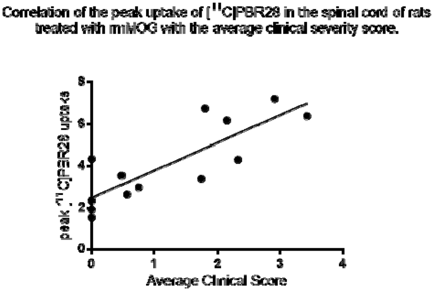
References
90
BRAIN-0454
BrainPET
[18F]DPA-714 PET IMAGING IN PRE-CLINICAL MODELS OF HUMAN GLIOMAS
1Inserm/CEA/Université Paris Sud UMR 1023 – ERL 9218 CNRS IMIV, CEA, Orsay, France
2European Institute for Molecular Imaging, Westfälische Wilhelms University Münster, Münster, Germany
Abstract
Objectives
The 18 kDa translocator protein (TSPO) is a protein of the outer mitochondrial membrane widely expressed in peripheral organs. TSPO expression in the brain is low but its expression is dramatically increased after glial cell activation and has become a well-characterized marker for neuroinflammation. Moreover, TSPO expression has also been shown in some cancers including gliomas. TSPO imaging can be performed using positron emission tomography (PET) with the specific radioligand [18F]DPA-714. As demonstrated in a rat glioma model, TSPO imaging in glioma monitors TSPO-positive neoplastic and to a lesser amount TSPO-positive inflammatory cells (Winkeler et al., 2012). To study a) the use of [18F]DPA-714 PET imaging in human gliomas and b) the impact of the tumor microenvironment, in particular glioma-associated inflammation, we have started to characterize an angiogenic and an invasive human glioma model for in vivo imaging using [18F]DPA-714 PET as well as immunohistochemistry (IHC) with dedicated human and murine TSPO antibodies.
Methods
2x10⁵ human glioma cells (angiogenic, U87dEGFR or infiltrative) were stereotactically implanted in the striatum of nude mice (n=4 or n=6, respectively). To monitor tumor growth, 30 minutes [18F]DPA-714 PET scans were acquired 10 days (U87dEGFR) or 1, 3, 5, 7 and 9 weeks (infiltrative) post-inoculation. Validation of PET findings was done by ex vivo autoradiography and IHC using specific human and murine TSPO antibodies.
Results
Ten days after cell implantation significantly higher uptake of [18F]DPA-714 has been demonstrated in human U87dEGFR tumors as compared to the contralateral brain (%ID/cc: 0.9 ± 0.1 and 0.4 ± 0.; p < 0.01) with a tumor-to-brain ratio of 2.2 ± 0.1. Furthermore, immunohistochemistry confirmed TSPO expression within the tumor. IHC using human and mouse specific TSPO antibodies allowed distinction between tumoral and stromal TSPO, indicating the presence of TSPO-positive stromal cells within the glioma (see Figure). In contrast tumor-to-contralateral-brain ratios in the invasive model ranged from 1.2 to 1.7, with a significantly higher uptake in the tumor only after 9 weeks post implantation (p<0.05), which was confirmed by ex-vivo autoradiography (p< 0.0001).
Conclusion
First results demonstrate that imaging of TSPO expression in vivo using [18F]DPA-714 PET is feasible in the human U87dEGFR glioma model. Distinction between tumoral and stromal TSPO could be achieved by ex vivo IHC, which will be of use to help studying changes of tumoral and stromal TSPO expression during tumor progression and therapy. However, kinetics of TSPO imaging in the infiltrative glioma model have to be further investigated and compared with ex vivo IHC and autoradiography at earlier time points.
This work was supported by the European Union's Seventh Framework Programme [FP7/2007-2013] INMiND (Grant agreement no. 278850)
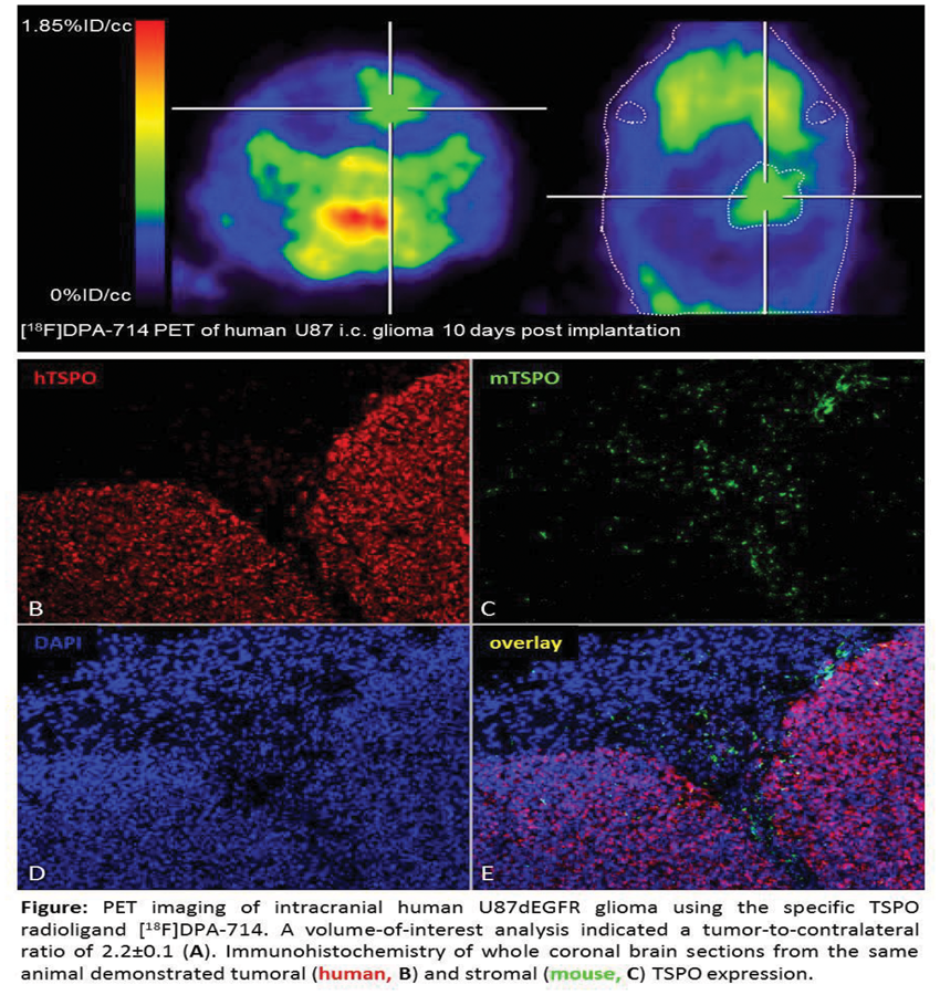
References
93
BRAIN-0964
BrainPET Symposium
Advances in MR/PET for studies in brain Function
1q, Harvard, Boston, USA
Abstract
The simultaneous acquisition of both positron emission tomography (PET) and magnetic resonance imaging (MRI) data offers an exciting opportunity to merge anatomical, physiological, metabolic, and molecular information in a single examination setting. This is especially relevant in studies of the brain, where many fundamental questions remain on the linkage between structure and function, and where even basic principals of the interactions between physiological changes such as cerebral blood flow, neuronal “activity”, and neuroreceptor function are yet to be fully elucidated. This talk will first describe the technological principals underpinning such combined instruments, including changes in both the PET and MRI architectures which allow such instruments to be cross-compatible. The talk will then discuss how combining information from both modalities can be used to explore structure/function relationships, and how the simultaneous acquisition of data can provide information that is difficult to obtain using sequential acquisitions. Finally, some clinically relevant examples will be presented to highlight the future potential of this tool for advanced diagnostic evaluation of neurolopsychiatric diseases.
94
BRAIN-0961
BrainPET Symposium
Molecular tools for imaging misfolded proteins
1Radiology, University of Pittsburgh, Pittsburgh, USA
Abstract
A variety of neurodegenerative diseases are pathologically characterized by the aggregation of specific proteins, such as extracellular aggregates of amyloid-β in senile plaques and intracellular aggregates of hyperphosphorylated tau in neurofibrillary tangles observed in Alzheimer's disease or intracellular aggregates of α-synuclein in Lewy bodies and Lewy neurites in Parkinson's disease. The utilization of positron emission tomography (PET) radioligands for the in vivo characterization of these aggregated proteins likely will play a critical role in defining pathological processes and guiding therapeutic treatment of these diseases and others characterized by abnormal protein aggregations. [C-11]-Labelled Pittsburgh compound B (PiB) was the first successful PET imaging agent to selectively target one of these aggregated proteins (amyloid-β), and selective targeting is important because many neurodegenerative diseases contain two or more different types of aggregated proteins. Worldwide investigations using PiB and four closely related F-18 analogues, now numbering in the tens of thousands, have helped identify the time course and pattern of amyloid-β plaque deposition characteristic of Alzheimer's disease a decade or more before the clinical symptoms of dementia. While recent progress has been realized in developing selective and potent tau PET tracers, efforts to develop selective α-synuclein radioligands remain a work in progress. The talk will highlight the considerations and challenges of designing PET radioligands for advancing scientific knowledge of the role aggregated proteins play in neurodegenerative diseases.
95
BRAIN-0982
BrainPET Symposium
Beyond a single target: Multi-modal & neural pathway imaging
1Psychiatry, NYSPI/Columbia UNiversity, New York, USA
2Yale University School of Medicine, Yale PET Center, New Haven, USA
Abstract
The goal of this study was to use Positron Emission Tomography (PET) to investigate the neurobiology of cocaine addiction using the radiotracer [11C]GR103545 (1) to detect changes in the kappa opioid receptor (KOR) dynorphin system in cocaine abuse, based on animal experiments showing that binge cocaine administration significantly increases levels of endogenous dynorphin.
Cocaine abusers and matched control subjects were scanned with the radiotracer [11C]GR103545, in order to compare kappa receptor availability between groups. In addition to this baseline scan, the cocaine abusers underwent PET scans following three days of binge smoked cocaine (300 mg/day), to investigate effect of cocaine on brain dynorphin levels. We also investigated the association between kappa receptor binding and stress-induced cocaine self-administration using the cold-pressor test. In these sessions, cocaine abusing subjects were allowed to chose between smoked cocaine and an alternative reinforcer (money) following a stress.
Data are available now for 14 cocaine abusers and 10 controls. The cocaine abusers were heavy, chronic users of smoked cocaine, and the controls were matched for age, gender, ethnicity, and cigarette smoking. The outcome measure for the PET scans was the volume of distribution (VT) of the regions of interest, which included the striatum (and its subdivisions), cortical regions (including prefrontal cortex), and medial temporal structures. The results were as follows: 1) No difference in [11C]GR103545 VT in any of the regions of interest was seen between the cocaine abusers and controls. 2) A positive correlation was seen between kappa receptor binding in the striatum and stress-induced cocaine self-administration: higher values of VT were associated with more choices for cocaine following the cold pressor test (Spearman correlation coefficient of 0.53, p = 0.03). 3) In the cocaine dependent subjects, a significant decrease (-17.7% ± 2.3%) in VT was measured in the stratum when comparing the baseline and post-binge scans, and similar decreases were seen in other brain regions (-12.9% ± 5.1%).
There was no between-group difference in kappa receptor VT in the baseline condition. However, there was a positive correlation between striatal kappa receptor VT and cold-pressor induced cocaine self-administration, showing that higher kappa receptor binding is associated with a greater vulnerability to stress induced choices for cocaine. The decrease in [11C]GR103545 VT seen following binge dosing of cocaine suggests that levels of dynorphin might be significantly upregulated, which replicates in humans the finding that cocaine increases brain levels of dynorphin. These data indicate that altered the kappa receptor/dynorphin system is significantly altered in the human brain in response to cocaine exposure.
Supported by NIDA 1R01 DA027777
96
BRAIN-0957
BrainPET Symposium
Awake animal imaging
1Central Research Laboratory, Hamamatsu Photonics, Hamamatsu, Japan
Abstract
Objectives; In PET imaging with experimental animals, anesthetics are indispensable to fix animals during data acquisition. However, anesthetics have been reported to affect the brain function [1], kinetics of PET probes [2], and pharmacological effects of drugs [2, 3], which may humper PET brain imaging research. To solve the problem, we developed a unique PET scanner, which can tilt its gantry up to 90 degrees [4]. This PET scanner allowed us to measure monkey brain function in conscious condition. In the presentstudy, we applied it to develop the novel PET probes for mitochondria complex-I (MC-I) to evaluate the brain aging. We previously demonstrated that Ischemic neuronal damage in the brains of rat [5] and monkey[6] could be detected using 18F-BCPP-EF 7 days post ischemic insul, twhen 18F-FDG could not do because of neuroinflammation with activated microglia [5, 6].
Methods; We conducted medicinal chemistry research based on BMS-747158 as a lead compound [7], developed 18F-BCPP-EF [5], and evaluated its specificity and affinity for MC-1 in the living monkey brain [8]. Then, PETscans using 11C-PIB for amyloid-β (Aβ) deposition,11C-DPA-713 for translocator protein (TSPO) activity as an inflammatory index, 18F-FDG for regional cerebral metabolism of glucose (rCMRglc), and 18F-BCPP-EF for MC-1 were performed in young and aged monkeys (Macaca mulatta) in conscious condition [9].
Results; Since isoflurane anesthesia affected rCMRglc and standard uptake value (SUV) of 11C-DPA-713, not total distribution volume (VT) of 18F-BCPP-EF and SUV ratio (SUVR) of 11C-PIB, the following analyses including rCMRglc and 11C-DPA-713 were conducted using data obtained in conscious condition only. When plotted VT of 18F-BCPP-EF against SUVR of 11C-PIB in the cerebral cortical regions, it showed a significant negative correlation between them. Plotting of SUV of 11C-DPA-713 against SUVR of 11C-PIB resulted in a significant positive correlation, suggesting that Aβ deposition-induced inflammatory effects with microglial activation. In contrast, plotting of rCMRglc against SUVR of 11C-PIB did not reach statistically significant level because of unexpected 18F-FDG uptake into the activated microglia.
Conclusions; The present study demonstrated that anesthesia and neuroinflammation significantly affected rCMRglc as measured by 18F-FDG. In contrast, 18F-BCPP-EFcould be a potential PET probe for quantitative imaging of age-related neurodegenerative alterations as a change in MC-1 activity in the living brain without being disturbed by anesthesia and neuroinflammation.
References;
98
BRAIN-0955
Symposium
Potential role of pluripotent fat pericytes in cell replacement in neurodegenerative disease
1Neurology, Wayne State University School of Medicine, Detroit Michigan, USA
Abstract
Adult stem cells (somatic cells) are undifferentiated cells found throughout the body after development. They have the potential to self renew and multiply indefinitely and have primary responsibility to maintain and repair the tissue of origin. The identity of the adult undifferentiated pluripotent stem cell is not known. Most experimental data has been gleaned from progenitor populations with limited ability to differentiate and an inherent genetic imprinting, or from differentiated adult cells induced to be pluripotent by the insertion of four embryonic genes (induced pluripotent cells [IPS]). The use of these cells seemed a promising answer to the debates over embryonic-stem-cell research. However IPS as well as progenitors have limited replicative ability and have early genetic senescence compared with embryonic stem cell derived tissue. This points to the continuing need to study true quiescent resting pluripotent cells of adult origin. We have identified an NG2+/nestin+/PDGFbR+ adult stem cell population within the CNS as well as other capillary networks (pericyte). CNS pericytes express all four embryonic antigens and are pluripotent having both neural and mesenchymal stem cell potential and can be isolated and expanded from brain capillaries. CNS pericytes as well as pericytes from many different organs have now been shown to be pluripotent and have the ability to form tissue specific progenitors. We have developed techniques to isolate human fat pericytes and pre-adipocyte progenitor cells and developed human cell lines that self renew and have pluripotent capacity. Two of these lines have been in culture for 8 months. Human populations retain pluripotent stem cell activity and self renew. Fat stem cells are easily obtained and expanded and can be frozen and thawed. Pericyte fat stem cell populations have been tested in an animal model of the human CNS degenerative disease multiple sclerosis to determine the potential therapeutic usefulness of human fat stem cells in cell replacement therapies for this disease.
100
BRAIN-0949
Symposium
Pericyte regulation of the blood-brain barrier
1Pharmacology and Neuroscience, UCSD, San Diego, USA
Abstract
Vascular endothelial cells in the central nervous system (CNS) form a barrier that restricts the movement of molecules and ions between the blood and the brain. This blood-brain barrier (BBB) is crucial to ensure proper neuronal function and protect the CNS from injury and disease. Although the properties of the BBB are manifested in the endothelial cells, transplantation studies have demonstrated that the BBB is not intrinsic to the endothelial cells, but is induced by interactions with the neural cells. Here we use a genomic, genetic and molecular approach to elucidate the cellular and molecular mechanisms that regulate the formation of the BBB. We have identified a critical role for pericytes in regulating the permeability of CNS vessels by inhibiting the properties that make endothelial cells leaky. In particular pericytes limit the rate of transcytosis through endothelial cells as well as the expression of leukocyte adhesion molecules in CNS endothelial cells, which limits CNS immune infiltration. Furthermore, we have developed methods to highly purify and gene profile endothelial cells from different tissues, and by comparing the transcriptional profile of brain endothelial cells with those purified from the liver and lung, we have generated a comprehensive resource of transcripts that are specific to the BBB forming endothelial cells of the brain. We have further examined the profile of CNS endothelial cells following injury and disease and have identified molecular mechanisms by which pericytes control BBB formation, which are then disrupted during neurological disease leading to BBB dysfunction.
101
BRAIN-0948
Symposium
Control of blood flow by capillary pericytes and the origin of BOLD fMRI signals
1NPP, UCL, London, United Kingdom
Abstract
Brain blood flow is regulated to ensure adequate power for neuronal computation. Blood flow is increased to areas where neurons are active, and this increase underlies non-invasive brain imaging using BOLD fMRI. Blood flow is controlled at the arteriole level by smooth muscle, but there is controversy over whether it is also regulated by pericytes at the capillary level. I will demonstrate that neuronal activity and the excitatory neurotransmitter glutamate evoke an outward membrane current in pericytes and dilate capillaries, and that this dilation is caused by active relaxation of pericytes rather than a passive expansion of capillaries downstream of arteriole relaxation. Glutamate-evoked dilation is mediated by prostaglandin E2, but requires nitric oxide release to suppress synthesis of the vasoconstrictor 20-HETE. In vivo, when sensory input to the neocortex releases glutamate and increases blood flow, capillaries dilate first, followed by arterioles. Capillary dilation is estimated to generate ∼80% of the increase of blood flow occurring. Capillaries also have a role in pathology. Ischaemia was previously shown to lead to a contraction of pericytes. I will report that this is followed by their death, which is expected to produce an irreversible constriction of capillaries. Pericyte death is reduced by block of glutamate receptors or calcium removal, and increases with reperfusion, but is not blocked by scavenging of reactive oxygen species. These data establish, for the first time, pericytes as major regulators of cerebral blood flow, and initiators of the BOLD fMRI response. They also focus attention on prevention of pericyte death as a therapeutic strategy to reduce the long-lasting blood flow decrease which contributes to neuronal death after stroke.
104
BRAIN-0431
Brain Oral Communication
LOCAL NEURONAL AND GLIAL OXIDATIVE METABOLISM DURING FOCAL CORTICAL ACTIVITY
1Laboratory for Functional and Metabolic Imaging (LIFMET), Ecole polytechnique fédérale de Lausanne, Lausanne, Switzerland
2Centre d’Imagerie Biomédicale – Animal and Technology Core (CIBM-AIT), Ecole polytechnique fédérale de Lausanne, Lausanne, Switzerland
Abstract
This work was supported by Swiss National Science Foundation (#148250), Centre d’Imagerie BioMédicale and National Competence Center in Biomedical Imaging.
105
BRAIN-0856
Brain Oral Communication
GLUCOSE CONSUMPTION ASSOCIATED WITH RESTING STATE FMRI ACTIVITY IN HUMAN BRAIN
1Robarts Research Institute, Western University, London, Canada
Abstract
Objectives: The energy demand of the human brain is primarily met by ATP derived from the oxidation of glucose. An energy audit (1), the Magistretti model (2) and 13C-MRS results (3) support the hypothesis that the metabolic steps involved in neurotransmitter cycling consume 5–10% of the total energy used by the brain (4). At 'rest” brain metabolism consists of a tonic component and a temporally varying (on the scale of seconds to minutes) component which likely supports the fluctuations responsible for the resting state functional magnetic resonance imaging (rs-fMRI) signal. The objective of this study was to determine the fraction of brain glucose consumption that supports the rs-fMRI signal.
Methods: We used data from 9 subjects in the ADNI database. All participants were required to have had at least one FDG-PET scan, one structural MRI scan and one rs-fMRI scan. The FDG-PET scans were acquired on a single Phillips scanner. All MRI images were preprocessed using FSL (5). Each PET image was processed using the recommended ADNI processing stream (6). PET and rs-fMRI images were registered to each other in MNI space after registering each to their respective structural MRI. Each subject’s BOLD time-series underwent ICA using MELODIC, followed by denoising by discarding artifactual components. The remaining fMRI signal was expected to be neuronal in origin and was characterized by its temporal variance on a voxel-vise basis. A joint-histogram analysis compared this voxel-wise rs-fMRI variance with the FDG-PET derived glucose consumption.
Results: The joint histogram for one subject is shown in Fig. 1. All 9 subjects looked similar and a group analysis was also performed. These joint histograms (JH) demonstrate that the glucose consumption associated with the rs-fMRI signal fluctuations was 5-10% of the total glucose consumption (see y-intercept), in line with prior estimates for task based fMRI (4). Furthermore, by mapping back the voxels in the far right of the JH (high FDG/high BOLD variance), we were able to show that these voxels corresponded to rich hubs (network hubs with high connectivity - Fig. 2). The JH approach allows the metabolic cost of each resting state network to be measured and compared as a fraction of total brain FDG consumption and to each other.
Abstract
Conclusions: Our JH analysis demonstrated that the metabolic cost of the rs-fMRI signal represents about 5-10% of the total metabolism of the brain. Regions of high rs-fMRI variance correspond to the rich hubs in the brain. Furthermore, the linear relationship between rs-fMRI variance and FSG-PET suggests that fMRI variance associated with the sum of the resting state networks can be used as a surrogate for FDG-PET of neural activity.


References
106
BRAIN-0562
Brain Oral Communication
RESTING-STATE BRAIN ENERGY METABOLISM PREDICTS LEVEL AND CONTENT OF CONSCIOUSNESS AFTER SEVERE BRAIN INJURY
1Department of Neuroscience and Pharmacology, University of Copenhagen, Copenhagen, Denmark
2Cyclotron Research Unit and Department of Neurology, University of Liège, Liège, Belgium
3Department of Computer Science, University of Copenhagen, Copenhagen, Denmark
Abstract
Objectives
Differential diagnosis between the minimally conscious and vegetative states (MCS, VS) remains an unresolved clinical challenge with important ethical and therapeutic implications. Current research addresses physiological correlates of perceptual awareness in these patients (1).
Assessment of cerebral glucose metabolism with fluorodeoxyglucose in positron emission tomograms (FDG-PET) serves as a diagnostic tool in disorders of consciousness (2). Here, we present a novel method of FDG-uptake normalization to non-cerebral tissue, which solves the issue of FDG-PET quantification in severely injured brains. We hypothesize that baseline metabolism predicts the level of consciousness, while regional variations relative to the baseline reflect preservation of specific functions, such as vision and language comprehension (3).
Methods
We included 138 patients clinically diagnosed with VS (n=49), MCS (n=65) or emergence from MCS (EMCS; n=17), and 28 healthy control subjects. We obtained one-year outcome (conscious/unconscious) for all patients.
All subjects had FDG-PET. Regions of interest were chosen as Brodmann areas and combined to cortical masks for each hemisphere. We normalized images by fitting the intensity distributions of extracerebral cranial tissue of individual patients to that of healthy subjects. A scaling parameter was found by minimizing the Jensen-Shannon divergence between the distributions. We used the average metabolic index of the least affected hemisphere as diagnostic marker, validated against behavioral diagnosis and outcome. Classification accuracy between VS and MCS was assessed by receiver operating characteristic (ROC) analysis. Brainwide interregional variability was assessed by the coefficient of variance (CV; std/mean). Following further scaling of the images to the global mean, we tested Pearson correlations between relative metabolic activity in visual cortex and language-related areas with visual responsiveness and command following capacity, respectively.
Results
Area under the ROC curve was 0.89 (0.82-0.96). The optimal classification threshold that distinguished MCS from VS was 41% of normal metabolism, which diagnosed 88% of the patients correctly, with 95% sensitivity and 78% specificity to MCS. All EMCS and healthy subjects were identified as conscious. The model predicted 86% of known outcomes in MCS and VS, with 97% sensitivity and 70% specificity to consciousness at follow-up. Notably, 8 out of 9 recoveries from VS were associated with above-threshold metabolism (Figure 1). No individual regions provided diagnostic classification superior to the hemisphere mean value. Interregional variance was 0.03 CV in VS, 0.093 in MCS, 0.097 in EMCS and 0.11 in controls. Relative metabolism in the visual cortex correlated with visual responsiveness (R=0.2443, p=0.0028), whereas relative metabolism in language-related areas correlated with command following capacity (R=0.486, p<0.0001).
Abstract
Conclusion
Global resting-state brain metabolism accurately predicted levels of consciousness. Regional metabolic variations relative to the baseline are characteristic of the conscious state and reflect perceptual contents. The metabolic level accounted for either the presence or imminent rise of consciousness in 95% of the patients. The results suggest that disorders of consciousness can be understood as abnormal neuroenergetic states and provides a unifying pathophysiological basis for these syndromes.

References
107
BRAIN-0510
Brain Oral Communication
METABOLIC CHANGES IN EARLY POST STATUS EPILEPTICUS MEASURED BY MR SPECTROSCOPY IN RATS
1Developmental Biology, University of Pittsburgh, Pittsburgh, USA
2Neurology, University of Pittsburgh, Pittsburgh, USA
3Biology, Carnegie Mellon University, Pittsburgh, USA
4Neurosurgery, Yale University, New Haven, USA
Abstract

REFERENCES
108
BRAIN-0096
Brain Oral Communication
NEURO-PROTECTIVE ROLE OF ASTROGLIA IN PARKINSON’S DISEASE ARISING FROM A REDUCTION IN OXYGEN STRESS THROUGH THE DOPAMINE-INDUCED ACTIVATION OF THE PENTOSE-PHOSPHATE PATHWAY
1Department of Neurology, Keio University School of Medicine, Tokyo, Japan
Abstract
Oxygen stress plays an important role in the onset and progression of Parkinson’s disease (PD). Mitochondrial overload induced by the pace-making activity of dopaminergic neurons to maintain an optimal dopamine (DA) concentration in the striatum (tonic release) might be a major cause of oxygen stress.1 Dopaminergic neurons also release additional DA as the need arises (phasic release). Another source of oxygen stress seems to be DA per se, since DA is auto-oxidized to form dopamine quinone, resulting in reactive oxygen species (ROS) production.1 Therefore, reducing oxygen stress seems to be an important strategy for the treatment of PD. Especially, the neuro-protective roles of astroglia have been recognized.1,2 Although most of the released DA is collected by dopaminergic neurons themselves, a part of DA is presumed to be collected by astroglia.3 DA may activate astroglial glucose metabolism via a mechanism similar to that induced by glutamate1-3 and may contribute to the reduction of oxygen stress. The present study examined DA-induced astroglial protective function through the activation of the pentose-phosphate pathway (PPP) to reduce ROS.
In vitro experiments were performed using striatal neurons and cortical or striatal astroglia prepared from Sprague-Dawley rats. The rates of glucose phosphorylation in astroglia were evaluated using a modification4 of the [14C]deoxyglucose method. PPP activity was measured using a modification4 of the method described by Hothersall et al.5 based on the determination of the difference in 14CO2 production from [1-14C]glucose and from [6-14C]glucose after acute (60 min) or chronic (15 hours) exposure to different concentrations of DA (10, 100 μM). ROS production was measured using H2DCFDA. The involvement of the Keap1/Nrf2 system was evaluated using immunohistochemistry.
Acute exposure to DA (10, 100 μM) elicited increases in astroglial glucose consumption (123.3±6.0,108% and 130.4±11.5, 115%, respectively) with lactate release, indicating an enhancement of glycolysis probably because of Na+-dependent DA uptake by a norepinephrine transporter. PPP activity in astroglia was enhanced robustly (2.9±0.4, 166% and 120.7±3.4, 6873%, respectively). In contrast, chronic exposure to DA induced moderate increases in PPP activity (2.0±0.1, 118% and 2.7±0.1, 163%, respectively). Chronic exposure to DA induced Nrf2 translocation to the nucleus in astroglia, indicating the Nrf2-dependent transcriptional up-regulation of G6PDH, a rate limiting enzyme of PPP. ROS production induced by DA in the absence of astroglia increased gradually over 12 hours, whereas it decreased in the presence of astroglia (86% and 75%, respectively).
DA released from dopaminergic neurons enhanced astroglial PPP activity in both an acute and a chronic manner, which may reduce neuronal oxygen stress and play an important role in preventing the onset and progression of PD. Further enhancement of the protective roles of astroglia could lead to an important therapeutic strategy for the treatment of PD.
References
109
BRAIN-0772
Brain Oral Communication
AN ALZHEIMER-LIKE PATTERN OF LOW BRAIN GLUCOSE UPTAKE ASSOCIATED WITH MILD INSULIN RESISTANCE IN YOUNG WOMEN WITH POLYCYSTIC OVARY SYNDROME: AN FDG PET/MRI STUDY
1Clinical Axis, Research Centre on Aging, Sherbrooke, Canada
2Endocrinology, Université de Sherbrooke, Sherbrooke, Canada
3Sherbrooke Molecular Imaging Center, Université de Sherbrooke, Sherbrooke, Canada
4Memory Clinic, Health and Social Sciences Center - Geriatrics Institute, Sherbrooke, Canada
5Nuclear Medicine and Radiobiology, Université de Sherbrooke, Sherbrooke, Canada
Abstract
References:
112
BRAIN-0967
Symposium
Supply-demand mismatch transients trigger spreading depolarizations within penumbral hot-zones
1Neurology and Radiology, Massachusetts General Hospital, Charlestown MA, USA
Abstract
Peri-infarct depolarizations (PIDs) are seemingly spontaneous spreading depression-like waves that negatively impact tissue outcome in both experimental and human stroke. Factors triggering PIDs are unknown. Here, we show that somatosensory activation of peri-infarct cortex triggers PIDs when the activated cortex is within a critical range of ischemia. We show that the mechanism involves increased oxygen utilization within the activated cortex, worsening the supply-demand mismatch. We support the concept by clinical data showing that mismatch predisposes stroke patients to PIDs as well. Conversely, transient worsening of mismatch by episodic hypoxemia or hypotension also reproducibly triggers PIDs. Therefore, PIDs are triggered upon supply-demand mismatch transients in metastable peri-infarct hot zones due to increased demand or reduced supply. Based on the data, we propose that minimizing sensory stimulation and hypoxic or hypotensive transients in stroke and brain injury would reduce PID incidence and their adverse impact on outcome.
113
BRAIN-0013
Symposium
Does age have an impact on spreading depolarization?
1Department of Medical Physics and Informatics, University of Szeged, Szeged, Hungary
Abstract
Waves of spreading depolarization (SD) spontaneously occur minutes after the onset of ischemia in the brain, and keep generating for a number of days to follow. It has become widely accepted that ischemia-related SDs are part of the pathophysiology of cerebrovascular diseases and predict worse outcome. SDs may exacerbate ischemic injury via related atypical hemodynamic responses. Aging is the most important risk factor for the incidence of cerebral ischemic stroke; yet, the effect of aging on the evolution of SDs and related hemodynamic responses has remained largely unexplored. Our recent investigations have aimed to identify the impact of aging on the elicitation threshold, duration, and propagation of SD, and the typical features of the related neurovascular coupling during ischemia. We have shown that the electric threshold to trigger SDs increases with progressing life time. Likewise, spontaneous SDs occur at lower frequency in the ischemic brain of aged animals, as compared with their young counterparts. However, once elicited, SDs tend to be persistent in the aged brain (as compared with predominantly transient SDs in the young brain), and their long duration may indicate metabolic crisis. While ischemia clearly compromises the kinetics of the SD-associated cerebral blood flow response (i.e. lower magnitude and longer duration of the distinct elements of the response), age exerts an additional shift to injurious CBF response types, especially inverse neurovascular coupling. We propose that an age-specific pattern of the SD-associated hemodynamic response must be involved in augmenting the expansion of ischemic brain damage in the elderly, and that structural and functional (mal)adaptation of the cerebrovascular system with aging serves as a potential basis for compromised vascular reactivity and subsequent tissue damage.
115
BRAIN-0024
Symposium
Spreading depolarizations in human brain trauma: a causal pathomechanism?
1Neurosurgery, University of Cincinnati, Cincinnati, USA
Abstract
Excitotoxicity and loss of ion homeostasis are well established as key mediators of secondary injury and lesion development after traumatic brain injury (TBI). Recent experimental evidence, however, suggests that these processes are actively induced and propagated in mass tissue events known as spreading depolarizations (SD), providing a novel basis for patient monitoring and therapeutic targeting. While SD scarcely occurs in rodent models of TBI, clinical studies now show that SD is a dominant pathophysiologic mechanism in many patients with severe TBI. Surgical patients monitored with subdural electrode strips have a 55% incidence of SD, and patterns vary widely from occasional events to continuous, repetitive events lasting hours to days. The basis of this heterogeneity is largely unknown, as the occurrence or severity of SD is not predicted by other clinical variables. Patients with and without SD are similar in terms of classical prognostic factors, indicators of injury severity, and pathoanatomic subtypes of TBI. Furthermore, the occurrence of SDs is largely independent of other physiologic ‘secondary insults’ such as hypotension or intracranial hypertension. Nonetheless, the occurrence of SD carries a significant and substantial increased risk for worse outcome, with estimated odds ratios in the range of 1.4 to 2.5. The risk is higher yet for patients with more severe SD patterns. A causal adverse impact of SD on injury progression is evidenced more directly by real-time monitoring. In some patients, SDs induce a persistent isoelectric “flatline” of cortical activity, analogous to the ischemic penumbra, lasting hours to days. Furthermore, simultaneous monitoring of local cerebral blood flow has shown that SDs can induce a spreading hypoperfusion, which limits delivery of metabolic substrates at a time of maximal metabolic demand. Together, these data suggest that SD as a sequela of TBI is not simply a marker of injury severity or other known pathologic processes, but is an independent factor with adverse impact on TBI recovery. As the first method to assess a heterogeneous mechanism in individual patients, monitoring of SD may allow for novel clinical trials based on selective inclusion, mechanistic targeting, and tailored therapeutic dosing.
116
BRAIN-0097
Symposium
The stroke-migraine depolarization continuum
1Center for Stroke Research Berlin, Charité University Medicine Berlin, Berlin, Germany
Abstract
The term spreading depolarization describes a mechanism of abrupt, near-complete ion translocation between neurons and the interstitial space which leads to a cytotoxic edema in the gray matter of the brain above the thalamic/hypothalamic boundary zone. In energy compromised tissue, spreading depolarization is preceded by a non-spreading silencing (depression of spontaneous activity) because of a neuronal hyperpolarization. By contrast, in non-energy compromised tissue, spreading depolarization causes spreading silencing (spreading depression) of spontaneous activity by a depolarization block. It is assumed that the non-spreading silencing translates into the initial clinical symptoms of ischemic stroke and the spreading silencing (spreading depression) into the symptoms of migraine aura. In energy compromised tissue, spreading depolarization is long-lasting and facilitates neuronal death whereas, in healthy tissue, it is short-lasting and relatively innocuous. Therapies targeting spreading depolarization in metabolically compromised tissue may potentially treat conditions of acute cerebral injury such as aneurysmal subarachnoid hemorrhage, ischemic stroke and traumatic brain injury. Moreover, neuromonitoring of spreading depolarizations has increasingly become routine practice in neurointensive care as it may offer unprecedented opportunities for treatment stratification.
119
BRAIN-0984
Symposium
Neurophysiological basis of spontaneous fluctuations in BOLD signal: Correlations of non-linear couplings between bands of the local-field potentials
1McConnell Brain Imaging Centre, Montreal Neurological Institute McGill University, Montreal, Canada
Abstract
Experimental evidence (Shmuel and Leopold, 2008) suggests that spontaneous blood oxygenation signals correlate with slow local fluctuations in amplitude of the gamma-band (30-100 Hz) local field potentials (LFP). In contrast, blood-oxygenation-based inter-areal correlations are associated with correlations of low-frequency rhythms of <20 Hz (Lu et al, 2007).
Phase-Amplitude coupling (PAC), in which the phase of a low-frequency rhythm modulates the amplitude of a higher frequency rhythm, has been proposed as the mechanism linking these two phenomena. However, it remains unclear how the various spontaneous rhythms interact and whether their interactions predict spontaneous fluctuations in blood oxygenation. Here we investigated intra- and inter-laminar PAC during spontaneous activity, and its correlation with cortical blood oxygenation. To this end, we simultaneously recorded spontaneous LFP using laminar probes and optical imaging signals (OIS) in the forelimb representation of area S1 in anesthetized rats.
We observed both intra- and inter-laminar spontaneous PAC in a cortical column, with the highest measures of interaction obtained for the 2 pairs of delta/fast-gamma and theta/fast-gamma bands. Intra- and inter-laminar PACs involving layers 2/3–5a were higher than those involving layer 6. Current sinks (sources) in the delta band were associated with increased (decreased) amplitudes of high-frequency signals in the beta to fast gamma bands throughout layers 2/3–6. Spontaneous sinks (sources) of the theta and alpha bands in layers 2/3 to 5a were on average linked to dipoles completed by sources (sinks) in layer 6, associated with high (low) amplitudes of the faster rhythms in the entire cortical column.
To determine whether PAC predicts spontaneous fluctuations in blood oxygenation, linear and nonlinear correlation between band-limited-amplitude and OIS and separately between PAC and OIS were then computed. For the majority of frequency-band combinations, PAC is positively correlated with spontaneous fluctuations in blood oxygenation, with the highest correlation values found between phases of delta to beta and amplitudes of low - and middle-gamma. Furthermore, nonlinear models of PAC are better predictors of OIS than linear models of band-limited-amplitude, although worse than nonlinear models of band-limited-amplitude.
Our study links recent theories on the involvement of PAC in resting-state functional connectivity with previous work that revealed lamina-specific thalamo-cortico-cortical anatomical connections. Our results highlight the importance of nonlinear mechanisms in the generation of spontaneous fluctuations in blood oxygenation.
120
BRAIN-0978
Symposium
Principles of energy demand in gray and white matter of the human brain
1Physiology and Biophysics, Fudan University, Shanghai, China
Principles of energy demand in gray and white matter of the human brain
School of Life Science and the Collaborative Innovation Center for Brain Science
Center for Computational Systems Biology
Fudan University, Shanghai, China
The mammalian brain is organized on multiple scales of network connections and functionally dependent regions for sensory and cognitive functions. Normal mammalian brain functions are mainly supported by high ATP yield from glucose oxidation (CMRglc(ox)). How the metabolic energy used at the subcellular level is related to the large swathes within the brain are still not well-known yet. We created a comprehensive CMRglc(ox)-derived energy budget for gray and white matter from the bottom-up, using biophysical properties of neurons and glia in conjunction with species-specific electrophysiological and morphological data. Comparing metabolic measurements by PET, 13C MRS, and autoradiography in rat and human brain with budget-derived CMRglc(ox) revealed conserved properties. Signaling events (e.g., synaptic transmission, propagation of action potentials, glutamate recycling) in gray and white matter respectively demanded ∼65% and ∼25% of awake state CMRglc(ox) values in the respective tissues, whereas remaining portions were used for nonsignaling functions (e.g., resting membrane potential, housekeeping). GABAergic neurons and glia each demanded ×5 less energy than glutamatergic neurons. Although rat brain possesses smaller cells, due to higher firing rates in rat brain their functions demand 15-20% greater energy than their human brain counterparts. While neuronal energetics was governed by events associated with synaptic transmission and propagation of action potentials, glial energetics was dominated by nonsignaling needs. Then, we feed the energy budget calculations of individual neurons and glias into a 3-D human brain cell density map calculated based on BigBrain cell-stained histological data, formulating a 3-D brain energy map with energy calculations in unit of either glucose in micromole/min/gram or ATP/min/gram in order to compare with PET measurement of actual brain. The quantitative comparison demonstrated a perfect match, indicating a first successful reconstruction of brain energy map fully consistent with PET glucose imaging measurement. Human brain CMRglc(ox) maps assembled using the budget with BigBrain cell-stained histological data showed voxel-wise correspondence with CMRglc(ox) maps measured (by PET, awake state) across large swathes of gray and white matter, requiring 0.5-1.5 Hz firing rates across the entire cerebrum. A validated bottom-up energy budget of heterogeneous cellular functions spanning gray and white matter of healthy human brain can provide opportunities to reveal testable microscopic-level anomalies underlying metabolic neuroimaging of brain diseases and disorders.
121
BRAIN-0985
Symposium
Blood flow & oxidative metabolism coupling in the awake and resting human brain: On the signaling roles of aerobic glycolysis and lactate
1Dept Neuroscience and Pharmacology, University of Copenhagen, Copenhagen DK-2200, Denmark
Abstract
Lactate appears to serve several roles as a signal in mammalian brain (Gjedde &Magistretti 2010, Bergersen & Gjedde 2012). Recent data suggests that lactate is a signal responsible in part or in whole for the coupling of blood flow to oxidative metabolism. In addition, there is emerging evidence that lactate also serves as a volume transmitter engaged, among other effects, in memory consolidation by interaction with the lactate receptor GBR81. Lactate is the product of aerobic glycolysis when oxygen tensions in the tissue are adequate for complete oxidation of glucose to CO2. Aerobic glycolysis, also known as the Warburg Effect, results in values of the Oxygen-Glucose Index (OGI) that are significantly less than 6. Conversely, when OGI values exceed 6, the phenomenon sometimes is referred to as the Inverse Warburg Effect. Recent evidence shows that OGI values change as function of age, rising from 4 or less during childhood to 7 or more in old age (Goyal et al, 2014). The consequences of this change are disputed, but here we claim that the change reflects evolving functions of lactate as a volume transmitter involved in learning in mammalian brain.
The theory of lactate as a volume transmitter claims that the presence of lactate in the tissue at certain concentrations imparts information to the regulatory mechanisms of the tissue. By consideration of the steady- and non-steady-states that brain tissue metabolism may occupy, we describe the processes of release of lactate to the tissue as tonic during steady-state and as phasic during non-steady-states, respectively, when temporary increases of the concentration of lactate occur, in keeping with the terminology used for the monoaminergic volume transmitters.
Lactate is the product of aerobic glycolysis in response to glutamate uptake into astrocytes in the steady-state and glycogenolysis in response to noradrenaline (NA) acting on beta 2 receptors in astrocytes in non-steady-states. The role of NA in the breakdown of glycogen suggests that this monoamine plays a special role in the phasic release of lactate. This role is reminiscent of NA's role in working memory and memory consolidation by interaction with alpha2 adrenoceptors that limit cAMP formation. Lactate similarly interacts with the GPR81 (or HCA1) G-protein receptor that also mediates an inhibition of cAMP formation. These actions imply that NA and lactate collaborate in second-messenger effects associated with working memory and memory consolidation. Further evidence for this control comes from actions of lactate in gene expression that are related to its positive modulatory effect on NMDA signaling in neurons (may be related also to the limitation of cAMP formation). The putative tonic and phasic release patterns of lactate further suggest that lactate concentrations in brain could reflect the memory and learning loads that brains face in different states.
In this context it is remarkable that OGI and OEF measurements together with measures of glucose and oxygen consumption rates as functions of age predict that lactate concentrations in brain tissue are particularly high during childhood (0-15 years) and low above the age of 60, in keeping with the memory, learning and plasticity loads that characterize these ages.
122
BRAIN-0986
Symposium
Glucose oxidation in normal human brain across different resting states
1Departments of Diagnostic Radiology & Biomedical Engineering, Magnetic Resonance Research Center Yale University, New Haven CT, USA
Abstract
Pioneering efforts in the past decades with PET and fMRI have laid strong foundations for network mapping in the human brain. Early PET work concentrated on quantitative imaging of resting state blood flow and metabolism. By the 1990s fMRI became popular because it was possible to conduct repeated mapping in the same session, where the functional hyperemic changes provided the contrast. In this presentation I will focus on two specific issues of network mapping by fMRI and PET (Hyder et al, 2013) that impact interpretations of what networks mean.
First, recent proposals that certain resting-state fMRI networks have higher rates of glycolysis imply regional variation of ATP regeneration rates. Furthermore, if regional glucose utilization is mismatched with oxygen delivery, then those areas can have impaired ATP production. These predictions are testable by measured differences between the oxygen to glucose index (OGI) and the oxygen extraction fraction (OEF). We used quantitative PET measurements of blood flow and rates of oxygen and glucose consumption in normal human brain (resting awake, eyes closed) to test whether values of OEF and OGI differ regionally. Similar OEF and OGI values and patterns prevailed as networks regardless of their size, location, and origin showed OEF and OGI values that matched the gray matter means. The excellent spatial agreement between oxygen transport and glucose metabolism in normal human gray matter suggests that no specific region is preferentially vulnerable to impaired ATP production. In normal human gray matter, uniform OGI and OEF sustained globally high total ATP turnover rates, with less than 1% contribution of total ATP from non-oxidative glycolysis. We conclude that the intrinsic network activity in healthy human brain covers the entire gray matter with ubiquitously high-energy demands met by glucose oxidation.
Second, human neuroimaging studies aim to isolate brain networks that differ between health and disease. But network differences revealed by methods that utilize global mean normalization remain difficult to interpret because it is unknown what fraction of total metabolic (or neuronal) activity the state-dependent variations reflect. To quantitatively characterize global vs. regional metabolic variations from a control state of awake with eyes closed, we examined PET-measured glucose consumption (CMRglc) in other states. The different states included awake with eyes open, awake but congenitally blind, healthy but sedated with anesthetics, and patients with disorders of consciousness. In relation to the control state, quantified CMRglc maps for all states (except congenitally blind) revealed significant global changes, ranging from +10% with eyes open, -20% with sedation, and -60% with disorders of consciousness. Statistical t-maps with quantified CMRglc data for each state (vs. control) revealed globally unidirectional metabolic changes. In contrast global mean normalized CMRglc t-maps exposed regionally bidirectional metabolic effects across different states. These results suggest that the hypothesized confounding effects from the global signal, in fact, contains metabolic information that depicts brain-wide deviations that are state-dependent.
In summary, quantitative metabolic mapping can be used to advance disease biomarker in human neuroimaging studies.
125
BRAIN-0261
Brain Oral Communication
INHIBITION OF BOTH THE ACTIVATED MICROGLIA/MACROPHAGE AND THE INFILTRATED NEUTROPHILS ATTENUATES BRAIN INJURY AND IMPROVES OUTCOMES FOLLOWING INTRACEREBRAL HEMORRHAGE
1Neurology, The First Affiliated Hospital of Henan University, Kaifeng, China
2Clinical Neurosciences and Hotchkiss Brain Institute, University of Calgary, Calgary, Canada
Abstract
OBJECTIVES: Microglia become activated following intracerebral hemorrhage (ICH). Monocytes from the circulation migrate into areas of CNS injury to become macrophages. These activated cells are often called 'microglia/macrophage” since it is difficult to differentiate between them. The neutrophils also infiltrate into the injured brain after ICH. Activated microglia/microphages and the infiltrated neutrophils are capable of releasing numerous detrimental mediators such as matrix metalloproteinases, free radicals, chemokines and cytokines, but they can also produce neurotrophic factors. We tested the hypothesis that the activation of microglia/macrophage and the infiltrated neutrophils promote brain injury in the acute period after ICH and that inhibition both of the activated microglia/macrophage and the infiltrated neutrophils attenuates brain injury and improves outcomes following ICH.
METHODS: 10-µl of autologous blood obtained from tail was introduced into the right striatum of adult male mice (7-9 weeks old) to produce ICH injury. C57/B6 wildtype mice were used to evaluate the time course of brain injury from 1-7 day(s). We determined the area of brain damage, the extent of neuronal death, the number of infiltrated neutrophils and microglia/macrophage activity. We also used transgenic CD11b thymidine kinase (CD11b-TK) mice where proliferating microglia/macrophage were removed by ganciclovir treatment. Functional grade purified anti-mouse Ly6G/Gr-1 antibodies or isotype-matched control antibodies were used to reduce the neutrophil infiltration. Wildtype and CD11b-TK mice were randomized into sham, injured treated, or controls and received either 4 mg/kg intraperitoneal injections of anti-LyG6/Gr-1 antibodies or isotype control at 2 and 24 h after SCI. Groups of CD11b-TK mice were treated with ganciclovir and/or anti-mouse Ly6G/Gr-1 antibodies. These mice were killed at 3 days after ICH injury and brain sections were stained for various parameters.
RESULTS: We found that ICH injury resulted in activation of microglia/macrophages through 1-7 days of brain insult. The area of brain damage peaked at 2-3 days; the number of dying neurons peaked at 1-3 days, the number of infiltrated neutrophils peaked at 1-3 days, while the activation of microglia/macrophages peaked at days 3-4. These findings led us to choose day 3 for further studies. We found that the activation of microglia/macrophage was significantly reduced in CD11b-TK transgenic mice with ganciclovir treatment after ICH compared to various injury control groups, including wildtype mice with ganciclovir treatment after ICH. The numbers of infiltrated neutrophils were significantly reduced in the group with anti-mouse Ly6G/Gr-1 antibodies treatment after ICH compared to various injury control groups, including wildtype mice with isotype treatment after ICH. Correspondingly, the area of brain damage and the extent of neuronal death were minimal in CD11b-TK mice with ganciclovir and anti-LyG6/Gr-1 antibodies treatment after ICH compared to all other groups.
CONCLUSIONS: Both activated microglia/macrophages and the infiltrated neutrophils in the early periods after ICH promote brain injury. Thus inhibition of their activities attenuates brain injury following ICH. These results shed light on the advent of new medications for ICH patients, including microglia deactivators and the antibodies to alleviate neutrophil infiltration.
126
BRAIN-0294
Brain Oral Communication
A NEW ANTIFIBRINOLYTIC, CM352, MEDIATES REDUCTION OF HEMATOMA VOLUME AND IMPROVES FUNCTIONAL RECOVERY IN EXPERIMENTAL INTRACEREBRAL HEMORRHAGE.
1Clinical Neurosciences Research Laboratory, Heath Research Institute of Santiago de Compostela (IDIS), Santiago de Compostela, Spain
2Atherosclerosis Research Laboratory, Center for Applied Medical Research (CIMA), Pamplona, Spain
3Clinical Neurosciences Research Laboratory, Health Research Institute of Santiago de Compostela (IDIS), Santiago de Compostela, Spain
4Small Molecule Discovery Platform, Center for Applied Medical Research (CIMA), Pamplona, Spain
Abstract
Hematoma and lesion volumes were assessed by means of magnetic resonance imaging (MRI). T2-weighted images were acquired at 1, 3, and 24 hours and at day 14 after ICH onset. T2-weighted images on each row correspond to one of 2D slices of the same animals within each group at different time points.
127
BRAIN-0640
Brain Oral Communication
RED BLOOD CELL-DERIVED MICROPARTICLES FOR THE TREATMENT OF INTRACEREBRAL HEMORRHAGE.
1Department of Neurology, University of Miami Miller School of Medicine, Miami, USA
2Division of Hematology/Oncology Department of Medicine, University of Miami Miller School of Medicine, Miami, USA
Abstract

References:
128
BRAIN-0310
Brain Oral Communication
EXPRESSION AND FUNCTION OF MICRORNAS IN HUMAN BRAIN ARTERIOVENOUS MALFORMATION
1Neuroscience and Neuroengineering Research Center, Med-X Research Institute, Shanghai, China
2Department of Neurosurgery, Hua Shan Hospital, Shanghai, China
3Department of Neurology, Ruijin Hospital, Shanghai, China
Abstract
, Workflow of the experiment. B, Heatmap ofthe significant differently expressed miRNAs in the discovery set. C, Principalcomponent analysis plots of all the 739 miRNAs. Green square=HB samples. Bluesquare=AVM samples. Red square=Ctrl samples. D, Overexpression of miRNA-137 ormiRNA-195* inhibited the AVM-SMC vasculogenesis. Scle bar=3mm. E, Overlapof proteins identified in the down regulated protein after the miRNA-137 ormiRNA-195* mimics transfered into the AVM-SMC. F, IPA predited regulatory mainnetwork of miRNA-137 down regulated proteins. G, IPA predited regulatorymain network of miRNA-195* down regulated proteins.
References:
129
BRAIN-0455
Brain Oral Communication
HISTOPATHOLOGICAL INVESTIGATION OF INTRACRANIAL ARTERIAL DISSECTIONS
1Department of Neurosurgery, The University of Tokyo, Tokyo, Japan
Abstract
[Objectives]
Intracranial arterial dissections (IAD) are mainly divided into hemorrhagic and non-hemorrhagic ones, the former is subarachnoid hemorrhage (SAH) and the latter are headaches or ischemic stroke. For patients with non-hemorrhagic IAD, conservative treatments has been advocated, while early repair of the affected vessels by open surgery or endovascular technique tends to be performed for SAH patients. Recently as the endovascular techniques advance, open surgery gradually decreased, and the chance of histopathological examination of the IAD gets rare. In this study, we conducted a histopathological investigation of intracranial arterial dissections.
[Methods]
From April 1980 to December 2000, 143 patients were diagnosed with acute IADs at our institutions. All cases satisfied one of the three diagnostic criteria for acute IAD: 1) the typical pearl and string or double lumen sign at a non-branching site of the intracranial cerebral arteries on angiography; 2) fusiform dilatation with retention of contrast medium or angiographic steno-occlusive lesions accompanied by intramural hemorrhage detected on MRI at the same region; or 3) histopathologically confirmed IAD. Among these cases, we obtained the tissue samples of IAD vessels from 13 patients at various intervals from onset and conducted histopathological investigation.
[Results]
17 pathological specimens were obtained from 13 IAD patients. 14 speciemens were hemorrhagic and 3 were non-hemorrhagic. The time to histological examination from the initial onset varied from 0 days to 8 months. Four characteristic features were identified: 1) internal elastic layer (IEL) disruption and intramural hemorrhage (IMH) causing additional medial disruption; 2) replacement of IMH within the pseudolumen by granulation tissue; 3) reactive intimal thickening around the pseudolumen; and 4) recanalizing vessel formation in the thickened intima. Disruption of the IEL and media were found in all cases. Replacement of IMH within the pseudolumen by granulation tissue was observed in all 6 samples obtained more than 14 days after onset. In addition, intimal thickening was observed in 4 samples obtained 26 days from onset. Recanalizing vessel formation within the thickened intima was observed in the 2 samples obtained more than 30 days after onset. Three IADs presenting with cerebral ischemia but showing aneurysmal enlargement demonstrated subintimal hemorrhage, medial disruption, and subadventitial hemorrhage.
[Conclusions]
Based on the results, we propose the following hypothesis for the mechanism of IAD. The first change in IAD seems to be IEL and medial disruption. The earliest repair process seems to be replacement of intramural hemorrhage within the pseudolumen by granulation tissue, which is rapidly followed by intimal hyperplasia. When intimal thickening reaches a sufficient level, neovascularization within the thickened intima seems to start. The new vessels within the intima seem to be fragile and cause repetitive intramural hemorrhage leading to dolichoectatic aneurysm. Although further studies are needed to confirm this hypothesis, knowledge of this possible mechanism of formation, repair, and recurrence of IAD would help understand the clinical features of IAD at different stages and would also help determine the appropriate treatment strategy.
[Reference]
130
BRAIN-0696
Brain Oral Communication
INVOLVMENT OF BLOOD CONSTITUENTS ON LESION DEVELOPMENT AFTER ACUTE SUBDURAL HEMORRHAGE IN RATS
1Inst. Neurosurgical Pathophysiology, University Medical Center, Mainz, Germany
2Dept. Neurosurgery, Horst-Schmidt Clinic, Wiesbaden, Germany
Abstract
References:
133
BRAIN-0084
Brain Oral Communication
DISRUPTION OF CAVEOLIN-1 FUNCTION COMPROMISES NEURAL PROGENITOR CELL REPONSE TO STROKE: IMPLICATIONS FOR THE DEVELOPMENT OF COGNITIVE DEFICITS.
1Anatomy and Cell Biology, University of Illinois at Chicago, Chicago, USA
2Pharmacology and Anesthesiology, University of Illinois at Chicago, Chicago, USA
3Anesthesiology, University of Illinois at Chicago, Chicago, USA
Abstract
Objectives: Stroke patients are at risk for the development of cognitive deficits and Alzheimer’s disease. However, the vascular component underlying lack of brain repair and cognitive deterioration is unknown. Neural progenitor cells have the capacity to replace dying neurons and support brain repair following stroke, but they fail to do so. Neural progenitor cells play a role in learning and memory and in the maintenance of brain plasticity. The neurogenic niche greatly depends on brain vasculature and angiogenesis. Endothelial cells intimately interact with neural stem cells and their progeny in vascular foci termed “hot spots”. Caveolin-1 (Cav-1) is a cholesterol binding protein, which plays a major role in endothelial cell function. Cav-1 knockout mice exhibit oxidative stress, greater susceptibility to stroke, as well as enhanced aging and Alzheimer’s disease-like neuropathology. The objective of this study is to examine the role of Cav-1 in the neurogenic response to stroke.
Methods: Extent of neurogenesis was assessed in Cav-1 knockout mice (Cav-1-/-). Expression of Cav-1 and neurogenic response following stroke were examined in adult mice following middle cerebral artery occlusion.
Results: We observed that Cav-1 expression is downregulated in adulthood as a function of age. Further, neurogenesis is altered in Cav-1 knockout mice (Cav-1-/-). In addition, lack of Cav-1 induces upregulation of amyloid precursor protein (APP). Mutations in APP cause familial Alzheimer’s disease. Intriguingly, the expression of Cav-1 is reduced in the neurogenic microenvironment following cerebral ischemia (MCAO) in adult mice. Lastly, we show that hypoxic conditions induce Cav-1 degradation.
Conclusions: These results suggest that following stroke Cav-1 function in endothelial cells is disrupted, leading to compromised vascular neurogenic niche and may underlie, at least in part, the failure of neurogenesis to support brain repair.
134
BRAIN-0350
Brain Oral Communication
CALORIC RESTRICTION INCREASES KETONE BODIES METABOLISM AND PRESERVES BLOOD FLOW IN AGING BRAIN
1Sanders-Brown Center on Aging, University of Kentucky, Lexington, USA
2Research Imaging Institute, University of Texas Health Science Center at San Antonio, San Antonio, USA
3Institutional Mass Spectrometry Laboratory, University of Texas Health Science Center at San Antonio, San Antonio, USA
Abstract
Objectives:
Caloric restriction (CR) has been shown to increase the lifespan and healthspan of a broad range of species. However, CR effects on in vivo brain functions are far from explored. In the study, our goal was to identify CR effects on cerebral metabolism and blood flow in aging rats. We used multi-metric neuroimaging methods and mass spectroscopy to characterize the CR-induced changes of brain metabolic and vascular functions.
Methods:
Experiments were conducted using male Fischer 344 Brown-Norway F1 (F344BNF1) rats. Young control (5 months, N = 6), old control, and old calorie-restricted rats (24 months, N= 6 for each group) were obtained from the NIA Caloric Restricted Colony. Cerebral metabolic rate of glucose (CMRGlc) was measured using 18FDG PET with 0.5 mCi of 18FDG dissolved in 1mL of physiologic saline solution injected through the tail vein. CMRGlc was determined using the mean standardized uptake value equation. Cerebral blood flow (CBF) was measured using MRI-based arterial spin labeling. The rats were scarified after MRI scans. We used high-performance liquid chromatography-electrospray ionization-mass spectrometry to measure brain metabolites in the cortex and hippocampus, including Glucose 1-Phosphate/D-Fructose 6-Phosphate (G1P-F6P), lactate, ketone bodies (β-Hydroxybutyrate; BHB). We used one-way, repeated measures ANOVA to determine the difference of the measured indices between the three groups. Post-hoc testing was performed by Newman-Keuls test.
Results:
We found that old control rats had significantly lower CMRGlc in the whole brain compared to the young controls; the old CR had significantly lower global CMRGlc relative to both control groups. Similar observations were also found in cortex, hippocampus and hypothalamus. This suggests that CMRGlc declines with age, and CR further reduces glucose utilization in aging. Old control rats overall had significantly lower CBF than that of the young controls. However, old CR rats had indistinguishable CBF compared to the young controls. Similar CBF patterns were also found in cortex, hippocampus and hypothalamus. This indicates that CBF reduces with age, but CR is able to impede the decline. In the brain tissue, we observed significantly reduced G1P-F6P and lactate concentrations, two glycolytic metabolites, in the old CR rats compared to the age-matched controls. In contrast, ketone bodies (BHB) were significantly elevated in the old CR group. Taken together, we found that CR reduces glycolysis, increases ketone bodies level and preserves cerebral blood flow in aging F344BNF1 rats.
Conclusion:
We demonstrated that CR shifts brain metabolism from glucose to ketone bodies metabolism, and preserves CBF in aging brain. These changes may play a crucial role in preserving neuronal healthspan under CR. Our results are consistent with previous findings that CR impedes age-related decline in mitochondrial functions and neuronal activity (1) and thus preserve memory in aging F344BNF1 rats (2). These results provide a rationale for CR-induced sustenance of brain health with extended lifespan. Understanding nutritional effects on brain function may have profound implications in human aging and other age-related neurodegenerative disorders.
References:
135
BRAIN-0445
Brain Oral Communication
SPONTANEOUS WHITE MATTER DAMAGE, COGNITIVE DECLINE AND NEUROINFLAMMATION IN MIDDLE-AGED HYPERTENSIVE RATS: AN ANIMAL MODEL OF EARLY-STAGE CEREBRAL SMALL VESSEL DISEASE
1Department of Cell Therapy, Fraunhofer Institute for Cell Therapy and Immunology, Leipzig, Germany
2Department of Radiology, University Hospital University of Leipzig, Leipzig, Germany
3Department of Neuroradiology, University Hospital University of Leipzig, Leipzig, Germany
4Department of Neurology, University of Münster, Münster, Germany
Abstract
Objectives
Cerebral small vessel disease (cSVD) is one of the most prevalent neurological disorders. The progressive remodeling of brain microvessels due to arterial hypertension or other vascular risk factors causes subtle but constant cognitive decline and substantially increases the risk for stroke. Preliminary evidence suggests a contribution of the immune system to disease initiation and progression. Since most cSVD animal models are biased towards the hemorrhagic component of the disease1, a more detailed understanding is currently impaired by the unavailability of appropriate animal models. Here, we investigated the spontaneously hypertensive rat (SHR) as a possible model for early onset cSVD.
Male SHR and normotensive Wistar Kyoto rats (WKY, n=16 each, 11 weeks at enrolment) were used in this study. Animals were assigned to four experimental groups (Fig. 1). In group 1 (n=3/3), blood brain barrier (BBB) integrity was assessed by FITC-lectin and Evans Blue at 24 weeks. A brain tissue leukocyte profile of was obtained from group 2 (n=3/3) by fluorescence activated cell sorting (FACS) in week 35. In groups 3 and 4 (n=5/5 each), blood pressure was measured biweekly from week 12 to 22. Animals were further subjected to the novel object recognition (week 30) and Morris Water Maze (week 34) tests. Total brain, ventricle and corpus callosum (CC) volumes were determined by cerebral T2 MRI (3T) in week 35. Postmortem analyses included detailed cerebrospinal fluid, peripheral blood analysis by FACS and gene expression studies (laser microdissection and PCR), as well as detailed brain histology (neural cells, white matter density (Luxol Fast Blue), vessels and macro-/microglia).
Blood pressure in SHR was significantly higher and increased over time (p<0.01 each). In contrast to age-matched normotensive WKY, SHR exhibited non-spatial memory deficits (p<0.01). MRI showed brain atrophy (increased ventricle volumes and decreased CC/brain volumes; p<0.01). An increased myelin index, indicating myelin loss in SHR (p<0.01; Fig. 2). Histological analyses confirmed white matter demyelination and unveiled a circumscribed BBB dysfunction in conjunction with micro- and macrogliosis in deep cortical regions (DCR; p<0.05 or below; Fig. 3). FACS and histological analyses further revealed substantial disparities in cerebral CD45high leukocyte counts and distribution patterns between SHR and WKY. SHR showed lower T cells counts in the choroid plexus and meningeal spaces as well as decreased interleukin-10 levels in the cerebrospinal fluid (p<0.05 or lower). Moreover, both T and NK cells were significantly augmented in the SHR brain microvasculature.
Our results indicate that SHR share behavioral and neuropathological characteristics with human cSVD patients and further undergird the relevance of immune responses for the initiation and progression of cSVD2.
Experimental setup. SHR exhibited increased ventricle (A), decreased CC and brain volumes (B, C), and a higher myelin index (D) indicating white matter loss. Circumscribed BBB integrity loss in DCRs 


References
136
BRAIN-0549
Brain Oral Communication
CEREBRAL SMALL VESSEL DISEASE, OBSTRUCTIVE SLEEP APNEA, AND GUT DYSBIOSIS
1Anesthesiology, Baylor College of Medicine, Houston, USA
Abstract
Individuals suffering from obstructive sleep apnea (OSA), a condition where the upper airway repeatedly collapses during sleep to produce apnea, are at risk for either developing cerebrovascular diseases or accelerating their progression. One of these cerebrovascular diseases, cerebral small vessel disease (CSVD), involves pathological alterations to the small vessels of the brain. CSVD produces cognitive impairment, resulting from damage to cerebral white and deep grey matter. In the present study we tested the hypothesis that CSVD is accelerated by pathological alterations to the gut microbiome (i.e., dysbiosis) resulting from OSA. 15 to 20 week old spontaneously hypertensive stroke prone rats (SHRSP), an animal model for CSVD, were chronically instrumented with a tracheal balloon that could be remotely inflated. Rats were randomly assigned to an OSA group, where the balloon was inflated 60 times/hr for 10 sec to produce apneas (corresponding to severe OSA in humans) or to a sham control group that were instrumented with a tracheal balloon but were not subjected to apneas. In the OSA group, apneas continued each day for 8 hr during the sleep cycle for 2 weeks. Analysis of the gut microbiome, through sequencing of the bacterial 16s rRNA gene in fecal samples, revealed a significant shift in the Bacteroidetes:Firmicutes ratio (from 0.76 to 0.48, p=0.046) indicating dysbiosis. Systolic blood pressure was significantly increased (n=7-10, p<0.05) in SHRSP with OSA by 20 ± 4 mm Hg above the sham rats. Blood brain barrier permeability, as assessed by IgG extravasation, was significantly increased (n=6, p<0.05) in small vessels of the OSA group compared to sham controls. Activation of microglia, the resident immune cells in brain was significantly greater (as determined by morphometric analysis) in the OSA group compared to sham controls (n=6,p<0.05). When pretreating the SHRSP OSA group with antibiotics (ampicillin [1gm/L] and neomycin [0.5 gm/L] in drinking water), which almost abolished the Firmicutes, OSA had no significant effect on the systolic blood pressure (n=4-9,p=0.478), damage to the blood brain barrier (n=4-9, p=0.015) was reduced, and activation of microglia was attenuated (n=4-9, p=0.001). Our studies link gut dysbiosis, occurring as a result of OSA, to hypertension, blood-brain barrier integrity, and activation of resident immunity in the brain. Our study further suggests that bacteriotherapy could possibly blunt some of the pathological consequences of OSA.
137
BRAIN-0508
Brain Oral Communication
INSULIN REGULATED AMINOPEPTIDASE: A POTENTIAL CEREBROVASCULAR AND NEUROPROTECTIVE MECHANISM IN A MOUSE MODEL OF ALZHEIMER’S DISEASE
1Laboratory of Cerebrovascular Research, Montreal Neurological Institute McGill University, Montreal, Canada
2Department of Human Anatomy, Nanjing Medical University, Nanjing, China
Abstract
138
BRAIN-0836
Brain Oral Communication
TOR-DEPENDENT NEUROVASCULAR DYSFUNCTION IN ALZHEIMER'S AND RELATED DEMENTIAS
1Barshop Institute/Physiology, University of Texas Health Science Center, San Antonio, USA
2Cellular and Structural Biology, University of Texas Health Science Center, San Antonio, USA
3Neurology, Washington University in St Louis School of Medicine, St Louis, USA
4Barshop Institute/Physiology, University of Texas Health Science Center at San Antonio, San Antonio, USA
5College of Medicine, University of Kentucky, Lexington, USA
6Biology, University of Alabama at Birmingham, Birmingham, USA
7Biochemistry, University of Texas Health Science Center at San Antonio, San Antonio, USA
Abstract
We recently showed that chronic treatment with the target-of-rapamycin (TOR) inhibitor rapamycin, a drug that extends lifespan and delays aging in mice, halted and even reversed Alzheimer’s (AD)-like memory deficits and reduced Aβ accumulation in brains of hAPP(J20) mice modeling the disease. Using multi-metric imaging we showed that attenuating TOR activity restored cerebral blood flow (CBF) and vascular density (VD) via eNOS activation in brain vascular endothelium, reduced CAA and microhemorrhages, and reestablished blood-brain barrier (BBB) integrity. As expected from the critical role of BBB in the clearance of Aβ peptide from brain, in vivo brain microdialysis studies showed that chronic TOR attenuation with rapamycin reduced the half-life of interstitial Aβ peptide in pre-plaque hAPP(J20) mice. Reducing TOR activity also restored cognitive function, CBF and VD in mice modelling atherosclerosis, as well as in aged rats, in which reduced TOR activity was associated with a complete recovery of cortical network activation and functional hyperemia evoked by somatosensory stimulation. Further, we showed that acute in vivo mTOR attenuation elicited endothelium-dependent NO-mediated vasodilation, and that endothelium-dependent NO release was required for the restoration of CBF and VD in AD mice. Taken together, our data suggest that TOR-dependent mechanisms of neurovascular dysfunction may be common to different age-associated neurological diseases and to brain aging, and single out endothelium-dependent NO release as a critical mechanism by which chronic TOR attenuation restores neurovascular competence and blocks age-related brain dysfunction or disease in rodent models. Thus, relieving mTOR-dependent inhibition of NO release may be a critical mechanism by which chronic rapamycin preserves brain vascular integrity and function during aging and in models of age-associated brain disease.
141
BRAIN-0293
Brain Oral Communication
RAGE IS A KEY RECEPTOR MEDIATING PERIPHERAL IMMUNE ALTERATIONS AFTER STROKE
1Institute for Stroke and Dementia Research (ISD), Klinikum der Universität München, München, Germany
2German Research Center for Environmental Health, Helmholtz Zentrum München, München, Germany
Abstract
142
BRAIN-0546
Brain Oral Communication
GENETIC ENGINEERING OF PRIMARY GLIAL RESTRICTED PROGENITORS FOR IMPROVED INTRAARTERIAL TARGETING TO THE ISCHEMIC BRAIN
1Radiology, Johns Hopkins School of Medicine, Baltimore, USA
2Department of Chemical & Biomolecular Engineering, Johns Hopkins School of Medicine, Baltimore, USA
Abstract
Objectives: Stroke is a leading cause of severe, long-term disability and it lacks effective therapy. Stem cells transplantation is an attractive strategy to repair damage following ischemia. In our model of mild stroke we observed loss of oligodendrocytes, while the integrity of axons was still retained. It provides rationale for treatment using glial restricted precursors (GRPs) to prevent delayed axonal degeneration. Our earlier work indicates positive effect of GRPs transplantation in rat models of transverse myelitis and dysmyelinated mice. However, clinical translation lacks methodology facilitating efficient targeting and broad cell distribution in the lesion. Local injections, effective in small animals, failed in several clinical trials. Intraarterial route may result in efficient distribution of cells within the lesioned area; however, it requires diapedesis. Studies with immortalized GRPs showed that overexpression of VLA4 adhesion molecule results in their capture on activated brain endothelium in rats. Though immortalized cells are relatively easy to transfect they are not suitable for clinical applications.
Methods: Primary glial restricted progenitor cells were genetically engineered to transiently overexpress VLA4 and functionality of the transgene has been assessed in vitro using microfluidic adhesion assays and in vivo following intraarterial injection in MCAO mouse model of stroke. Moreover, dynamics of oligodendrocyte damage following stroke has been assessed in PLP-GFP reporter mice using intravital two-photon microscopy
Results: Transfection efficiency with DNA plasmids for integrin α4 and β1 optimized for the primary GRPs reached 60%. Functionality of the transgene was tested in microfluidic adhesion assays. Perfusion of VLA4+GRPs through microfluidic channels coated with VCAM1 protein showed their slowed rolling compared to naïve GRPs. Adhesion experiment with brain endothelial cells coated channels revealed higher number of VLA4+GRPs binding to endothelial cells activated with TNFα. Based on these data we initiated animal studies with assessing endothelial capture and diapedesis in rodent model of stroke using intravital multi-photon microscopy. Using time-lapse multi-photon microscopy and immunohistochemistry we have shown that degradation of oligodendrocytes starts within minutes after ischemia as evidenced by loss of green fluorescence in transgenic PLP-GFP mice, leaving surviving axons denuded for extended periods after stroke. With immunohistochemistry we demonstrate degradation of basic myelin protein (MBP) as early as 24 hours after stroke and this effect was amplified over time. Moreover, using time-laps 2-photon microscopy with visualization of brain vasculature based on systemic injection of TxRed Dextran® (Life Technologies) we demonstrated that intraarterially injected VLA4+GRPs effectively bind to endothelial cells and extravasate (Fig. 1). Ex vivo analysis of brain slices 3 hours after transplantation showed significantly greater number of arrested mouse GRPs transfected with VLA4 compared to naïve GRPs.
Conclusions: We demonstrated that overexpression of VLA4 in primary GRPs results in their improved adhesion to endothelium both in vitro and in vivo. We also provided evidence that intra-arterially delivered GRPs are indeed capable of extravasation.
143
BRAIN-0539
Brain Oral Communication
NEUROGENESIS MANIPULATION AFTER STROKE CAN AFFECT A PREVIOUS ACQUIRED MEMORY
1Pharmacology, Complutense University of Madrid, Madrid, Spain
Abstract
References
144
BRAIN-0509
Brain Oral Communication
PURKINJE CELL DEATH AND CEREBELLAR DEFICITS RESULTING FROM EXPERIMENTAL CARDIAC ARREST AND CARDIOPULMONARY RESUSCITATION
1Anesthesiology, University of Colorado, Denver, USA
2Pharmacology, University of Colorado, Denver, USA
Abstract
Objectives: Purkinje cell death in the cerebellum is a well-accepted consequence of global cerebral ischemia resulting from cardiac arrest and cardiopulmonary resuscitation (CA/CPR). However, the mechanisms of injury and consequences of Purkinje cell loss are poorly understood. The goal of this study was to assess Purkinje cell injury and cerebellar function in adult and pediatric mice following cardiac arrest and cardiopulmonary resuscitation. We also tested the hypothesis that calcium/calmodulin-dependent kinase (CAMKII) activation contributes to Purkinje cell death in pediatric and adult mice.
Methods: Pediatric (21-25 day old) and adult (8-12 week) male mice were subjected to 8 minutes cardiac arrest followed by cardiopulmonary resuscitation or sham surgery. Purkinje cells were labeled using anti-calbindin antibody and cell density was analyzed to examine neuronal injury. To examine synaptic function, whole-cell voltage clamp recording on acute cerebellar slices was performed at 1 and 7 days after CA/CPR or sham surgery. Excitatory postsynaptic currents (EPSCs) resulting from parallel (PF) or climbing fiber (CF) stimulation were recorded. To assess motor function rotarod and gait analysis was performed. Long-term depression (LTD), a form of synaptic plasticity that underlies aspects of motor learning, was induced by simultaneous parallel and climbing fiber stimulation (1 Hz, 5 min).
Results: At 7 days following CA/CPR, Purkinje cell loss was observed in pediatric (24% cell loss) and adult (32% cell loss) mice. Administration of the novel CAMKII peptide inhibitor, tat-CN19o, provided significant protection against Purkinje cell loss in adults and pediatric mice compared to vehicle controls. In adult mice, latency to fall on rotarod testing was significantly faster following CA/CPR, demonstrating motor coordination impairments. Similarly, gait abnormalities were observed in adult and pediatric mice at 7 days after CA/CPR. Long-term depression, a characteristic reduction of synaptic strength of PF EPSCs, was observed in adult and pediatric sham controls following PF+CF stimulation (46.6 ± 10% of baseline n=5 and 55.4 ± 6.1% n=3, respectively). LTD was absent in pediatric and adult mice at 24 hours after CA/CPR (96.1 ± 5.2% n=6 and 89.4 ± 4.5% n=2, respectively). At 7 after CA/CPR LTD was absent in adult mice (81.6 ± 10.8% n=6 and 86.5 ± 8.9% n=4) but had returned in pediatric mice at 7 days after CA/CPR (70 ± 5.4% n=3).
Conclusions: The results of this study suggest that CA/CPR results in significant Purkinje cell loss in pediatric and adult mice and that CAMKII activation contributes to cell death. Motor coordination impairments were observed in adult and pediatric mice following cardiac arrest and these impairments likely correlate with Purkinje cell death. In contrast, differences were observed in synaptic plasticity with pediatric mice showing enhanced recovery of LTD compared to adult mice. Therefore, future studies will be aimed at determining whether there are motor learning impairments that recover in pediatric, but not adult mice.
145
BRAIN-0676
Brain Oral Communication
SENSORY DEPRIVATION FOLLOWING CORTICAL FOCAL ISCHEMIA FACILITATES REMAPPING AND ACCELERATES BEHAVIORAL RECOVERY
1Neurology, Washington University, St. Louis, USA
2Radiology, Washington University, St. Louis, USA
3Radiology Physics Biomedical Engineering, Washington University, St. Louis, USA
4Neurology Radiology Biomedical Engineering, Washington University, St. Louis, USA
Abstract

References:
146
BRAIN-0867
Brain Oral Communication
CONCURRENT ASSESSMENT OF FORELIMB FUNCTION AND MESOSCOPIC CORTICAL NETWORKS IN MOUSE STROKE MODELS
1Department of Psychiatry, University of British Columbia, Vancouver, BC, Canada
2Department of Biomedical and Molecular Sciences, Queen’s University, Kingston, ON, Canada
Abstract
Introduction:
The need for reliable animal models of small vessel disease is great, as the vascular and neuronal changes that produce this condition in humans are difficult to study in patient populations. Our approach here was to first develop and characterize a mouse model of diffuse microinfarction that produces occlusions in penetrating arterioles throughout the brain. We then developed an automated forelimb motor task that allows us to perform simultaneous imaging of brain activity using the GCaMP6 calcium indicator, and behavioural assessment. We validate this approach in a longitudinal study where we assess brain activity and behavioural performance after recovery from focal ischemia.
Methods:
To assess the structural impact of micro-occlusions, ∼20µm fluorescent microspheres were injected in the common carotid artery of Thy1-GFP transgenic mice and histological sections were examined at 9-day survival. In parallel experiments we developed a forelimb functional assessment tool that requires head-fixed mice to pull and hold a lever to receive a water reward. The task was designed to be compatible with wide-filed imaging in GCaMP6 transgenic mice, thus allowing longitudinal monitoring of cortical activity during stroke recovery. Imaging was performed transcranially through a chronic window that did not require removal or thinning of the skull.
Results:
We found that injecting ∼2,000 microspheres produces 423±218 micro-occlusions per brain, with the majority located in the cortex (34%±5.6) or thalamus (31%±5.7; N=6 mice). Although the cortex contained more occlusions than other structures, labeled layer 5 neurons were resistant to structural damage, with < 2% of the lodged microspheres producing obvious neuronal damage. The mean diameter for blocked vessels was 12.36±0.5 µm in the cortex, 11.04±0.2 µm in the striatum and 10.26±0.1 µm in the thalamus, consistent with measures of penetrating arteriole diameter in vivo.
Mice trained on the lever-pulling task completed an average of 128±27 successful lever pulls per session (N=7 mice). Calcium imaging revealed a time-locked increase in activity of the forelimb sensorimotor region (8.5% ΔF/F) while barrel cortex and other unrelated sensory areas showed much less activity (1.8% ΔF/F) during pulling. After a 1mm diameter photothrombotic stroke in the forelimb sensorimotor cortex, successful pulls decreased significantly during the first week (48% of baseline, p=0.022), indicating that motor cortex at least in part contributes to task performance. The calcium activity within the forelimb region was substantially diminished during the first post-stroke week (1.5% ΔF/F), however peri-infarct motor regions such as the rostral forelimb area maintained their baseline level of activity (5% ΔF/F).
Conclusions:
Our results demonstrate that endovascular injection of fluorescent microspheres produces identifiable microinfarcts throughout the brain, thus opening the possibility of assessing the functional impact of this form of stroke. To this end, we describe a novel method of wide-field imaging of cortical activity during a simple forelimb motor task and quantify deficits in both behaviour and cortical activity after a relatively small focal ischemic stroke. Future studies will apply these tools to assess deficits in motor function after diffuse microinfarction.
149
BRAIN-0813
BrainPET
MEASUREMENT OF DOPAMINE RELEASE WITH PET/FMRI
1Radiology, Massachusetts General Hospital Harvard Medical School, Boston, USA
Abstract
Abstract
(A,B) Unified PET-fMRI model fit to experimental rhesus data. [11C]Raclopride was administered by bolus-plus-infusion (Kbol=50 min) and i.v. amphetamine (0.15 mg/kg) was injected at 25 minutes. Predicted baseline PET curve is obtained using cerebellar input and estimated parameters to extrapolate to later time points in absence of DA increase. Dopamine release profile (C) and receptor occupancies at D1-like and D2-like receptors (D) estimated by the model.
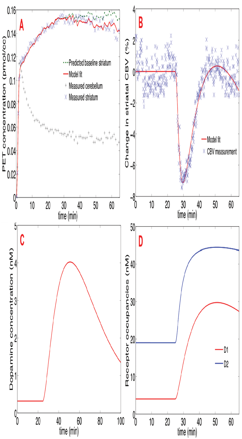
References
150
BRAIN-0488
BrainPET
CATEGORIZING NETWORKS OF VOXELS INTO BRAIN STATES BASED ON SEGMENTATION OF DOPAMINE LATENCY IMAGES
1Biomedical Engineering, Yale University, New Haven, USA
2Diagnostic Radiology, Yale University, New Haven, USA
3Psychiatry, Yale University, New Haven, USA
Abstract
A. Overall 'Latency” maps averaged across subjects and Brain State images segmented by time. 
References:
151
BRAIN-0662
BrainPET
INVESTIGATION OF THE VARIABILITY OF THE TSPO RADIOLIGAND [11C]PBR28 IN HUMAN BRAIN
1Imanova Centre for Imaging Sciences, Imanova Ltd, London, United Kingdom
2Division of Brain Sciences, Imperial College London, London, United Kingdom
Abstract
Objectives
[11C]PBR28 is a 2nd generation Translocator Protein (TSPO) radioligand for imaging neuroinflammation. Although [11C]PBR28 has demonstrated good radioligand properties, the variability in total volume of distribution (VT) and standardized uptake value (SUV) is high. Demonstration of the rs6971 single nucleotide polymorphism (SNP)[1] has accounted for a large component of this variability, but the remaining variability is still higher than expected from a ‘well behaved’ reversible ligand. Here, we investigate the inter- and intra-subject variability in a range of outcome measures and explore the contributions from blood and tissue.
Dynamic [11C]PBR28 PET scans from 34 healthy volunteers (19 HABs, 10 MABs and 5 LABs) with arterial sampling were analysed. 5 HABs and 3 MABs received a second scan approximately 3 months after baseline.
Inter- and intra-subject variability was investigated for 4 outcome measures across regions: I) VT using a 2-tissue compartmental model (2-TCM) with parent plasma input; II) distribution volume ratio (DVR) from 2-TCM VT with cortical grey matter as pseudo reference; III) SUV between 60-90 min (SUV60-90) and IV) SUVR derived as the ratio of SUV60-90 in tissue and cortical grey matter.
Individual variability in blood and tissue data was investigated. Plasma over blood ratios (pob) were fitted to an exponential plus constant model while controlling for the SNP, and cluster analysis was employed to investigate heterogeneity in tissue time activity data.
In HABs, the inter-subject variability was 28.2% in VT, 1.0% in DVR, 24.3% in SUV60-90 and 1.2% in SUVR in brain. The intra-subject variability was 19.8±14.0% in VT, 0.9±0.4% in DVR, 7.9±4.9% in SUV60-90, and 0.6±0.3% in SUVR (Figure 1A). The variability of AUC0-90min in total plasma for HABs was 21.2%/21.9% (Inter/Intra), 17.7%/18.8% in parent plasma, 18.2%/18.0% in whole blood, and 21.8%/13.8% in parent fraction. This suggests that ∼63% of the variability in total VT could be attributed to blood.
The pob curves were significantly different by genotype (Figure 1B) suggesting that access of [11C]PBR28 to blood cells was mediated by the affinity for TSPO. The tissue delivery of [11C]PBR28 (2-TCM) also varies across genotypes, with K1=0.17±0.05 in HABs, 0.11±0.03 in MABs and 0.09±0.02 mL/cm3/min in LABs.
Within HABs, two distinct tissue kinetic classes were observed with significant difference in K1 (0.23±0.05 vs. 0.18±0.05 mL/cm3/min, p<0.05) and VT (7.19±1.06 vs. 4.66±1.01 mL/cm3, p<0.001)(Figure 1C&D).
[11C]PBR28 variability in blood and tissue leads to the high variability observed for VT and SUV as compared to DVR and SUVR. Similar variability has been observed for other TSPO ligands (e.g. [11C](R)PK11195[2] and [18F]PBR111[3]) implicating unaccounted factors in TSPO biology rather than individual radioligand characteristics.
Our data highlights the challenges with identifying the correct input function, the existence of distinct kinetic tissue classes beyond the rs6971 SNP and opens the question of whether TSPO acts as a transporter for PET radioligands into cells.

References
152
BRAIN-0848
BrainPET
REFERENCE TISSUE-BASED KINETIC EVALUATION OF [F-18]AV-1451 IN AGING AND DEMENTIA
1Radiotracer development and imaging technology, Lawrence Berkeley National Laboratory, Berkeley, USA
2Radiology, University of Pittsburgh, Pittsburgh, USA
3Helen Wills Neuroscience Institute, University of California Berkeley, Berkeley, USA
4Memory and Aging Center, University of California San Francisco, San Francisco, USA
Abstract
Abstract
SRTM produced unrealistically large BPs (>15) or failed to converge for brainstem (n=28), entorhinal cortex (n=13), caudate (n=12), occipital (n=9), and anterior cingulate (n=6). The k2 values were higher in subcortical regions and lower in cortical. SRTM2 yielded reasonable values across all subjects and regions. DVR and SUVR values continued to increase over time (Figure 2) in cortical regions known to accumulate aggregated tau (temporal, parietal, occipital and frontal) in AD subjects, without plateau within the 150 minute scan.
Abstract
Subcortical regions (putamen, thalamus, caudate, and pallidum), assumed to exhibit off-target binding, showed a decrease in SUVR and DVR values over time and faster tracer washout relative to the reference region. Intermediate kinetics were observed in medial regions (hippocampus, precuneus, anterior and posterior cingulate).


References:
153
BRAIN-0587
BrainPET
MODEL TO DESCRIBE THE SPATIOTEMPORAL DISTRIBUTION OF MISFOLDED PROTEINS IN ALZHEIMER'S DISEASE
1Brain Sciences, Imperial College London, London, United Kingdom
2McConnell Brain Imaging Centre, Montreal Neurological Institute, Montreal, Canada
Abstract
The accumulation of misfolded proteins (β-amyloid and tau) in Alzheimer’s Disease (AD) follows a stereotypical pattern. We introduce a model to describe the spatiotemporal distribution of misfolded proteins and apply it to cross-sectional β-amyloid data obtained from the Alzheimer’s Disease Neuroimaging Initiative (ADNI).


from the ADNI database. The model was implemented in Matlab and nonlinear parameter optimisation was achieved using Levenberg-Marquardt.
The model provided good fits to the cross-sectional data (see Figure 1) both with and without spreading. The decrease in the residual sum of squares with distal spreading was -0.1% with a similar AIC for both models indicating that spreading is minimal.
The predicted regional production rates varied across regions, vi=0.2±0.05 years−1 [range:0.03-0.35] and the clearance rate was 0.31 years−1. The estimated parameter t*, ranged between 46.3 and 91.4 (72.0±7.7) years. Estimated values of T (= age at time of scan – t*) for the 4 groups are given in Table 1 along with results from an SUVr in anterior cingulate gyrus.
The presented model accurately describes β-amyloid data taken from ADNI. Inclusion of distal spreading did not significantly improve the performance suggesting that the behaviour is governed by local production and clearance. Future studies will investigate longitudinal data and application to tau where spreading may play a greater role.
Spatiotemporal model (without spreading) fit to β-amyloid data from 733 subjects (4 of 76 cortical regions displayed).
154
BRAIN-0584
BrainPET
SPECTRAL ANALYSIS OF [18F]FLUTEMETAMOL GREY AND WHITE MATTER KINETICS
1Nuclear Medicine and PET, Uppsala University, Uppsala, Sweden
2Life Sciences, GE Healthcare, Amersham, United Kingdom
3Nuclear Medicine & Molecular Imaging, KU Leuven Hospital, Leuven, Belgium
4Experimental Neurology, KU Leuven Hospital, Leuven, Belgium
Abstract
Most amyloid imaging PET tracers exhibit high uptake in the white matter (WM) to different degrees in spite of absence of fibrillar β-amyloid which is the intended target of the tracers. Although WM uptake has been hypothesized to be non-specific binding of equal magnitude in amyloid positive and negative subjects, recent studies suggest that the uptake of the amyloid imaging agent [11C]PiB in WM is due to specific binding to myelin basic protein β-sheet structures1,2, and as such could be useful for identifying WM lesions3. The aim of this study was to investigate kinetics of the uptake of the amyloid imaging agent [18F]flutemetamol in the WM in comparison with grey matter (GM).
Dynamic [18F]flutemetamol PET scans were performed in three elderly healthy volunteers (HV) and three patients with probable Alzheimer’s disease (AD) between 0-90 min, together with arterial blood sampling. Subjects underwent T1 MRI which was used to segment the image data into GM and WM. The data was analyzed with and without correction for partial volume effects (PVE) and time activity curves for total WM and GM of the whole brain were extracted. Kinetic components of total WM and GM uptake were identified using spectral analysis enabling analysis without a pre-defined assumption of the number of compartments and compared to the total volume of distribution (VT).
Three categories of exponential components were identified in GM and two in WM. The slowest component was detected in both GM and WM; the average contribution to VT of this was 35%(±25) in GM and 77%(±6) in WM when the data was uncorrected for PVE. After correction, the average contribution to VT was 16%(±18) in GM and 90%(±4) in WM. Two of the HV subjects and one AD subject had zero contribution of the slow component in GM, whereas in the remaining HV and two AD subjects, visually recognized as amyloid positive, 21-42% of VT was attributable to the slowest component.
The intermediate of the three components, contributing on average 40%(±16) to the VT in GM, was not detected in WM. The fastest component had an average contribution to the VT of 43%(±9) in GM and 10%(±4) in WM.
Spectral analysis indicated the presence of a dominating slow binding process of [18F]flutemetamol in the WM, much less prominent in GM uptake and following PVE correction completely absent in the GM in amyloid negative brains. Kinetic properties of GM and WM are thus easily distinguishable, but further studies are needed in order to identify the exact mechanism of the WM binding. If WM lesions alter tracer binding and the potential use of [18F]flutemetamol as a marker for WM injury remains to be investigated.
References
157
BRAIN-0969
Symposium
Neurovascular coupling in health and disease: the next chapter
1Brain and Mind Research Institute, Weill Cornell Medical College, New York, USA
Abstract
Brain function requires a finely regulated homeostatic balance between delivery of nutrients and clearance of waste products through blood flow. If the blood flow delivery does not match the dynamic energy requirements imposed by neural activity, brain dysfunction and damage may ensue. The mechanisms of the increase in blood flow evoked by neural activity have been the subject of extensive investigation over the past two centuries, and our appreciation of their complexity has increased dramatically over the time. Whereas early models emphasized direct metabolic or synaptic interactions between neurons on cerebral blood vessels, current constructs have evolved towards a coordinated interaction among multiple cell types, neurons, astrocytes, vascular-perivascular cells, etc., converging on the microvasculature to regulate flow. These cells, collectively termed the “neurovascular unit”, work in concert to implement the changes in microvascular network resistance underlying the temporally and spatially focused increases in cerebral blood flow triggered by neural activation. Increasing evidence implicates neurovascular unit dysfunction in brain diseases associated with cognitive deficits. Although cognitive dysfunction caused by vascular factors (vascular cognitive impairment) or neurodegeneration (Alzheimer’s disease, AD) have traditionally been considered distinct, recent data suggest that alterations in cerebral blood vessels play a role in both. In addition to blood flow, alterations in cerebral blood vessel ultrastructure resulting in disruption of the blood-brain barrier may also play a role, particularly in AD, vascular dementia due to hypertension, and frontotemporal dementia, the major cause of dementia in the middle aged. These observations, collectively, indicate that neurovascular dysfunction is involved in a wide variety of diseases affecting cognitive function and support the use of approaches to safeguard cerebrovascular health in their prevention. At the same time, research efforts need to continue to unravel the cellular basis of the hemodynamic response induced by activation and gain a better insight into how its dysfunction leads to the synaptic alterations underlying cognitive impairment.
158
BRAIN-0988
Symposium
Blood brain barrier alterations in neurodegenerative diseases: towards a molecular understanding
1Keck School of Medicine, Zilkha Neurogenetic Institute, Los Angeles, USA
Blood brain barrier alterations in neurodegenerative diseases: Toward a molecular understanding
Zilkha Neurogenetic Institute, Keck School of Medicine University of Southern California, Los Angeles, CA 90089-2821
Abstract
Blood vessels in the brain are organized with surprising precision, patterned in parallel with the major brain circuits tasked with sensation, memory and motion. This tight interrelationship may reflect key functional roles in neuronal normal function, disease and brain aging. I will discuss the cellular and molecular mechanisms within the neurovascular unit and the blood-brain barrier (BBB) including aberrant pericyte-endothelial and pericyte-astrocyte signal transduction that can lead to BBB breakdown, dysregulated and reduced cerebral blood flow responses, diminished oxygen and glucose supply leading to cerebral microvascular degenerative changes and causing secondary neuronal injury, neurodegeneration, loss of connectivity and neuronal loss. Examples in animal models and human studies using molecular and imaging biomarkers of neurovascular and brain functions will be provided. I will also discuss how highly validated genetic risk factors for late onset Alzheimer’s disease (APOE4, PICALM) and early autosomal Alzheimer’s disease (PSEN1) affect the neurovascular unit and BBB functions and brain function and structure.
159
BRAIN-0980
Symposium
Neurovascular and cognitive failure in Alzheimer's disease
1McGill University, Montreal Neurological Institute, Montréal QC, Canada
Abstract
163
BRAIN-0738
Brain Oral Communication
IN SILICO INVESTIGATION OF REGIONAL CEREBRAL BLOOD FLOW TO ACCOUNT FOR THE DISCREPANCY AMONG MEASURES BY DIFFERENT PERFUSION IMAGING METHODS
1Department of Bio and Brain Engineering, Korea Advanced Institute of Science and Technology, Daejeon, Korea
Abstract
164
BRAIN-0355
Brain Oral Communication
LONGITUDINAL MONITORING OF MICROGLIAL ACTIVATION AFTER STROKE WITH A NOVEL SPION-ENHANCED MRI METHOD IN A RAT MCAO MODEL
1Neurology, University of New Mexico, Albuquerque, USA
2College of Pharmacy, University of New Mexico, Albuquerque, USA
Abstract
165
BRAIN-0796
Brain Oral Communication
LABEL-FREE OPTICALLY QUANTIFIED CORTICAL METABOLIC RATE OF OXYGEN CONSUMPTION
1Biomedical Engineering, University of California Davis, Davis, USA
Objectives
Abstract
Positron emission tomography (PET) with the 150 labeled gas tracer 0150, while technically complex, remains the gold standard measure of oxygen consumption1. Here, we present a new all-optical method of measuring mouse cortical cerebral oxygen metabolism (CMRO2) quantitatively that uses measurements of three key parameters: cortical cerebral blood flow (CBF), arteriovenous oxygen saturation difference (Ya-Yv), and hematocrit ([Hbtotal]). This method involves scanning a visible light beam of a few milliwatts average power (the same as a laser pointer) on the sample, and does not require an exogenous oxygen probe.
A visible light Optical Coherence Tomography (OCT) system was constructed to image the mouse cerebral cortex. Individual measurements were performed using Doppler2 and Spectroscopic3 algorithms through a thinned skull cranial window4. Detailed and systematic ex vivo validation of saturation and hematocrit measurements were performed, demonstrating the capability to quantify both. A series of in vivo experiments were performed to verify that CMRO2, saturation, and hemoglobin concentrations behaved as expected under a range of physiological manipulations including modulation of the fraction of inspired oxygen (FiO2), modulation of the fraction of inspired carbon dioxide (FiCO2), and cardiac arrest.
Baseline metabolic measurements in mice are shown to be consistent with literature values (∼100-300 µmol/100g/min). CMRO2, as measured using our method, does not change during minor variations in FiO2 and FiCO2, in spite of much larger changes in oxygen saturation and flow. In particular, the figure shows that during mild hypoxia (FiO2=16%), saturation drops in both arteries and veins (A). Overall, there is a slight increase in oxygen extraction during mild hypoxia (B). However, there also is a slight decrease in blood flow during mild hypoxia (C). These results are consistent with the assertion that CMRO2 remains constant during mild hypoxia. During mild hypercapnia, vessels dilate and flow increases. Accordingly, the venous saturation increases considerably due to the lower tissue oxygen extraction (D), consistent with the assertion that CMRO2 remains unchanged. Complete conversion of oxy-hemoglobin to deoxy-hemoglobin after loss of brain blood flow upon cardiac arrest was also verified.
In conclusion, a method of measuring CMRO2 using three optical measurements of cortical blood flow, arteriovenous oxygen saturation difference, and hematocrit using a single instrument was presented. Several key validation experiments were performed. To provide further methodological validation, these methods will be compared against gold standard measurements such as PET in the future.

References
166
BRAIN-0141
Brain Oral Communication
MAGNETIC RESONANCE ADVECTION IMAGING (MRAI): SENSITIVITY TO PULSATILE FLOW
1Radiology, Weill Cornell Medical College, New York, USA
2Stochastic Algorithms and Nonparametric Statistics, WIAS, Berlin, Germany
3Neurology, Weill Cornell Medical College, New York, USA
Abstract
Objectives
Magnetic Resonance Advection Imaging (MRAI, [1]) is an imaging modality to obtain velocity vector maps related to vascular dynamics directly from spatiotemporal differential modeling of EPI data. It has the potential to provide precisely localized, in relation to BOLD-EPI data, information about vascular dynamics, which might aid the interpretation of (resting state) functional MRI experiments affected by vascular anatomy [2, 3]. Up to now, the biophysical origins of the estimated velocity vectors were unknown. Here we provide evidence that for a certain spatiotemporal scale MRAI maps depict areas affected by pulsatile flow [4].
It is assumed that the BOLD signal ρ(
∂ρ(
with the flux having only an advective component, i.e.,
Abstract
∂ρ(
Parameters such as the velocity can be estimated from spatiotemporal data by multiple regression [6], in this case from
Y = const + ux X1 + uy X2 + uz X3 + ε,
with Y = ∂ρ(
Abstract
In order to compute coherence between the cardiac cycle and estimated velocities for each voxel in the brain, pulse-oximetry data, pulse(t), was recorded from the left hand of an adult male volunteer (rest, eyes closed) during a multiband-EPI acquisition [7] on a 3.0 T MRI scanner (TR = 187 ms, 2 mm isotropic resolution, 1000 repetitions, scan time 3:07 min).
MRAI maps of estimated average velocities showed venous, arterial, and CSF components that correspond to anatomy visible in a time-of-flight (TOF) angiogram of the same subject (see Figure). A map displaying max[coherence(pulse(t), ρ(
MRAI partially reproduces vascular anatomy. For the chosen temporal and spatial resolution, the observed MRAI maps are mainly affected by pulsatile flow components but are more specific to vascular anatomy than maps correlating pulse with voxel intensity; the latter one probably picking up motion in adjacent tissue as well. We believe that MRAI at this spatiotemporal scale can contribute to the modeling of the cerebrovascular system with potential for application as a biomarker for cerebrovascular disease.

References
167
BRAIN-0543
Brain Oral Communication
MULTI-EXPOSURE, CONTINUOUS LASER SPECKLE CONTRAST IMAGING OF MOUSE BRAIN ENABLED BY A NOVEL SINGLE PHOTON AVALANCHE DIODE (SPAD) ARRAY
1Medical Optics, ICFO-Institut de Ciencies Fotonique, Castelldefels, Spain
2Dip. Elettronica Informazione e Bioingegneria, POLIMI-Politecnico di Milano, Milano, Italy
3Department of Brain Ischemia and Neurodegeneration Institute for Biomedical Research (IIBB) Spanich Council Research (CSIC), IDIBAPS - Institut d´Investigacions Biomediques August Pi i Sunyer, Barcelona, Spain
Abstract
Fitted values of τ C and changes of cerebral blood flow for each challenge

References:
170
BRAIN-0611
BrainPET
ENDOTOXIN ENHANCES METHYLPHENIDATE-INDUCED DOPAMINE RELEASE MEASURED WITH [11C]-RACLOPRIDE PET
1Biomedical Engineering, Yale University School of Engineering Arts and Sciences, New Haven, USA
2Chemistry, Yale PET Center, New Haven, USA
3Research Coordination, Yale PET Center, New Haven, USA
4Clinical Neuroscience, Yale University School of Medicine, New Haven, USA
Abstract
It is known that the brain monitors inflammatory signals from the periphery, but details on how the brain incorporates and uses this information is lacking. Endotoxin-induced systemic inflammation in rodents causes an increase in striatal dopamine (DA). Here, we used methylphenidate to induce DA release and [11C]-raclopride PET to measure it in the human striatum. The change in DA release in the presence of endotoxin was the primary outcome measure.
Five healthy subjects received [11C]-raclopride PET scans in 2 conditions: methylphenidate (MP) alone and methylphenidate plus endotoxin (MP+E). MP (40 mg) was given 60 minutes before tracer administration in each condition, and endotoxin (0.8 ng/kg) was given intravenously 30 minutes before tracer administration in the MP+E condition. Each condition was performed on a separate day, and each drug scan followed a baseline scan (total of 4 scans per subject). PET images were obtained on a Siemens HR+ and corrected for motion, scatter, randoms, and dead time. Modeling of regional PET time activity curves was performed using the Simplified Reference Tissue Model with the cerebellum as the reference region to obtain regional non-displaceable binding potential (BPND abbreviated here as BP). DA release was measured as percent change of BP from baseline (ΔBP) and was calculated as:
ΔBP = (BPbaseline - BPcondition)/(BPbaseline) * 100
Subjective sickness symptom ratings (0-4 from negligible to severe) were recorded for each subject in each scan every 30 minutes. Levels of peripheral inflammatory cytokines were measured from blood plasma every 30 minutes in drug scans.
Striatal regions displayed high test-retest reliability based on a comparison of each subject’s two separate baseline scans (intraclass correlation coefficient = 0.97, fitted linear slope = 0.95, r2 = 0.96). Whole striatum mean ΔBP was 13.9% +/- 1.3% in the MP+E condition compared to 3.5% +/- 1.5% in the MP condition. The MP+E condition displayed significantly greater mean ΔBP in whole striatum (p<0.005), caudate (p<0.005), and right putamen (p<0.05) as compared to the MP condition. As expected, endotoxin produced mild sickness symptoms and a robust increase in blood levels of inflammatory cytokines.
This study shows for the first time that acute systemic inflammation induced by endotoxin enhances MP-induced synaptic DA levels in the human striatum, a finding that is consistent with rodent studies and with the fact that striatal dopamine release can signal both rewarding and aversive stimuli.

171
BRAIN-0477
BrainPET
DETECTING DOPAMINERGIC MODULATION INDUCED CAMP CHANGES IN MONKEY BRAIN BY PDE10A PET IMAGING
1Department of Clinical Neuroscience, Karolinska Institutet, Stockholm, Sweden
2Neuroscience Research Unit, Pfizer Inc., Cambridge MA, USA
3Division of Synaptic Transmission, H. Lundbeck A/S, Valby, Denmark
4Division of Discovery Chemistry and DMPK, H. Lundbeck A/S, Valby, Denmark
Abstract
rent patterns of relative change in BPND after dopaminergic modulation between substantia nigra (SN) and caudate nucleus (CN).
References:
172
BRAIN-0365
BrainPET
CEREBRAL SEROTONIN 4 RECEPTOR BINDING IS NEGATIVELY ASSOCIATED WITH THE CORTISOL AWAKENING RESPONSE IN HEALTHY VOLUNTEERS
1Neurobiology Research Unit, Copenhagen University Hospital Rigshospitalet, Copenhagen, Denmark
Abstract
Serotonin signaling is considered critical for an appropriate adaptation to stress. Previously we have shown that prefrontal serotonin transporter (SERT) binding is positively associated with hypothalamic-pituitary-adrenal (HPA)-axis output as indexed by cortisol awakening response (CAR) (Frokjaer V.G. et al. 2013). This implies that prefrontal SERT, which regulates synaptic serotonin, is coupled to CAR. However, it remains elusive if the coupling is mediated through synaptic serotonin. CAR is a distinct feature of the HPA-axis output and reflects both the increase in response to awakening and the following decrease due to inhibitory feedback regulation. CAR is thus considered an index of HPA-axis reactivity and its response to, e.g., psychosocial stressors; CAR has for example been found blunted in mood disorders (Fries E. et al. 2009). Cerebral serotonin 4 (5-HT4) receptor binding as measured with [11C]SB207145 PET has in preclinical and clinical studies recently been shown to index serotonin levels (Haarh M.E. et al 2014). Here, we investigated in healthy individuals if cerebral 5-HT4 receptor binding - as a probe for serotonin levels – is associated with CAR.
Thirty healthy volunteers (25 male, 5 female, age range 20 to 56 years) underwent PET imaging with [11C]-SB207145, genotyping of the serotonin-transporter-linked polymorphic region (5-HTTLPR), and performed serial home sampling of saliva to assess CAR, defined as the area under curve with respect to increase from baseline from 0-60 minutes after awakening (unit: nmol/l*min). The association was tested in a multiple linear regression model. The primary model included CAR as the predictor, 5-HT4 binding (BPnd) as the outcome, and adjusted for age and 5-HTTLPR genotype. Three regions of interest (ROIs) were defined a priori: prefrontal cortex, anterior cingulate cortex and pallidostriatum.
CAR was negatively associated with 5-HT4 receptor binding in pallidostriatum (p=0.01), frontal cortex (p=0.03) and anterior cingulate cortex (p=0.002) with parameter estimates of -3.28, -14.6, and -12.4 nmol/l*min per 0.01 BPnd, respectively. The 5-HTTLPR status did not show a main effect on CAR, nor did it moderate the association between CAR and 5-HT4 binding as tested in an interaction analysis. The CAR association with 5-HT4 proved robust, remaining significant in age-restricted subsamples (<40 years) and when taking into account other potentially relevant covariates (sex, BMI, Cohen’s perceived stress scale, Neuroticism, absolute cortisol concentrations at wake-up or sleep length on the day of the samples).
In healthy volunteers, we find a robust negative association between CAR and 5-HT4 receptor binding in prefrontal and anterior cingulate cortex, and pallidostriatum. We speculate that high synaptic serotonin, as indexed by low 5-HT4 binding, is of importance to maintain HPA-axis dynamics, i.e. a robust CAR. Future studies with experimental manipulation of synaptic serotonin must elucidate if acute and/or subacute changes in serotonergic tone affect CAR and, further, if direct 5-HT4 receptor signaling plays a role in HPA-axis regulation including in relevant clinical populations, e.g. major depression.
173
BRAIN-0578
BrainPET
SIMULTANEOUS PET/MRI MEASUREMENTS OF HEMODYNAMIC RESPONSE TO µ-OPIOID RECEPTOR OCCUPANCY
1Department of Radiology, Massachusetts General Hospital Harvard Medical School, Charlestown, USA
Abstract
The molecular mechanisms underlying the development of opioid addiction remain incompletely understood. Interaction between the brain’s opioid and dopamine systems has been highlighted as potentially pivotal in opioid addiction. Simultaneous PET/MRI could be used to investigate inter-related receptor system interactions and to dissect complex fMRI signals into neurochemical constituents. In this study, we measure the relationship between μ-opioid receptor occupancy and fMRI response, and to examine how different receptor systems contribute to a composite fMRI signal.
Ten PET/MRI scans were acquired on two macaques (male, ∼12 kg). Animals were anesthetized with isoflurane and ventilated. Images were acquired on a 3T Siemens BrainPET with an 8-channel coil. PET/MRI scans were acquired from each animal using a μ-opioid selective radiotracer (∼10 mCi [11C]carfentanil) given as a bolus-infusion. PET data were stored in list mode and binned into 1-min frames. CBV-fMRI data were obtained following an iron oxide (Feraheme, 10 ug/kg, i.v.)1-3 injection. Graded doses of an opioid receptor antagonist, naloxone (baseline, 0.01, 0.03, and 0.05 mg/kg) were given intravenously at 35 min post radiotracer bolus injection. In addition, one dose of a potent μ-opioid agonist, remifentanil (10 μg/kg) was used as the challenging drug. PET data was analyzed for binding potentials referenced to a non-displaceable compartment (BPND) using the simplified reference tissue model4. A gamma-variant function was used to model the PET and fMRI temporal response to drug challenge. Changes in fMRI signal were converted to CBV changes1-3.
Baseline PET BPND maps showed a high-level of specific binding in the thalamus, caudate, putamen, frontal cortex, which corresponded well to known distribution of μ-opioid receptors in NHPs (Figure). μ-Opioid receptor BPND and percent CBV reduced in a dose-depended manner to naloxone challenges (Figure). A dose of 0.05 mg/kg naloxone achieved >90% receptor occupancy. The largest BPND reductions were observed in the thalamus and caudate, while the largest CBV changes were observed in the putamen (Figure). Regional analysis of the BPND and CBV data revealed a 2nd-order polynomial coupling relationship. Naloxone induced a negative CBV response, which could be due to activating the inhibitory neurotransmitter (GABA) and/or its downstream effects (i.e. GABA depletes basal level of dopamine in the basal ganglia). At a given receptor occupancy, the ratio of putamen:caudate %CBV change was ∼1.7, implying the possibility that the CBV responses observed in the basal ganglia are dominated by the indirect opioid-dopamine mechanism1,2. Remifentanil challenge showed the opposite sustained CBV-fMRI responses3. Future pharmacological studies modulating the GABA and dopamine systems are needed to confirm the opioid direct vs. indirect modulations on the fMRI signals
Using simultaneous PET/MRI with pharmacological challenges in NHPs, we determined the engagement of the opioid system and the resulting CBV-fMRI implicated a dopaminergic component in limbic basal ganglia. Simultaneous PET/MRI provides the unique opportunity to directly relate neurochemical events to functional responses and has great potential to facilitate drug development.
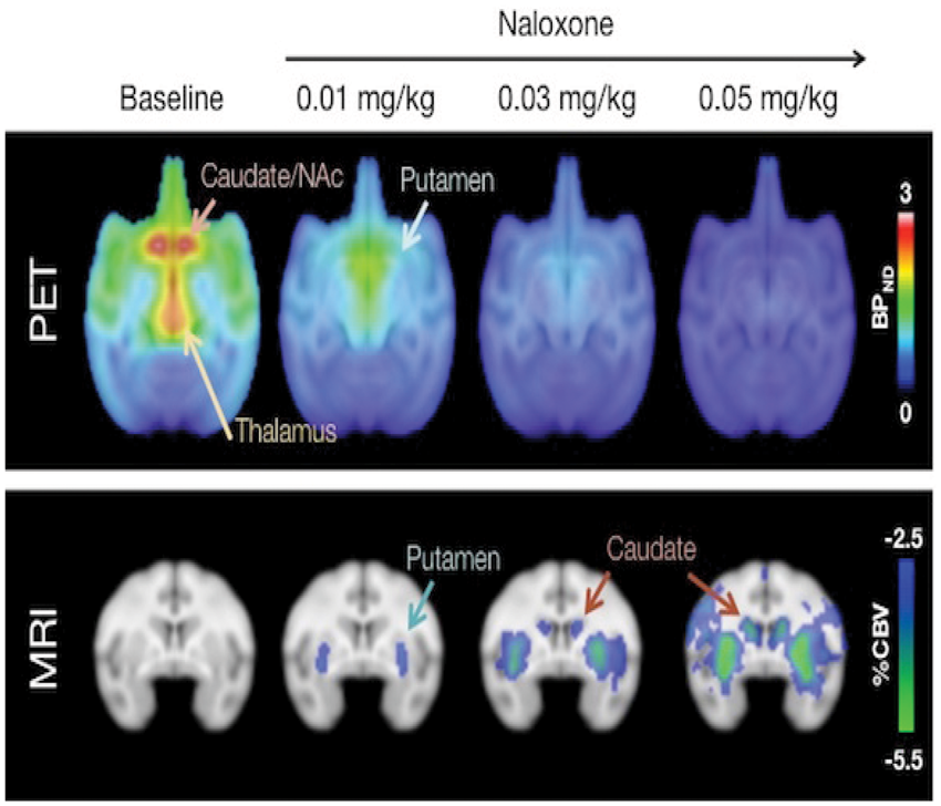
References:
174
BRAIN-0742
BrainPET
BLOCKADE OF TRANSLOCATOR PROTEIN (TSPO) BY XBD173 TO MEASURE SPECIFIC BINDING OF 11C-(R)-PK 11195 IN HUMAN BRAIN: AN ONGOING STUDY
1National Institute of Mental Health, National Institutes of Health, Bethesda, USA
Abstract
Figure: Lassen plots with VT and VT/fP on HABs
175
BRAIN-0702
BrainPET
PK-PD ANALYSIS OF OCCUPANCY-CONCENTRATION-TIME CHARACTERISTICS OF A NOVEL GLYCINE TRANSPORTER-1 (GLYT1) INHIBITOR EXHIBITING PHARMACOLOGICAL HYSTERESIS IN NON-HUMAN PRIMATES AND HUMANS USING [18F]CFPYPB
1., AbbVie Inc., North Chicago, USA
2., AbbVie Deutschland GmbH & Co KG, Ludwigshafen, Germany
Abstract
Objective: The glycine transporter-1 (GlyT1) has been implicated in the pathophysiology of schizophrenia and is pursued as a target for therapeutic drug development. The purpose of this study was to model the receptor occupancy-concentration-time relationship of a novel GlyT1 inhibitor in non-human primates and in human volunteers.
Methods: [18F]CFPyPB [1] dynamic PET data were collected in 3 cynomologus monkeys (cynos), 4 baboons and 9 healthy volunteers with arterial sampling prior to and at multiple time points (0h-32h) post administration of a novel GlyT1 inhibitor at varying dose levels. The 3 cynos received test-retest scans separated by 1-3 weeks. Subject-space atlas based on anatomical MRI was coregistered to PET images of each animal to derive regional time activity curves for high binding regions: brainstem, pons, midbrain, thalamus, cerebellum and low binding regions: frontal, temporal and occipital cortex. Volume of distribution (VT) values were derived using both Logan and 2TCM+Vb models. Receptor occupancy was determined using Lassen occupancy plot [2] and pseudo reference tissue method [3] using occipital and whole cortex as the pseudo reference region in primates and humans respectively. The occupancy-concentration-time relationship were modeled using a population based biophase model [4] and an indirect model [5]. The model relationship derived from primate data was applied to human PK time course to predict the human occupancy time course and confirmed with observed human occupancy. The model was then fit to human data to estimate the half-life, kon, koff and Kd at the effect site.
Results: Logan and 2TCM+Vb methods were able to fit the data well. The test-retest variability of VT across all regions were 6%, 8% and 20% in the 3 cynos tested. The VT (ml/cm3) ranged between 3-14 in cynos, 2-14 in baboons and 2-9 in humans. Dose-dependent receptor occupancy was observed following different doses of the GlyT1 inhibitor. Hysteresis was observed in the plasma concentration-occupancy relationship. The indirect model and the biophase model with a semi-mechanistic effect-site compartment fit the data well.
Conclusion: Dose-dependent and time-variant GlyT1 receptor occupancy was successfully modeled. Such PK-PD modeling approach can be leveraged to predict human occupancy time course from preclinical species and to predict repeat-dose occupancy using occupancy data obtained after administration of a single dose [5].
Disclosures: All authors are employees of AbbVie. The design, study conduct, and financial support for this research were provided by AbbVie. AbbVie participated in the interpretation of data, review, and approval of the publication.
References:
178
BRAIN-0533
Brain Oral Communication
CD69 PLAYS A BENEFICIAL ROLE IN ISCHEMIC STROKE POTENTIALLY VIA THE MODULATION OF LEUKOCYTE RECRUITMENT AND SECONDARY MICROTHROMBOSIS
1Institut d’Investigacions Biomèdiques de Barcelona (IIBB), Consejo Superior de Investigaciones Científicas (CSIC) Institut d’Investigacions Biomèdiques August Pi i Sunyer (IDIBAPS), BARCELONA, Spain
2Hemotherapy and Haemostasis Service, Hospital Clinic, BARCELONA, Spain
3Grupo de Activación Inmunológica Centro Nacional de Microbiología, Instituto de Salud Carlos III (ISCIII), MADRID, Spain
Abstract
Objectives: Expression of CD69 is a hallmark of lymphocyte activation and the majority of research on CD69 has focused on its effects on the immune system. Several lines of evidence support that inflammatory and immune responses are involved in stroke brain damage. The aim of this study was to examine whether CD69 plays a role in stroke outcome and to investigate the underlying mechanisms.
Methods: Cerebral ischemia was produced by 45-min intraluminal middle cerebral artery occlusion (tMCAo) followed by reperfusion in male CD69 KO (n=56) and Wt (n=68) mice. In addition, permanent distal MCAo (pMCAo) was induced in male CD69 KO (n=28) and Wt (n=41) mice and in Rag2-/- CD69-/- (n=27) and Rag2-/- CD69+/+ (n=31) mice. Neurological impairment was assessed and brain infarct and edema volume were measured using MRI (T2 maps). Flow cytometry was used to measure changes in immune cell populations in the brain, spleen, cervical lymph nodes (CLNs) and the blood. PCR was performed to measure changes in inflammatory gene expression in the brain. Circulating von Willebrand factor (vWF) levels (ELISA) and function (Collagen Binding Assay) were studied in plasma, and fibrin(ogen) deposition was also studied in cerebral blood vessels (Western Blot). In other mice, we performed the tail-bleeding test. Animal work was carried out in compliance with Spanish law and with approval of the Ethics Committee (CEEA) of the University of Barcelona.
Results: CD69 KO mice had a significantly larger infarct volume compared to Wt mice at 24 and 72h after tMCAo (P<0.05). Also, CD69 deficiency increased infarct volume 24h after pMCAo (P<0.05). Following tMCAo, CD69 KO mice were more functionally impaired using a neurological score and tape test than the Wt mice. 96h after tMCAo, CD69 KO mice had a greater percentage of T cells (CD45hi CD3+) in the CLNs, spleen and brain compared to WT mice and less B cells (CD45+ CD45R+) in the CLNs. There was also a greater number of neutrophils (CD11b+ Ly6G+) in the brain. To test whether the absence of CD69 in lymphocytes was responsible for the observed effects, we carried out pMCAo in lymphocyte deficient Rag2-/- mice. Rag2-/- mice showed smaller infarct volumes than the Wt mice (P<0.001) and CD69 deficiency in Rag2-/- mice increased infarct volume (P<0.001) demonstrating that the absence of CD69 in cells other than lymphocytes was playing a role. In addition to lymphocytes, CD69 is expressed in platelets, which led us to hypothesize that CD69 deficiency might affect thrombosis. Preliminary findings in the tail-bleeding test showed a trend for less total bleeding time and less re-bleeds in the CD69 KO mice compared to the Wt mice. In the plasma, the concentration and activity of vWF was higher 6h after pMCAo in CD69 KO mice compared to Wt mice (P<0.05). In cerebral blood vessels, CD69 deficiency enhanced vWF expression and fibrin(ogen) deposition after ischemia.
Conclusions: Our results suggest that CD69 may play a beneficial role in cerebral ischemia by regulating leukocyte recruitment and secondary local microthrombosis.
Funding: Supported by the Spanish Ministry of Economy (SAF2011-30492).
179
BRAIN-0065
Brain Oral Communication
INCREASED TONE OF BRAIN PARENCHYMAL ARTERIOLES DURING EARLY POST-ISCHEMIC REPERFUSION IS ASSOCIATED WITH INCOMPLETE REPERFUSION AND GREATER INFARCTION
1Neurological Sciences, University of Vermont College of Medicine, Burlington, USA
Abstract

References:
180
BRAIN-0237
Brain Oral Communication
PREDICTORS OF MORTALITY IN ACUTE ISCHEMIC STROKE INTERVENTION: ANALYSIS OF THE NASA REGISTRY
1Miami Cardiac & Vascular Institute, Baptist Health South Florida, Miami, USA
2Center for Research & Grants, Baptist Health South Florida, Miami, USA
3Neuroscience Center, Baptist Health South Florida, Miami, USA
4Radiology Division of Diagnostic & Interventional Neuroradiology, Massachusetts General Hospital, Boston, USA
5Neurosurgery, Methodist Neurological Institute, Houston, USA
6Neurology, Baptist Health Louisville, Louisville KY, USA
7Neurology, Medical College of Wisconsin/Froedtert Hospital, Milwaukee WI, USA
8Neurology, Emory University School of Medicine, Atlanta, USA
9Neurology, Delray Medical Center, Delray Beach FL, USA
10Neurointerventional Services, California Pacific Medical Center, San Francisco, USA
11, Alexian Brothers Medical Center, Elk Grove Village IL, USA
12Neurology, Wayne State University School of Medicine, Detroit, USA
13Radiology, West Virginia University Hospital, Morgantown WV, USA
14Neurology Neurosurgery and Radiology, Vanderbilt University Medical Center, Nashville, USA
15Neurology Neurosurgery and Radiology, Boston Medical Center, Boston, USA
16Radiology and Neurology, UT Southwestern Medical Center, Dallas, USA
Abstract
In acute ischemic stroke from large vessel occlusion (LVO), failure to recanalize strongly predicts mortality. We used the retrospective North American Solitaire Acute Stroke (NASA) registry (approved by each entity's Institutional Review Board with waiver of consent) to investigate baseline characteristics, recanalization parameters, and symptomatic intracranial hemorrhage (sICH) in association with mortality (90-day mRS=6).
Fisher’s exact test compared 90-day mortality between patients with successful and failed recanalization. In successfully recanalized patients (TICI≥2b), logistic regression evaluated baseline characteristics and recanalization outcomes for association with 90-day mortality. A multivariable model was developed based on backwards selection with retention criteria of p<0.05 from factors with at least marginal significance (p≤0.10) on univariate analysis. This model was refit to minimize the number of excluded cases; the c-statistic was used as a measure of predictive power.
Patients who were successfully recanalized had lower mortality compared to those whose recanalization failed [25.2% (59/234) vs. 46.9% (38/81) p<0.001], but there was no difference in the incidence of sICH between groups [9% (21/234) vs. 14% (11/79), p=0.205]. However, mortality was significantly higher in patients with sICH compared to those without [72% (23/32) vs. 26% (73/281), p<0.001]. In successfully recanalized patients, univariate analysis identified increased risk of mortality for: proximal occlusion (ICA or vertebrobasilar), initial NIHSS≥18, use of rescue therapy (p<0.05), final TICI 2b and 3+ passes (p<0.10). In the multivariate model with good predictive power (c-index=0.72), proximal occlusion, initial NIHSS≥18 and use of rescue therapy were significant independent predictors of 90-day mortality.
In the NASA registry, failure to recanalize and presence of sICH resulted in increased mortality. Furthermore, despite successful recanalization, proximal occlusion, severe neurological deficit at stroke onset, and need for rescue therapy were predictors of mortality.
181
BRAIN-0328
Brain Oral Communication
NEUROPROTECTIVE ROLE OF CARBON MONOXIDE RELEASE FROM CELL-FREE PEGYLATED HEMOGLOBIN DURING REPERFUSION FROM TRANSIENT FOCAL CEREBRAL ISCHEMIA
1Department of Anesthesiology and Critical Care Medicine, Johns Hopkins University, Baltimore, USA
Abstract
Reference:
182
BRAIN-0118
Brain Oral Communication
EFFECT OF NEURONAL NO SYNTHASE INHIBITION BY 7-NI IN A JUVENILE CEREBRAL ISCHEMIC MODEL: PENUMBRAL REPERFUSION IMPROVEMENT
1PICU- Armand-Trousseau, AP-HP, PARIS, France
2Physiology unit- Lariboisière, AP-HP, PARIS, France
3U1141, INSERM, PARIS, France
4PICU- Necker, AP-HP, PARIS, France
5PICU- Robert Debré, AP-HP, PARIS, France
Abstract
183
BRAIN-0693
Brain Oral Communication
IN VIVO IMAGING OF COLLATERAL BLOOD PERFUSION DYNAMICS DURING FOCAL STROKE
1Bioengineering, University of Washington, Seattle, USA
Abstract
BACKGROUND AND OBJECTIVE
Changes in blood perfusion in highly interconnected pial arterioles provide important insights about hemodynamic homeostasis at the ischemic region in brain. Penetrating arterioles are other important components of blood flow regulation, as they play an essential role in delivering blood from a highly collateralized pial arteriole network to capillaries in cerebral cortex. In case of stroke, the source of blood to these vital pathways to penumbra is regulated by pial arterioles. Within the highly collateralized pial arteriole network, major cerebral arteries, such as middle cerebral artery and anterior cerebral artery, are interconnected by arteriolo-arteriolar anastomoses (AAAs), with penetrating arterioles attaching to them as T-junctions. Here, we evaluate vessel diameter, red blood cell (RBC) velocity, and total flow changes in significant number of pial and penetrating arterioles in relation with AAA to provide an insight about AAA’s role in active hemodynamic regulation during focal stroke.
A fiber-based spectral domain optical coherence tomography system1 is used in this study to acquire volumetric images of mouse cerebral cortex in vivo through a cranial window. The system operates on a central wavelength of 1340 nm with an A-line rate of 92 kHz, and provides ∼7 μm axial and ∼10 μm lateral resolution with 0.12 mm depth of field. We apply optical microangiography (OMAG) techniques1,2 to evaluate vessel diameter and RBC velocity changes in a large number of pial and penetrating arterioles within AAA abundant and AAA scarce regions overlie the stroke penumbra in the parietal cortex after middle cerebral artery occlusion (MCAO). In contrast to other techniques, our methods make it possible to image a larger set of vessels with a high resolution in a short imaging time without administering exogenous contrast agents during a time-constrained MCAO experiment.
We evaluated diameter and flow fluctuations in total 143 surface and 127 penetrating arterioles overlie stroke penumbra on four mice during MCAO. Three main findings are highlighted: (1) Flow reversals occur in pial arterioles through anastomosis, and ACA takes over blood supply to the penetrating arterioles attaching to MCA side. (2) Penetrating arterioles dilate near strong AAAs, and sufficiently restore flow to the ischemic region. (3) The flow compensation grows weaker as getting further away from AAA, resulting in poorly recovered penetrating arterioles residing further from AAA connections during reperfusion.
Thanks to the high sensitivity and the large field of view provided by OMAG, we compare areas in mouse cerebral cortex closer to, or further away from AAAs during MCAO in mouse cerebral cortex after focal stroke. The results suggest that AAA plays a major role in active regulation of the pial arterioles during stroke, providing blood flow for active dilation of penetrating arterioles to rescue stroke penumbra.

REFERENCES
186
BRAIN-0710
Brain Oral Communication
INFLAMMATORY AND ANGIOGENIC RESPONSE TO CHRONIC HYPOXIA IN MOUSE BRAIN EVALUATED BY POSITRON EMISSION TOMOGRAPHY AND HISTOLOGICAL STUDIES
1Molecular Imaging Center, National Institute of Radiological Sciences, Chiba, Japan
2Département de Chimie Moléculaire, CNRS-Université Joseph Fourier, Grenoble, France
3Institut des Biomolécules Max Mousseron, UMR 5247 École Nationale Supérieure de Chimie de Montpellier, Montpellier, France
4Brain Science Inspired Life Support Research Centre, University of Electro-Communications, Chofu, Japan
Abstract
[Objectives] We reported morphological and functional changes in cerebral vascular system in mice induced by chronic hypoxia (Takuwa 2013, Masamoto 2014, Sekiguchi 2014). The present study aims to evaluate the occurrence of chronic hypoxia-induced inflammatory and angiogenic responses in mouse brain using positron emission tomography (PET) and histological studies.
[Methods] Thirty-five mice, 8 to 12 weeks of age, kept in hypoxic chamber with 8-9% oxygen concentration for 0, 4, 7 and 14 days, were used for the subsequent assays. Firstly, the in vivo expression of translocator protein 18 kDa (TSPO), a known biomarker of brain inflammation, was examined by dynamic PET scans with 11C-PK11195. In vitro autoradiography (ARG) studies were performed to evaluate the specific binding of 11C-PK11195 and the distribution of TSPO in brain tissues. Up-regulated Iba1 expression in activated microglia, as a sign of inflammation, was examined by immunohistofluorescence staining. Secondly, the expression of αVβ3 integrin, a known angiogenesis biomarker, was examined by PET imaging and ex vivo ARG using the αVβ3 integrin-specific radiotracer 64Cu-cyclam-RAFT-c(-RGDfK-)4 (Jin 2012). Double immunohistofluorescence staining of the pan-endothelial cell marker CD31 and the mouse β3 integrin subunit CD61 was conducted to determine the hypoxia-induced changes in microvasculature and αVβ3 integrin expression in mouse brain.
[Results] Standardized uptake value (SUV) of 11C-PK11195 PET revealed chronologically 50% higher at 4 days than others at 0, 7 and 14 days, and anatomically uniform across brain regions within +/- 10%. These were supported by in vitro ARG of 11C-PK11195 with 2.5 times higher in the specific binding at 4 days, and by Iba1 immunofluorescence with the highest staining at 4 days. It is considered that homogeneous microglia activation precedes vascular remodeling, which we observed at 7 days or after the chronic hypoxia (Yoshihara, 2013). Regarding the angiogenesis study in mouse brain, 64Cu-cyclam-RAFT-c(-RGDfK-)4 PET and ARG studies did not show distinctive tracer uptake except for the ventricles in all the mice studied. This is corresponded with the weak or negative expression of β3 integrin on CD31-stained brain microvessels. The lack of detectable expression of vascular αVβ3 integrin is consistent with our previous measurement of low frequency of the vascular sprouting with less than 30 vessels per mm3 (0.02% of microvessels) in the mouse cortex after exposure to chronic hypoxia (Masamoto, 2013). Therefore, it is estimated that the vascular remodeling under chronic hypoxia was achieved mostly by vascular morphological changes and very little by the vascular sprouting based on αVβ3 integrin expression.
[Conclusion] In chronic hypoxia mouse brain, homogenous inflammation occurred at 4 days after induction of hypoxia, indicating a transient microglial activation prior to the vascular remodeling, with very little angiogenesis.
[References]
187
BRAIN-0759
Brain Oral Communication
GLASGOW OXYGEN LEVEL DEPENDENT (GOLD) TECHNOLOGY AS A NOVEL THERANOSTIC IN ACUTE ISCHEMIC STROKE
1Institute of Neuroscience & Psychology, University of Glasgow, Glasgow, United Kingdom
2Institute of Neurological Sciences, NHS Greater Glasgow & Clyde, Glasgow, United Kingdom
Abstract
A potential solution to this unmet medical need is Glasgow-Oxygen-Level-Dependent (GOLD) technology, a novel stroke management product. GOLD offers unique theranostic benefits through simultaneous diagnostic and therapeutic applications. Diagnostic: two complementary MRI-based techniques are combined with intravenous perfluorocarbon (Oxycyte®) and an oxygen challenge (OC, through increased inspired oxygen) to identify ischaemic penumbra. Technique-1utilises a T2* signal1 based on the different magnetic properties of deoxy- and oxyhaemoglobin in blood, while technique-2 uses Lactate Change (LC) Imaging2, which differentiates between anaerobic/aerobic metabolism2. Diagnostic proof-of-concept was established using 100%O2 alone in rodent stroke models with T2*OC translated to 3T clinical scanners and tested in healthy volunteers and stroke patients3. However both techniques were subject to limitations (poor signal-to-noise, artefacts related to 100% O2 on T2*scans and long scan times for LC) which were overcome with the addition of Oxycyte. Therapeutic: Oxygen-carrying perfluorocarbon particles (ca. 200nm) enable enhanced oxygen transport through the microcirculation via any remaining plasma flow, improving oxygen levels in penumbra and limiting damage.
Abstract
Study design: serial scanning (Bruker 7T Biospec) included diffusion-, perfusion-weighted imaging, T2*OC & LC imaging following permanent MCAO (n=9). Results confirmed previous findings4 that Oxycyte (4.5ml/kg, i.v) combined with 40-50%O2 resulted in a strong T2*OC signal within penumbra (Figure A.i). In all animals increased lactate levels detected within the penumbral zone decreased in response to hyperoxia+Oxycyte® (Figure A.iii). On returning to normoxia, lactate increased within this tissue (Figure A.iv). The addition of Oxycyte® improved sensitivity to detect LC to OC, when compared to hyperoxia (100%O2) alone2. Terminal [14C]2-deoxyglucose (2-DG) autoradiography confirmed ongoing glucose metabolism in tissue identified as penumbra.
Serial diffusion-weighted MRI, acutely following permanent MCAO (30mins-4hrs post-stroke), revealed less lesion growth in rats administered 3ml/kg Oxycyte®+hyperoxia compared to normoxia or hyperoxia alone controls. In neuroprotection studies Oxycyte®+hyperoxia reduced infarct size and improved functional outcome following transient and permanent MCAO (Figure B,C).
Oxycyte® provided by Tenax Therapeutics Inc. (Morrisville, NC, USA).

References:
188
BRAIN-0594
Brain Oral Communication
QUANTITATIVE BETA MAPPING FOR HIGH-FIELD CALIBRATED FMRI IN RAT BRAIN
1Biomedical Engineering, Yale University, New Haven, USA
2Diagnostic Radiology, Yale University, New Haven, USA
Abstract
Abstract

REFERENCES:
189
BRAIN-0682
Brain Oral Communication
FUNCTIONAL CONNECTIVITY IN THE MOUSE BRAIN DURING TRANSITIONS FROM AWAKE TO DEEP ANESTHESIA
1Radiology, Washington University in Saint Louis, Saint Louis, USA
2Biomedical Imaging, Washington University in Saint Louis, Saint Louis, USA
3Physics, Washington University in Saint Louis, Saint Louis, USA
4Anesthesiology, Washington University in Saint Louis, Saint Louis, USA
Abstract

References
190
BRAIN-0172
Brain Oral Communication
MONITORING THE FUNCTIONAL PARCELLATION AND TOPOGRAPHY WITH MESOSCOPIC CALCIUM IMAGING OF RESTING STATE CORTICAL ACTIVITITY IN MICE
1Psychiatry, UBC, VANCOUVER, Canada
Abstract
Strong reciprocal connections exist between the primary (S1) and secondary somatosensory cortex (S2) and primary motor cortex (area M1) within and between both hemispheres. These areas are organized in somatotopic maps of the body. In this study, the topology of functional connections between these areas was explored by using wide field calcium imaging in Emx1-cre X R26-GCaMP3 or CaMK2-tTA/TIGRE-GCaMP6 mice expressing the genetically encoded calcium indicator GCaMP3 in excitatory neurons.
Wide field green fluorescence imaging measured cortical calcium responses in anesthetized and awake mice. Spontaneous activity was recorded to establish connectivity relations between arbitrary cortical points using a 'seed pixel” approach.
During sequences of spontaneous activity, calcium signals recorded of each location of the area S1 were correlated with localized activity in region of homotopic contralateral area S1, ipsilateral area S2 and bilateral areas of M1. Comparably, activity within each location of area S2 was correlated with activity localized in ipsilateral area S1 and M1. Activity within each location of area M1 was correlated with localized activity in region of homotopic contralateral area M1, bilateral areas of S1, and ipsilateral area S2.
The K-means clustering revealed 3 mains clusters of brain areas corresponding to locomotion (sensorimotor regions of hindlimb and forelimb), oro-facial activity (e.g. somatosensory and motor barrel cortex) and vision. By increasing the number of cluster, the symmetries existing between area S1 and M1 was clearly revealed. These results are consistent with this idea that connections between areas of the somatomotor cortex link similar somatotopic regions and that these maps are reflected across the cortex and hemispheres as mirror images of each other that flipped in orientation and scaled in size.
This study demonstrated that several degrees of imbricated mesoscopic cortical functional organization co-exist and should have a major contribution in the spatial component of neural coding. We anticipate that calcium imaging of functional connections using spontaneous activity will enable longitudinal studies during plasticity paradigms or after models of CNS disease such as stroke where the weighting within these connectivity maps could be altered.
191
BRAIN-0806
Brain Oral Communication
CHANGES IN FUNCTIONAL CONNECTIVITY OF THE CONTRALESIONAL SENSORIMOTOR SYSTEM IN RATS RECOVERING FROM UNILATERAL STROKE TO THE MOTOR CORTEX
1Biomedical MR Imaging and Spectroscopy Group, University Medical Center Utrecht, Utrecht, Netherlands
2Laboratory for Experimental Brain Research, Lund University, Lund, Sweden
Abstract
Figure: A. Anatomical MR images at 3, 8 and 60 days after photothrombotic stroke. ROIs in the contralesional hemisphere, i.e. M1 (red), M2 (blue), S1FL (yellow) and CPU (green), are overlaid on the anatomical images. B. Forelimb use asymmetry before, and up to 60 days after stroke. C. Functional connectivity (z’) between contralesional M1 and contralesional M2, S1FL, CPU and Th at 3, 8 and 60 days after stroke (p<0.05 vs. 3 days* or 8 days$post-stroke).
References:
194
BRAIN-0634
BrainPET
ALPHA SYNUCLEIN MODEL OF PARKINSON’S DISEASE DISPLAYS SYNAPTIC DISRUPTION
1Department of Nuclear Medicine and PET centre, Aarhus University Hospital, Aarhus, Denmark
2Department of Neuroscience and Pharmacology, University of Copenhagen, Copenhagen, Denmark
3Department of Biomedicine, Aarhus University, Aarhus, Denmark
Abstract
Objectives
Investigation of the pathogenesis of Parkinson’s disease (PD) has extensively progressed through the development of gene modification techniques in order to reproduce features of PD in animal models. The misfolding and aggregation of the alpha synuclein protein is linked to both sporadic and familial PD, and multiplication of the alpha synuclein gene is identified in families with an autosomal dominant presentation. The aim of this study is to apply PET imaging with [11C]DTBZ, the radioligand of the vesicular monoamine transporter (VMAT-2), to detect disrupted synaptic function in rat models of PD that overexpress alpha synuclein in nigrostriatal dopaminergic projections. In order to support the in vivo findings, we included in vitro autoradiography and immunohistochemistry.
Methods
PD was reproduced in female Sprague Dawley rats by unilateral intranigral injection of recombinant adeno-associated viral vectors, engineered to overexpress the human wild type alpha synuclein gene. The control group received a viral vector designed to overexpress green fluorescent protein (GFP). We performed 90 minute dynamic [11C]DTBZ tomography at 12 weeks after gene transduction using the MEDISO nanoScan PET/MRI imaging system,. Binding potentials (BPND) in left and right striatum were quantified by Logan plot using cerebellum as input function. To support the in vivo findings, in vitro autoradiography with tritium labeled DTBZ was performed on post-mortem brains at the coronal level -0.3 mm from bregma. Total binding was determined by incubation with [3H]DTBZ and nonspecific binding was obtained by co-incubation with non-radioactive DTBZ.
Results
Immunohistochemical staining confirmed the sustained unilateral expression of alpha synuclein and GFP expression at 12 weeks after transduction (
Conclusions
Our data indicate that alpha synuclein overexpression induces toxicity and disrupts the integrity of dopaminergic axonal projections in an adult trangenic rat model of PD. In agreement with recent investigations in human,1 and other VMAT studies in rodent models of PD,2-4 our data indicate that [11C]DTBZ is powerful marker for monitoring early degenerative processes. This is beneficial for future investigations of the dynamics of alpha synuclein pathology in PD as well in evaluating response to treatment.


References
195
BRAIN-0372
BrainPET
LOSS OF PHOSPHODIESTERASE 10A SIGNALLING IS ASSOCIATED WITH PROGRESSION AND SEVERITY IN PATIENTS WITH PARKINSON’S DISEASE
1Department of Basic & Clinical Neuroscience, King's College London, London, United Kingdom
2Sobell Department of Motor Neuroscience, UCL Institute of Neurology, London, United Kingdom
3Department of Psychosis Studies, King’s College London, London, United Kingdom
4School of Psychology, Cardiff University, London, United Kingdom
5Centre for Imaging Sciences, Imanova, London, United Kingdom
6Division of Brain Sciences, Imperial College London, London, United Kingdom
Abstract
OBJECTIVES
To assess the availability of phosphodiesterase 10A (PDE-10A) in vivo in Parkinson’s disease (PD) patients, using [11C]IMA107 PET.
BACKGROUND
PDE-10A is a dual substrate enzyme highly expressed in the striatal medium spiny neurons, where it regulates cAMP/cGMP signaling cascades, thus having a key role in the regulation of the striatal output pathways, and in promoting neuronal survival.
We have quantified the availability of PDE-10A in 24 patients with levodopa-treated PD (13 males, mean-age: 67 years, mean-PD-duration: 9.7 years, H&Y range: 1-4, UPDRS-III: 36.2) and compared to a group of 12 healthy controls. Parametric images of [11C]IMA107 binding potential relative to non-displaceable binding (BPND) were generated from the dynamic [11C]IMA107 scans using implementation of the simplified reference tissue model with the cerebellum as the reference tissue. To facilitate anatomical delineation of regions of interest (ROIs), PET images were co-registered and resliced to the corresponding volumetric MRI using the Mutual Information Registration algorithm in SPM8. Striatum (caudate and putamen) and globus pallidus ROIs were manually delineated on the co-registered MRIs using ANALYZE11.
PD patients had significantly lower mean [11C]IMA107 BPND in the caudate (28.4%; P<0.001), putamen (25.5%; P<0.001) and globus pallidus (14.2%; P<0.05) compared to healthy controls. Longer PD duration correlated with lower [11C]IMA107 BPND in caudate (r=-0.65; P<0.01;), putamen (r=-0.51; P<0.01), and globus pallidus (r=-0.47; P<0.05;). Higher UPDRS-III scores correlated with lower [11C]IMA107 BPND in caudate (r=-0.54; P<0.05), putamen (r=-0.48; P<0.05), and globus pallidus (r=-0.70; P<0.001;). Higher UDysRS scores in those Parkinson’s patients with levodopa-induced dyskinesias (n=12), correlated with lower [11C]IMA107 BPND in caudate (r=-0.73; P<0.05) and putamen (r=-0.74; P<0.05).
Our findings suggest loss of PDE-10A signalling in PD, which is associated with the progression and severity of the disease. [11C]IMA107 PET may provide a valuable tool to understand the pathophysiology of PD. PDE-10A is an enzyme that could be targeted with novel pharmacotherapy, which may help to alleviate PD symptoms and complications.
Axial summed [11C]IMA107 PET images for the striatum of a healthy male showing normal striatum [11C]IMA107 binding (BPND = 2.24) (top); a Parkinson’s disease male patient (disease duration: 16 years; H&Y: 4; UPDRS-III: 64) showing decreases in striatal [11C]IMA107 binding (BPND = 1.09) (bottom).
196
BRAIN-0750
BrainPET
GRAY MATTER VOLUME ASSOCIATIONS WITH AMYLOID-BETA DEPOSITION AND AGE IN DOWN SYNDROME
1Medical Physics, University of Wisconsin-Madison, Madison, USA
2Biostatistics, University of Pittsburgh, Pittsburgh, USA
3Radiology, University of Pittsburgh, Pittsburgh, USA
4Psychiatry, University of Pittsburgh, Pittsburgh, USA
5Waisman Center, University of Wisconsin-Madison, Madison, USA
6Internal Medicine, University of Pittsburgh, Pittsburgh, USA
7Psychology, New York Institute for Basic Research in Developmental Disabilities, Albany, USA
8Medicine-Geriatrics, University of Wisconsin-Madison, Madison, USA
Abstract
Clusters of significant negative association between GM volume and age (top row) and between GM volume and SUVR (middle row) regardless of PiB status, alongside a PiB(+) SUVR image (bottom row).
197
BRAIN-0383
BrainPET
EVIDENCE FOR EXACERBATED NEUROINFLAMMATION FOLLOWING AN INFLAMMATORY TRIGGER IN THE G2019S LRRK2 RAT MODEL USING LONGITUDINAL PET IMAGING
1PHAS, University of British Columbia, Vancouver, Canada
2PPRC, University of British Columbia, Vancouver, Canada
3UBC/TRIUMF, University of British Columbia/TRIUMF, Vancouver, Canada
4Neuroscience, University of British Columbia, Vancouver, Canada
5Medical Genetics, University of British Columbia, Vancouver, Canada
Abstract
OBJECTIVES. The G2019S LRRK2 mutation is the most common mutation associated with an increased risk of Parkinson’s disease (PD); the related pathogenic mechanisms are however still not well understood. Here we test the hypothesis that increased risk could be mediated by an exaggerated neuroinflammatory response to an inflammatory trigger, ultimately resulting in selective degeneration of the dopaminergic system.
METHODS. 11 male wild-type (WT) and 13 male LRRK2 G2019S (BAC) transgenic (TG) littermates [1] were scanned at baseline (4 month of age) with 11C-PBR28, a marker of microglia activation, and 11C-DTBZ, a marker of VMAT2 density, an indicator of dopaminergic integrity. Each group was then divided into two subgroups; approximately half of the animals in each group were administered 3mg/kg of LPS i.p., while the other half was given saline. Animals were rescanned at 1, 6 and 12 weeks and 6 and 10 months post-LPS administration. Logan derived tissue input binding potential (BPND) was the primary outcome variable for DTBZ, while standard uptake values (SUV), previously validated against 2-compartment based total distribution volume (DV), were used to quantify 11C-PBR binding. Behavioural tests were administered at 4-6 months post-LPS administration.
RESULTS. No difference in any of the markers was observed at baseline. Likewise the acute response to LPS was similar in the two LPS-treated groups. 11C-PBR binding was similar in the four groups up to the 12 weeks time point. At 6 months post LPS a significant interaction between genotype and treatment was observed (p=0.02), with the LPS-TG group showing the highest 11C-PBR28 binding reflecting a higher degree of neuroinflammation. Preliminary data indicate such effect to persist at 10 months, while no difference in dopaminergic integrity is being observed. The only relevant outcome of the behavioral studies was an almost significant (p=0.07) genotype effect for the time spent on the rotarod consistent with earlier observations[1].
CONCLUSION. The results of this longitudinal in-vivo study seem to support the hypothesis that the G2019S LRKK2 mutation mediates an exacerbated response to an inflammatory trigger, consistent with what was observed in cross-sectional studies in a mutant alpha-synuclein overexpression mice model [2]. These preliminary results however do not provide evidence for selective dopaminergic deficit following an acute inflammatory insult, as previously observed in [2]. Further studies should explore effects of repeated rather than single acute insults and possible effects on other systems other than the dopaminergic.
Definition of boundary conditions.
REFRENCES
198
BRAIN-0516
BrainPET
SPECT-IMAGING OF DISTURBED BRAIN FUNCTIONS IN DEMENTIA USING A LIPOPHILIC CHELATE COMPLEX OF THE K+-PROBE 201THALLIUM
1PG Neuropharmacology, Leibniz Institute for Neurobiology, Magdeburg, Germany
2Department of Radiology and Nuclear Medicine, University Hospital Magdeburg, Magdeburg, Germany
3Special Lab Non-Invasive Brain Imaging, Leibniz Institute for Neurobiology, Magdeburg, Germany
4Emeritus Group Lifelong Learning, Leibniz Institute for Neurobiology, Magdeburg, Germany
5Department Systemphysiology, Leibniz Institute for Neurobiology, Magdeburg, Germany
Abstract
We have previously shown that the CNS K+-metabolism can be studied using thallium diethyldithiocarbamate (TlDDC) (Stöber et al. 2014). TlDDC is a lipophilic chelate complex that crosses the BBB. After crossing the BBB, Tl+ is released from TlDDC.
When animals are intravenously injected with TlDDC, neurons in the CNS take up Tl+ in an activity-dependent manner from the extracellular space. With increasing Tl+-uptake, Tl+-efflux increases in return and Tl+ redistributes over time. When Tl+-influx and –efflux equilibrate, the intra- to extracellular Tl+-gradients are related to the intra- to extracellular K+-gradients and to the membrane potentials.
We here tested, whether dynamic single-photon emission computed tomography (SPECT) imaging after a single intravenous injection of 201TlDDC could be used to detect in vivo alterations in 201Tl-uptake patterns and redistribution kinetics in mouse models of dementia.
References:
199
BRAIN-0511
BrainPET
LONGITUDINAL 6-[18F]FLUORO-L-M-TYROSINEPET IMAGING OF DOPAMINE FUNCTION FOLLOWING ADMINISTRATION OF OXB-102, AN ENHANCED GENE THERAPY FOR PARKINSON'S DISEASE
1Commissariat à l’Energie Atomique et aux Energies Alternatives (CEA), Département des Sciences du Vivant (DSV) Institut d’Imagerie Biomédicale (I2BM) MIRCen Centre National de la Recherche Scientifique (CNRS) Université Paris-Sud Université Paris-Saclay UMR 9199 Neurodegenerative Diseases Laboratory, Fontenay-aux-Roses, France
2Oxford BioMedica, (UK) Ltd, Oxford, United Kingdom
3Commissariat à l’Energie Atomique et aux Energies Alternatives (CEA), Département des Sciences du Vivant (DSV) Institut d’Imagerie Biomédicale (I2BM) MIRCen Neurosurgery Henri Mondor Hospital APHPInstitut Mondor de Recherche Biomédicale (IMRB) INSERM/Université Paris Est Creteil (UPEC), Fontenay-aux-Roses/Créteil, France
Abstract
202
BRAIN-0977
Symposium
Everything you need to know about behavioral testing in rodent models of stroke.
1School of Life Science, Univeristy of Nottingham, Nottingham, United Kingdom
Abstract
Or, What I learnt moving from behavioural assessment in Huntington’s disease to Stroke.
In order to test whether therapeutics will be beneficial for stroke functional assessments are required. I will discuss how behavioural assessment of models of other basal ganglia disorders, such as Parkinson’s and Huntington’s disease can be utilized to assess the MCAO model of stroke. This will included a discussion of the pit falls of the functional tests, and how some of these can be overcome. We assessed 14 different behavioural tests in the rat filament model to establish which provides the most robust and reliable method for assessing long term functional impairments in the model. In the majority of commonly used tests deficits are not maintained over the 2 months following stroke, rendering them unsuitable for assessments of therapeutics. However, a number of tests are robust and therefore suitable for assessing treatments over the long term; these included skilled reaching behaviour, assessment of response to whisker stimulation and lateralised stepping. Using data from stroke and examples from other disease, I will also talk about how these tests can inform us about the disease process and treatment strategies, and how we can develop more reliable and relevant tests of function.
203
BRAIN-0093
Symposium
FXII INHIBITION PROTECTS FROM EXPERIMENTAL BRAIN TRAUMA BY REDUCING THROMBUS FORMATION
1Neurology, University Hospital Würzburg, Würzburg, Germany
2Pharmacology and Toxicology, CSL Behring GmbH, Marburg, Germany
3Neurosurgery, University Hospital Würzburg, Würzburg, Germany
Abstract
Traumatic brain injury (TBI) is a result of an outside force causing mechanical disruption of brain tissue. In addition, delayed pathogenic events occur, which collectively exacerbate the injury. Although TBI is a devastating neurological condition and a frequent cause of permanent disability in young adults, causal treatment options are lacking. Recent studies indicate that thrombus formation in the brain microvasculature is an important pathologic feature of TBI, probably contributing to the immediate decline in regional cerebral blood flow that can also affect remote brain areas. Thrombus formation may be triggered either by the tissue factor-driven extrinsic coagulation pathway or by the coagulation factor XII (FXII)-driven intrinsic coagulation pathway. We examined the pathophysiological role and drugability of FXII in different mouse models of brain trauma.
Male C57Bl/6 wild-type mice (wt), FXII-deficient mice (FXII-/-), and FXII-/- mice reconstituted with human FXII (FXII-/-/hFXII) were subjected to cortical cryolesion or weight drop injury resulting in focal or diffuse TBI, respectively. Pharmacological inhibition of activated FXII (FXIIa) was achieved by intravenous application of 200mg/kg recombinant human albumin-fused Infestin-4 (rHA-Infestin-4; provided by CSL Behring GmbH, Marburg) 1 hour after trauma. Neurological recovery after brain trauma was assessed by the neurological severity score and lesion size was calculated by volumetry from brain slices stained with 2,3,5-triphenyltetrazolium chloride. The extent of microvascular thrombosis was quantified from H&E stained brain sections and Western Blot against the platelet surface marker glycoprotein Ib. Animal experiments were approved by legal state authorities (Government of Lower Franconia) and conducted in accordance with the recommendations of the European Convention for the Protection of Vertebrate Animals used for Experimentation.
Genetic deficiency of FXII was protective after focal TBI. Male FXII-/- mice subjected to cryolesion showed significantly reduced lesion volumes on day 1 when compared with wt mice or FXII-/-/hFXII mice. This protective effect was preserved in female mice and at later stages of trauma (day 3). Reduced microthrombus formation in the brains of FXII-/- mice could be identified as underlying mechanisms. Similar results could be observed in the weight drop model or following pharmacological inhibition of FXIIa with rHA-Infestin-4 1 hour after induction of TBI in both animal models. Importantly, blocking of FXII did not increase the risk of intracranial bleeding after TBI as assessed by serial magnetic resonance imaging (MRI).
We here for the first time describe a prominent role of the intrinsic coagulation cascade during TBI. Blocking of FXII effectively reduced thrombotic processes in the injured brain and this was not accompanied by an increased bleeding risk. Hence, targeted interference with the intrinsic coagulation cascade might become an effective and safe strategy to treat TBI and other neurological disorders associated with increased cerebrovascular thrombosis.
204
BRAIN-0976
Symposium
Elevated intracranial pressure following stroke: there’s more to the story than cerebral oedema.
1School of Biomedical Sciences & Pharmacy, The University of Newcastle and Hunter Medical Research Institute, Callaghan, Australia
Abstract
References:
205
BRAIN-0009
Symposium
The role of Src family kinases in traumatic and hemorrhagic brain injury
1Neurology, University of California at Davis, Sacramento, USA
2Neurosurgery, University of California at Davis, Sacramento, USA
Abstract
References:
206
BRAIN-0954
Symposium
Investigating capillary dynamics with aging
1Génie Biomédical, École Polytechnique Montréal, Montreal, Canada
Abstract
Using various optical techniques: intrinsic imaging, optical coherence tomography and two photon microscopy, we investigate changes in the brain functional response with age and its relation to microvasculature. We identify brain vessel pulsatility as a contributor to flow mediated dilation and investigate the consequences of these changes on brain oxygenation. Finally, a model of the BOLD response is built from first principles to translate these results to the macroscopic world.
209
BRAIN-0164
Brain Oral Communication
MULTIMODAL PERCEPTION OF SUGAR MODULATES CEREBRAL BLOOD FLOW IN THE HEDONIC CIRCUIT DIFFERENTLY THAN INDEPENDENT ORAL OR INTESTINAL PERCEPTION
1UR1341 ADNC, INRA, St Gilles, France
2UMR1348 PEGASE, INRA, St Gilles, France
3US1395 Ani-Scans, INRA, St Gilles, France
Abstract
The characterization of brain networks contributing to the processing of oral and/or intestinal sugar signals in a relevant animal model might help to understand the neural mechanisms related to the control of food intake in humans and suggest potential causes for impaired eating behaviours. The aims of this study were 1) to investigate whether the combination or dissociation between oral and post-oral sucrose perception influence the brain activity in reward-related brain structures, and 2) to determine whether duodenal infusion of sucrose in the absence of sweet taste, and conversely, induce specific activity in the brain reward circuit.
Seven growing pigs underwent four brain single photon emission computed tomography to assess cerebral blood flow (CBF) modifications further to oral stimulation with neutral or sucrose artificial saliva paired with saline or sucrose infusion in the duodenum. For each brain imaging session, animals were anaesthetised after an overnight fasting and the injection of 99Tc-HMPAO (740 MBq) was synchronized with the oral stimulation, 50 min after the duodenal infusion (timeline corresponding to the peak of glycemia after sucrose duodenal infusion). CBF changes were investigated via SVC analyses using SPM8 in several regions of interest including cortical and subcortical areas of the hedonic/limbic circuit. An uncorrected value of P=0.01 was set as threshold for the clusters’ peak and clusters comprising a minimum of 25 contiguous voxels were considered significant.
Oral and/or duodenal sucrose sensing induced differential CBF changes in brain regions known to be involved in memory, reward processes and hedonic (i.e. pleasure) evaluation of sensory stimuli, including the dorsal striatum, prefrontal cortex, cingulate cortex, insular cortex, hippocampus, and parahippocampus cortex. Sucrose duodenal infusion only and combined sucrose stimulation induced similar activity patterns in the putamen, ventral anterior cingulate cortex and hippocampus. Some brain deactivations in the prefrontal and insular cortices were only detected in the presence of oral sucrose stimulation. Finally, activation of the right insular cortex was only induced by combined oral and duodenal sucrose stimulation, while specific activity patterns were detected in the hippocampus and parahippocampal cortex with oral sucrose dissociated from caloric load.
We demonstrated that oral, duodenal and the bimodal perception of sucrose induced different patterns of brain metabolism in structures involved in memory, reward processes and hedonic evaluation of sensory stimuli. Using controlled conditions in a pertinent animal model for human nutrition and nutrient sensing, we managed to demonstrate that some CBF changes are specific to oral or duodenal sucrose sensing, and that bimodal sucrose stimulation can even have a synergetic effect in some brain areas. We identified brain areas that are probably involved in the congruence between the sweet oral perception and internal state. All these results have important implications for discussions related to caloric vs. non-caloric sweeteners consumption and impact of sugars on the brain hedonic circuits and motivational processes.
References
210
BRAIN-0315
Brain Oral Communication
OBSTRUCTIVE SLEEP APNEA ATTENUATES THE CEREBROVASCULAR CIRCADIAN CLOCK AND RHYTHMS IN VASCULAR FUNCTION
1Anesthesiology, Baylor College of Medicine, Houston, USA
Abstract
Objectives: Molecular circadian clock components oscillate in cells of the cardiovascular system. These clocks allow the cell to respond to a stimulus with proper timing and magnitude. Alterations in these rhythms are associated with and/or contribute to various cardiovascular diseases. We tested the hypothesis that obstructive sleep apnea (OSA) disrupts the normal rhythms of the cerebrovascular circadian clock and vascular function.
Methods: OSA was produced in rats by remotely inflating a balloon placed in the trachea. Unanesthetized rats underwent 60 apneas/hour for 8 hours/day (sleep phase). Following 2 weeks of sham or OSA, cerebral arteries were isolated at; the transition from dark-to-light (i.e., zeitgeber time (ZT) 0), 6, 12, or 18, for mRNA and functional analysis.
Results: We identified significant diurnal rhythms in mRNA expression levels of the circadian clock genes period 1 (per1), period 2 (per2), and the clock controlled gene albumin d-site binding protein (dbp) in cerebral vessels of sham rats, which were significantly attenuated following OSA (p<0.05 for each). Perfused posterior cerebral arteries (PCA) from sham rats exhibited a significant diurnal rhythm in the sensitivity to ATP induced vasodilation, that was most responsive at the beginning of the awake phase (p<0.05). This rhythm was abolished in OSA arteries, which exhibited diminished ATP sensitivity independent of the time-of-day (see Figure; p<0.05). In the presence of L-NAME the diurnal rhythm in ATP sensitivity was abolished in sham vessels and not different from OSA.
Conclusions: Cerebral arteries possess a functional circadian clock and exhibit a diurnal rhythm in nitric oxide induced vasodilation. In our model of OSA, 2 weeks of apnea significantly attenuates the diurnal rhythms of the cerebrovascular circadian clock and vascular function.
211
BRAIN-0329
Brain Oral Communication
MODELLING THE FLOW IN CEREBRAL CAPILLARY NETWORKS WITH DISCRETE RBC TRACKING
1Institute of Fluid Dynamics, Swiss Federal Institute of Technology, Zurich, Switzerland
2Institute of Pharmacology and Toxicology, University of Zurich, Zurich, Switzerland
Abstract
A profound understanding of the distribution of red blood cells (RBCs) in the cerebral microvasculature is crucial to analyze oxygen delivery in the microcirculation and the capabilities of the vasculature to locally up-regulate the oxygen supply. It is well established that neuronal activation leads to dilations of penetrating arterioles. However, the regulation mechanisms at smaller scales are still fairly unknown. Recently, evidence was found that pericytes can alter the diameter of capillaries [1]. Our work focuses on investigating the role of capillary dilation in microvascular networks.
We developed a numerical model, which simulates blood flow in the microcirculation with discrete RBC tracking [2]. It accounts for three well-known hemodynamic effects: Fahraeus-, Fahraeus-Lindqvist and phase-separation effect [3]. Furthermore, transient effects are captured and due to discrete RBC tracking we are able to comment on phenomena such as transit time heterogeneity.
First we studied the impact of RBCs on flow in artificial capillary networks. We showed that the interaction of RBCs and flow promotes well-balanced divergent bifurcations (bulk flow velocities in the outflow branches are equalized). Furthermore, our results suggest that at well-balanced bifurcations capillary dilation leads to an accumulation of RBCs in the dilated branch without significantly influencing the flow rates [4]. For example, in a hexagonal network the dilation of a 40 µm long segment by 15% increases the number of RBCs on average by 32%. Hence, we postulate that capillary dilation is an effective mechanism to locally alter the distribution of RBCs.
To confirm our results in realistic cerebral networks, appropriate boundary conditions need to be defined. This constitutes a major challenge for flow simulations in realistic networks. We present a new approach to define boundary conditions: the realistic network is implanted in a large artificial vascular bed. A Kirchhoff-simulation (without RBC tracking) is performed to obtain boundary conditions for the simulations with discrete RBC tracking (Figure 1). We validated the method by comparing the resulting boundary conditions computed from different artificial vascular networks and different locations for the realistic implant.
We could confirm that the presence of RBCs increases the number of well-balanced divergent capillary bifurcations. Whereas in networks without RBCs only 23% of divergent capillary bifurcations are well-balanced, for networks with an inflow tube hematocrit of 0.2 (0.3) 31% (34%) are well-balanced. As shown for the artificial network, capillary dilation leads to an increased number of RBCs in the dilated branch for all investigated cases. However, the changes in flow rate are more complex in the realistic networks and need to be investigated in more detail. All in all, this supports our hypothesis that capillary dilation may play an important role in neurovascular-coupling on the small scale.
Currently, we are working on analyzing capillary transit time heterogeneity and on deriving mechanisms to explain the homogenization of hematocrit during activation.


212
BRAIN-0690
Brain Oral Communication
TEMPORAL DIAMETRIC CHANGE OF INTRACORTICAL PENETRATING ARTERIES IN RESPONSE TO CORTICAL SPREADING DEPRESSION OBSERVED WITH TWO-PHOTON MICROSCOPY IN ANESTHETIZED MICE
1Neurology, Keio University School of Medicine, Tokyo, Japan
2Brain Science Inspired Life Support Research Center, University of Electro-Communications, Chofu, Japan
3Molecular Imaging Center, National Institute of Radiological Sciences, Chiba, Japan
Abstract
[Objective] Cortical spreading depression (CSD) is a parenchymally propagating mass depolarization of neuronal and glial cells, followed by sustained suppression of spontaneous neuronal activity. CSD induces at least three vasomotor elements, vasoconstrictive tone, profound hyperemia and long-lasting hypoemia(1). We observed marked constriction, followed by dilation and subsequent mild constriction in pial arteries, and the vasoreactivity was diminished with subsequent CSD passage in spite of the constant response of marked hyperperfusion(2). To further understand the microcirculatory responses associated with CSD, we examined the temporal diametric changes and its relationship with baseline diameter of intracortical penetrating arteries associated with KCl-induced CSD in mice.
[Methods] Male Tie2-green fluorescent protein transgenic mice, in which fluorescent vascular endothelial cells can be specifically identified, were used (n=11). Under urethane-anesthesia and artificial ventilation, intracortical images were obtained with a two-photon microscope through a cranial window made on the parieto-temporal cortex. After acquisition of volume images up to 350 µm from the cortical surface with a z-step size of 5 µm, a region of interest was selected to follow a single penetrating artery. Sequential images at three depths (0, 150, 300 µm) were acquired every 6 sec with continuous recording of DC potential during passage of subsequent CSD induced by application of KCl. Vessel diameter was measured with Image Pro®. Regional cerebral blood flow (rCBF) was measured with laser Doppler flowmetry during subsequent CSD.
[Results] Passage of CSD could be identified by a rapid fall of intracortical background intensity of the images. First CSD elicited rapid and marked constriction throughout the arteries (mean of maximum response; 0 µm, -21.1±24.3%; 150 µm, -22.2±19.9%; 300 µm, -14.7±13.8%) with subsequent dilation (0 µm, 30.5±15.8%; 150 µm, 14.4±16.4%; 300 µm, 8.3±12.5%) (Figure Aa). Second or later CSD elicited smaller or no constriction followed by marked dilation at any depth (23.7 to 41.8%) (Figure Ab). Slight dilation preceded the transient constriction throughout the arteries, suggesting that transient vasoconstriction might counteract lasting vasodilation. Vasodilation and the decrease of intracortical background intensity temporally corresponded to the change of rCBF and the DC potential deflection, respectively (Figure B), suggesting that dilation of penetrating arteries may provoke rCBF increase, while the decrease of background intensity may be related to parenchymal cellular activity. Furthermore, first CSD-induced initial constriction and subsequent dilation of the entrance of the penetrating artery were well correlated with the basal diameter (r=0.581 and r=-0.558, respectively). Second or later CSD-induced dilation was also well correlated with the basal diameter throughout the arteries (r=-0.538 to -0.867). This correlation indicates that smaller arteries showed higher vasoreactivity.
[Conclusion] These results indicate that CSD-induced rCBF changes mainly reflect changes of the diameter of intracortical arteries, and the effect seems to be higher in smaller arteries.

[References]
213
BRAIN-0843
Brain Oral Communication
EFFECT OF ELECTRICAL FOREPAW STIMULATION ON CAPILLARY TRANSIT TIME HEREOGENEITY
1CFIN, Aarhus University Hospital, Aarhus C, Denmark
2Optics Division, Martinos Center for Biomedical Imaging MGH/Harvard Medical School, Charlestown, USA
Abstract
Introduction:
Reduction of capillary transit time heterogeneity (CTH) was recently shown to counteract the inherent reduction in oxygen extraction efficacy as CBF increases.1 Indeed, it may be an intrinsic property of passive microvascular networks that CTH falls in proportion to the mean capillary transit time (MTT) during functional hyperemia2. In this study we adapted a method for CTH estimation3 for use with bolus tracking by two-photon microscopy (TPM)4 to quantify changes in plasma MTT and CTH during forepaw stimulation in mice.
Methods:
Mice were anesthetized with Isofluorane and femoral catheters were placed to monitor blood gases and for bolus injection of intravascular dye (30 µl, Texas-Red dextran). Mice were mechanically ventilated and TPM of the somatosensory cortex through a cranial window. Feeding and draining vessels were then defined based on a dye bolus, and line-scan positions defined. Line-scans during additional dye injections were acquired in each vessel during rest and forepaw stimulation (6 trials per subject). Arteries and veins, as well as their closest arteriole and venule, were identified by vessel segmentation. For these corresponding inputs and outputs, we estimated the distribution of capillary transit times by fitting to a gamma distribution 2. MTT and CTH were then determined as the mean and standard deviation of the distribution. Red blood cell velocities (RBCv) and flux (RBCf) were measured to confirm the area of neuronal activation. We also analyzed the relative dispersion, CTH:MTT (CoV), to examine whether transit time homogenization occurred beyond that expected as a result of hyperemia.This study were approved by the Danish Animal Experiments Inspectorate (Dyreforsøgstilsynet), and handling of the animals was performed following the guidelines for ethical animal treatment.
Results:
1. Neuronal activation produced an increase on RBCv and RBCf of 29.5% ± 8.3 (p<0.001) and 15.9% ± 1.7% (p<0.001) on 295 capillaries in 8 mice.
2. Electrical forepaw stimulation produced a 16.0% ± 1.6% (s.e) reduction of MTT (p<0.001) and 24.6% ± 4.0% reduction of CTH (p<0.001) throughout all segments. Thus, CoV fell by 14.3% ± 3.5% (p<0.001).
3. From artery to vein, MTT and CTH showed significant reductions of 15.6% ± 2.7% (p=0.001) and 15.4 ± 7.8% (p<0.001), respectively. However, no CoV changes were observed. From arteriole to venule, MTT fell by 13.7% ± 3.7% (p=0.012) and CTH by 38.4% ± 3.6% (p<0.001). This corresponded to a significant reduction in CoV (20.3% ± 5.8%, p=0.021).
Conclusions:
MTT and CTH decreased by the electrical forepaw stimulus, in agreement with earlier observations5. When combined with the Jespersen-Østegaard model, our results suggest that CTH indeed co-vary with MTT to maintain efficient oxygen extraction in the brain. The decrease on CoV by neuronal activation suggests homogenization beyond that expected by a passive microvascular network. Our results suggest that active reduction of CTH occurs between arterioles to venules, consistent with active pericyte dilation6.
214
BRAIN-0871
Brain Oral Communication
Investigating the Spatiotemporal Characteristics of the BOLD and the Non-BOLD Response Across Cortical Layers in Awake Marmosets
1NINDS, National Institutes of Health, Bethesda, USA
Abstract
Blood oxygenation level dependent (BOLD) functional magnetic resonance imaging (fMRI) has a strong prognostic potential in ischemic stroke due to its non-invasive nature, high spatial resolution in differentiating cortical layers, and high sensitivity to changes in hemodynamics. However, reports of the spatiotemporal characteristics of the cortical fMRI response are non-conclusive. For instance, there is no consensus on onset time (OT) values of the BOLD fMRI response across each cortical layer. One source of contamination to BOLD fMRI is the inflow effect, which may contribute differently at the pial surface versus parenchyma due to the different orientation of tangential pial vessels and penetrating cortical vessels.
To address these concerns, we adopted a dual-echo sequence to separate the fMRI signal into pure-BOLD (deoxyhemoglobin-related) component and non-BOLD component, including inflow effect and partial volume effect from the cerebrospinal fluid, and used it to map laminar BOLD responses with high spatiotemporal resolution in awake marmosets, a small New World non-human primate model. Four adult male marmosets were acclimated to head restraint by custom-built helmets in the sphinx position (as shown in figure 1) inside a horizontal 7T MRI spectrometer. High-resolution BOLD functional images were obtained every 200ms using a dual-echo EPI sequence from a single coronal slice. The median nerve was stimulated by pairs of electrode pads placed across both wrists. The stimulus paradigm was 4s of pulse train with 50Hz of pulse frequency, 0.4ms of pulse duration and 1.5mA of stimulus intensity. AFNI and Matlab were used to process the data.
In one representative animal shown in figure 2, robust BOLD responses were bilaterally detected in primary somatosensory area (S1), secondary somatosensory area and caudate nucleus, in support of previous work [Liu et al Neuroimage 2013]. In S1, Laminar OT values were determined to be 1.4s in layers 1 to 3, 1.2s in layer 4 and 1.6s in layers 5 to 6, which was consistent with the order previously reported in rodents [Silva et al PNAA 2002]. Robust pure-BOLD responses were observed across all layers except layers 1 and 2, which showed an insignificant pure-BOLD response but a strong and delayed non-BOLD response.
Abstract
We have demonstrated the feasibility of measuring the dynamics of pure-BOLD and non-BOLD changes across the cortical layers of awake marmosets. A significant contribution of the non-BOLD component was found on the superficial layers, which may be attributed to the inflow effect from pial vessels running perpendicular to the imaging plane. Therefore, caution should be exercised when measuring the laminar onset time of BOLD fMRI.

217
BRAIN-0783
BrainPET
EFFECT OF MODAFINIL ON THE DOPAMINE TRANSPORTER AND ITS CLINICAL EFFECTIVENESS IN COCAINE DEPENDENT PATIENTS: A PET STUDY
1UMR 1000 Research Unit “Neuroimaging & Psychiatry”, INSERM Paris Sud University Paris Descartes University, Orsay, France
2Laboratoire Imagerie Moléculaire In Vivo (IMIV), INSERM CEA CNRS Paris Sud University, Orsay, France
3Addictology Department, Paul Brousse Hospital AP-HP, Villejuif, France
4UMR 8165 Laboratoire Imagerie et Modélisation en Neurobiologie et Cancérologie, CNRS Paris Sud University Paris Diderot University, Orsay, France
5Service Hospitalier Frédéric Joliot, CEA I2BM, Orsay, France
Abstract
Although Modafinil has shown promise in the treatment of cocaine dependence, its mechanism of action remains to be elucidated. Recently, PET studies confirmed that it produces a significant level of dopamine transporter (DAT) blockade in healthy humans. Cocaine acts as a dopamine reuptake inhibitor and its action on the DAT is usually considered critical for its psychoactive effects and the establishment of an addiction. Thus, the potential therapeutic effect of modafinil in cocaine users could be mediated by its pharmacological mechanism. So far, the DAT blockade produced by modafinil has never been directly studied in cocaine-dependent patients. The main aims of our study were to evaluate the level of the DAT occupancy produced by therapeutic dosage of modafinil in cocaine users and to examine if this blockade was correlated with the clinical outcome of the patients.
Thirty treatment seeking cocaine-dependent patients recently abstinent were hospitalized for seventeen days within the framework of a three-month longitudinal randomized modafinil vs placebo controlled study. Cerebral DAT availability was investigated using high resolution PET imaging (HRRT) and a specific DAT radioligand, [11C]-PE2I, before (PET1) and after (PET2) two weeks of treatment. Neuropsychological tests and clinical measures as craving, depression rates and urinary cocaine amount were carried out during and after hospitalization. Voxel-wise analyses were performed using Statistical Parametric Mapping (SPM8).
Analysis of the [11C]-PE2I binding potential indicated a significant interaction between time and group factors. Modafinil-treated patients showed a significant decrease of the [11C]-PE2I binding after two weeks of treatment in regions that are known to have high DAT density levels whereas no significant difference was found in the placebo-treated group. Additionally, a positive correlation has been found between a risk-taking behavior and DAT availability of these cocaine-dependent patients. During hospitalization, a significant time effect showing an improvement in clinical outcomes was displayed for both modafinil and placebo groups. Nevertheless, the outpatients’ follow-up displayed more therapeutic failures in the modafinil-treated patients than in the placebo-treated patients beyond hospitalization.
For the first time in cocaine outpatients, we confirm the high level of DAT occupancy by therapeutic dosage of modafinil, previously reported in healthy subjects. However, despite its pharmacological effect on DAT, modafinil did not improve the clinical outcome of the patients.
Acknowledgments: PHRC 2007 - French ministry of health; MILDT/INCa Inserm 2006
Dackis et al. A double-blind, placebo-controlled trial of modafinil for cocaine dependence. J Subst Abuse Treat. 2012; 43(3):303-12
Kim et al. In vivo activity of modafinil on dopamine transporter measured with PET and [18F]FE-PE2I. Int J Neuropsychopharmacol. 2014; 17(5): 697-703
Volkow et al. Effects of Modafinil on Dopamine and Dopamine Transporters in the Male Human Brain: Clinical Implications. JAMA. 2009; 301(11): 1148-1154
218
BRAIN-0326
BrainPET
SEASONAL VARIATION OF MONOAMINE OXIDASE A QUANTIFIED WITH [11C]HARMINE AND POPULATION-BASED INPUT FUNCTIONS
1Department of Psychiatry and Psychotherapy, Medical University of Vienna, Vienna, Austria
2Department of Biomedical Imaging and Image-guided Therapy Division of Nuclear Medicine, Medical University of Vienna, Vienna, Austria
Abstract
OBJECTIVES
Monoamine oxidase A (MAO-A) is responsible for the degradation of various neurotransmitters, hence, it plays an important role in mood disorders [1]. However, potentially relevant seasonal variations, such as of the serotonin transporter [2], have not yet been evaluated. On the other hand, quantification of MAO-A requires an arterial input function (AIF) due to lack of a reference region. Therefore, the aims of this study were to investigate the seasonal variation of MAO-A and an alternative quantification with population-based input functions (PBIF).
METHODS
15 healthy subjects (35.4±10.3 years, 10 females) underwent a single 90min PET scan with [11C]Harmine for PBIF evaluation. Another 12 subjects (partly overlapping, 35.7±10.7, 10 females) were scanned two times, once during the summer and once in early (November, n=6) or late winter season (February, n=6). Arterial blood sampling was done automatically for 10min and manually thereafter (5, 10, 20, 30, 45, 60, 80min). For AIFs, whole blood activity was corrected for plasma-to-whole blood ratio and radioactive metabolites [3]. To construct PBIFs, AIFs were normalized by injected dose/kg, shifted in time that maxima coincide and averaged with a leave-one-out procedure. PBIFs were scaled with the average of 10min and 60min samples as this showed highest correlation with AIF area under the curve (r=0.99). For the evaluation of seasonal effects, PBIFs were applied prospectively.
Quantification of total volume of distribution (VT) was carried out with the two-tissue compartment model (2TCM) with K1/k2 coupled across regions as well as the Logan plot [3] for 12 regions of interest (frontal, temporal, parietal, occipital, cingulate, insulary cortices, amygdala-hippocampus complex, thalamus, striatum, midbrain, cerebellar gray and white matter).
The study was approved by the ethics committee of the Medical University of Vienna, Austria, and all participants provided written informed consent.
RESULTS
Comparison between AIF and PBIF showed good agreement for Logan VT (r=0.95, slope=0.86, intercept=1.95, Fig 1a), which was slightly reduced for 2TCM VT (r=0.90, slope=0.85, intercept=2.17). Assessment of MAO-A seasonal variations with the Logan plot showed significant main effects of the factors summer/winter scan (F=86.5) and early/late winter (F=48.8) as well as an interaction between summer/winter scan * early/late winter across all regions (F=98.4, all p<0.001). The same result was observed when using PBIFs (summer/winter scan F=163.8, early/late winter F=58.0, summer/winter * early/late F=80.0, all p<0.001) and for the 2TCM independent of the input function (all p<0.001). Post-hoc evaluation revealed that MAO-A VT increased throughout the year from late winter to summer and from summer to early winter (Fig 1b).
CONCLUSIONS
MAO-A of healthy individuals is subject to substantial changes throughout the year, which might have potential implications in mood disorders [1, 2]. Although some individual variability remains, PBIFs seem to be able to capture these seasonal changes, hence, they may represent a promising alternative for quantification [4].
DISCLOSURE
The study was supported by a grant from the Austrian Science Fund (FWF 24359) to D. Winkler.

REFERENCES
219
BRAIN-0699
BrainPET
TREATMENT RESPONSE EVALUATION USING 18F-FET PET IN GLIOBLASTOMA PATIENTS AT FIRST RECURRENCE TREATED WITH A COMBINATION OF BEVACIZUMAB PLUS LOMUSTINE
1Dept. of Neurology, University of Cologne, Cologne, Germany
2Inst. of Neurosciene and Medicine, Research Center Juelich, Juelich, Germany
3Dept. of Clinical Physiology Nuclear Medicine & PET, Rigshospitalet Copenhagen, Copenhagen, Denmark
4Dept. of Nuclear Medicine, University of Cologne, Cologne, Germany
Abstract
Objectives:
The recent BELOB trial suggests that the combination of bevacizumab plus lomustine (BEV/LOM) is a valuable treatment option for glioblastoma patients at first recurrence (1).
The objective of this study was to prospectively compare MRI response assessment based on RANO criteria (2) with metabolic O-(2-18F-fluoroethyl)-L-tyrosine (FET) PET response evaluation during BEV/LOM treatment in glioblastoma patients at first recurrence.
Fifteen patients (median age, 55 years; range, 34-75 years) with a first recurrence of a primary glioblastoma (MGMT promoter not methylated in 87%, unmutated IDH status in 100%) after radiotherapy with concomitant and adjuvant temozolomide chemotherapy were treated with LOM 90 mg/m2 every 6 weeks and BEV 10 mg/kg every 2 weeks. Additionally, 5 patients (33%) underwent surgery at time of first recurrence.
Dynamic FET PET and standard MRI scans were performed immediately before BEV/LOM treatment and after 10 weeks. FET PET metabolic tumor volumes (MTV), maximum and mean tumor/brain ratios (TBRmax, TBRmean) as well as dynamic FET PET parameters such as time-to-peak (TTP) and curve pattern of FET uptake were obtained.
The predictive abilitiy of FET PET parameters and standard MRI on treatment response assessment was evaluated with regard to the disease-free survival (DFS), defined as time between first glioblastoma recurrence and clinical deterioration/palliative care.
Using receiver-operating characteristic (ROC) curve analyses, threshold values for FET PET parameter changes between baseline and follow-up imaging were obtained. For MRI changes, the RANO criteria for “Stable Disease”, “Partial Response” and “Complete Response” were considered as treatment response. Subsequently, Kaplan-Meier survival analyses were performed to assess their predictive power for DFS.
At the time of data evaluation, 12 of 15 patients (80%) expirienced clinical deterioration/needed palliative care (median DFS, 5 months; range, 2-13 months), while the remaining 3 patiens were disease-free (median follow-up, 7 months; range, 4-8 months).
Treatment response as assessed by standard MRI based on RANO criteria was not predictive for a significant longer DFS. Responders on MRI had a 1.2 times longer DFS (6 months vs. 5 months; P = 0.17) than nonresponders (8 patients vs. 7 patients, respectively).
In contrast, the FET PET parameters MTV (MTV decrease > 30%) and TBRmax (TBRmax decrease > 12%) were predictive for treatment response to BEV/LOM. Changes of MTV predicted best a longer DFS. Responders with a MTV decrease > 30% had a 1.6 times longer DFS (8 months vs. 5 months; P = 0.008) than nonresponders (7 patients vs. 8 patients, respectively).
However, both TBRmean and dynamic FET PET parameters were not predictive for treatment response.
FET PET appears to be helpful to identify treatment responders to BEV/LOM early after initiation of treatment.
References:
220
BRAIN-0817
BrainPET
DECREASED DOPAMINE TRANSPORTER EXPRESSION IN MAJOR DEPRESSIVE DISORDER MEASURED WITH [11C]ALTROPANE PET
1Center for Advanced Medical Imaging Sciences & Department of Radiology, Massachusetts General Hospital & Harvard Medical School, Boston, USA
2Center for Depression Anxiety and Stress Research, McLean Hospital, Belmont, USA
3McLean Imaging Center, McLean Hospital & Harvard Medical School, Belmont, USA
4Biological Psychiatry Laboratory, McLean Hospital & Harvard Medical School, Belmont, USA
5Center for Depression Anxiety and Stress Research & McLean Imaging Center, McLean Hospital & Harvard Medical School, Belmont, USA
Abstract
221
BRAIN-0077
BrainPET
REDUCED INSULIN SENSITIVITY IS RELATED TO LESS ENDOGENOUS DOPAMINE AT D2/3 RECEPTORS IN THE VENTRAL STRIATUM OF HEALTHY NON-OBESE HUMANS
REDUCED INSULIN SENSITIVITY IS RELATED TO LESS ENDOGENOUS DOPAMINE AT D2/3 RECEPTORS IN THE VENTRAL STRIATUM OF HEALTHY NON-OBESE HUMANS
1Research Imaging Centre, Centre for Addiction and Mental Health, Toronto, Canada
Abstract

References:
222
BRAIN-0215
BrainPET
KINETIC ANALYSES OF 11C-PIB WITH 3 COMPARTMENTAL MODEL IN PATIENTS WITH SYMPTOMATIC SMALL VESSEL DISEASE: PET STUDIES
1Department of Cerebrovascular Medicine, National Cerebral and Cardiovascular Center, Suita, Japan
2Department of Investigative Radiology, National Cerebral and Cardiovascular Center, Suita, Japan
3Department of Radiology, National Cerebral and Cardiovascular Center, Suita, Japan
4Department of Integrative Stroke Imaging Center, National Cerebral and Cardiovascular Center, Suita, Japan
Abstract
References:
225
BRAIN-0950
Journal Highlights
Bone-marrow derived endothelial progenitor cells protect postischemic axons after traumatic brain injury
1Neurovascular group, Create Fertility Research, Toronto, Canada
2Critical Care and Anaesthesia St Michael's Hospital, Keenan Research Centre Li Ka Shing Knowledge Institute, Toronto, Canada
3Critical Care and Anaesthesia St Michael's Hospital, University of Toronto, Toronto, Canada
Abstract
White matter sparing after traumatic brain injury (TBI) is an important predictor of survival and outcome. Blood vessels and axons are intimately associated anatomically and developmentally. Neural input is required for appropriate vascular patterning, and vascular signaling is important for neuron development and axon growth. Owing to this co-dependence between vascular cells and axons during development and the contribution of endothelial progenitor cells (EPCs) in ischemic injury, we hypothesized that EPCs are important in axonal survival after TBI. We examined the effects of allogenic-cultured EPCs on white matter protection and microvascular maintenance after fluid percussion injury in adult Sprague-Dawley rats. We used two in vitro models of injury, mechanical stretch and oxygen-glucose deprivation (OGD), to examine the effects of EPCs on the mechanical and ischemic components of brain trauma, respectively. Our results indicate that EPCs improve white matter integrity and decrease capillary breakdown after injury. Cultured cortical neurons exposed to OGD had less axon degeneration when treated with EPC-conditioned media, whereas no effect was seen in axons injured by mechanical stretch. The results indicate that EPCs are important for the protection of white matter after trauma and represent a potential avenue for therapy.
228
BRAIN-0956
Journal Highlights
Pathophysiology of vascular cognitive impairment: From animal models to patients
1Neurology, University of New Mexico, Albuquerque, USA
Pathophysiology of vascular cognitive impairment: From animal models to patents
Gary A. Rosenberg, MD, Professor and Chair of Neurology, University of New Mexico Health Sciences Center, Albuquerque, NM
Abstract:
Vascular cognitive impairment (VCI) is the diagnostic term used to describe a heterogeneous group of sporadic and hereditary diseases of the large and small blood vessels.1 Subcortical small vessel disease (SVD) leads to lacunar infarcts and progressive damage to the white matter. Patients with progressive damage to the white matter, referred to as Binswanger’s disease (BD), constitute a spectrum from pure vascular disease to a mixture with neurodegenerative changes.2 BD patients are a relatively homogeneous subgroup with hypoxic hypoperfusion lacunar infarcts and inflammation that act synergistically to disrupt the blood-brain barrier and break down myelin. Identification of this subgroup can be facilitated by multimodal disease markers obtained from clinical, CSF, neuropsychological, and imaging studies. Spontaneously hypertensive stroke prone rat (SHR/SP) is an animal model for BD that has been used to test treatments. We showed that hypoxia measured with electron paramagnetic resonance triggered an inflammatory response that damaged the white matter as shown on MRI,3 and that the anti-inflammatory tetracycline derivative, minocycline, reduced the white matter injury.4 A pilot clinical trial to demonstrate the effect of minocycline on the blood-brain barrier has been started, demonstrating the translation of results from animal studies into patients.
REFERENCES
231
BRAIN-0666
Niels Lassen Award
ASSESSING RECEPTOR DESENSITIZATION AND INTERNALIZATION PARAMETERS WITH SIMULTANEOUS PET/FMRI
1A. A. Martinos Center for Biomedical Imaging, Massachusetts General Hospital, Charlestown, USA
Abstract

References:
232
BRAIN-0174
Niels Lassen Award
ENDOTHELIAL CYTOSKELETAL REORGANIZATION CONTRIBUTES TO SURPRISINGLY EARLY BLOOD-BRAIN BARRIER DISRUPTION AND PERMANENT ISCHEMIC/REPERFUSION BRAIN INJURY
1Neurology, University of Pittsburgh, Pittsburgh, USA
2Mylan School of Pharmacy, Duquesne University, Pittsburgh, USA
3Neurosurgery, University of Michigan, Ann Arbor, USA
Abstract

References:
233
BRAIN-0297
Niels Lassen Award
RESULTS OF THE FIRST PRECLINICAL MULTICENTER TRIAL: ANTI-CD49D TREATMENT IN ACUTE BRAIN ISCHEMIA
1Institute for Stroke and Dementia Research, Klinikum der Universität München, Munich, Germany
2Department of Brain Ischemia and Neurodegeneration, IDIBAPS, Barcelona, Spain
3Neuroscience Department, Mario Negri Institute, Milan, Italy
4GIP Cyceron, INSERM UMR-S U919, Caen, France
5Center for Stroke Research Berlin, Charité Universitätsmedizin Berlin, Berlin, Germany
Abstract
Due to the unprecedented sample size from 6 leading stroke research centers, this study enabled additionally to objectively review the performance of widely used methods in experimental stroke research. Major methodological difficulties were identified including the operational complexity to harmonize experimental methods across centers, the exceedingly high variability of the fMCAO model, differences in mortality between centers and surprisingly low sensitivity of common behavior tests.
234
BRAIN-0522
Niels Lassen Award
EVALUATION OF [11C]UCB-J AS A NOVEL PET RADIOLIGAND FOR IMAGING SYNAPTIC VESICLE GLYCOPROTEIN 2A (SV2A) IN THE HUMAN BRAIN
1Department of Diagnostic Radiology, Yale University, New Haven, USA
2Medicinal Chemistry, UCB Pharma S.A., Braine-l’Alleud, Belgium
3Global Exploratory Development, UCB Pharma S.A., Braine-l’Alleud, Belgium
4Department of Laboratory Medicine, Yale University, New Haven, USA
5Department of Neurology, Yale University, New Haven, USA
Abstract
Figure: (Left top) Parametric VT maps of test and retest PET measurement with [11C]UCB-J in a healthy subject. (Left bottom) PET summation images (40-60 min) post [11C]UCB-J injection in epilepsy patient, with arrows indicating areas with reduced binding in the right mesial temporal lobe. (Right top) Mean regional time activity-curves of [11C]UCB-J. (Right bottom) Fitting of regional time activity-curves using 1TC and 2TC models.
References:
239
BRAIN-0018
Symposium
Capillary flow patterns and cerebral oxygenation
1Center of Functionally Integrative Neuroscience, Institute of Clinical Medicine Aarhus University Denmark, Aarhus, Denmark
2Center of Functionally Integrative Neuroscience, Institute of Clinical Medicine and Institute of Physics and Astronomy Aarhus University Denmark, Aarhus, Denmark
Abstract
240
BRAIN-0185
Symposium
The effects of elevated intracranial pressure on capillary perfusion patterns
1Neurosurgery, University of New Mexico, Albuquerque, USA
Abstract
Despite considerable evidence for the existence of microvascular shunts (MVS) in the brain, few investigators acknowledged their existence: “Red veins” in contused brains; “shunt peaks” in cerebral blood flow (CBF) measurements; and hyperemia in infarcted brain. In 1972 Miller et al (Prog Brain Res. 1972; 35: 411-432) reported that increasing intracranial pressure (ICP) decreased the critical CPP from 60 to 30 mmHg. The reason for this decrease in critical CPP was unknown. We hypothesized that it was due to an elevated CBF due to MVS. We tested this hypothesis using two-photon laser scanning microscopy (2PLSM) studying brain microvascular flow in rats during progressive decrease in CPP by either reducing arterial pressure or by increasing ICP. Reduction of CPP by decreasing arterial pressure reduced flow in all microvessels whereas by increasing ICP compromised capillary flow and increased flow in microvessels >8-25 um diameter at flow rates > 1.0 mm/s; consistent with of microvascular shunt flow (Bragin et al, J Neurotrauma. 2011 May;28(5):775-85.). For the first time we showed a graded transition from capillary (CAP) to MVS flow. We also showed that increasing CPP delayed the conversion to MVS at high ICP (Bragin et al, Stroke. 2013 Jan; 44(1): 177-81). Using this same high ICP model with microvascular shunting, we showed that passive pressure as compared to dynamic pressure measurement of the critical CPP threshold of CBF autoregulation failed at high ICP (Bragin et al, Crit Care Med. 2014 Dec; 42(12): 2582-90). These observations on capillary flow transitions to MVS can be used to study its relationship to the loss of CBF autoregulation clinically and in laboratory animals (Nemoto et al, Acta Neurochir Suppl. 2013; 118:205-9).
241
BRAIN-0415
Symposium
Capillary perfusion patterns during cortical spreading depressions
1Department of Neurology, Keio University School of Medicine, Tokyo, Japan
Abstract
Red blood cell (RBC) behaviour in capillariesis important, since RBCs are the predominant oxygen carrier from lung to brain tissue. However, the dimensions of RBCs in bloodare of the same order as the diameter of capillaries, so RBCs do not behavelike solutes, but their passage is constrained by local changes in extracellular environment, viscosity, morphological transformation and other factors. Cortical spreading depression (CSD) is a mass depolarization of neurons and excitationof glial cells, followed by sustained suppression of spontaneous neuronal activity, characterized by suppression of electrical activity, negative deflection of direct current (DC) potential and redistribution of ions between extracellular and intracellular compartments. We examined microcirculatory changes, including RBC movements, associated with neuronal dysfunction during CSD with ahigh-speed camera laser-scanning confocal fluorescence microscope system, as well as a two photon microscope,in anesthetized animals. Application of KCl ontothe surface of rat brain elicited concentrically propagating CSD, accompanied with a biphasic response of regional cerebral blood flow (rCBF), namely initial oligemia with capillary flow cessation followed by hyperemia(1). Rapid and prominent constriction of pial arteries propagated in the distal direction with transmission of the CSD wave. RBCs flowing in intraparenchyal capillaries were globally decelerated during passage of CSD, but a small population of RBCs was remarkablyaccelerated(2). The decrease of RBC velocity corresponded temporallyto DC potential deflection and a transient fall of rCBF. Since pial arteriesand intracortical penetrating arteries exhibited prominent constriction followedby slight dilation during first CSD passage in mice(3), the RBC slowdown might be due, at least in part, to the arterial behavior. Diametric changes of pial veins and parenchymal capillaries were much smaller and were heterogeneous. Vasoconstriction of both pial and penetrating arteries was considerably diminished and prominent vasodilation of intracortical penetrating arteries was elicited by repeated CSD. RBC velocity was generally high and stable during repeated passage of CSD, in spite of neuronal depolarization and metabolic enhancement. On the other hand, RBC flow in single capillaries fluctuated constantly and the changes associated withCSD were heterogeneous(4). Unpredictable redistribution of RBCs at branches of capillaries was commonly observed, even though nochange in diameteror arteriolar-venule pressure difference was apparent. Sluggish movements of RBCs in capillaries and occasional full stops or evenbackflow were observed. Local changes, such as morphological changes of astroglial endfeet, swelling of neurons and contraction of pericytes may play a role in this phenomenon. Thus, CSD-induced global activation of neurons and glia might alter adjacent capillary resistance through a physical and/or hemorheological mechanism, so-called neuro-capillarycoupling(4).
[References]
242
BRAIN-0974
Symposium
The role of pericytes in incomplete microcirculatory reperfusion in the brain and retina
1Institute of Neurological Sciences and Psychiatry, Hacettepe University, Ankara, Turkey
Abstract
Increasing evidence shows that re-opening of the occluded artery within the accepted therapeutic time window does not ensure a good outcome without achieving microvascular reperfusion. Unlike other parts of the circulation, pericyte density is high in retina and CNS microvessels in line with their role in maintenance of the blood-brain/retina barrier (BBB) and fine control of local microcirculatory blood flow in these tissues. Capillary pericytes dilate or contract in response to physiological stimuli and vasoactive agents. Ischemia induces persistent contraction of microvascular pericytes in the brain and retina in vivo within two hours, which persists after recanalization. Capillary pericytes are particularly vulnerable to disturbances in calcium homeostasis compared to upstream pericytes or smooth muscle cells. Subtle luminal diameter decreases due to pericyte contractions disrupts tissue oxygenation by restricting capillary transit of erythrocytes. Increased capillary transit time heterogeneity can catastrophically impair tissue oxygenation and promote tissue lactic acidosis and infarction. In addition to the pericyte contraction, astrocytic end-feet and endothelial swelling can also contribute to incomplete microcirculatory reflow and BBB dysfunction after ischemia. Restoring pericyte function and reducing swelling of end-feet and endothelia may then favorably impact the outcome of recanalization therapies in stroke by improving microcirculatory blood flow and BBB integrity. Indeed, we have recently shown that sustained release of adenosine to circulation from squalenoyladenosine nanoparticles reduced ischemia-induced erythrocyte entrapment and improved microcirculatory reflow by relaxing contracted pericytes after 2 hours of MCA occlusion. Adenosine also reversed the retinal no-reflow 2 hours after ischemia before irreversible injury to pericytes. These developments bring about the exciting possibility that effective prevention of the injury to neurovascular unit during pharmacological or interventional re-opening of the occluded artery may significantly improve the outcome of recanalization therapies by promoting microcirculatory reflow. They also point to the critical (but partly neglected) importance of the microcirculation in neuroprotection.
243
BRAIN-0962
Symposium
Imaging the cerebral microcirculation and oxygenation
IMAGING THE CEREBRAL MICROCIRCULATION AND OXYGENATION
1Martinos Center Optics Division, Massachusetts General Hospital and Harvard Medical School, Charlestown, USA
Optics Division, Martinos Center for Biomedical Imaging, Massachusetts General Hospital and Harvard Medical School, Charlestown, Massachusetts, USA
Abstract
Objectives
The global architecture of the blood supply to the cortex consists of a planar mesh of pial arteries and veins that dive into the cortex supplying the complex microvascular network and draining the blood back to the surface. However, in spite of extensive efforts in brain and in other organs, the detailed intravascular oxygen distribution along the microvascular paths that connect pial arteries and veins remains largely unknown. Therefore, we have limited knowledge about the mechanisms that secure sufficient oxygen delivery in microvascular domains during brain activation, and provide some metabolic reserve capacity in diseases that affect either microvascular networks or the regulation of cerebral blood flow (CBF). Such information is therefore critical for our understanding of not only normal brain physiology, but also the relation between progression of microvascular dysfunction and neurodegeneration in various brain diseases, and for attempts to develop a quantitative interpretation of existing and emerging brain imaging modalities.
Methods
To start addressing these questions, we used Two-Photon Microscopy (Sakadžić et al., 2010; Lecoq et al., 2011) to measure PO2 in a large subset of arterioles, venules, and capillaries at different levels of CBF, and to obtain microvascular morphology. We exploited a Doppler Optical Coherence Tomography to acquire CBF in penetrating arterioles and surfacing venules. The measurements were combined with a detailed analysis of the microvascular morphology and with computation of oxygen delivery from an anatomical vascular model under different levels of oxygen metabolism.
Results and Conclusions
We have found that arterioles are responsible for 50% of the extracted O2 at baseline activity (Sakadžić et al., 2014). Most of the remaining O2 exchange is taking place at the level of the first few capillary branches after precapillary arterioles, while majority of the capillaries (those of higher branching orders) on average release little O2 at rest. Our measurements and modeling results support this finding showing that high branching order capillaries may act as a dynamic O2 reserve that is recruited on demand to ensure adequate tissue oxygenation during increased neuronal activity or decreased blood flow. Our results challenge the common perception that O2 is almost exclusively released from the capillaries and provide a novel understanding of the distribution and dynamics of O2 extraction along the capillary paths in the cortex.
References
246
BRAIN-0330
Symposium
Mechanisms of intrinsic ischemic tolerance in the arctic ground squirrels
1Institute of Arctic Biology, University of Alaska Fairbanks, Fairbanks Alaska, USA
2Department of Emergency Medicine, University of Michigan Medical School, Ann Arbor Michigan, USA
Abstract
Objectives: Hibernating species exhibit unparalleled resistance to brain injury including ischemia/reperfusion. This resistance is enhanced by cold during torpor and restorative processes ensue upon rewarming1, 2. Animals enter hibernation, in part, by suppressing thermogenesis which conserves energy and allows body temperature (Tb) to approach ambient temperature (Ta). Activation of A1 adenosine receptors (A1AR) in the CNS induces hibernation in hibernating species and a hibernation-like state in rats, principally by attenuating shivering and nonshivering thermogenesis3-5. However, with systemic drug administration direct effects of A1AR agonists on the heart may produce unwanted bradycardia. Thus, we tested the hypothesis that central A1AR could be targeted by combined drug administration.
Methods: We targeted A1AR within the CNS by IP delivery of the A1AR agonist 6N-cyclohexyladenosine (CHA) along with 8-(p-Sulfophenyl) theophylline (8-SPT), a non-specific adenosine receptor antagonist with poor blood brain barrier permeability. After 8 min of asphyxial cardiac arrest animals that were resuscitated within 120 sec and met additional inclusion criteria were randomly allocated to therapeutic hypothermia (TH) or normothermia control (NC). Treatment commenced 70 min after ROSC. Animals assigned to the TH group were moved to 16oC and CHA and 8-SPT delivered every 4h for 24h. Animals assigned to the NC group were moved to a neonatal incubator at 29oC and vehicles delivered in the same manner as CHA and 8-SPT. At the end of 24h, all rats were housed at 20oC for 7 days until tissue collection. Body temperature was monitored throughout treatment and daily thereafter using subcutaneous IPTT-300 transponders.
Results: CHA (1mg/kg, IP every 4 hours for 20 hours at Ta of 16℃) with 8-SPT induced and maintained Tb between 29-31℃ for 24 h in both naïve rats and rats subjected to 8 minutes of asphyxial cardiac arrest. More stable hypothermia was achieved by continuous infusion of CHA delivered subcutaneously via iPRECIO® minipumps. 8-SPT (25mg/kg,IP) produced a Tb dependent increase in heart rate without affecting Tb. Animals subjected to cardiac arrest and cooled by CHA survived better and showed less neuronal cell death than controls where the highest Tb recorded was 37.1℃.
Conclusions: Systemic administration of 8-SPT reverses A1AR agonist and cooling-induced bradycardia without interfering with hypothermia. Depth and rate of cooling is easily controlled by the temperature gradient. A1AR agonists show promise as effective pharmacological adjuncts for therapeutic hypothermia.
References
247
BRAIN-0947
Symposium
SUMO2/3 conjugation as an endogenous neuroprotective mechanism.
1Department of Experimental Neurology, Center for Stroke Research Berlin (CSB) Charité-Universitätsmedizin, Berlin, Germany
2Department of Experimental Neurology, Clinic for Neurology Charité-Universitätsmedizin, Berlin, Germany
3Department of Experimental Neurology, Cluster of Excellence NeuroCure Charité-Universitätsmedizin, Berlin, Germany
SUMO2/3 conjugation as an endogenous neuroprotective mechanism
Abstract
Small ubiquitin-like modifier (SUMO) conjugation to a broad range of target proteins was identified in animal models of hibernation and ischemia as an endogenous brain protective mechanism (Lee et al., 2007; Yang et al., 2008). SUMOylation predominantly targets chromatin-associated transcription factors and epigenetic modifiers and thereby changes transcriptional profiles (Nayak & Müller, 2014). The maintenance of SUMO-conjugation levels in post-mitotic neurons during the course of multiple and multicellular damaging cascades following an ischemic insult may therefore increase the survival rate of neurons and improve the outcome after stroke. Ischemic tolerance might be induced by inhibition of SUMO specific proteases (sentrin-specific isopeptidase; SENP).
We investigated the vulnerability of mouse cortical neurons to combined oxygen-glucose deprivation (OGD) after RNA interference with SUMO2/3 (Datwyler et al., 2011) or SENP7 using neuronal-specific lentiviral gene delivery. We applied RNA sequencing in conditions of SENP7 loss of function at baseline and early after OGD and assessed intracellular localizations and kinetics of SUMO species upon various SENP7 mutations. Finally, we addressed neurite outgrowth after SENP7 depletion in retina explants as a model of regeneration.
• Results
The loss of free SUMO2/3 in cortical neurons reduced cell viability and survival already in case of sub-threshold OGD were almost all control neurons maintained healthy morphology. While SENP7 depletion in cortical neurons improved their survival during OGD, the number of neurites and the length of neurite outgrowth was reduced in retina explants.
• Conclusions
SUMO2/3 conjugation to nuclear proteins is an important mechanism to modify cellular transcriptional activities. Insufficient amounts of SUMO2/3 limit the cellular stress tolerance of cortical neurons to OGD. However, maintenance of the pool of SENP7-cleavable SUMO2/3 conjugation levels enhances survival after OGD. Loss of SENP7 had a detrimental effect in a neurite outgrowth assay in retina explants. This illustrates the complex and intricate interplay between endogenous neuroprotection and endogenous regeneration and recovery after stroke.
Datwyler AL, Lättig-Tünnemann G, Yang W, Paschen W, Lee SL, Dirnagl U, Endres M, Harms C (2011). SUMO2/3 conjugation is an endogenous neuroprotective mechanism. J Cereb Blood Flow Metab. 31(11):2152-9.
Lee YH, Miyake S, Wakita H, McMullen DC, Azuma Y, Auh S, Hallenbeck JM (2007). Protein SUMOylation is massively increased in hibernation topor and is critical for the cytoprotection provided by ischemic preconditioning and hypothermia in SHSY5Y cells. J Cereb Blood Flow Metab. 27 (5):950-62.
Nayak A, Müller S (2014). SUMO-specific proteases/isopeptidases: SENPs and beyond. Genome Biol. 15(7):422.
Yang W, Sheng H, Warner DS, Paschen W (2008). Transient focal cerebral ischemia induces a dramatic activation of small ubiquitin-like modifier conjugation. J Cereb Blood Flow Metab. 28 (5):892-6.
249
BRAIN-0010
Symposium
Ischemic pre-condiitioning alters cerebral microRNA that are upstream to neuroprotective signaling pathways
1Neurological Surgery, University of Wisconsin School of Medicine and Public Health, Madison, USA
Abstract
Cerebral gene expression is known to be significantly influenced by a sublethal ischemic event (preconditioning; PC) that induces tolerance to future damaging ischemic events. Small non-coding RNAs known as microRNAs (miRNAs) were recently shown to control the mRNA translation. We currently profiled cerebral miRNAs in the cerebral cortex of rats subjected to PC. The miRNAome reacted quickly and by 6h following PC, levels of 51 miRNAs were altered (26 up- and 25 downregulated; >1.5 fold change). 20 of these stayed at the altered level even at 3 days after PC. At least 9 miRNAs showed >5 fold change at one or more time points between 6h to 3 days after PC compared to sham. Bioinformatics analysis showed 2007 common targets of the miRNAs that were up-regulated and 459 common targets of the miRNAs that were down-regulated after PC. Pathways analysis showed that MAP-kinase and mTOR signaling are the top 2 KEGG pathways targeted by the upregulated miRNAs, and Wnt and GnRH signaling are the top 2 KEGG pathways targeted by the down-regulated miRNAs after PC. We hypothesize that alterations in miRNAs and their down-stream mRNAs of signaling pathways might play a role in the induction of ischemic tolerance.
252
BRAIN-0408
Brain Oral Communication
EFFECT OF B2 LYMPHOCYTE DEFICIENCY ON OUTCOME AFTER TRANSIENT FOCAL ISCHEMIA IN MICE
1Department of Pharmacology, Monash University, Melbourne, Australia
2Vascular Biology and Atherosclerosis Laboratory, Baker IDI Heart and Diabetes Research Institute, Melbourne, Australia
3Department of Medicine, Centre for Inflammatory Diseases Monash University, Melbourne, Australia
Abstract
References:
253
BRAIN-0446
Brain Oral Communication
AIM2 AND NLRC4 INFLAMMASOMES CONTRIBUTE WITH ASC TO ACUTE BRAIN INJURY INDEPENDENTLY OF NLRP3
1Faculty of Life Sciences, University of Manchester, Manchester, United Kingdom
2Laboratory of Molecular Neuroendocrinology, Institute of Experimental Medicine, Budapest, Hungary
3Inflammation and Experimental Surgery Unit, Murcia Biohealth Research Institute, Murcia, Spain
Abstract
Inflammation that contributes to acute cerebrovascular disease is driven by the pro-inflammatory cytokine interleukin-1 (IL-1) and is known to exacerbate resulting injury [1]. Multi-molecular protein complexes called inflammasomes, which are activated in a diverse range of diseases, regulate IL-1 activity [2]. However, the nature of the inflammasomes involved in brain injury is currently unknown, and we set out here to test this using mice deficient (-/-) in specific inflammasome components.
Experiments were carried out in 12-16 week old male mice (n=71), all on a C57BL/6 background (WT, NLRP3-/-, NOD2-/-, ASC-/-, NLRC4-/-, AIM2-/-). Cerebral ischemia was induced via transient (45min) middle cerebral artery occlusion (tMCAo), followed by 24h reperfusion. Neurological deficit was assessed using the Bederson score [3], with subsequent post-mortem measurement of infarct volume (cresyl-violet) and neuroinflammation (IL-1 (α and β) expression, microglial (Iba1, CD45) and vascular activation (tomato lectin)) on coronal brain sections.
ASC-/-, AIM2-/- and NLRC4-/- mice showed significantly reduced (∼50% for all) ischemic injury when compared to WT, whereas there was no difference in mice with deletion of NLRP3 or NOD2. Reductions in ischemic injury were accompanied by improved behavioural outcomes. Microglial activation and leukocyte recruitment were also reduced in the brains of ASC-/-, AIM2-/- and NLRC4-/- mice, though numbers of microglial expression IL-1α and IL-1β were similar across all strains.
These data identify the NLRC4 and AIM2 inflammasomes as new potential therapeutic targets for stroke, and provide new insights into how the inflammatory response is regulated after an acute injury to the brain.
References
254
BRAIN-0569
Brain Oral Communication
SHAPE DESCRIPTORS AS TOOLS TO INVESTIGATE THE FUNCTIONAL COMMITMENT OF MYELOID CELLS IN ACUTE BRAIN INJURY MODELS
1Department of Neuroscience, IRCCS - Istituto di Ricerche Farmacologiche Mario Negri, Milan, Italy
Abstract
Myeloid cells in the brain (microglia and macrophage) respond to acute injury developing either harmful or beneficial functions, making their manipulation an attractive tool to define novel therapeutic strategies. Morphological changes can provide information on microglia/macrophage functional commitment. A morphological description is thus a priority to clarify the specific functional significance of these cells in brain pathology. Here we identified quantitative morphological parameters that can be used to explore myeloid cell functions in acute brain injury.
Procedures involving animals and their care were conducted in conformity with the institutional guidelines that are in compliance with national and international laws and policies for ethical animal treatment. We used three different clinically relevant murine models of focal injury, namely controlled cortical impact brain injury (TBI), transient and permanent occlusion of middle cerebral artery (tMCAo and pMCAo, respectively). Twenty four hours after injury, myeloid cells were labeled by CD11b, CD45 or CD68 and 40x photomicrographs were acquired by unbiased sampling of the lesion core using a motorized stage microscope1. CD45 staining can distinguish between microglia (faint expression and ramified cells, CD45low) and infiltrated leukocytes (strong labeling and round cells, CD45high), therefore it was employed for a differential count. Images were processed with Fiji software to obtain shape descriptors.
We validated several parameters, including area, perimeter, Feret’s diameter (caliper), circularity, aspect ratio and solidity on CD11b-stained sections, providing quantitative information on myeloid cell morphology over wide tissue portions2. We showed that the shape descriptors that best represent ramification/elongation are area and perimeter, while circularity and solidity provide information on the ameboid shape. We also provide evidence of the involvement of different populations in local inflammatory events, with macrophages replacing microglia into the lesion core when reperfusion does not occur. Increasing the duration of transient ischemia from 30 to 60 minutes of occlusion caused a larger ischemic damage as expected. In the ischemic territory (striatum) after 60 minute-tMCAo, myeloid cells had increased round-shape parameters (circularity and solidity), larger cell area and caliper than after 30 minute-tMCAo. This indicates a reactive ameboid phenotype with massive leukocyte infiltration after 60 minute-tMCAo, as confirmed by increased CD68 expression (lysosomial marker) and larger amount of infiltrated CD45high cells than after 30 minute-tMCAo.
We have defined specific morphological features that myeloid cells acquire in response to different acute insults by applying a sensitive and readily applicable approach to cell morphological analysis in the brain tissue. These morphologies were associated with specific functions, e.g. phagocytic behavior, that define the myeloid cell commitment. Potential application of this method can be extended to all cell types able to change shape following activation, e.g. astrocytes, or to different disease states, including chronic pathologies.
SF is funded by Fondazione Cariplo, project 2012-0590.
References
255
BRAIN-0747
Brain Oral Communication
BRAIN INFLAMMATION DURING ANGIOTENSIN II-INDUCED HYPERTENSION
1Pharmacology, Monash University, Clayton, Australia
Abstract
256
BRAIN-0596
Brain Oral Communication
SYSTEMIC IMMUNE CHALLENGE WITH ENDOTOXIN INDUCES A ROBUST INCREASE IN BRAIN MICROGLIAL ACTIVATION: A C-11 PBR28 PET STUDY IN HUMANS
1Psychiatry, Yale University, New Haven, USA
2Diagnostic Radiology, Yale University, New Haven, USA
3Laboratory Medicine, Yale University, New Haven, USA
4Neurology, Yale University, New Haven, USA
5CNS Molecular Translational Medicine, UCB Pharma SA, Braine-l’Alleud, Belgium
Abstract

References:
257
BRAIN-0645
Brain Oral Communication
IN VIVO KINETIC ANALYSIS FOR WM LESIONS IN MULTIPLE SCLEROSIS WITH [11C]-PK11195
1Radiology, Weill Cornell Medical College, New York, USA
2Neurology, Weill Cornell Medical College, New York, USA
Abstract
Reference:
260
BRAIN-0555
Brain Oral Communication
PILOCARPINE ACTS ON DISTINCT MUSCARINIC ACETYLCHOLINE RECEPTORS EXPRESSED IN BRAIN MICROVASCULAR ENDOTHELIAL CELLS AND ALTERS BBB FUNCTIONALITY. A NEW PARADIGM IN EPILEPTOGENESIS
1Department of Neurological and Movement Sciences, University of Verona, Verona, Italy
2Department of Anatomy Animal Physiology and Biophysics, University of Bucharest, Bucharest, Romania
3Department of Public Health and Community Medicine, University of Verona, Verona, Italy
4Department of Pathology and Diagnostics, University of Verona, Verona, Italy
5Department of Physiology and Pharmacology, Sapienza University of Rome, Rome, Italy
Abstract
Blood brain barrier (BBB) alterations play an essential role in epilepsy and there is a pathogenetic link between leukocyte-vascular interactions, BBB damage and seizure generation [1,2]. Considering the pilocarpine-induced mice epilepsy model, in our study we aim to highlight the role of brain microvascular endothelial cells as the primary site of pilocarpine action in epileptogenesis.
Male Balb/c mice (6 weeks old, 25±5 g; Harlan-Nossan, Italy) were used in accordance to international animal welfare guidelines. Patch-clamp recordings on hippocampal transverse slices were performed in the absence/presence of pilocarpine.
Primary brain microvascular endothelial cells (BMVEC) and bEnd.3 (ATCC) cells have been used in control conditions and upon pilocarpine treatment. qRT-PCR was used to quantify chrm1-5 gene expression, and immunofluorescence (IF) to evaluate mAChR and tight junctions proteins (claudin-5 and ZO-1) expression after pilocarpine treatment. The pharmacological response evaluation of endothelial cells upon treatment with acetylcholine (ACh), pilocarpine, M1R antagonists (telenzepine, VU-0255035) and M3R antagonists (J104129-fumarate, 4-DAMP) was done by Fura-2-based calcium imaging assay. Pilocarpine-induced cytokine production (IL-1β, IL-6, TNF-α, CCL2, CXCL1) was evaluated by Luminex xMAP Technology, and the endothelial–leukocyte adhesion (ICAM-1, VCAM-1, P-selectin and E-selectin) expression changes were monitored by IF and flow-cytometry.
The application of pilocarpine (10μM, 200μM) on whole-cell voltage-clamped CA3 hippocampal pyramidal neurons in slices produced a 2-fold enhancement of the mean frequency of spontaneous excitatory (sEPSCs) and inhibitory (sIPSCs) postsynaptic currents, without changes in amplitude or without eliciting any paroxysmal depolarization or other epileptiform activity.
qRT-PCR analysis indicated the ranking expression: chrm3 > chrm4 > chrm5 > chrm2 > chrm1 in BMVECs and bEnd.3 cells. ACh induces a concentration-dependent increase in peak-amplitude cytosolic calcium transients, with EC50 0.21µM (BMVECs) and 0.29µM (bEnd.3). mAChR antagonists have the binding characteristics IC50(nM) and Ka (nM), respectively: telenzepine (4.13±1.02; 0.69), VU-0255035 (48.19±7.11; 8.56), J104129-fumarate (33.81±3.8; 6.00) and 4-DAMP (1.96±0.48; 0.42), correlating with high M3R and low M1R expression.
Abstract
Pilocarpine triggers cytosolic calcium transients in 13% (10μM) and 19% (100μM) of BMVECs. Pilocarpine (15μM; 2hrs, 6hrs) upregulates ICAM-1, VCAM-1 expression (leukocyte adhesion involvement) and slightly upregulates P-selectin expression (neutrophils adhesion involvement) in BMVECs, similarly to in vivo preclinical and clinical epilepsy data [2]. Pilocarpine (100μM; 2hrs, 6 hrs, 24hrs) significantly upregulates M1R, M3R, chrm1, chrm3 expression, slightly downregulates claudin-5 and ZO-1 expression, and decreases CXCL1, CCL2 production in BMVECs.
Pilocarpine acts on the brain endothelium by complex mechanisms: (i) activation of mAChR triggers cytosolic calcium signaling, (ii) adhesion molecules increased expression (probably correlated with leukocyte recruitment), (iii) modulation of tight junctions proteins expression, (iv) cytokine release. In conclusion, we might hypothesise that pilocarpine triggers BBB permeabilisation mechanisms, the primary event in pilocarpine-induced epileptogenesis model being its direct action on brain endothelium and the neuronal epileptiform activity being a secondary event.
References
261
BRAIN-0708
Brain Oral Communication
DRUG METABOLIZING ENZYMES P450S AND NUCLEAR RECEPTORS IN HUMAN EPILEPTIC BRAIN: IMPORTANT THERAPEUTIC TARGET TO TACKLE DRUG RESISTANCE
1Biomedical Engineering, Cleveland Clinic Lerner Research Institute, Cleveland, USA
2Department of Neuroscience, Institute of Functional Genomics CNRS, Montpellier, France
Abstract
262
BRAIN-0171
Brain Oral Communication
IDENTIFICATION OF P-GLYCOPROTEIN CO-FRACTIONATING PROTEINS AND BINDING PARTNERS IN RAT BRAIN MICROVESSELS SUGGESTING POTENTIAL REGULATORY MECHANISMS
1Pharmacology, University of Arizona, Tucson, USA
Abstract
One of the most difficult clinical challenges for treatment of pathologies with a CNS component is drug delivery across the blood brain barrier (BBB). The endothelial cells of the BBB provide a physical and biochemical barrier between the blood and the CNS. The barrier function is required to prevent CNS infection and toxicity; however, it must be selectively modulated to allow for effective CNS drug delivery. P-glycoprotein (PgP), a transmembrane ATP-driven efflux pump, is a major biochemical component of the BBB. PgP extrudes a wide range of substrates, including toxins and drugs, from the cytoplasm and lipid bilayer into the blood, effectively blocking CNS uptake of the major portion of these compounds. Agents that directly inhibit PgP activity have not proven clinically viable. Alternative strategies to selectively modulate PgP activity at the level of the BBB are needed to facilitate targeted drug delivery to the CNS.
Previously, we found that in response to an acute pain stimulus, increased PgP activity is accompanied by a redistribution of PgP in BBB endothelial membrane fractions and disassembly of high molecular weight PgP-containing complexes. These data suggest that post-translational regulation of PgP activity can occur via modulation of PgP-containing protein complexes and trafficking.
References:
263
BRAIN-0349
Brain Oral Communication
THE AQUAPORIN-MEDIATED MOLECULAR MECHANISMS OF MANNITOL IN BRAIN OEDEMA AND CEREBRAL WATER FLOW
1Biosciences, BMRC/Sheffield Hallam University, Sheffield, United Kingdom
2School of Clinical and Experimental Medicine, University of Birmingham, Birmingham, United Kingdom
Abstract
Elucidating the molecular mechanisms of mannitol action/resistance that could lead to an improvement in its effect as an osmotherapeutic agent in treating cerebral oedema and enhancing cerebral water flow. This will be achieved through the understanding of the functional and expressional profile of cerebral AQPs and their distribution in astrocytes.
An osmotic swelling assay has been used to investigate the effects of mannitol on astrocytes by studying the effects of four different mannitol incubation times including mannitol effective half-life incubation period on the cross-sectional diameter of U373 MG astrocytes and primary rat astrocytes in response to hypotonic stress.
Real-time PCR has been used to assess the expression level of natively expressed cerebral AQPs in primary rat astrocytes under nomoxic and hypoxic conditions as experienced following traumatic brain injury (TBI). These data have been used as a platform to investigate the effects of different clinically used mannitol concentrations on the expression of cerebral AQPs.
Western blotting analysis has been used to investigate the translation capacity of the expressed mRNAs and cell surface biotinylation has been used to determine the membrane localisation of investigated AQPs.
Plasmid DNA encoding AQP 1, 4, 5 and 9-GFP fusion protein-was transfected into HEK293 cell line and also into a more physiologically relevant cell line of U373 MG astrocytes. The translocation responses of these AQP-GFPs were visualised following hypotonic stimulation using confocal microscopy.
Our data suggest the possible protective effect of mannitol following sudden hypotonic stress conditions that experimentally mimic those occurring in an acute brain oedema.
Primary rat astrocytes were shown to endogenously express all of the investigated cerebral AQPs but at different levels. The changes that happen following mannitol incubation could indicate the possible involvement of cerebral AQPs in the mannitol mechanism of action and/or loss of its initial effect.
In our study, AQP-GFPs undergo rapid and reversible trafficking to the cell membrane in HEK293 and show changes in the distribution profiles in U373 astrocytes following hypotonic stress.
Mannitol is a cornerstone therapy in the treatment of acute brain oedema. Part of its initial mechanism of action could be related to AQP1&4. The upregulation of AQP9; which is permeable to mannitol, under the stressful conditions seen in brain oedema such as hypoxic stress and lack of nutrients could have a role in mediating the resistance to mannitol that is seen 48-72 h following treatment. Hypotonicity has been shown to regulate the translocation of AQP1, 4, 5 and 9 within a time-scale of seconds. Applying these findings together with the possible protective role of mannitol on astrocytic swelling following hypotonic stimuli could help in the discovery of targeted pharmacological modulation of water and solute transport using AQPs that would appear to provide novel opportunities for therapeutic interventions in a variety of human disorders.
REFERENCES
264
BRAIN-0151
Brain Oral Communication
THE BLOOD-BRAIN BARRIER AFTER STROKE: STRUCTURAL STUDIES AND THE ROLE OF CAVEOLAE IN TRANSCELLULAR PERMEABILITY
1Faculty of Life Sciences, University of Manchester, Manchester, United Kingdom
Abstract
265
BRAIN-0565
Brain Oral Communication
REGULATING THE BLOOD BRAIN BARRIER AFTER STROKE WITH TPA INHIBITORS
1Internal Medicine, University of Michigan, Ann Arbor, USA
2Vascular Biology, Karolinska Institutet, Stockholm, Sweden
3Internal Medicine, Unversity of Michigan, Ann Arbor, USA
Abstract
Stroke is a leading cause of morbidity and the third leading cause of mortality in the United States. Treatment of acute ischemic stroke with the thrombolytic agent tissue plasminogen activator (tPA) can improve patient outcomes. However, the number of patients receiving tPA is limited due to the association of thrombolytic therapy with a significant risk of intra-cerebral hemorrhage (ICH). Recently, we demonstrated that tPA in the neurovascular unit (NVU) can exacerbate blood brain barrier (BBB) leakage and promote ICH via activation of latent platelet derived growth factor CC (PDGF-CC) in a plasminogen independent manner. This is in contrast to blood where the primary tPA substrate is plasminogen, and suggests that the regulation of tPA activity may be different in CNS than it is in the blood. This compartmentalization of tPA activity suggests that regulators of tPA activities may have different functions depending on which side of the NVU they reside. Since it is well established that in the blood tPA activity is controlled by plasminogen activator inhibitor 1 (PAI-1) whereas in the CNS it has been suggested that the related inhibitor, neuroserpin, regulates tPA activity, we have investigated if modulating the level of these two inhibitors either genetically or pharmacologically promotes different outcomes after middle cerebral artery occlusion (MCAO) in mice. Our results showed that infarct volume, BBB permeability, and ICH were exacerbated in Ns-/- mice compared to wild type mice after MCAO, whereas in PAI-1-/- mice, infarct volume was reduced but BBB permeability was not worsened, suggesting differential roles for these two tPA inhibitors in stroke. These studies suggest that tPA has opposing activities in the cerebrovasculature, promoting both fibrinolysis and BBB permeability, and that these two activities overlap in complex ways that have made the practical application thrombolytic therapy in stroke difficult. Our data also support the concept that promoting the fibrinolysis in the vessel lumen while limiting tPAs activity in the brain can be beneficial in ischemic stroke.
268
BRAIN-0811
Brain Oral Communication
AGING IS ASSOCIATED WITH IMPAIRED CEREBRAL ENDOTHELIAL FUNCTION THAT IS GREATER IN MEN THAN WOMEN
1War Related Illness and Injury Study Center, New Jeresy VA Hospital, East Orange, USA
2Neurology, Brigham and Women's Hospital, Boston, USA
3Gerontology, Beth Israel Deaconess Medical Center, Boston, USA
Abstract
References:
269
BRAIN-0145
Brain Oral Communication
PERIVASCULAR AQUAPORIN-4 POLARIZATION IS COMPROMISED IN THE AGING HUMAN BRAIN AND ASSOCIATED WITH AMYLOID BETA PLAQUE DEPOSITION
1Department of Anesthesiology and Perioperative Medicine, OHSU, Portland, USA
2Department of Pathology, OHSU, Portland, USA
Abstract
Objectives: Advanced age is the strongest risk factor for the development of chronic neurodegenerative disorders including Alzheimer’s disease (AD) and cerebral amyloid angiopathy (CAA). A common feature of these disorders is the deposition of aberrant proteins throughout the brain, yet the factors that render the aged brain susceptible to protein aggregation and the development of dementia are poorly understood1. In recent studies, we demonstrated that clearance of soluble amyloid β protein (Aβ) along the brain-wide ‘glymphatic' pathway relies on aquaporin-4 (AQP4) expression that is localized specifically to perivascular astrocytic endfeet2. This perivascular localization is lost during aging in rodents and is associated with the failure of glymphatic function and the slowed clearance of Aβ3. We sought to define the pattern of AQP4 expression in the cortex of patients with AD, CAA, or cognitively healthy aging controls.
Methods: Sections of the frontal cortex from cognitively-healthy adults aged 20-45, 60-85, and >85 years and from AD patients aged 60-85 and >85 years were obtained from the Oregon Brain Bank and immunostained for Aβ1-42, Aβ1-40, AQP4, and GFAP. Wide field montages of the cortex were acquired by confocal microscopy. Individual vessels were characterized as plaque-bearing or plaque-spared and AQP4 density surrounding the perivascular space was quantified and characterized as polarized or non-polarized.
Results: Perivascular AQP4 polarization in the frontal cortex declined significantly as a function of subject age, and loss of perivascular polarization was associated with both greater Aβ plaque burden and neurofibrillary tangle pathology. Vascular Aβ deposits (CAA) were associated only with perivascular spaces featuring low AQP4 expression. In a subpopulation of cognitively-intact “super-agers” over the age of 85, perivascular AQP4 polarization was preserved and indeed was preserved to levels seen in young adults.
Conclusions: Loss of perivascular AQP4 polarization is a feature of the aging human brain and is associated with Aβ plaque burden. This suggests that loss of AQP4 localization may be one of the events that render the aging human brain vulnerable to neurodegeneration. The association of the loss of perivascular localization with vascular Aβ deposits in subjects exhibiting CAA suggests that AQP4 mis-localization may promote the deposition of Aβ1-40 into paravascular plaques.
References:
270
BRAIN-0803
Brain Oral Communication
DEPLETION OF MEMORY B CELLS IN THE CSF IS ASSOCIATED WITH DYSREGULATION OF AB CLEARANCE IN SUBJECTS WITH AMNESTIC MILD COGNITIVE IMPAIRMENT
1Neurology & Neurotherapeutics, University of Texas Southwestern Medical Center, Dallas, USA
2Surgery, University of Texas Southwestern Medical Center, Dallas, USA
3Institute for Exercise & Environmental Medicine, Texas Health Presbyterian Hospital, Dallas, USA
Abstract
References:
271
BRAIN-0385
Brain Oral Communication
INFLAMMATORY BLOOD-BRAIN BARRIER DISRUPTION IN PATIENTS WITH SUBCORTICAL ISCHEMIC VASCULAR DISEASE OF THE BINSWANGER TYPE
1Neurology, University of New Mexico, Albququerque, USA
2Translational Science, MIND Research Network, Albququerque, USA
Abstract
272
BRAIN-0544
Brain Oral Communication
A COMPARATIVE METABOLIC PET AND PERFUSION MRI STUDY FOR ASSESSING ABNORMAL METABOLIC NETWORK ACTIVITY IN PATIENTS WITH MULTIPLE SYSTEM ATROPHY
1PET center, Huashan Hospital, Shanghai, China
2Center for Neurosciences, Feinstein Institute for Medical Research, Manhasset New York, USA
3Department of Neurology, Huashan Hospital, Shanghai, China
Abstract
Objectives:
Multiple system atrophy (MSA) is one of the most common atypical parkinsonism which is associated with a disease-related metabolic brain network (MSARP). Network activity of MSARP was found to be elevated in patients with MSA relative to healthy controls as well as patients with Parkinson’s disease (PD), suggesting that it can assist in differential diagnosis of parkinsonism [1-2]. In this study we compared the metabolic network activity in MSA patients using FDG PET images and cerebral blood flow images acquired with perfusion MRI.
Methods:
A Chinese cohort of 12 MSA patients (age 59.8 ± 9.0 years) and 14 healthy control subjects (age 60.5 ± 6.0 years) underwent FDG PET and perfusion MRI with arterial spin labeling (ASL). All subjects signed written informed consent for the study which was approved by the IRB. Network scores were computed using MSARP identified in a Chinese cohort of 10 MSA patients and 10 healthy controls. The computation was performed for both types of images as described previously [3-4]. The network scores were compared between the groups for each imaging measure and correlated between two modalities.
Results:
MSARP was characterized by covarying metabolic decreases in the putamen, thalamus, midbrain, pons, occipital regions and cerebellum, covarying with metabolic increases in the precuneus, cingulate, insular and temporal areas. MSARP scores were significantly elevated (P < 0.001; unpaired t-test) in the patients relative to the controls in the derivation cohort. Individual scores of MSARP measured prospectively in both FDG PET and ASL MRI were higher (P < 0.002) in the MSA patients than in the controls. Of note, MSARP network activities obtained from both imaging modalities were comparable (P = 0.37; paired t-test) and correlated significantly (R = 0.728, P <0.001) in the combined cohort.
Conclusions:
Network activity of MSARP can be quantified using FDG PET and ASL MRI to differentiate MSA patients from normal controls, very similar to the analogous findings reported in patients with PD using the PD-related pattern [4]. These results further suggest that images of cerebral blood flow and glucose metabolism are comparable for assessing abnormal metabolic network activity as well as for deriving disease-related brain networks [5]. Both measures may provide viable markers to discriminate MSA patients in differential diagnosis of parkinsonism.

References:
273
BRAIN-0768
Brain Oral Communication
FADD PHOSPHORYLATION LINKS TO ABNORMAL MITOCHONDRION PROLIFERATION IN MITOCHONDRIAL ENCEPHALOMYOPATHY
1Pathology Department, Affiliated Drum Tower Hospital of Nanjing University Medical School, Nanjing, China
2Neurology Department, Affiliated Drum Tower Hospital of Nanjing University Medical School, Nanjing, China
Abstract
Mitochondrial encephalomyopathy(ME) is a group of metabolic neuromuscular diseases that are mainly involved in skeletal muscle. Mitochondrial proliferation is the most important pathological change of these diseases [1]. Fas-associated protein with death domain (FADD) is a critical adaptor protein for Fas induced apoptosis. Recently, FADD has been found to participate in non-apoptotic activities, such as modulation of cell proliferation and cycle progression [2]. However, the influence of FADD on mitochondrial metabolic disorders, especially in mitochondrial encephalomyopathy, has not been evaluated to date. This study elucidates a previously unexplored role of FADD in mitochondrial diseases that were independent of its well-known effect in triggering apoptosis.
Patients: We recruited 16 patients with definite mitochondrial encephalomyopathy by gene mutation and muscle biopsy. Common symptoms were muscle weakness, exercise intolerance, and/or muscular atrophy, as well as other secondary manifestations including numbness, bulbar palsy, intention tremor, depression, ataxia, and cardiac conduction defects cardiomyopathy. The average age was 27.6±1.4 years (range from 6 to 54 years).
Analysis: The quadriceps femoris muscle (vastus lateralis) was chosen as the most suitable muscle for biopsy. Serial cryostat sections of fresh frozen tissue were stained with routine histochemical reactions, including HE, MGT, PAS, NADH-TR, COX, and SDH. Real-time PCR (qPCR) was used to detect mRNA expressions of FADD. Protein levels of FADD and p-FADD were detected by western blot. Immunostaining were used to observe the co-expression of p-FADD in pathological mitochondrial proliferation .

Reference:
276
BRAIN-0866
Brain Oral Communication
DRAG REDUCING POLYMERS IMPROVE MICROVASCULAR PERFUSION IN THE TRAUMATIZED BRAIN WITH HIGH INTRACRANIAL PRESSURE
1Department of Neurosurgery, University of New Mexico School of Medicine, Albuquerque, NM, USA
2McGowan Institute for Regenerative Medicine University of Pittsburgh, Pittsburgh, PA, USA
Abstract
Objectives: High intracranial pressure (ICP) is a frequent complication of traumatic brain injury (TBI) which, as we previously shown in the healthy brain, compromised capillary (CAP) flow and increased non-nutritive microvascular shunt (MVS) flow, associated with ischemia, edema and blood brain barrier (BBB) damage (Bragin et al, J Neurotrauma, 2011). Therapies for TBI with high ICP have not explored methods aimed to improve cerebral microvascular circulation. Nanomolar intravenous concentrations of linear, blood-soluble drag-reducing polymers (DRP) have been shown to improve circulation and survival in animal models of the ischemic myocardium and limbs, but not yet in the brain. Recently, we have demonstrated that DRP enhanced hemodynamics and tissue oxygenation in the healthy rat brain. Here we examined the effects of DRP on microcirculation in traumatized rat brain with high ICP.
Methods: Two models were employed: 1) intracranial pressure (ICP) increased by artificial cerebrospinal fluid reservoir connected to the cisterna magna; 2) TBI resulting in an increase in ICP, by fluid percussion injury (FPI) with a custom built gas-driven device (1.5 ATA, 100 ms pulse). Using in vivo 2-photo laser scanning microscopy over the rat parietal cortex, we studied the effects of DRP on microvascular blood flow velocity, tissue oxygenation (NADH) and BBB permeability at elevated ICP or post-TBI. DRP solution (2 µg/ml blood) was injected i.v. at 30 minutes after raising ICP or TBI. Doppler cortical flow, rectal and cranial temperatures, intracranial and arterial pressures, blood gases and electrolytes were monitored.
Results: DRP applied at high ICP model (40 mmHg) enhanced capillary flow and reduced MVS flow, reflected by a decrease in the MVS/CAP ratio from 1.07 ± 0.22 to 0.68 ± 0.17 (Mean ± SEM, n=5, P<0.01) compared to a baseline ratio of 0.43 ± 0.16 at an ICP of 10 mmHg. The relative change in NADH autofluorescence (ΔF/Fo(ICP 10 mmHg)) at an ICP of 40 mmHg decreased from 0.17 ± 0.03 to 0.13 ± 0.01 after DRP injection, reflecting improved tissue oxygenation (P<0.05). The TBI increased ICP to 30.8 ± 4.7 mmHg (n=8, p<0.05), and increased the MVS/CAP ratio from 0.43 ± 0.09 before injury to 0.92 ± 0.25, 0.96 ± 0.21, 1.18 ± 0.26, 1.32 ± 0.20 and 1.39 ± 0.23 at 0, 1, 2, 3 and 4 hours after injury, respectively (P<0.01). The increase in the MVS/CAP ratio was associated with an increase in NADH (ΔF/Fo=0.59 ± 0.06, P<0.01), reflecting reduced tissue oxygenation. DRP reduced ICP to 24.6 ± 5.6 mmHg, restored flow in collapsed capillaries and decreased MVS/CAP ratio to 0.67 ± 0.22 (p<0.05). Improved capillary flow mitigated tissue hypoxia, as reflected by a decrease in NADH (ΔF/Fo=0.24 ± 0.05, p<0.05) and reduced BBB degradation.
Conclusions: DRP enhanced and restored capillary flow, decreased MVS flow, reduced tissue hypoxia and BBB degradation after FPI induced TBI with a high ICP. DRP might be effective in improving cerebral microvascular flow and an invaluable therapeutic intervention for the treatment of high ICP after TBI.
277
BRAIN-0434
Brain Oral Communication
ASTROCYTE ACTIVATION BY BRAIN METASTASES ALTERS NEUROVASCULAR COUPLING
1CRUK and MRC Oxford Institute for Radiation Oncology Department of Oncology, University of Oxford, Oxford, United Kingdom
2FMRIB Centre Nuffield Department of Clinical Neurosciences John Radcliffe Hospital, University of Oxford, Oxford, United Kingdom
3MIRcen UMR9199 CEA CNRS, University of Paris Sud, Fontenay-aux-Roses, France
Abstract
Methods: Two cohorts of BD-IX rats were injected intracortically in one node of the whisker barrel cortex pathway (the barrel field somatosensory cortex) with either (i) a lentivirus expressing ciliary neurotrophic factor (Lv-CNTF) known to switch astrocytic phenotype to an activated state, or (ii) a metastatic N-ethyl-N-nitrosourea-induced mammary adenocarcinoma cell line (ENU1546). Lv-CNTF injected animals were studied 6 weeks after intracortical injection, and ENU1546 injected animals were studied 1 week after injection. All animals underwent multiphase pseudo Continuous Arterial Spin Labelling (pCASL) imaging to measure basal CBF and LSCI to measure the CBF response, to both electrical stimulation of the whisker pad and hypercapnic (CO2) challenge. LFP measurements of neuronal responses to electrical stimulation of the whisker pad were acquired from the same animals. Histological assessment of metastatic burden, astrocyte activation and neuronal death was performed post-mortem.
Results: In both cohorts, persistent activation of astrocytes, revealed by strong upregulation of GFAP, was observed in the injected cortex, which was not associated with neuronal damage. Basal CBF was decreased (∼30%) in the injected cortex compared to the non-injected cortex, for both Lv-CNTF and ENU1546 injected animals. Notably, in the ENU1546 injected rats, the area of metastasis was considerably more restricted than the area of altered CBF, whilst the area of astrocyte activation correlated closely with the area of reduced CBF. CBF responses to whisker pad stimulation or CO2 challenge were greatly reduced (∼50%) in the injected cortex compared to the non-injected cortex for both Lv-CNTF and ENU1546 injected animals (Fig 1). Interestingly, LFP responses to whisker pad stimulation were not reduced in either cohort compared to the control contralateral cortex, indicating modulation of neurovascular coupling.
Conclusion: Our findings suggest that astrocyte activation occurring in brain metastasis is the major cause of both decreased basal CBF and altered neurovascular coupling. Our findings also provide evidence to suggest that astrocytes play an important role in the physiological control of CBF. Together these findings support the concept that astrocytes are key mediators of cerebrovascular function and that reactive astrocytes are unable to perform this function in brain metastasis.
- Both neurovascular and physiological coupling is reduced in brain metastasis as demonstrated by LSCI.
278
BRAIN-0441
Brain Oral Communication
TIME COURSE OF PERICYTE CONSTRICTION OF CAPILLARIES FOLLOWING TRANSIENT MIDDLE CEREBRAL ARTERY OCCLUSION
1Acute Stroke Programme Radcliffe Department of Medicine, University of Oxford, Oxford, United Kingdom
2School of Psychology, University of Sussex, Brighton, United Kingdom
3Department of Neuroscience Physiology & Pharmacology, University College London, London, United Kingdom
Abstract
Pericytes, located on the extra-luminal surface of brain capillaries, regulate cerebral blood flow (CBF), blood-brain barrier integrity and vascular stability. After stroke, recanalisation of an artery does not necessarily lead to reperfusion of the brain parenchyma, which could be explained by capillary narrowing1, caused by pericyte contraction2,3 and their subsequent death in rigor2. We wished to determine the time course of capillary constriction after stroke and whether this narrowing was due to pericyte contraction.
All procedures were in accordance with the Animal (Scientific Procedures) Act, 1986 (UK). Adult male Sprague-Dawley rats (250-330g) were subjected to 90 minutes middle cerebral artery occlusion (MCAO) using an intraluminal filament. CBF was monitored using laser Doppler flowmetry. At various times after recanalisation, rats underwent a gel-filling procedure4 to determine capillary diameter and define perfused regions of the brain. This involved intra-aortic perfusion of 0.5% FITC-albumin in 5% gelatin in PBS, and solidifying the gelatin in situ on ice. Brains were sectioned into 200μm slices and labelled with propidium iodide (for cell death) and Alexa 647-isolectin B4 (vessel basement membrane marker for pericyte identification2). Slices were subsequently fixed, and imaged using confocal microscopy. Capillary diameter was determined at regions adjacent to and distant from pericytes. For each image, non-perfused regions and pericyte death were also quantified. Sham MCAO (with no change in blood flow) or naïve animals were used as controls. All imaging analysis was conducted in the striatum and cortex, where ischaemia occurs during MCAO.
Throughout MCAO, CBF decreased to 30% of pre-MCAO levels and returned to approximately 70% upon filament retraction, indicating hypoperfusion even though full recanalisation had occurred. CBF did not change from baseline in sham animals. Within 5 mins of recanalisation after MCAO, there was no change in capillary diameters in the ipsilateral striatum and cortex compared to controls. At 1.5 h following recanalisation, capillary diameters were significantly (p<0.001) reduced in both the ipsilateral striatum and cortex compared to controls, with a significantly (p=0.035) greater constriction at pericyte locations. The length of non-perfused regions in the ipsilateral cortex and striatum increased (p=0.001) following MCAO compared to controls. At these early timepoints, there was very little cell death in pericytes (8%) compared to 24 h following 1.5 h MCAO (70%)2.
These results show that at 1.5 h post-recanalisation (but not immediately after MCAO), pericytes constrict capillaries, presumably leading to no reflow and post-ischaemic hypoperfusion. Therefore, there is a time window of less than 1.5 h after recanalisation to pharmacologically prevent pericyte constriction of capillaries and improve perfusion to the brain after ischaemia, but pharmacological reversal of constriction once it has occurred may be efficacious at later times, before the pericytes die.
Supported by the Fondation Leducq, ERC, MRC, Wellcome Trust, Henry Smith Charity and Rosetrees Trust.
References
279
BRAIN-0307
Brain Oral Communication
INTERNEURON DEFICIT LINKS ATTENUATED NETWORK SYNCHRONIZATION TO MISMATCH OF ENERGY SUPPLY AND DEMAND IN AGING
1Neuroscience and Pharmacology & Center for Healthy Aging, University of Copenhagen, Copenhagen, Denmark
2Neuroscience and Pharmacology, University of Copenhagen, Copenhagen, Denmark
3Neuroscience and Pharmacology and Clinical Neurophysiology, University of Copenhagen and Glostrup Hospital, Copenhagen, Denmark
Abstract
During aging global cerebral blood flow (CBF) is reduced, while the cerebral metabolic rate of oxygen (CMRO2) is relatively preserved in humans. The neuronal substrate for this change in function is incompletely understood. We here examined the hypothesis that an overall reduction in activity of cortical interneurons explained the decline in CBF and CMRO2. We focused on parvalbumin positive (PV) interneurons, which are fast spiking and induce neuronal network oscillations in the gamma frequency range. Gamma oscillations correlate strongly with hemodynamic responses in both humans and rodents and evoke large increases in CMRO2. Gamma oscillations are believed to underlie higher cognitive functions and disruptions in gamma rhythm and function of parvalbumin positive interneurons are associated with cognitive decline seen in CNS disorders, such as schizophrenia and Alzheimer’s disease.
Using 2-photon microscopy, we examined PV interneuronal Ca2+ signals in vivo in relation to CBF and CMRO2 in adult and aged mice. We report, that evoked CBF responses, synaptic activity and gamma oscillations decreased in old mice as compared to adult mice. The decline in gamma activity was consistent with a decrease in Ca2+ activity in PV perisomatic boutons, and in neurons receiving PV perisomatic innervation in old as compared to adult mice. In comparison, the stimulation-induced rise in CMRO2 became larger; suggesting that the metabolic costs of evoked synaptic activity was increased in old animals.
These results suggest that PV interneurons are selectively affected by aging and that changes in function of PV interneurons may be lead to frailty and cognitive decline. The age-dependent increase in energetic costs of synaptic activity is consistent with a decline in energetic reserve capacity with age and disrupted network deficiency leading to age-related cognitive decline.
280
BRAIN-0506
Brain Oral Communication
INTERRELATIONSHIP BETWEEN NIRS MEASUREMENTS OF CEREBRAL CYTOCHROME-C-OXIDASE AND SYSTEMIC CHANGES INDICATES INJURY SEVERITY IN NEONATAL ENCEPHALOPATHY
1Medical Physics and Biomedical Engineering, University College London, London, United Kingdom
2Institute for Women's Health, University College London, London, United Kingdom
Abstract
CCA score of CCO dependency on the systemic signals plotted against MRS measured Lac/NAA per subject. A Lac/NAA ratio of greater than 0.3 indicates severe injury (black line). Dotted boxes show injury groupings.
References:
281
BRAIN-0665
Brain Oral Communication
PERIOPERATIVE CEREBRAL HEMODYNAMICS AND METABOLISM IN NEONATES WITH SINGLE-VENTRICLE PHYSIOLOGY
1Medicine, Boston Children's Hospital/Harvard Medical School, Boston, USA
2Cardiology, Boston Children's Hospital/Harvard Medical School, Boston, USA
3Radiology, Athinoula A. Martinos Center for Biomedical Imaging Massachusetts General Hospital/Harvard Medical School, Charlestown, USA
4Neurology, Boston Children's Hospital/Harvard Medical School, Boston, USA
Abstract
Newborns with congenital heart disease have brains that are different than controls possibly due to impaired in
utero cerebral perfusion and oxygen delivery leading to impaired cerebral maturation.1 Our goal was to characterize preoperative brain hemodynamics in neonates with single-ventricle (SV) physiology compared to normal neonates by measuring cerebral blood volume (CBV), indices of cerebral blood flow (CBFi) and cerebral metabolic rate of oxygen consumption (CMRO2i) using near infrared spectroscopy (NIRS) techniques. We also sought to measure these parameters postoperatively to determine if they provide additional information compared to NIRS measures of cerebral oxygen extraction fraction (OEF) and cerebral hemoglobin oxygen saturation (SO2).
Frequency-domain NIRS and Diffuse Correlation Spectroscopy were used to quantify CBV and CBFi, respectively, and to derive CMRO2i. Measurements were made preoperatively and postoperatively until discharge in 11 neonates with SV defects (Stage I with Blalock-Taussig shunt or Sano modification). Median age at surgery was 4 days, median gestational age was 39 weeks, and median length of stay in the Hospital was 31 days including 15 days (median) in the Cardiac Intensive Care Unit (CICU). No patients experienced significant adverse events.
Preoperative measurements in SV patients were compared to normals (N=20, median gestational age: 39.9 weeks). Relative changes in CBV, CBFi, and CMRO2i as well as OEF and SO2 were calculated by dividing postoperative by preoperative values. Relative changes when neonates were critically ill with vasoactive scores2 (VAS≥10) and when well (after discharge from CICU and VAS<10) were compared. Relative changes were also correlated with temperature.
Preoperatively, SV patients had significantly lower CMRO2i and CBFi and higher OEF compared to normals (Fig 1). SO2 was also lower (p<0.001). Relative changes in CMRO2i, CBFi and OEF were lower when patients were sick compared to well (Fig 2). However, no differences were found in cerebral SO2 and CBV. Only when patients were well did changes in CMRO2i and CBFi positively correlate with temperature whereas OEF and SO2 did not correlate with temperature in either sick or well neonates (see Fig 3).
If baseline CMRO2i is dominated by synaptic density,3 lower CMRO2i in SV patients compared to normals suggests impaired in utero synaptic development, consistent with other studies.4 The lower baseline CBFi suggests that in utero the brain is in a flow limited state that is not completely compensated by the increased OEF. CMRO2i, CBFi, and cerebral OEF became significantly reduced when patients were sick, however SO2 was not significantly different suggesting that our additional measures provide complimentary information. The lack of temperature correlation with CMRO2 in sick unlike well neonates, suggests other factors dominate neuronal metabolism in sick neonates. Thus, our measures provide additional new information about cerebral hemodynamics not provided by SO2 that may in future prove useful in monitoring neonatal CICU course in congenital heart disease.


References
285
BRAIN-0014
Symposium
Investigating cerebral energy metabolism in vivo using two-photon imaging and FRET nanosensors
1Institute of Pharmacology and Toxicology, University of Zurich, Zurich, Switzerland
Abstract
The lack of adequate methods to investigate brain energy metabolism with the required spatio-temporal resolution in the intact organism has hampered significant advances in the field. Förster resonance energy transfer (FRET) sensors specific for energy substrates, such as glucose, lactate and pyruvate have been developed and successfully used in cultured cells and in brain slices. A major advantage of these FRET sensors is that they do not interfere with the intrinsic metabolite concentrations and pathways. In addition to unsurpassed spatial resolution, FRET microscopy can also detect fast metabolic dynamics. Furthermore, these sensors have great potential for in vivo studies in combination with two-photon microscopy.
We present here first results using the genetically encoded FRET sensors for glucose, lactate and pyruvate in vivo. Recombinant adeno-associated virus (AAV) was used with appropriate promoters to express the sensors in astrocytes and neurons. Experiments were carried out under anesthesia and in awake, head-fixed mice. Various pharmacological interventions were developed and applied to compare the basal concentration and transients of energy substrates in single cells. We demonstrate that FRET sensors for energy substrates are powerful tools for in vivo investigations of the cellular compartmentalization of energy metabolism. We will present novel evidence for a significant lactate concentration gradient from astrocytes to neurons. This gradient is in support of a vectorial flux of lactate from astrocytes to neurons, as suggested by the astrocyte-neuron lactate shuttle hypothesis.
286
BRAIN-0032
Symposium
Contribution of oligodendrocytes to brain energy metabolism assessed by genetically encoded sensors for metabolites
1Carl-Ludwig-Institute for Physiolgy, University of Leipzig, Leipzig, Germany
2Dept. of Neurogenetics, Max-Planck-Institute for Experimental Medicine, Göttingen, Germany
3Institute of Pharmacology & Toxicology, University of Zurich, Zurich, Switzerland
Abstract
Metabolic cooperation between different types of brain cells has been a major topic of research in brain energy metabolism for many years. However, this research has mainly focussed on astrocytes and neurons for a long time and only recently oligodendrocytes have entered the stage provoked by some seminal discoveries. This includes for example the finding that oligodendrocytes lacking functional mitochondria are still able to support the axons enwrapped by their myelin sheaths, most likely via release of metabolites via MCT1. Nevertheless, the precise pathways of metabolic support of oligodendrocytes to axons as well as its regulation and physiological impact are still to be elucidated. To further address this contribution of oligodendrocytes to brain metabolism, genetically encoded fluorescent sensors for metabolites like ATP and glucose were used to image these metabolites and their dynamics both in cultured primary cells and tissue preparations. Intracellular glucose concentrations in oligodendrocytes were found to be regulated by neurotransmitters by direct action on receptors present on oligodendrocytes. Using novel transgenic mice expressing a fluorescent ATP sensor in neurons in combination with a setup allowing simultaneous confocal imaging of axons as well as electrophysiological recordings of compound action potentials in ex vivo optic nerves, ATP content was assessed in myelinated axons. We found that ATP content in axons is indeed subject to fast and reversible changes during physiological activity, which are modulated by neighbouring oligodendrocytes. Therefore, genetically encoded fluorescent biosensors for metabolites will contribute to a detailed understanding of brain energy metabolism as well as of the pivotal involvement of oligodendrocytes.
287
BRAIN-0017
Symposium
Measuring brain oxygen consumption with a micron-scale resolution
1Laboratory of Neurophysiology and new microscopies, Inserm U1128, paris, France
Abstract
The brain is extremely sensitive to hypoxia. Yet, the physiological values of oxygen concentration in the brain remain elusive because resolute measurements have only been performed during anesthesia, which affects two main parameters modulating tissue oxygenation, i.e. neuronal activity and blood flow. Using the recent finding that measurements of capillary erythrocyte-associated transients (EATs), i.e. fluctuations of oxygen partial pressure (Po2) associated with individual erythrocytes, can be used to infer Po2 in the nearby neuropil, we report the first non-invasive micron-scale mapping of Po2 in the brain of awake resting mice. We find that within two brain regions, the olfactory bulb and the somato-sensory cortex, tissue Po2 is about half of that under mild isoflurane anesthesia, ranges from very low values, few mm Hg, up to 50 mm Hg, and displays regional layer-specificity. Our study stresses the importance of measuring energy parameters non-invasively in physiological conditions to precisely quantify and model brain metabolism.
292
BRAIN-0970
Symposium
Novel Animal Models to Mechanistically Link SUMOylation to Neuroprotection
1Anesthesiology, Duke University Medical Center, Durham, USA
Abstract
Impairment of brain functions due to cerebral ischemia is associated with many pathological states of high clinical significance including stroke, cardiac arrest following resuscitation, and various surgical procedures involving cardiopulmonary bypass. New pharmacological tools are still urgently needed to increase the resistance of the brain to transient ischemia. Neuroprotective pathways that are activated under ischemic conditions could be promising targets for novel therapeutic strategies. One such promising target is the small-ubiquitin-like modifier (SUMO1-3) pathway, which is massively activated after ischemia (1). SUMO conjugation (SUMOylation), a post-translational modification, modulates almost all major cellular processes. Mounting evidence from in vitro and in vivo studies suggests that increased global SUMOylation protects the brain against ischemic damage. To explore the potential for manipulating SUMOylation for preventive and therapeutic purposes, a better understanding of this pathway and its impact on ischemic outcome is important. To facilitate this line of research, we have created several genetically modified mouse models that will allow us to investigate the mechanisms that link SUMOylation to neuroprotection. First, to study the effect of SUMOylation by all 3 SUMO isoforms (SUMO1-3), we have generated a novel SUMO transgenic mouse (Sumo-KD) in which a Thy1 promoter drives expression of 3 distinct microRNAs to silence Sumo1–3 expression, specifically in neurons (2). The Sumo-KD mouse line 27 shows widespread transgene expression of Sumo1–3 microRNAs and a corresponding marked decrease in levels of SUMO1 and SUMO2/3 protein in neurons of the cerebral cortex, hippocampus, and amygdala, as well as in isolated motor neurons of the spinal cord. To clarify the role of individual SUMO isoforms in pathological states, SUMO knockout mice are preferred. Two groups have generated SUMO1 knockout mice, and found that they are viable and lack any overt phenotype, since SUMO2/3 provides functional compensation. We, therefore, generated SUMO2 and SUMO3 knockout mice (3). While SUMO3 knockout mice are healthy, deletion of SUMO2 is embryonic lethal. Currently, we are in the process of generating conditional SUMO2 knockout mice. To uncover the mechanisms that link SUMOylation to neuroprotection, we must identify proteins that are SUMOylated in post-ischemic brains. Profiling the SUMO-modified proteome is, however, still challenging because the levels of SUMOylated proteins are low. To overcome this challenge, we have generated a novel conditional SUMO transgenic mouse model (CAG-SUMO) expressing all 3 SUMO isoforms with different epitope tags to enable efficient enrichment of SUMOylated proteins from mouse tissue samples. Using these mice and proteomics techniques, we have characterized the SUMO3-modified proteome regulated by brain ischemia, and have identified putative protective proteins and pathways in post-ischemic brains (4). Thus, this novel mouse model will be an invaluable tool for in-depth analysis of the SUMO-modified proteome associated with pathological states. In summary, our new SUMO mouse models are important experimental tools for unraveling the mechanisms that link SUMOylation to a pathological state under investigation.
References:
294
BRAIN-0222
Symposium
Docosanoid-mediated neuroprotection for stroke: underlying mechanisms and clinical translation
1Neurosurgery and Neuroscience, LSUHSC, New Orleans, USA
2Neuroscience, LSUHSC, New Orleans, USA
3Pediatrics, Loma Linda, Loma Linda, USA
Abstract
Abstract
[LRR1]See previous comment.
295
BRAIN-0090
Symposium
OMEGA-3 FATTY ACIDS IN NEURODEGENERATIVE DISEASES: FOCUS ON MITOCHONDRIA
1Pharmacology, Nutritional Neuroscience Group, Frankfurt, Germany
2Neuroscience Center, Experimental Neurosurgery, Frankfurt, Germany
3LSU Neuroscience Center, Ophthalmology, New Orleans, USA
Abstract
Reference:
298
BRAIN-0818
Brain Oral Communication
HEMODYNAMIC CHARACTERISTICS OF CEREBRAL ARTERIOVENOUS MALFORMATION FEEDER VESSELS WITH AND WITHOUT ANEURYSMS.
1Neurosurgery, University of Illinois at Chicago, Chicago, USA
Abstract
linical and anatomical characteristics of AVMs with and without feeder aneurysms.
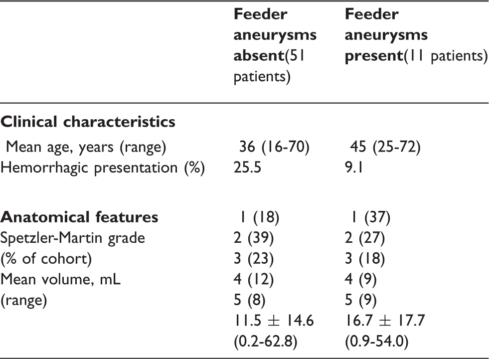
References:
299
BRAIN-0588
Brain Oral Communication
DOES EXTREME PREMATURITY AFFECT ADULT BRAIN VESSEL COMPLIANCE? A PRELIMINARY MRI STUDY.
1Institute of Neurology, University College London, London, United Kingdom
2Institute for Women's Health, University College London, London, United Kingdom
3Philips Medical Systems, Philips, Tokyo, Japan
4Centre for Medical Image Computing, University College London, London, United Kingdom
5C.J. Gorter Center for High Field MRI, Leiden University Medical Center, London, United Kingdom
6Centre for Medical Imaging, University College London, London, United Kingdom
Abstract
Image processing was based on CoW angiography analysis by van Osch et al.6. Control volumes were subtracted from label volumes for all 16 phases. These were then corrected for label T1 decay. For visualization of volumes, a 2D maximum intensity projection was taken in the axial plane for all 16 phases. For each pixel, the blood arrival time was defined as the phase at which signal increased most rapidly. Finally, non-vessel regions were removed from these blood arrival time maps using a vessel extraction method by Zuluaga et al.7, limited to a single image modality.
In each subject, mean blood arrival times were measured in a region of interest drawn on the bilateral M1 and M2 segments of the medial cerebral artery, terminating at its bifurcation.
In this small group, cerebral blood arrival times have been found to be significantly shorter in preterm-born adults compared to terms. This is consistent with cerebral hypertension and reduced brain vessel compliance in this population, which may affect health outcomes. It also suggests that prematurity-induced reductions in compliance are not rectified at adulthood. Further studies with larger sample sizes would need to be undertaken to confirm these results.
Blood arrival times (seconds) for each subject in axial MI projection.
References:
300
BRAIN-0420
Brain Oral Communication
DEFINING THE ISCHAEMIC PENUMBRA WITH COMBINED METABOLIC AND PERFUSION MR IMAGING
1Radcliffe Department of Medicine, Oxford University, Oxford, United Kingdom
2Acute Stroke Service, Oxford University Hospitals NHS Trust, Oxford, United Kingdom
3Department of Engineering, Oxford University, Oxford, United Kingdom
4Nuffield Department of Clinical Neurosciences, Oxford University, Oxford, United Kingdom
5Department of Neuroradiology, Oxford University Hospitals NHS Trust, Oxford, United Kingdom
Abstract
The original description of the ischaemic penumbra asserted that both cerebral blood flow (CBF) and metabolism would be required to monitor therapeutic intervention in acute stroke (Astrup et al 1981). However, investigation of the ischaemic penumbra in patients has generally relied on perfusion-weighted imaging alone. pH-weighted imaging has aided the preclinical definition of the penumbra (Sun et al 2007), with proof of principle of this approach recently demonstrated in man (Harston et al 2015). The aim of this clinical MRI study was to use serial measurements of pH-weighted imaging, in addition to CBF, to improve the understanding of tissue outcome in patients with acute stroke.
Patients with non-lacunar ischaemic stroke underwent serial MRI over 1 month. The imaging protocol included single slice pH-weighted imaging (quantified using amide proton transfer ratio, APTR*), and vessel-encoded pseudocontinuous arterial spin labeling (ASL) to quantify CBF. Repeatability measures were quantified in healthy volunteers using coefficients of variation. Diffusion-weighted imaging, T1-weighted structural and FLAIR imaging were used to define the tissue outcome in grey and white matter: ischaemic core, infarct growth, and oligaemic tissue that survived. Images and masks were registered to native image spaces for voxelwise and patient-level serial analyses. Thresholds were calculated using ROC curve and Youden analyses.
30 patients and 6 volunteers were included in this analysis. The repeatability of both CBF and APTR* demonstrated significant variation between healthy individuals (p<0.0001, p=0.002 respectively). Repeatability between time points was variable for CBF (p<0.0001), but not APTR* (p=0.3). Variability of CBF was within the accepted limits for ASL (<20%).
In the voxelwise analyses, the degrees of both hypoperfusion and acidosis were associated with tissue outcome in patients (<6hrs, ANOVA, p<0.0001). Presenting grey matter CBF predicted tissue at risk with an optimum threshold of 26ml/100g/min (AUC=0.70, sensitivity=73%, specificity=61%), and 24ml/100g/min for predicting acute infarction (AUC=0.74, sensitivity=77%, specificity=61%). The relationship between acidosis and tissue outcome was similar in grey and white matter.
In the patient-level analyses, neither hypoperfusion nor acidosis was significantly associated with tissue outcome (p=0.1, p=0.07 respectively). Patterns of serial CBF values within individuals were independent of tissue outcome. However, serial pH-weighted imaging was most severely acidotic in the infarct core acutely, remaining abnormal at 24 hours. Infarct growth demonstrated acidosis acutely, and both acidosis and alkalosis at 24 hours. Oligaemic regions that survived all had normal pH by 24 hours. Dynamics of intracellular pH were not related to reperfusion status.
Both sequences generated data in voxelwise analysis consistent with preclinical data, demonstrating thresholds associated with tissue outcome. However, following patient-level analysis, issues of sequence repeatability and the diversity of an individual’s response to ischaemia exposed the flaw of relying on a single parameter to define the ischaemic penumbra. These data strongly support Astrup’s contention of the need to combine CBF and metabolic measures, in this case pH, to define the ischaemic penumbra in the clinical setting, and, hence, opportunities for therapeutic intervention.
References
301
BRAIN-0826
Brain Oral Communication
DETECTING FUNCTIONAL ACTIVATION SEPARATED BY PERIODS UP TO 4 WEEKS BY ARTERIAL SPIN LABELING
1Imaging Program, Lawson Health Research Institute, London, Canada
2, Fraunhofer Institute for Medical Image Computing MEVIS, London, Germany
Abstract
Abstract


302
BRAIN-0627
Brain Oral Communication
FUNCTIONAL HYPEREMIA OF THE VISUAL CORTEX IN EARLY STAGE ALZHEIMER’S DISEASE
1Medical Biophysics, University of Toronto, Toronto, Canada
2Imaging Research, Sunnybrook Research Institute, Toronto, Canada
3Institute of Medical Sciences, University of Toronto, Toronto, Canada
4Neurology, Sunnybrook Health Sciences Centre, Toronto, Canada
Abstract
