
Abstract
The INHAND (International Harmonization of Nomenclature and Diagnostic Criteria for Lesions) Project (www.toxpath.org/inhand.asp) is a joint initiative of the Societies of Toxicologic Pathology from Europe (ESTP), Great Britain (BSTP), Japan (JSTP), and North America (STP) to develop an internationally accepted nomenclature for proliferative and nonproliferative lesions in laboratory animals. The purpose of this publication is to provide a standardized nomenclature for classifying microscopic lesions observed in most tissues and organs from the minipig used in nonclinical safety studies. Some of the lesions are illustrated by color photomicrographs. The standardized nomenclature presented in this document is also available electronically on the internet (http://www.goreni.org/). Sources of material included histopathology databases from government, academia, and industrial laboratories throughout the world. Content includes spontaneous lesions as well as lesions induced by exposure to test materials. Relevant infectious and parasitic lesions are included as well. A widely accepted and utilized international harmonization of nomenclature for lesions in laboratory animals will provide a common language among regulatory and scientific research organizations in different countries and increase and enrich international exchanges of information among toxicologists and pathologists.
Table of Contents
Figure Legends
References
Chapter 1. Introduction
The INHAND Project (International Harmonization of Nomenclature and Diagnostic Criteria) is a joint initiative of the societies of toxicologic pathology from Europe (European Society of Toxicologic Pathology [ESTP]), United Kingdom (British Society of Toxicological Pathologists [BSTP]), Japan (Japanese Society of Toxicologic Pathology [JSTP]), and North America (Society of Toxicologic Pathology [STP]) to update the existing World Health Organization/International Agency for Research on Cancer (WHO/IARC) and Society of Toxicologic Pathology/Standardized System of Nomenclature and Diagnostic Criteria (STP/SSNDC) nomenclature systems. The INHAND nomenclature and the related diagnostic criteria represent a consensus of experienced toxicologic pathologists and were reviewed by the INHAND-Global Editorial and Steering Committee (INHAND-GESC) for compliance with INHAND principles. Members of the societies of toxicologic pathology had the opportunity to comment on the draft versions of INHAND documents during a 60-day review period. The initial series of nomenclature publications was focused on lesions in rats and mice. With interest of the United States Food and Drug Administration (FDA) in the use of published terminology standards and the decision of the Clinical Data Interchange Standards Consortium (CDISC) initiative on Standard for the Exchange of Non-clinical Data (SEND) to model the controlled terminology (CT) based on the INHAND nomenclature, the INHAND project was extended to other laboratory animal species including the nonhuman primate, rabbit, minipig, dog, and fish.
Although the INHAND nomenclature and diagnostic criteria represent a preferred international standard nomenclature for lesions identified in nonclinical studies, recommendations for diagnostic criteria and preferred terminology may not be applicable in all situations. The purpose of specific experiments or the specific context of a given study may require deviation from this standardized nomenclature and diagnostic criteria. The appropriate diagnoses are ultimately based upon the scientific judgment of the study pathologist.
The present publication provides standardized terms and diagnostic criteria for histopathologic observations to be used in nonclinical toxicology studies conducted in the minipig (Sus scrofa), which is increasingly used as a nonrodent species in nonclinical toxicology studies. The different breeds of minipigs used in the field of nonclinical safety assessment include the Göttingen, Hanford, Yucatan, Wuzhishan, and Sinclair minipig, with the Göttingen as the most commonly used breed in North America, Japan, and Europe. The nomenclature, observations, and comments included in this article cover all these breeds, but most data are derived from the Göttingen minipig.
The focus of the present publication is on standardized terms and diagnostic criteria for histopathology findings that occur in nonclinical safety studies conducted in the minipig and will not include all background lesions of domestic pigs. References to domestic pigs have been included, where appropriate. Throughout this publication, findings applicable for use in general toxicology studies in minipigs are tabulated by organ system. The nomenclature and tables in this publication build on the existing INHAND rodent nomenclature. In most instances, the definition and description of the rodent lesion applies to the minipig and is not described further. This publication focuses on findings that are unique to the minipig and are not observed in rodents, findings in the minipigs that share the same terminology with a rodent finding but display different morphologic features, or findings for which additional commentary specific to the minipig is needed. Findings that are unique to rats or mice and are not found in minipigs are denoted accordingly in the tables in this article. The tabulated findings are categorized according to the following characteristics: “common,” “uncommon,” “not observed but potentially relevant,” and “not applicable.” The distinction between common and uncommon findings is based on the authors’ experience regarding the occurrence in untreated minipigs and, when available, published references are included. The uncommon category is reserved for changes only observed sporadically as spontaneous findings in minipig studies or those that are almost exclusively induced by xenobiotics. “Not observed but potentially relevant” are changes that have not been described or observed in minipigs; however, the use of these terms has been considered permissible should a lesion meet the diagnostic criteria. The category “not applicable” refers to rodent-specific findings and terms, and the use of these terms in minipigs is considered inappropriate; examples include chronic progressive nephropathy of the kidney or fibro-osseous lesion of bones. It should be noted that minipigs used in toxicologic studies are usually young in age and are kept on study only for a relatively short time frame, a fraction of the normal life span of the minipig. Prior to study initiation, the health status of individual minipigs is usually carefully checked and the individual minipigs selected for a study are in good health. For these reasons, the spectrum and frequency of changes are different from those encountered in domestic pigs and diagnostic laboratories, and common age-related findings including neoplasms are rare. Neoplasms described in this publication are limited to those observed by the authors or described in the literature. Whenever possible, the equivalent rodent term/SEND terminology should be used for any tumors not specifically addressed in this article, as appropriate.
In addition to this journal publication, the nomenclature and diagnostic criteria for the minipig are also available online (www.goreni.org). The online version contains additional images and useful links to differential diagnoses as well as trimming guides, making it a practical tool for toxicologic pathology and experimental work. In addition, all INHAND publications are available at www.toxpath.org and www.eurotoxpath.org/nomenclature.
Across the INHAND publications, the recommended nomenclature is generally descriptive rather than diagnostic. The criteria used are based only on standard hematoxylin and eosin (H&E)–stained paraffin-embedded sections. Histochemical or immunohistochemical staining, and occasionally electron microscopic, characteristics may be addressed in the comments section for the respective finding. Such special techniques may be required in some situations, but a comprehensive discussion of these methods is outside the scope of this publication. Systemic nonproliferative findings that occur across organ systems and are not specific to an organ are reviewed in the section on Systemic Pathology. Instead of “synonyms” for each term, the nonrodent publications have used the notation “other terms.” While these synonyms or other terms have been used historically, the primary listed term is the preferred term and will link to the controlled terminology (CT) in SEND.
Findings included in this nomenclature system may be further specified by modifiers. Criteria are given for modifiers that are of particular relevance. These modifiers should be consistently applied. Additional modifiers not provided in this nomenclature system may describe the location, tissue type, or duration, among others. General principles of the INHAND nomenclature have been published separately. 1 As new information becomes available, new terms and modification of current terms will be needed from time to time and a request for this new term or modification will be applied by “change control” (see goRENI and STP websites).
The minipig is a good animal model for the human with exceptional relevance in several organ systems, including skin, cardiovascular system, gastrointestinal tract, and kidney. In the introduction to each of the following organ systems, a short description of, or reference to, any specific features of the system that can potentially contribute to risk assessment is included, as consideration of features common to minipigs and humans can be important in the design or risk assessment of nonclinical safety studies.
Recommended general references regarding background pathology of minipigs include the following:
Dincer Z, Skydsgaard M. Spontaneous/Background Pathology of Göttingen Minipig. The Minipig in Biomedical Research. CRC Press; 2012:305-320.
Glerup P, Grand N, Skydsgaard M. The use of minipigs in non-clinical research. In: Haschek, Wanda M. C. G. Rousseaux and M. A. Wallig. Haschek and Rousseaux’s Handbook of Toxicologic Pathology. 3rd ed. Vol 2. Academic Press; 2013:461-475. Chapter 13.
Helke KL, Nelson KN, Sargeant AM, et al. Pigs in toxicology: Breed differences in metabolism and background findings. Toxicol Pathol. 2016;44:575-590.
Jeppesen G, Skydsgaard M. Spontaneous background pathology in Göttingen minipigs. Toxicol Pathol. 2015;43:257-266.
Mclnnes E. Minipigs. Background Lesions in Laboratory Animals. A Color Atlas. Mclnnes E, eds. Saunders Elsevier; 2012. Chapter 6.
Chapter 2. Systemic Pathology
There are a number of microscopic findings that may be seen across several organs and/or tissues and are not specific to just one organ system. There are also a number of different microscopic findings that are present across several organs and/or tissues that together constitute a syndrome. Those findings that occur in multiple tissues are listed here, and they are also described under the organ systems in which they occur if they have unique features. Syndromes specific to the minipig are mentioned in individual chapters, but their definitive descriptions are presented here.
The table gives an indication of how frequently the changes may be observed in the minipig, associated diseases, conditions, etiologies or inducing agents, and a list of tissues where they may be found. Where further explanation is deemed useful, selected lesions are discussed in more detail below the table.
Microscopic Findings of Systemic Pathology (Generally Used Preferred Terms): Minipig.
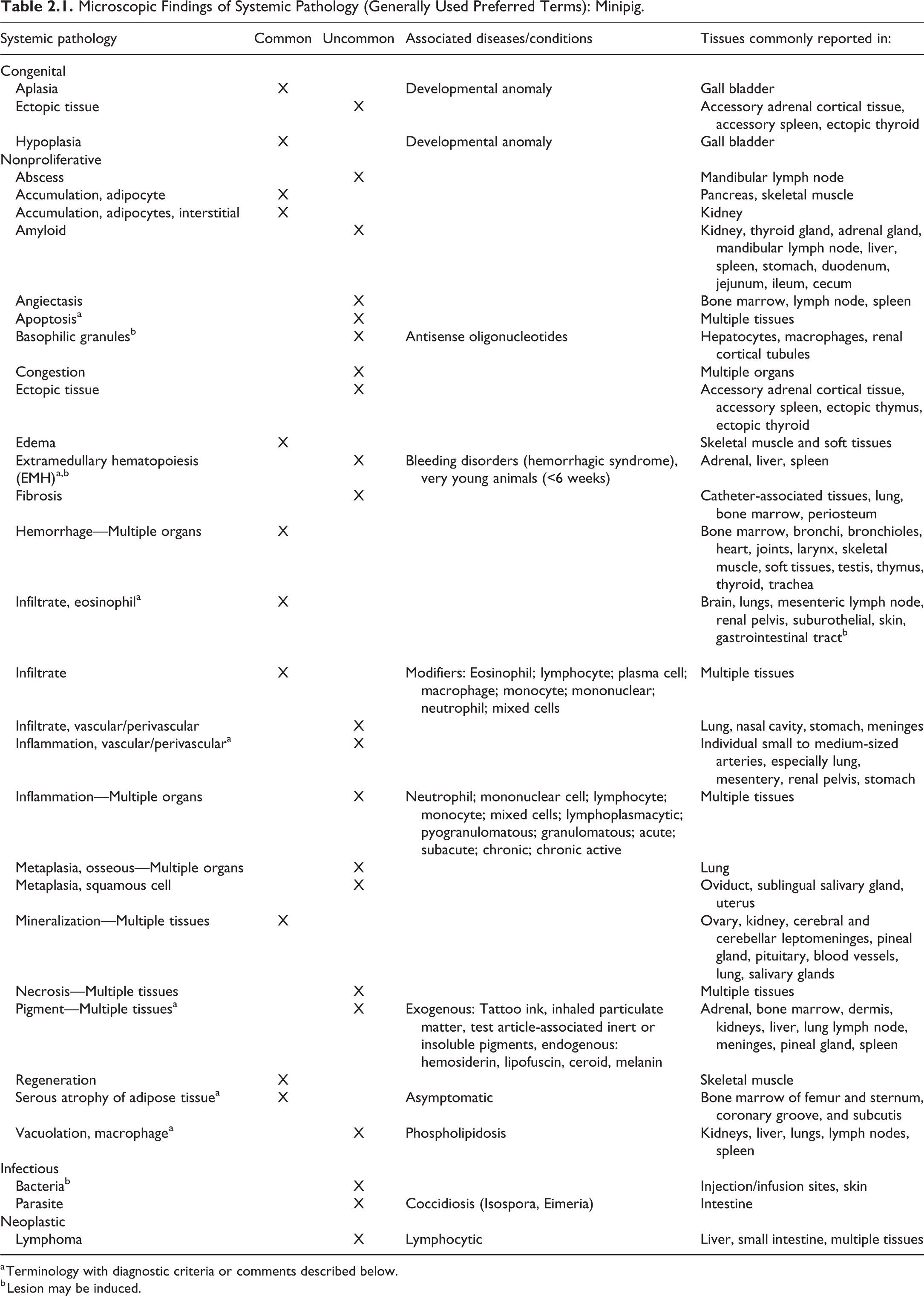
a Terminology with diagnostic criteria or comments described below.
b Lesion may be induced.
Amyloid
Eosinophilic, homogenous, fibrillar material that stains orange or red with Congo Red and exhibits apple green birefringence under polarised light has been observed in aged (8 years or older) Microminipigs. Interstitial deposition was noted in kidney, thyroid gland and adrenal gland, and in the splenic cords and the walls of arterioles of several organs. 2
Apoptosis
For a full discussion, see Elmore S. Apoptosis: a review of programmed cell death. Toxicol Pathol. 2007;35(4):495-516.
Ectopic Tissue
Comments
Accessory adrenal cortical tissue may be found adjacent to the adrenal gland or attached to it and surrounded by a fibrous capsule. 3 Ectopic spleen has been recorded in the pancreas of a cloned Yucatan minipig, and accessory spleens may be found in the gastrosplenic ligament. 4 As the parathyroid is often found embedded in the cervical thymus, it is not considered an ectopic tissue in this location.
Extramedullary Hematopoiesis
Comments
Extramedullary hematopoiesis is generally greatest at around 14 days postpartum, is decreased or absent by 35 days, and is absent after 63 days 5 or 6 weeks. 6,7 In older pigs, extramedullary hematopoiesis may be seen in response to severe anemia. The INHAND term for rodents is “extramedullary hematopoiesis, increased”; however, in minipigs over 6 weeks old, as this is not considered a normal feature of the spleen, the modifier “increased” has been dropped.
Infiltrate, Eosinophil (Figures 2.1–2.3)
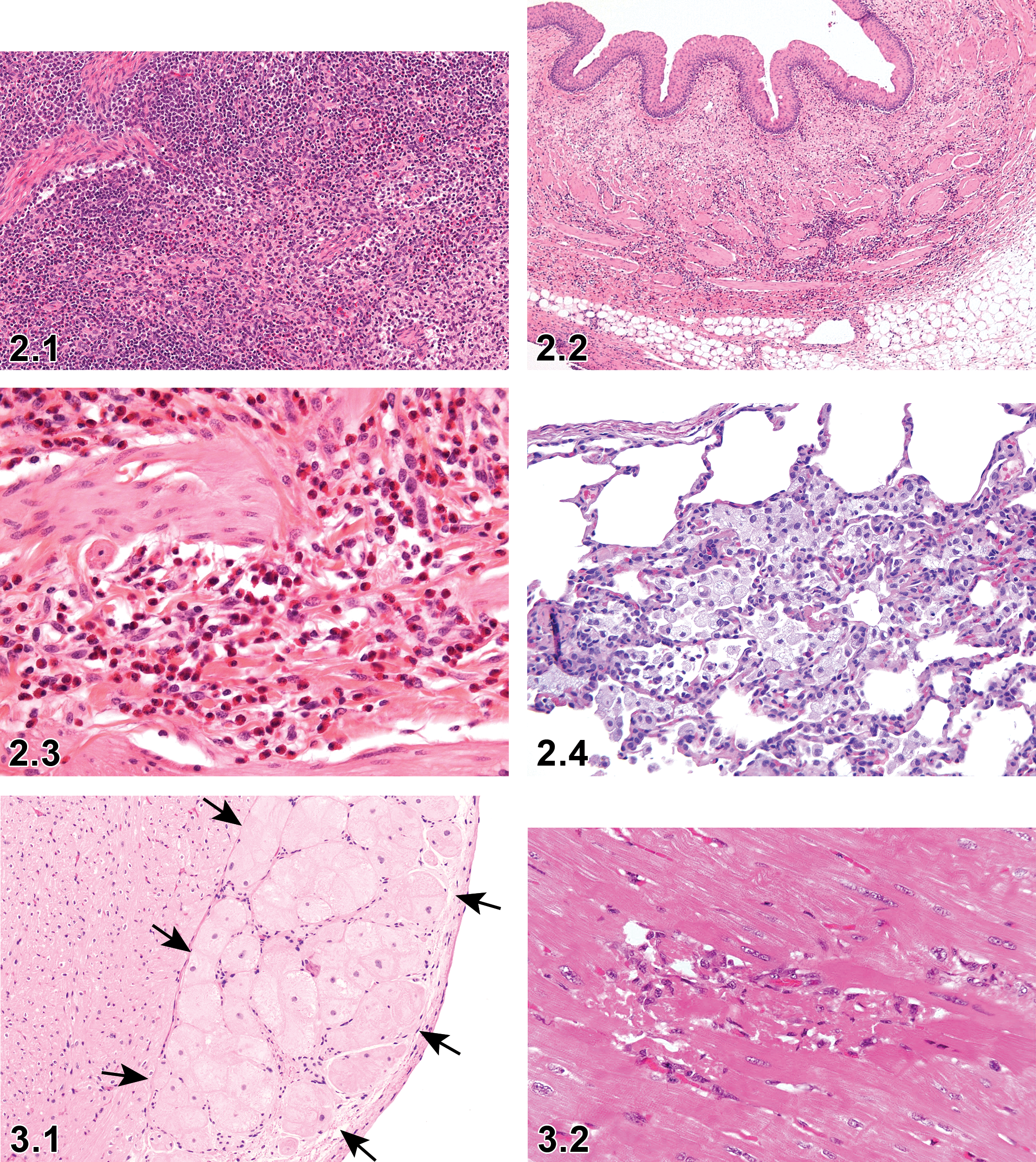
Minipig; Mesenteric lymph node; Infiltrate, eosinophil; H&E ×20.
Diagnostic features
Infiltration of a relatively pure population of eosinophils into the tissue with no other histological criteria of inflammation.
Comments
This finding has been highlighted due to the prominence of this cell type in certain tissues, especially the mesenteric lymph node, in minipigs. 8 This is also commonly seen in brain, lungs, renal pelvis, suburothelial, skin, and gastrointestinal tract. It is recommended to record this routinely when seen.
Inflammation, Vascular/Perivascular
Other term(s)
Arteritis, vasculitis, periarteritis, perivasculitis, degeneration/necrosis, media or wall, artery, necrosis/inflammation, media or wall, artery, inflammation, media or wall, artery fibrosis, perivascular
Diagnostic features
In the minipig, inflammation of the blood vessels usually has one of two appearances:
A fibrinoid necrotic form, where inflammatory cell infiltrates associated with necrosis and fibrin deposits in the vessel wall are present in all layers of the artery.
A chronic form, where the vessel wall is thickened by fibrosis and extends into the surrounding tissues with associated mononuclear cells.
Comments
Arteritis/polyarteritis is an occasionally observed spontaneously occurring background change in Göttingen minipigs. It can be present in a small- or medium-sized single artery of a single organ or several organs in an animal or in several animals in a study. The severity is generally minimal or mild but can occasionally be observed at moderate levels with no age or sex predilection. The prevalence of the finding ranges from 0.06% to 0.29%; it is most commonly recorded with descending frequency in the cardiac and extracardiac blood vessels, vagina, oviduct, rectum, epididymis, spinal cord, pancreas, urinary bladder, kidneys, and stomach. 9
The rodent terms “degeneration/necrosis, media or wall, artery,” “necrosis/inflammation, media or wall, artery,” “inflammation, media or wall, artery,” “infiltrate, inflammatory cell, perivascular”, and “fibrosis, perivascular” may be referred to for a detailed description. 3,7,10
Pigment
Comments
Endogenous pigments
The Göttingen minipig has unpigmented skin, but is not albino, so brown-black pigment (melanin) may be seen, for example, melanin has been recorded in the pineal gland. Pigmented breeds are also used in toxicology studies (eg, Sinclair, Yucatan, Micro Yucatan) and may contain melanin in various tissues. Lipofuscin is a yellow-brown, finely granular cytoplasmic pigment found predominantly in macrophages and can be confirmed with Schmorl’s stain. Ceroid is a golden cytoplasmic pigment, similar to lipofuscin, but does not stain with Schmorl’s. Hemosiderin is found within macrophages as golden brown granules.
Exogenous pigments
Tattoo ink is a black pigment localized to the skin and draining lymph node and is usually found in macrophages. The appearance of other exogenous pigments will vary with their nature.
Serous Atrophy of Adipose Tissue
Other term(s)
Gelatinous bone marrow transformation
Pathogenesis/cell of origin
This spontaneous lesion is most commonly observed in the femur and tibia of Göttingen minipigs used as control animals in toxicology studies. This lesion may occur in other marrow cavities; however, femur and tibia are the bones sampled as standard in most studies. It can occur as a physiological response to a normal degradation of adipose tissue under conditions where the minipig needs additional energy. 7,11,12
Diagnostic features
Focal or diffuse depletion of atrophied or degenerated adipocytes, often starting in the epiphysis. If only the epiphysis is affected, it is graded as minimal or slight. With increasing severity, the metaphysis and diaphysis are affected and the assigned grade would be moderate or marked.
Reduced cellularity of the hematopoietic cells.
Interstitial accumulation or total replacement of adipose tissue by homogenous eosinophilic gelatinous tissue (hyaluronic acid, mucopolysaccharides).
Differential diagnoses
Adipocyte atrophy in the absence of eosinophilic gelatinous tissue.
Decreased hemopoietic cells.
Comments
Serous atrophy is characterized by minimal to marked serous atrophy of the adipose tissue in the bone marrow. Serous atrophy can also be observed in the marrow of the sternum, in the adipose tissue in the sulcus coronarius in the heart, and in the subcutaneous adipose tissue. This is a well-described change seen especially in male Göttingen minipigs but is also observed in female Göttingen minipigs. 11,13 The etiology of the serous atrophy seen as background pathology is thought to be nutritional, likely related to a restricted feeding regimen. Microscopically, serous atrophy of the adipose tissue in the bone marrow is seen as atrophy of the adipocytes accompanied by increased interstitial acidic mucopolysaccharides which stains with Alcian blue. 3 Decreased cell density of the hematopoietic cells of the bone marrow often accompanies this change. It is important to state that minipigs showing this alteration are typically clinically unaffected. Serous atrophy is less commonly seen in minipigs in North America than in Europe 13 ; however, incidence appears to be decreasing in Europe. In humans, serous atrophy is observed in patients with anorexia nervosa, alcoholism, and AIDS and patients suffering with generalized severe illness (cachexia). 14 Serous atrophy in minipigs as described above is limited to the bone marrow, or in extreme, but still asymptomatic cases, the fat in the coronary groove and subcutaneous fat, whereas in humans, serous atrophy is usually systemic in the situation mentioned above. This finding may also be observed in nonhuman primates. In domestic pigs, serous atrophy of fat can be found in cases of emaciation or inanition associated with illness.
Vacuolation, Macrophage (Figure 2.4)
Other term(s)
Phospholipidosis.
Comments
Similar to the other laboratory species, the minipig is susceptible to phospholipidosis. Affected cells usually have pale, finely vacuolated cytoplasm and eccentric nuclei. 15 The role of type II cells in phospholipid metabolism makes them vulnerable to phospholipidosis induced by amphophilic cationic drugs. 16,17
Infectious Diseases
Comments
Minipigs are bred under barrier conditions, are microbiologically defined, and are kept in strictly controlled/biosecured facilities when on study, so infectious disease (parasitism, bacterial and viral diseases) is unlikely. Isospora and Eimeria spp have been observed in the past but have not been observed for several years. Domestic pigs, which are often used in medical device studies, are, however, susceptible to a plethora of infectious disease so one must be aware of the possibility of infectious disease. For the purposes of histology data collection, only entering the observation of “parasite” or a description of the inflammation present, and so on, is appropriate and an etiologic or disease diagnosis (eg, coccidiosis) is not appropriate as a morphologic term.
Syndromes
Syndromes that may occur in the minipig include hemorrhagic syndrome, porcine stress syndrome (PSS), and phospholipidosis, which are described below. The syndrome should not be recorded as a finding but should be mentioned as a histonote in the data capture system and/or in the pathology narrative. The individual findings that together make the syndrome should be recorded in their own right using INHAND terms where possible.
Hemorrhagic Syndrome
Other term(s)
Thrombocytopenic purpura
Pathogenesis/cell of origin
The etiology of hemorrhagic syndrome is unknown but may be due to thrombocytopenia due to an immune complex-associated disorder (type III hypersensitivity, although a type II reaction cannot be excluded.). Animals between 7 weeks and 1 year old have been affected. There does not appear to be a hereditary etiology.
Diagnostic features
On hematology, regenerative anemia and thrombocytopenia (≤20 000/μL) are consistent findings.
Macroscopically, generalized petechial to ecchymotic hemorrhages or hematomas in the skin, periosteum, muscle, mesentery and mucosal and/or visceral surfaces of skin, heart, urinary bladder, intestine, kidney, and lung, but also of other tissues, are seen.
Microscopically, findings consist of multifocal interstitial hemorrhages in multiple organs, edematous inflammation of the bladder, interstitial nephritis, membranoproliferative glomerulonephritis and interstitial fibrosis, hemorrhagic parenchymal necrosis of liver, reactive lymphoid hyperplasia in lymph nodes, apoptotic and immature megakaryocytes, and increased hematopoiesis in bone marrow and extramedullary hematopoiesis in liver and spleen. Complement (C1q) and IgG and IgM may be present in glomerular capillaries and mesangium. A wide range of degenerative to proliferative vascular lesions in small to medium muscular arteries may be seen, particularly in epicardial and intramural coronary arteries and pelvis and medulla of the kidney; however, this is not a consistent feature. Lesions range from endothelial hypertrophy, medial thickening, and vacuolation of myocytes to perivascular and/or vascular inflammation, necrosis of the tunica media, and concentric laminar thickening of the media. 18,19
Differential diagnoses
Exclude infectious diseases that cause vasculitis, for example, classical swine fever, porcine reproductive and respiratory virus, and erysipelas.
Exclude other hemorrhagic diatheses, for example, neonatal alloimmune thrombocytopenia, von Willebrand disease, disseminated intravascular coagulation.
Xenobiotic.
Comments
All microscopic observations should be recorded individually, and a diagnosis of hemorrhagic syndrome made as an animal comment in the data capture system and report text.
Porcine Stress Syndrome
Other term(s)
Malignant hyperthermia; back muscle necrosis; pale soft exudative pork; transport myopathy; fulminant hyperthermia stress syndrome
Pathogenesis/cell of origin
In susceptible pigs, a defective ryanodine receptor gene (RYR1) leads to defective ryanodine receptors which allow Ca2+ leakage into sarcoplasmic reticulum resulting in sudden increased sarcoplasmic calcium concentration on exposure to a trigger, with resulting skeletal muscle pathology. A single-nucleotide polymorphism in the dystrophin gene (DMD) has recently been identified in commercial breeds that causes decreased dystrophin in cardiac and skeletal muscle and PSS-like symptoms on exposure to isoflurane or stress. Pathology due to the DMD mutation is limited to cardiac myocytes. 20 -23
Diagnostic features
Histopathology and clinical history are diagnostic. Rapid rigor mortis (within 5 minutes) is characteristic.
Macroscopically, pale, wet, swollen muscles, and signs of cardiac failure including hepatic congestion, pulmonary edema and congestion, hydrothorax, and hydropericardium may be seen.
Microscopically, swollen myofibers, myocyte segmental hypercontraction, degeneration (of a floccular nature), necrosis, and edema between myofibers, particularly in the longissimus, psoas, and semitendinosus (predominantly type 2 fibers), are characteristic.
Subepicardial hemorrhage, cardiac myocyte degeneration, necrosis, and intramural hemorrhage may be present in the heart, particularly in the left ventricle.
In the kidney, acute tubular necrosis and hemorrhage (acute renal failure, shock kidney) may be seen.
There may be marked hepatic congestion and congestion, hemorrhage, and edema in the lungs.
Comments
All microscopic observations should be recorded individually, and a diagnosis of PSS made as an animal comment in the data capture system and report text.
Porcine stress syndrome is an autosomal recessive pharmacogenetic disorder usually seen in commercial herds, although PSS has also been reported in Vietnamese Pot-bellied pigs. The Göttingen minipig has German Landrace and Vietnamese Pot-bellied pig in its genetic makeup, and this is presumably where the susceptibility to PSS arises. It is extremely uncommon.
Triggers include exposure to halothane or isoflurane, increased physical activity, tension/stress, handling, transport, fighting, climatic conditions, high environmental temperature, low energy levels in diet, vitamin D deficiency, and caffeine.
One of the triggers causes an increased concentration of intracellular channel gating agents and a sudden sustained increase in cytosolic Ca2+ due to hypersensitive RYR1. This causes sustained muscle contraction leading to depletion of ATP by calcium homeostasis mechanisms and a switch to anaerobic metabolism. This leads to increased CO2 and lactic acid and results in metabolic and respiratory acidosis, thermogenesis, and peripheral vasoconstriction. Cytokine release may be seen, which is pyrogenic. Increased temperature, acidosis, and ATP depletion lead to rhabdomyolysis. Enzymes and electrolytes are released from damaged cells, and high potassium levels eventually lead to cardiac arrest.
Clinically, muscle tremors, dyspnea and open-mouth breathing, tachycardia, elevated body temperature, and alternate areas of blanching and erythema of skin are seen. Mortality is associated with signs of cardiac failure.
Clinical chemistry perturbations may include creatine phosphokinase elevation, metabolic and respiratory acidosis, hemoglobinuria, myoglobinuria, renal failure, or impaired blood coagulation.
Susceptible pigs may be identified by exposure to halothane (homozygous only) or by conducting a DNA polymerase chain reaction test for the defective gene (HAL 1843) available from commercial laboratories.
Chapter 3. Cardiovascular System
Anatomically the heart and great vessels of the pig are similar to human, the main exception being the presence of a left azygos vein draining the intercostal system into the coronary sinus. The coronary artery’s blood supply to the heart is almost identical to that of human in anatomy and function. As in humans, the pig has no collateral vessels in the myocardium, leading to increased susceptibility to cardiac infarcts. 3 A characteristic of the pig heart is fully differentiated and large Purkinje fibers (Figure 3.1) that can be found both subendocardially and intramurally within the heart. 24,25 Similar to humans, the porcine aorta also has vasa vasorum.
At necropsy, the heart is sampled with the root of the large vessels and fixed in buffered-formalin solution. The heart should be opened prior to sampling and immersion in fixative in order to ensure adequate fixation and eliminate large blood clots in the cardiac chambers. Several samples are collected from the heart including a section from left and right ventricles, atria and either tricuspid or bicuspid valves, left and right auricles, left and right papillary muscles, and interventricular septum. In addition, the pulmonary valve and pulmonary trunk, thoracic aorta, and caudal vena cava are also sampled. 26 This allows an accurate identification of changes in the cardiomyocytes, extracellular matrix, conduction system, and the vascular structures within the myocardium and the adjacent epicardial tissue. Special dissection procedures may be required to systematically evaluate the coronary arteries.
For detailed description of the cardiovascular system lesions, refer to the rodent publication. 27 While the type and classification of pathological findings of the blood vessels, heart, and heart valves are identical or very similar across species, their frequency in untreated individuals may differ substantially among various species.
I. Heart
Microscopic Findings of Cardiovascular System: Heart; Minipig.
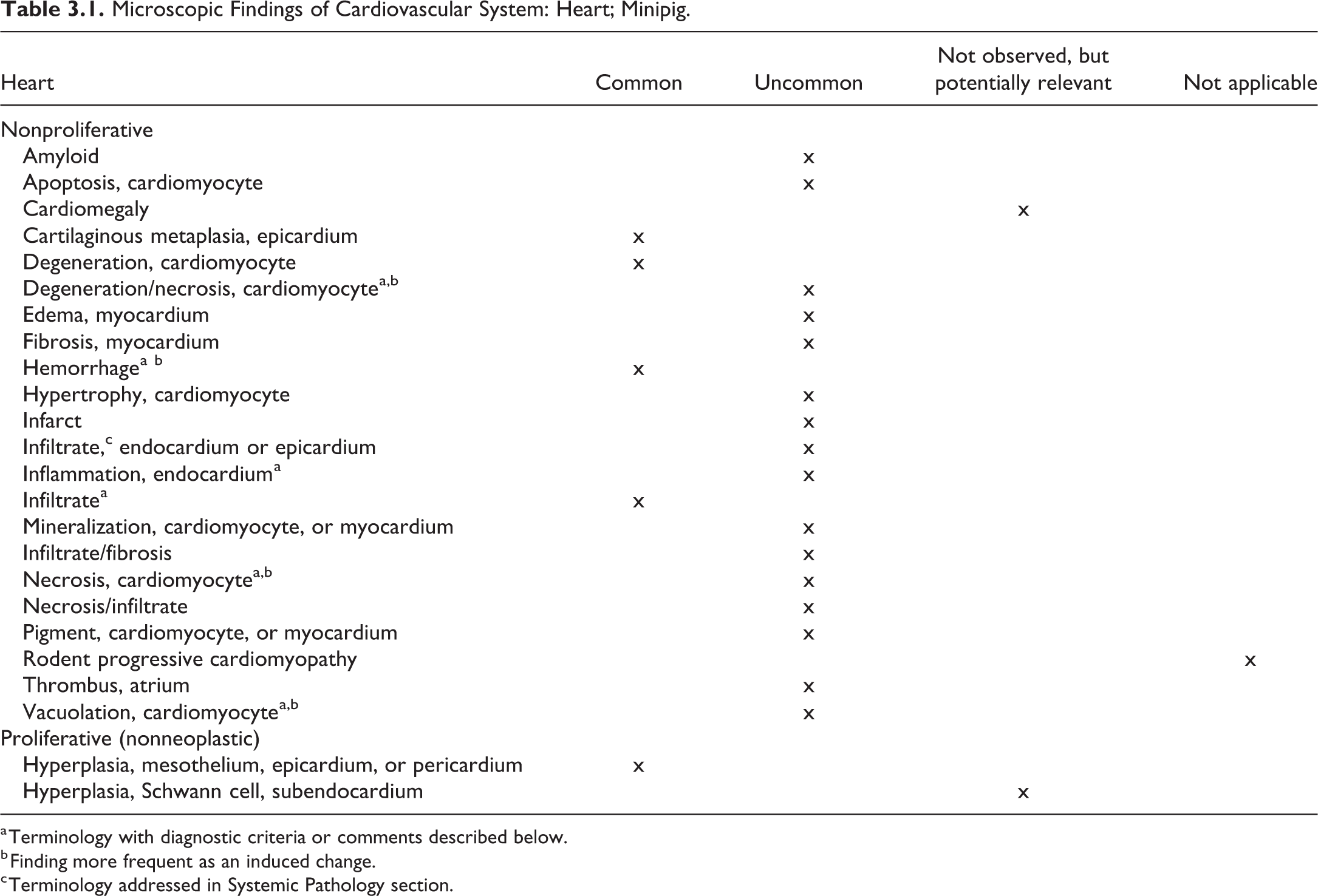
a Terminology with diagnostic criteria or comments described below.
b Finding more frequent as an induced change.
c Terminology addressed in Systemic Pathology section.
Degeneration/Necrosis, Cardiomyocyte, Heart (Figures 3.2 and 3.3)
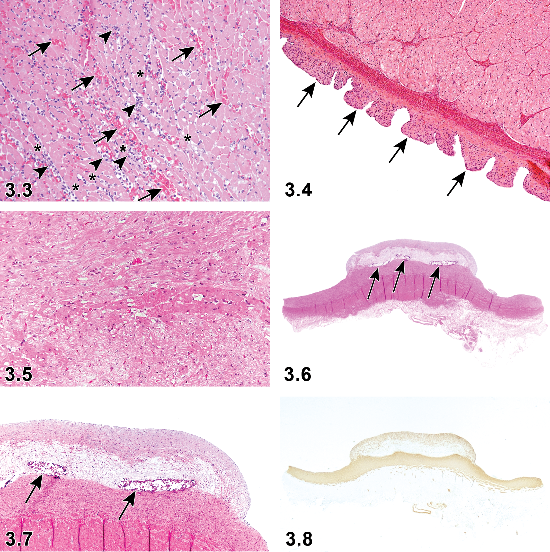
Heart: Hemorrhage (arrow), infiltrate (arrowhead), and degeneration/necrosis in cardiomyocyte (asterisk). H&E x20.
Species
Minipig.
Comment
Treatment with the vasodilating antihypertensive drug, Minoxidil, caused left ventricular papillary muscle necrosis. 3,9,28 The lesion may be observed with PSS, which is further described in Chapter 2, Systemic Pathology. 13,29
Hemorrhage, Heart (Figure 3.3)
Species
Minipig.
Comment
Treatment with the vasodilating antihypertensive drug, Minoxidil, caused diffuse atrial epicardial hemorrhage as well as myocardial necrosis in miniature swine, 9,28 while radiation caused multi-organ hemorrhages including in the heart of Göttingen minipigs. 30
The lesion may be observed with hemorrhagic syndrome and PSS, which is further described in Chapter 2, Systemic Pathology. 3,10,18,19,29
Inflammation, Endocardium, Heart (Figure 3.4)
Species
Minipig.
Comment
In intravenous infusion studies, focal subacute endocardial inflammation and thickening of the intima/endocardium in the right atrium are considered to be related to the method of treatment representing a local irritant effect at the tip of the intravenous catheter. 7 The Hanford breed commonly has subacute to chronic myocardial inflammation. 13,31
Infiltrate, Heart (Figure 3.3)
Species
Minipig.
Other term(s)
Infiltration; infiltrate, inflammatory cell; cell infiltration.
Modifier(s)
Type of inflammatory cell that represents the predominant cell type in the infiltrate: lymphocyte; plasma cell; mast cell; monocyte/macrophage; mononuclear; neutrophil; eosinophil; basophil; mixed.
Comment
Randomly distributed focal mononuclear infiltrates, predominantly lymphocytes are found primarily within the interstitial tissue. The mononuclear infiltrates can be in association with necrosis of myocytes. These lesions are usually focal and minimal. 3
Inflammatory cell infiltrates have been reported in the heart of both Göttingen and Yucatan minipigs as a background lesion. 7,10,13,31
Necrosis, Cardiomyocyte, Heart (Figures 3.2 and 3.3)
Species
Minipig.
Comment
Occasional foci of myocardial necrosis have been observed as a background lesion. Focal myocarditis was also sometimes present. 3,7 The lesion may be observed with PSS, which is further described in Chapter 2, Systemic Pathology. 19
Vacuolation, Cardiomyocyte, Heart (Figure 3.5)
Species
Minipig.
Comment
Cardiomyocyte vacuolation and myofibrillar loss was induced in miniature swine by chronic doxorubicin administration. Vacuolation may also be observed spontaneously. 32
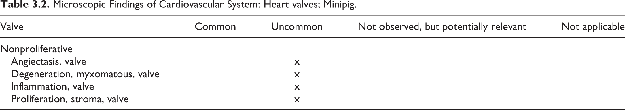
II. Heart Valves
Microscopic Findings of Cardiovascular System: Heart valves; Minipig.
III. Blood Vessels
Microscopic Findings of Cardiovascular System: Blood Vessels; Minipig.
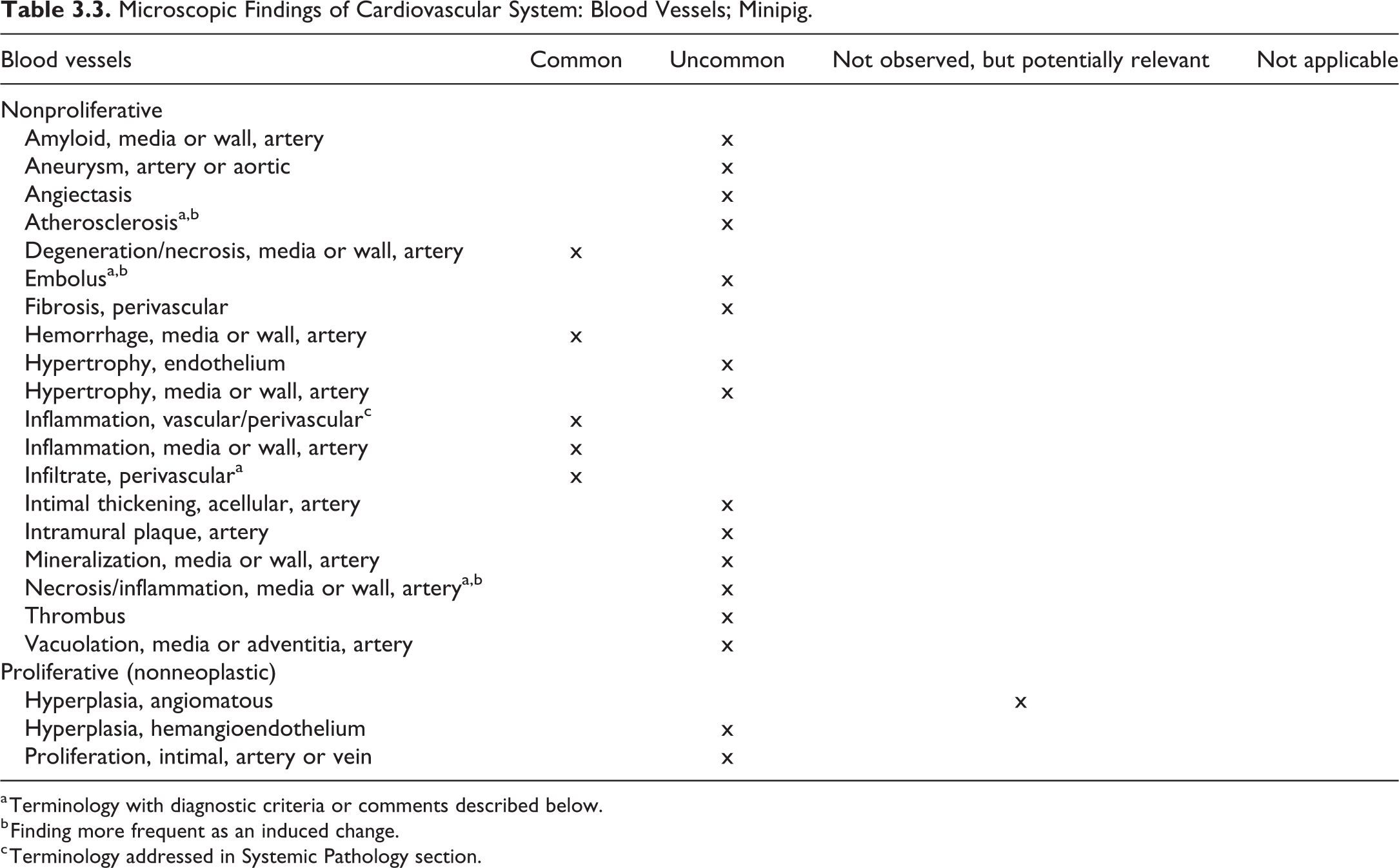
a Terminology with diagnostic criteria or comments described below.
b Finding more frequent as an induced change.
c Terminology addressed in Systemic Pathology section.
Atherosclerosis, Blood Vessels (Figures 3.6–3.10)
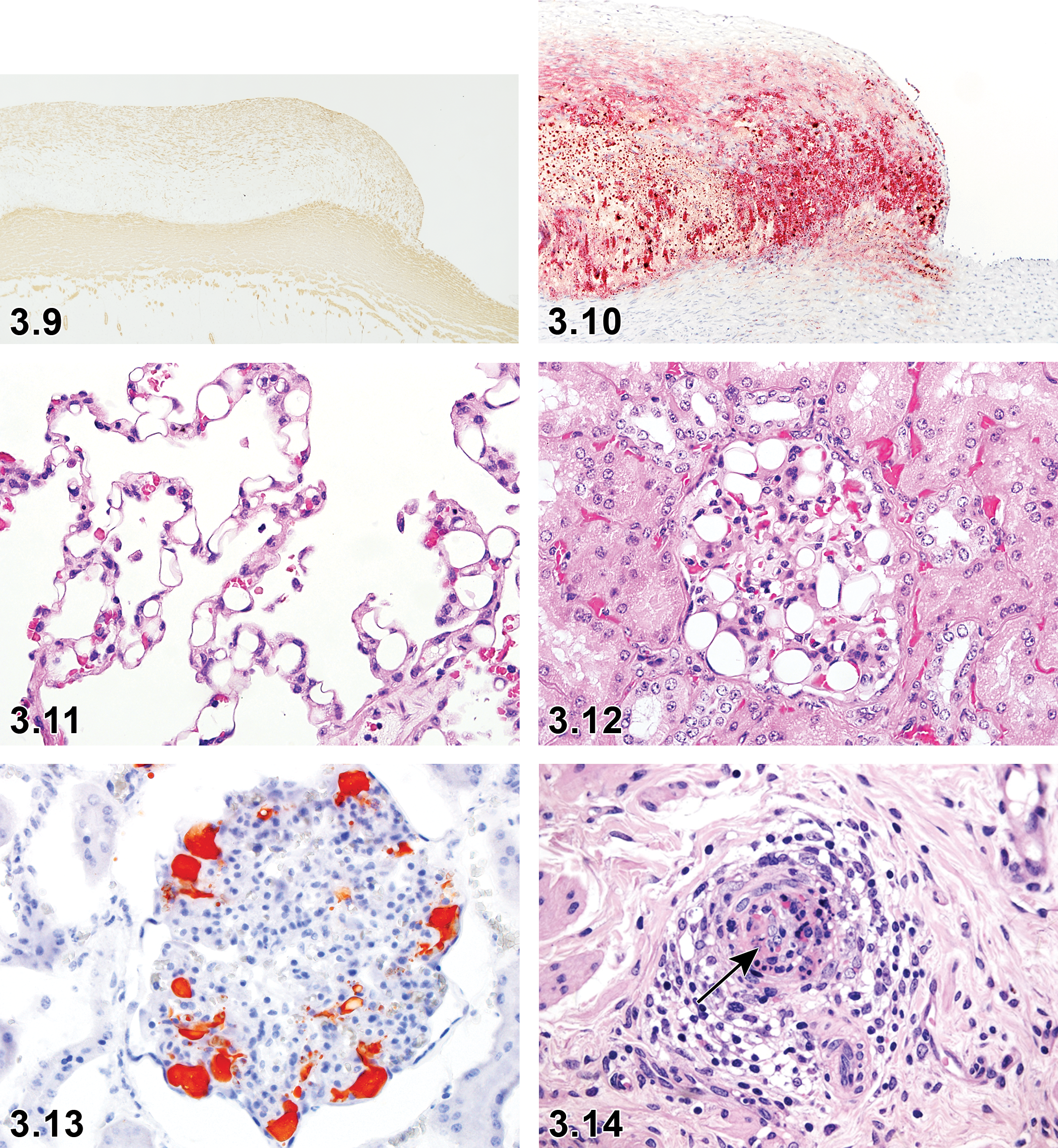
Blood vessels (induced with high cholesterol diet). α-smooth muscle actin. IHC x10.
Species
Minipig.
Pathogenesis/cell of origin
Atherosclerosis is a degenerative change that occurs along the tunica intima of large and small arteries, and several induced pig models were reported. Yucatan minipigs expressing human, liver-specific D374Y-PCSK9 exhibited reduced low-density lipoprotein (LDL) receptor levels, resulting in impaired LDL clearance and increased plasma LDL levels. The administration of high-fat/high-cholesterol diet for 46 weeks led to greatly increased cholesterol levels. At approximately 1 year of age, complex progressive human-like atherosclerotic lesions in the aortae and iliofemoral and left anterior descending coronary arteries were observed. 33
Feeding a high cholesterol diet to Göttingen minipigs for 3 months produced atherosclerotic lesions in the abdominal aorta, aortic arch, celiac artery, right coronary artery, and paracoronal interventricular branch. 34 A high-fat/high-cholesterol diet fed to microminipigs also causes this lesion. 35,36
A diabetes mellitus and high-fat/high-cholesterol diet atherosclerosis model has been developed in Yorkshire or Hanford pigs, and a metabolic syndrome-induced atherosclerosis model has been developed in Ossabaw pigs. 33,37 –39
Diagnostic features
In atherosclerosis models, macroscopically, areas of yellow-white discoloration may be observed grossly on the inner surface of vessels, some of which may be raised plaques (especially remarkable in abdominal arteries).
Microscopically, intimal thickening, infiltration of lipid-laden foamy macrophages, destruction of elastic layers, increased extra cellular matrix and spindle-haped smooth muscle cells, calcification, cholesterol clefts, fibrous cap, and necrotic core may be seen.
Special techniques for diagnostics
Basophilic granular material visible in H&E sections, confirmed with special stains: Von Kossa (mineral), Oil-Red-O (lipid), Elastic van Gieson (extracellular matrix and elastic layer changes), or α-smooth muscle actin.
Comment
Mineralization, proliferation (intimal), and thrombus may all be seen in conjunction with atherosclerosis. These terms are defined further in the rodent cardiovascular INHAND manuscript. 27
Embolus, Blood Vessels (Figures 3.11-3.13)
Species
Minipig.
Modifier(s)
Lipid.
Diagnostic features
In the obesity Göttingen minipig model, lipid embolism is reported. In the lung of this model, thickening of the alveolar septa with variable-sized vacuoles is observed. In some animals, the lipid changes are accompanied by focal alveolar edema and hemorrhage. In the kidney of same animal model, variable-sized vacuoles in glomeruli is observed and the glomerular capillaries are dilated due to lipid and sometimes erythrocytes are seen to be displaced in the capillaries. 40
Special techniques for diagnostics
Oil-Red-O (lipid).
Comment
Obesity is induced by gradually offering increasing amounts of food approaching ad libitum level after 9 to 10 months, aiming for a body weight of approximately 80 to 85 kg at study start. In general, the ad libitum fed pigs consumed 1 to 1.5 kg per day, which correspond to 2 to 3 times the recommended amount. The lipid embolism observed at approximately 6 to 12 months after initiation of the study. 40
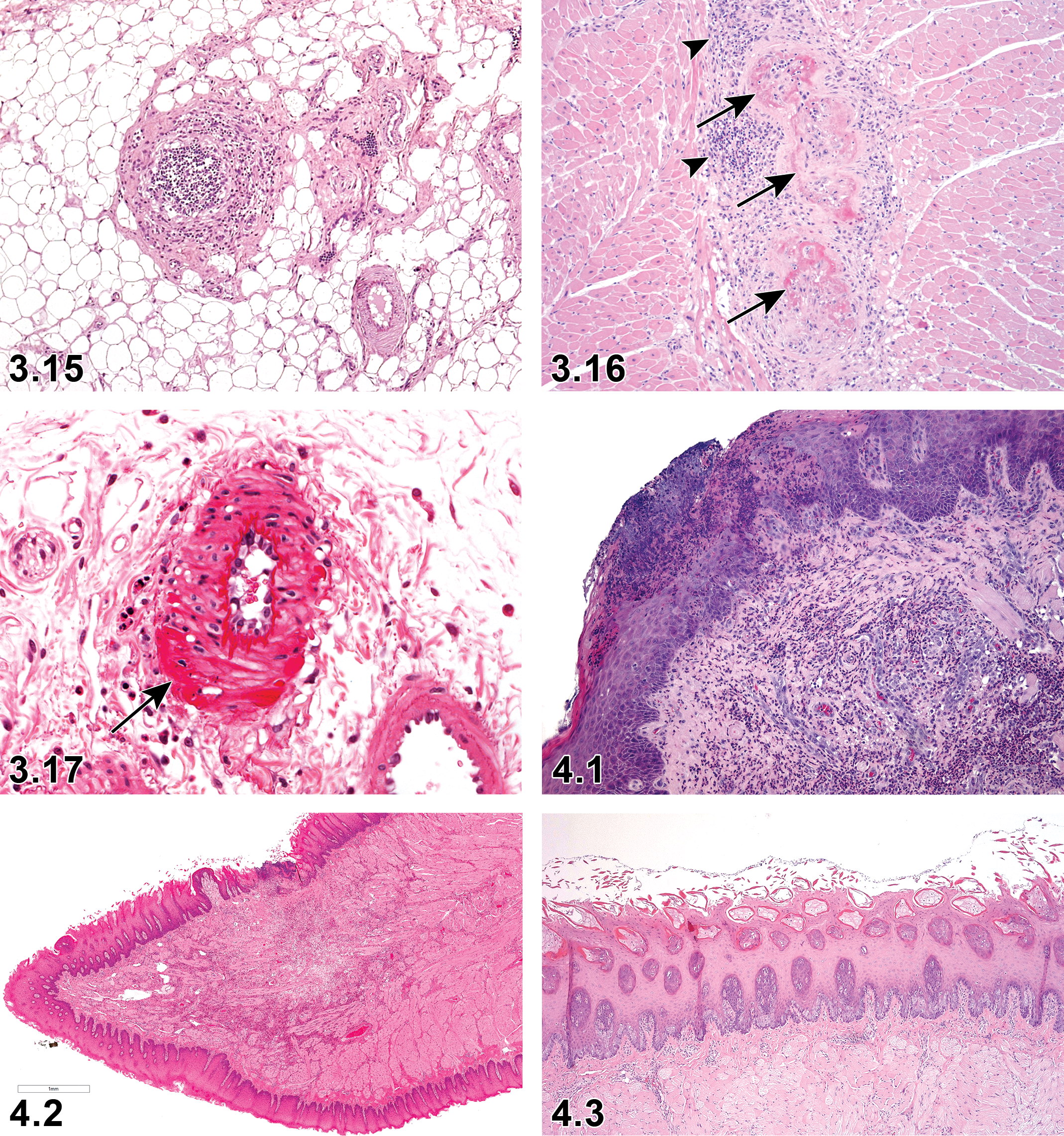
Infiltrate, Perivascular, Blood Vessels (Figures 3.14–3.16)
Blood vessels: Infiltrate, perivascular in intestine serosa. H&E x10.
Species
Minipig.
Comment
Arteritis/polyarteritis is an occasionally observed spontaneously occurring background change in Göttingen minipigs. It can be present in a small- or medium-sized single artery of a single organ or several organs in an animal or in several animals in a study. The severity is generally minimal or mild but can occasionally be observed at moderate levels with no age or sex predilection. The prevalence of the finding ranges from 0.06% to 0.29%; it is most commonly recorded with descending frequency in the cardiac and extracardiac blood vessels, vagina, oviduct, rectum, epididymis, spinal cord, pancreas, urinary bladder, kidneys, and stomach. 9 Mononuclear inflammatory cells have also been reported in the aorta. 41
The lesion may be observed with hemorrhagic syndrome, which is further described in the Chapter 2, Systemic Pathology. 18,19
Necrosis/Inflammation, Media or Wall, Artery, Blood Vessels (Figures 3.14–3.17)
Species
Minipig.
Diagnostic features
In spontaneous cases, inflammatory cells infiltrate the media and/or all other layers of the artery, associated with necrosis and fibrin deposits of the vascular wall. 3,7,9,10
Comment
Necrotizing vasculitis characterized by fibrinoid necrosis of the vascular wall and often associated with lymphocytic exudates or thrombosis was observed in Yucatan minipigs following endovascular radiation from 32P sources resulting in exposure between 6 and 40 Gy. 42
Treatment with the vasodilating antihypertensive drug, Minoxidil, caused endothelial injury, intramural accumulations of erythrocytes and platelets, periadventitial hemorrhage, fibrin deposits, and inflammatory cell reaction in epicardial small arteries. 9
Pigs treated with immunomodulating drugs, such as hydrocortisone or betamethasone, caused medial degeneration with fibrinoid necrosis in small/medium-sized arteries of intestines, peritoneum, spleen, pancreas, mesenteric plexus, liver, kidney, and adrenals. 9
The lesion may be observed with hemorrhagic syndrome and PSS, which is further described in Chapter 2, Systemic Pathology. 18,19
Chapter 4. Digestive System
For detailed description of the digestive system lesions, refer to the rodent publication. 43 While the type and classification of pathological findings of the digestive system are identical or very similar across species, their frequency in untreated individuals may differ substantially among various species.
The minimum recommendation for examination of the digestive system is to examine stomach (nonglandular and glandular), small intestine (duodenum, jejunum, ileum), and large intestine (cecum, colon). For minipig/pig nonclinical safety studies, it is recommended that the same samples of digestive system as in rats and mice are examined to adequately evaluate the digestive tract in routine studies. Considering the length of the porcine esophagus and intestine, the collection of additional samples is recommended. 26 However, in nonclinical safety studies, the number of samples examined is the same as in rodents. In investigational studies, additional sampling could be considered. 26 A detailed step-by-step sampling protocol of all organ systems including digestive system is presented by Albl et al. 26
The human and pig digestive systems are very similar to each other in many respects and are classified as monogastric (ie, having one stomach) or nonruminant. Pigs and humans are both omnivores and have comparable metabolic functions, pH changes, gastric cell types, secretion, intestinal transport times, and nutrient absorption characteristics. Therefore, pigs have become very useful in basic nutritional research. 44,45
Xenobiotic-induced toxic injury to the gastrointestinal system often results in degeneration, ulceration, or hemorrhage in many laboratory species. However, the porcine gastrointestinal tract does not appear to be as sensitive to the ulcer-inducing properties of nonsteroidal anti-inflammatory drugs as the gastrointestinal tract of dogs. 44
I. Upper Alimentary Tract (Oral Cavity, Tongue, Pharynx, and Esophagus)
Oral Cavity
The oral cavity is not routinely examined in nonclinical safety studies microscopically and therefore is not included in this chapter.
Tongue
Porcine nonclinical safety study protocols recommend the same sample generation as in rats and mice to adequately evaluate the tongue: one transverse section through the mid-portion of the tongue. 26
On microscopic examination, the basal parts of the epithelium of the tongue appear quite basophilic, often with vacuolated and swollen cells; focal differences in this basophilia can be present within the same animal and care should be taken not to misinterpret this as a test article-related finding. 7
Most often lesions in the tongue are found in studies with oral administration. The lesions are linked to mechanical damage, as the minipig is prone to biting the tongue during the dosing procedure (gavage). 7
Microscopic Findings of Digestive System: Tongue; Minipig.
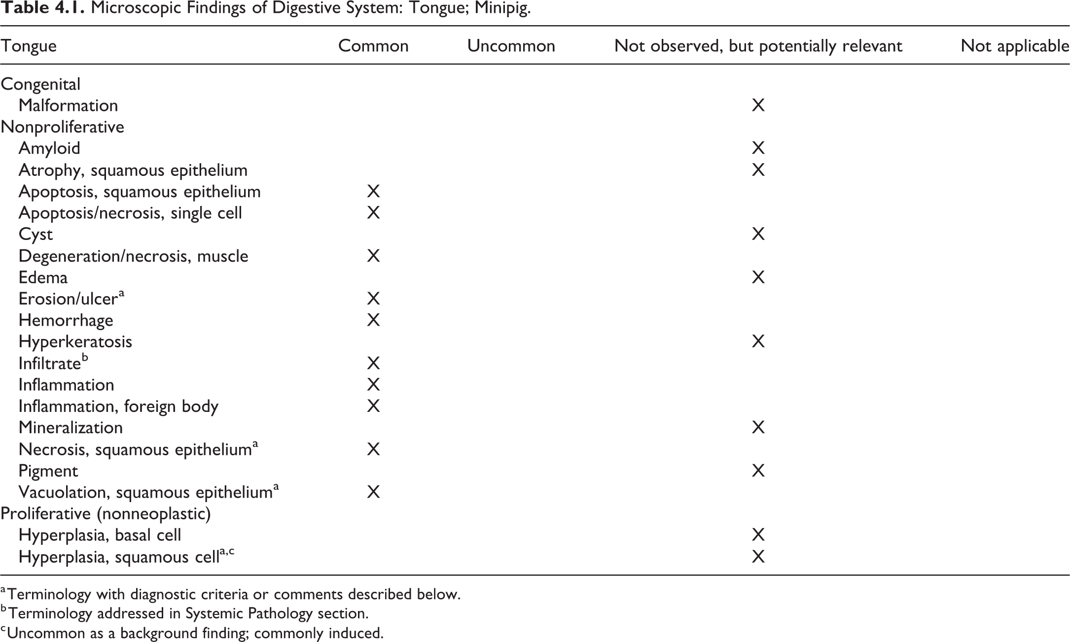
a Terminology with diagnostic criteria or comments described below.
b Terminology addressed in Systemic Pathology section.
c Uncommon as a background finding; commonly induced.
Erosion/ulcer, tongue (Figures 4.1 and 4.2)
Pathogenesis/cell of origin
Nonkeratinized stratified squamous epithelium.
Diagnostic features
Focal or multifocal.
Basal lamina may be intact under the erosion.
Ulcer is associated with mixed inflammatory cell infiltrates in the submucosa.
Differential diagnoses
Necrosis, squamous epithelium.
Comment
Erosion/ulcer in the fungiform papilla of nonkeratinized stratified squamous epithelium is commonly observed, generally focal and of slight to moderate severity. The lesion is caused by trauma due to biting the tongue during the dosing procedure (gavage). It can be associated with minimal to slight focal inflammation of the muscle and occasionally with focal necrosis. 3,7
Hyperplasia, squamous cell, tongue (Figure 4.3)
Comment
Hyperplasia of the squamous epithelium of the tongue is not observed in minipigs as a background change. However, hyperplasia of the squamous epithelium has been seen as an induced change with antioxidant inflammatory modulators (AIMs) (Jeppesen, unpublished data).
Necrosis, squamous epithelium, tongue (Figure 4.4)
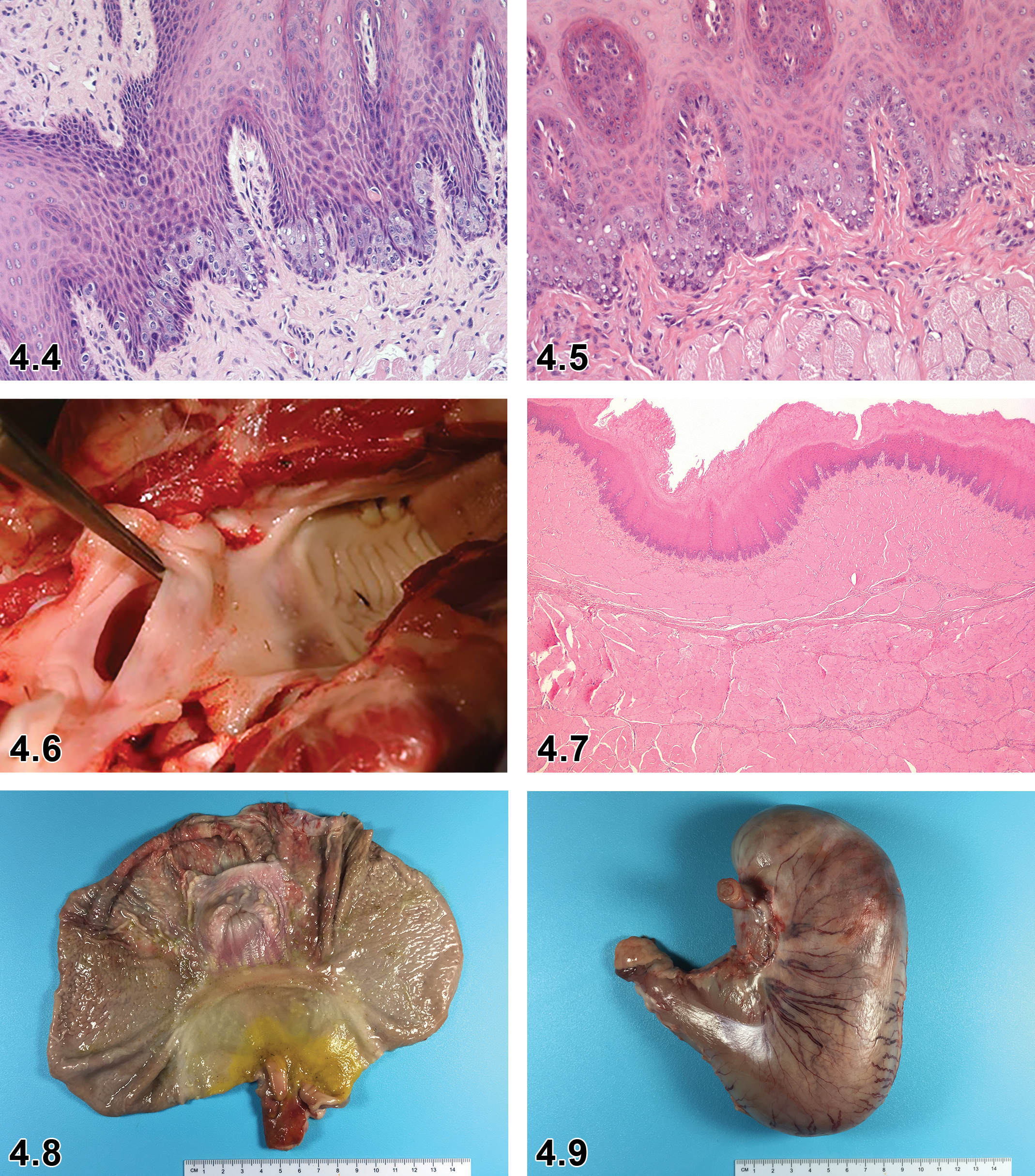
Tongue, necrosis and vacuolation of the squamous epithelium, ×20.
Comment
Necrosis of the squamous epithelium of the tongue is a commonly observed in minipigs as a background change and characterized by deaths of individual epithelial cells with fragmented nucleus (karyorrhexis or karyolysis), pale eosinophilic cytoplasm, and absence of inflammatory cells. Necrosis is generally present in the vicinity of vacuolated epithelial cells of the mucosa (Jeppesen, unpublished data).
Vacuolation, squamous epithelium, tongue (Figure 4.5)
Comment
Vacuolation of squamous epithelium in the tongue is a commonly observed background finding in minipigs. It is characterized by enlarged, fine or coarsely vacuolated epithelial cells with central or peripheral nucleus located predominantly in the basal parts of the epithelium and generally seen at minimal level. 7 The basal parts of the epithelium of the tongue appear quite basophilic often with vacuolation. Focal differences in this basophilia can be present within the same animal and care should be taken not to misinterpret this as a treatment-related finding. 7
Pharynx
The pharynx is not routinely examined in nonclinical safety studies. However, the pharynx can be occasionally sampled for examination of mucosal changes. The palatine tonsils which are located on the dorsolateral side of the pharynx can also be sampled. 26 When sampling of the pharynx is required, the complete nasopharynx is primarily sampled and the soft palate (the “roof” of the oropharynx) is included. A transverse section of the sampled circular structure (nasopharynx and the soft palate) is processed (Figure 4.6). Since pharynx is not routinely evaluated, information on lesion incidences is limited.
Microscopic Findings of Digestive System: Pharynx; Minipig.
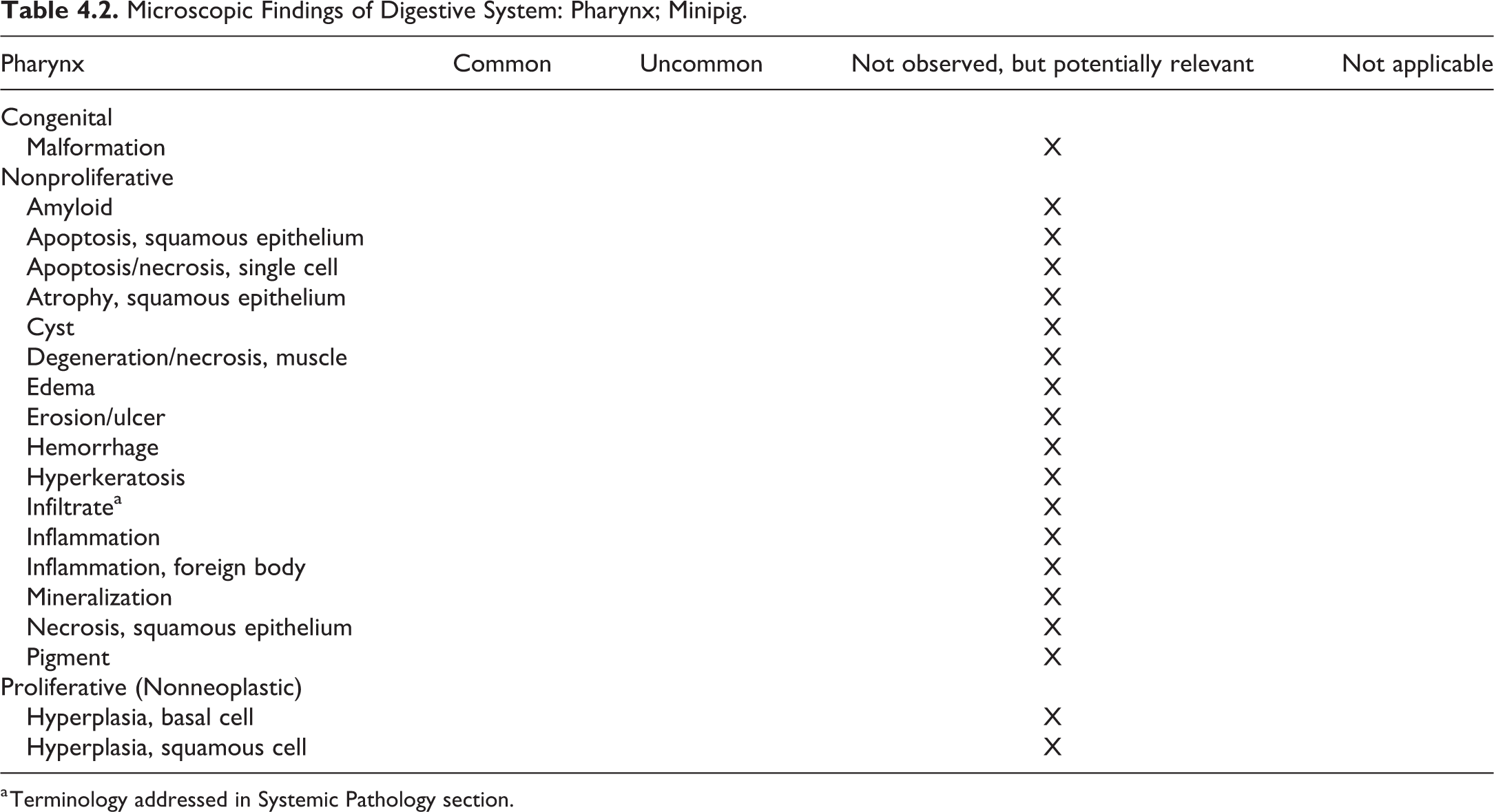
a Terminology addressed in Systemic Pathology section.
Esophagus
In spite of the length of the porcine esophagus, sampling from one location of the esophagus as in rats and mice is considered adequate for evaluation of the porcine esophagus in nonclinical toxicity studies: one transverse section through the esophagus and the trachea at the level of the thyroid gland is recommended. 26
The most commonly observed background change in the esophagus is focal inflammatory cell infiltrates, predominantly mixed cells, at minimal severity. 7
Periesophageal hemorrhage/inflammation occurring as a result of blood sampling procedures is often observed in toxicity studies. 7
Microscopic Findings of Digestive System: Esophagus; Minipig.
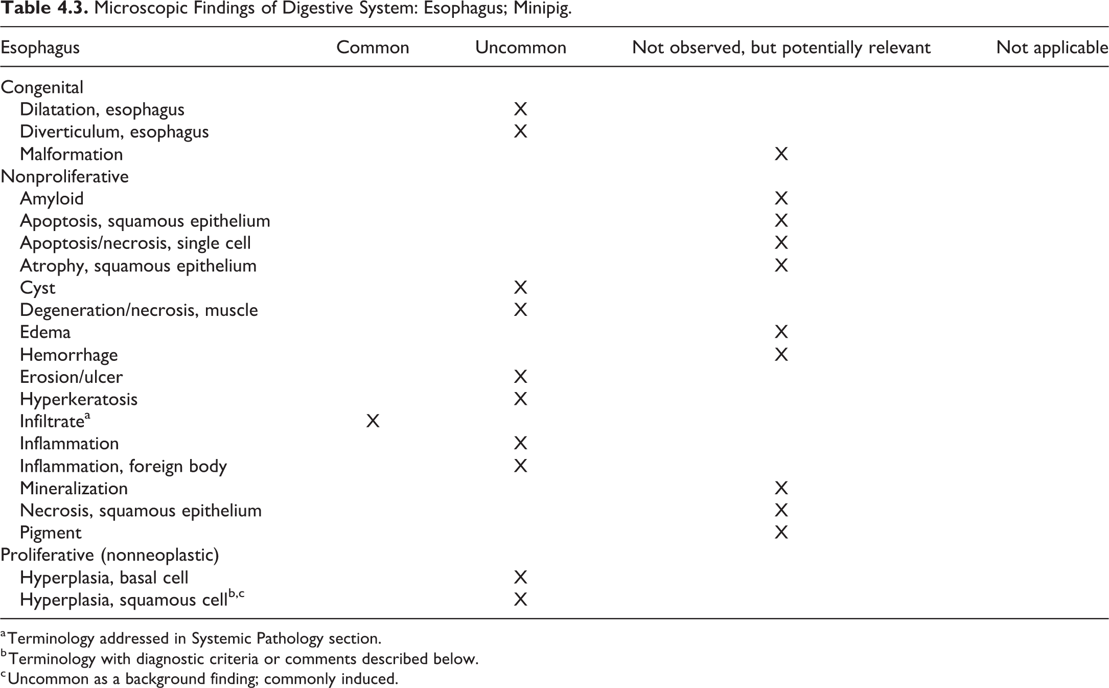
a Terminology addressed in Systemic Pathology section.
b Terminology with diagnostic criteria or comments described below.
c Uncommon as a background finding; commonly induced.
Hyperplasia, squamous cell, esophagus (Figure 4.7)
Comment
Squamous cell hyperplasia in the esophagus is uncommon as a background change. However, squamous cell hyperplasia has been observed as an induced change with AIMs (Jeppesen, unpublished data).
II. Stomach
The stomach is typical of monogastric species except for a prominent muscular protuberance, the torus pyloricus at the level of pylorus, and a more prominent cardia (Figures 4.8 and 4.9). Among domestic mammals, pigs and ruminants are the only species that have a pyloric torus. The pyloric torus is a pedunculated structure, forming a protuberance at the lesser curvature of the pylorus of the stomach. The histomorphologic features of the pyloric torus include a simple columnar mucus secretory cell lining, simple branched tubular mucous glands arranged into lobules by connective tissue septa in the lamina propria, large vascular spaces devoid of a muscular layer at the base of the lamina propria, an internal fibromuscular layer surrounding the glandular lamina propria, an inconstant layer of adipose tissue of variable thickness between the internal and external muscular layers, and serosa. 46
As in rodents, the minipig stomach consists of 2 parts: glandular and nonglandular. The nonglandular portion of the stomach, the pars esophagea, is well defined from the glandular portion as in the rodent, however, is less extensive compared with rodents. The glandular mucosa of the stomach is divided into 3 parts: the cardiac gland region (the largest), the fundic gland region, and the pyloric gland region. The stomach produces mucus, acid, peptic secretions, and gastric hormones from its glandular mucosa. The pH in the nonglandular pars esophagea and in the cardiac gland region is maintained above 5, while in the fundic and pyloric gland regions, where acid and pepsinogen are secreted, the luminal pH of 3.5 or lower is optimum for the proteolytic activity of pepsin, the hydrolytic product of pepsinogen. 47 The anatomic fundus in swine is lined by cardiac-type mucosa with mucous glands and not by true gastric glands (with zymogen and oxyntic cells). True gastric glands are present only in the body of the stomach, nearer the pylorus than the fundus. 48
For nonclinical safety studies, the same sampling of gastric regions as in rats and mice is recommended in minipigs: (1) junction between nonglandular and cardiac gland region, (2) fundic gland region, and (3) junction between pyloric gland region and duodenum.
Microscopic Findings of Digestive System: Stomach (Nonglandular); Minipig.
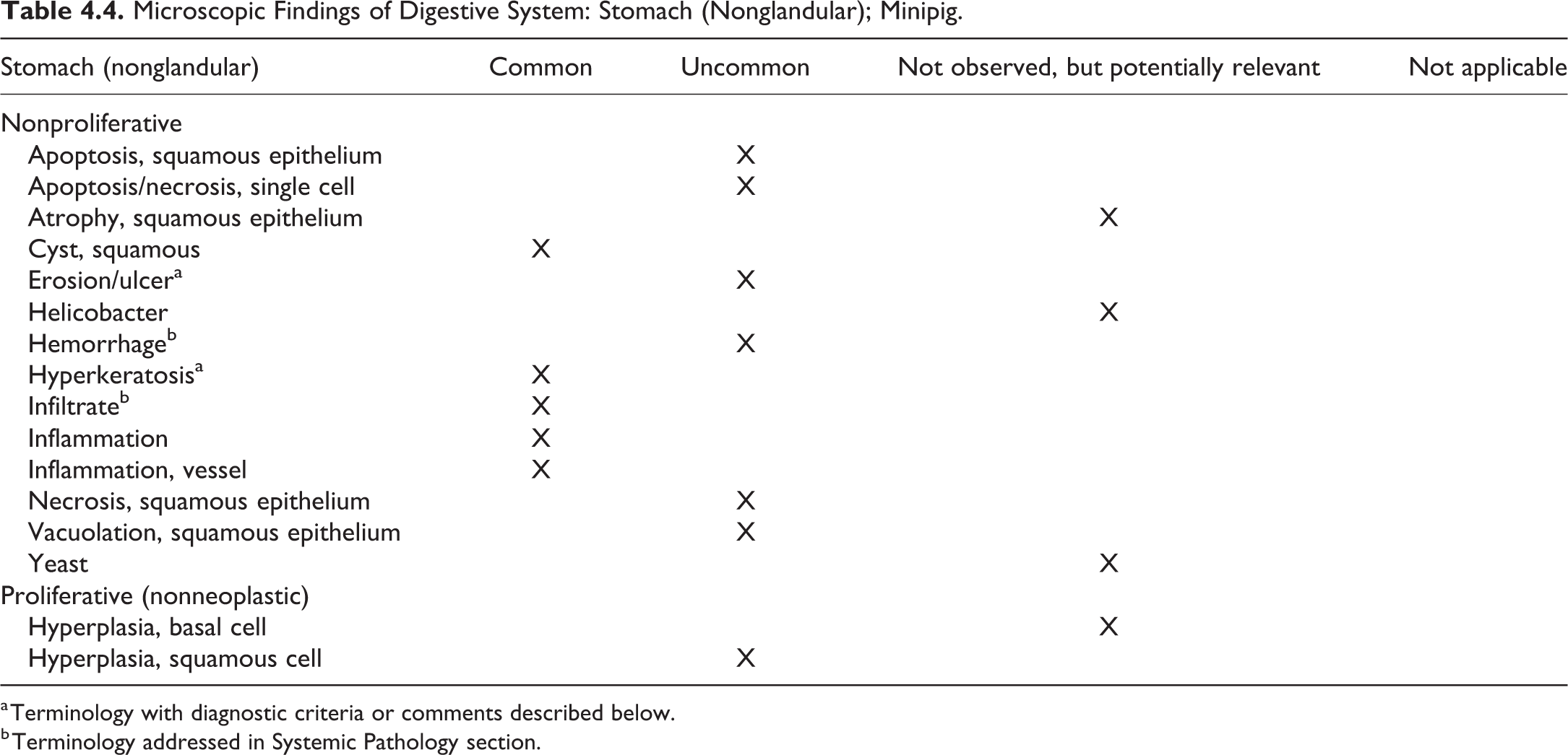
a Terminology with diagnostic criteria or comments described below.
b Terminology addressed in Systemic Pathology section.
Erosion/ulcer, stomach (nonglandular) (Figure 4.10)
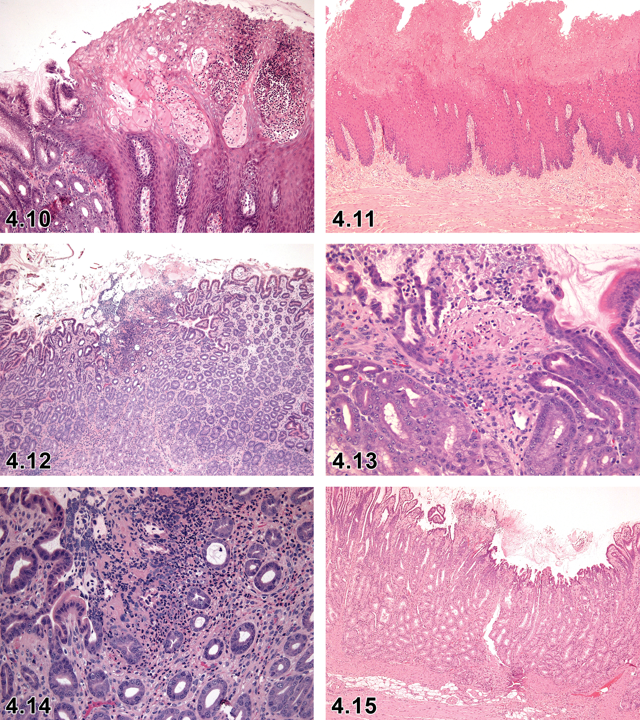
Stomach, nonglandular, erosion, ×10.
Comment
Erosion/ulcer in the nonglandular stomach is generally an uncommon background finding in nonclinical safety studies in minipigs. 44 However, it should be mentioned that the incidence may increase depending on the stress status of the animals during handling and dosing procedures. Erosions and ulcers are seen in the nonglandular portion of the stomach in Gottingens and Yucatans, whereas the glandular portion of the stomach in the Hanford displays erosions and ulcers. 7,13
Hyperkeratosis, stomach (nonglandular) (Figure 4.11)
Modifiers
Orthokeratotic
Parakeratotic
Pathogenesis/cell of origin
Keratinization of the squamous epithelium
Diagnostic features
Diffuse.
Increase in the thickness of the keratin layer with nonnucleated or nucleated keratinized cells on the luminal epithelial surface.
Hyperkeratosis, orthokeratotic: thickened keratin layer with nonnucleated keratinized cells.
Hyperkeratosis, parakeratotic: thickened keratin layer with nucleated keratinized cells.
Differential diagnoses
Hyperplasia, squamous cell: proliferation and thickening of the stratum spinosum (Figure 4.11)
Comment
Hyperkeratosis is a commonly observed background change in the absence of hyperplasia.
Microscopic Findings of Digestive System: Stomach (glandular); Minipig.
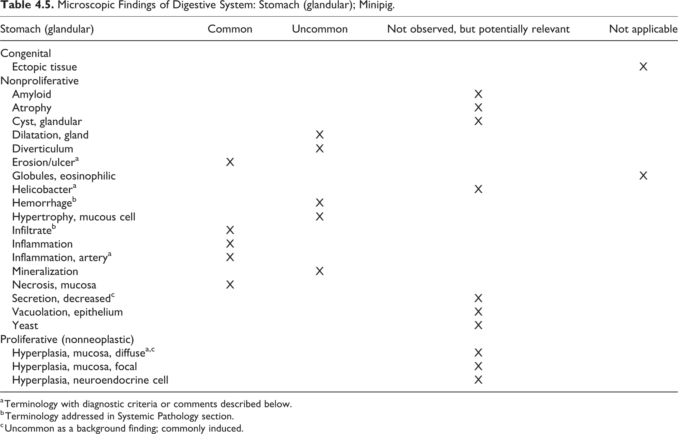
a Terminology with diagnostic criteria or comments described below.
b Terminology addressed in Systemic Pathology section.
c Uncommon as a background finding; commonly induced.
Erosion/ulcer, stomach (glandular) (Figures 4.12-4.14)
Cell of origin
Surface epithelial cells.
Diagnostic features
Loss of mucosal epithelium with preservation of muscularis mucosa (erosion) or with penetration of muscularis mucosa (ulcer) in association with inflammatory cell infiltrates and/or fibrosis in the lamina propria.
Focal or multifocal.
Acute or chronic.
Differential diagnoses
Autolysis: loss of cells at the luminal surface and absence of inflammatory infiltrate.
Comment
Erosions or ulcers are quite commonly seen in the glandular part of the stomach. They are generally graded as minimal or slight. Focal ulcers can be of moderate severity. These changes are predominantly observed at the pyloric–duodenal junction, possibly due to constant pH changes in the area, but can also be seen in the cardiac region. Administration of the xenobiotics and/or stress can increase the incidence and severity of such changes. 3,7
Helicobacter, stomach (glandular)
Comment
Production pigs are commonly infected with the zoonotic pathogen Helicobacter spp, particularly Helicobacter suis. This pathogen mainly colonizes the fundic and pyloric regions of the porcine stomach inducing inflammation and a decrease in daily weight gain. Alterations in hydrochloric acid production in the glandular region of the stomach, associated with chronic H suis infections, may play a role in the pathogenesis of swine gastric ulceration. 49 Helicobacter infections have not been observed or demonstrated in nonclinical safety studies; however, gastric erosion/ulcers are commonly observed and attributed to stress or constant pH changes in the area. 3
Hyperplasia, diffuse, mucosa, stomach (glandular)
Comment
Diffuse mucosal hyperplasia of the glandular stomach has not been noted as a background change in minipig nonclinical safety studies. However, it has been recorded as an induced change with an AIM (Jeppesen, unpublished data).
Inflammation, artery, stomach (glandular)
Comment
Inflammation of the arteries is commonly seen in the muscularis mucosa of the glandular stomach (see Cardiovascular section).
III. Small and Large Intestines
The small intestine is approximately 10% duodenum, 80% jejunum, and 10% ileum. In humans and nonhuman primates, the duodenum is relatively shorter and the ileum tends to be similar in length to the jejunum or even larger. The mesenteric vessels of the small intestine form vascular arcades in the muscularis mucosa of the intestine and not in the friable mesentery, as in other mammals. The presence of duodenal Brunner’s glands and jejunal Peyer’s patches allows easy histologic differential identification of these structures as it does in other species. In the pig, unlike other domestic animals, Peyer’s patches occur in a continuous band along much of the length of the small and large intestine and form a 2-cm-wide, thickened focus around the ileocecal opening referred to as the cecal tonsil. The large intestine of the pig is quite different anatomically from that of other common laboratory animals. The cecum, the ascending and transverse colon, and the proximal portion of the descending colon are arranged in a series of centrifugal and centripetal coils in the left upper quadrant of the abdomen, collectively forming the spiral colon. The cecum has 3 longitudinal muscular bands (tenia), and the proximal portion of the spiral colon has 2 bands. These result in a series of sacculations (haustra). 44
Pigs develop intestinal crypts prenatally, similar in timing to the human fetus. 50 However, presence of Paneth cells in the crypts of the small intestine is still debatable. Underwood states that pigs normally do not develop Paneth cells. 50 Gonzalez et al could not identify Paneth cells in the crypts of porcine small intestine by TEM and immunostainings, however a Paneth cell equivalent (Paneth-like cell), similar to that found in the mouse colon, may be present. 51 Therefore, background changes associated with Paneth cells in the rodent are recorded in minipig as occurring in Paneth-like cells.
Sampling the intestines for nonclinical safety studies is similar in minipigs as in rats and mice: one transverse (cross) section from the duodenum, jejunum, ileum, cecum, and colon and one longitudinal section from the rectum. 26 Albl et al recommended collection of additional samples in investigational studies (1 mid-segmental sample taken from the duodenum, 1 from the ileum, and 1 from the cecum. Two samples are prepared from the jejunum, 3 from the colon [1 from a centripetal turn, 1 from a centrifugal turn, and 1 from the descending colon], and 1 from the rectal mucosa). 26
Microscopic Findings of Digestive System: Small and Large intestines; Minipig.
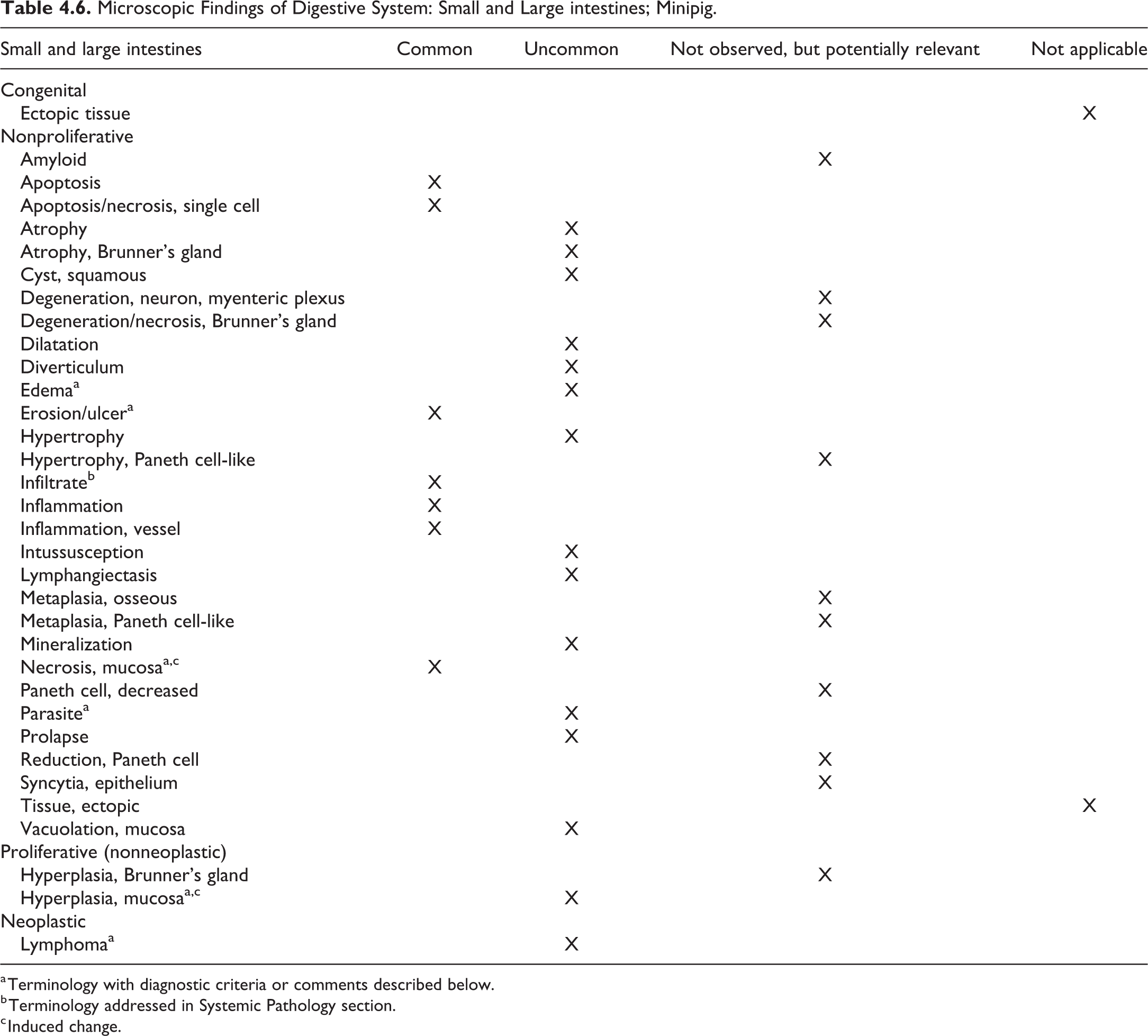
a Terminology with diagnostic criteria or comments described below.
b Terminology addressed in Systemic Pathology section.
c Induced change.
Edema, small and large intestine
Comment
Edema without inflammation/inflammatory cells is occasionally observed, predominantly in the large intestine. Edema can be accompanied by hemorrhage.
Erosion/ulcer, small intestine (Figure 4.15)
Diagnostic features
Focal loss of mucosal epithelium (enterocytes) with partial mucosal penetration (erosion) or with full penetration of the mucosa including muscularis mucosa (ulcer).
Epithelial cells are necrotic or absent from the superficial mucosa, and muscularis mucosa is intact; no edema or hemorrhage are present (erosion).
Epithelial cells are necrotic or absent and muscularis mucosa is destroyed (ulcer).
Erosion/ulcer is associated with an inflammatory cell infiltrate.
Differential diagnoses
Artifacts or autolysis
Comment
Erosion/ulcer is a commonly observed background finding in nonclinical safety studies. Erosion is seen more commonly than ulcer and is usually focal or multifocal at minimal to slight severity (occasionally can be of moderate severity) and is present most frequently in the duodenum. Focal ulcers can be of moderate severity. 3,7
Hyperplasia, mucosa, small intestine
Comment
In juvenile minipigs, treatment with Gattex (Teduglutide, a 33 amino acid recombinant analog of the human glucagon-like peptide-2 [GLP-2]) can result in microscopic changes in the small intestine (mucosal hyperplasia) as observed in mice and monkeys. 52
Lymphoma, small intestine (Figures 4.16 and 4.17)
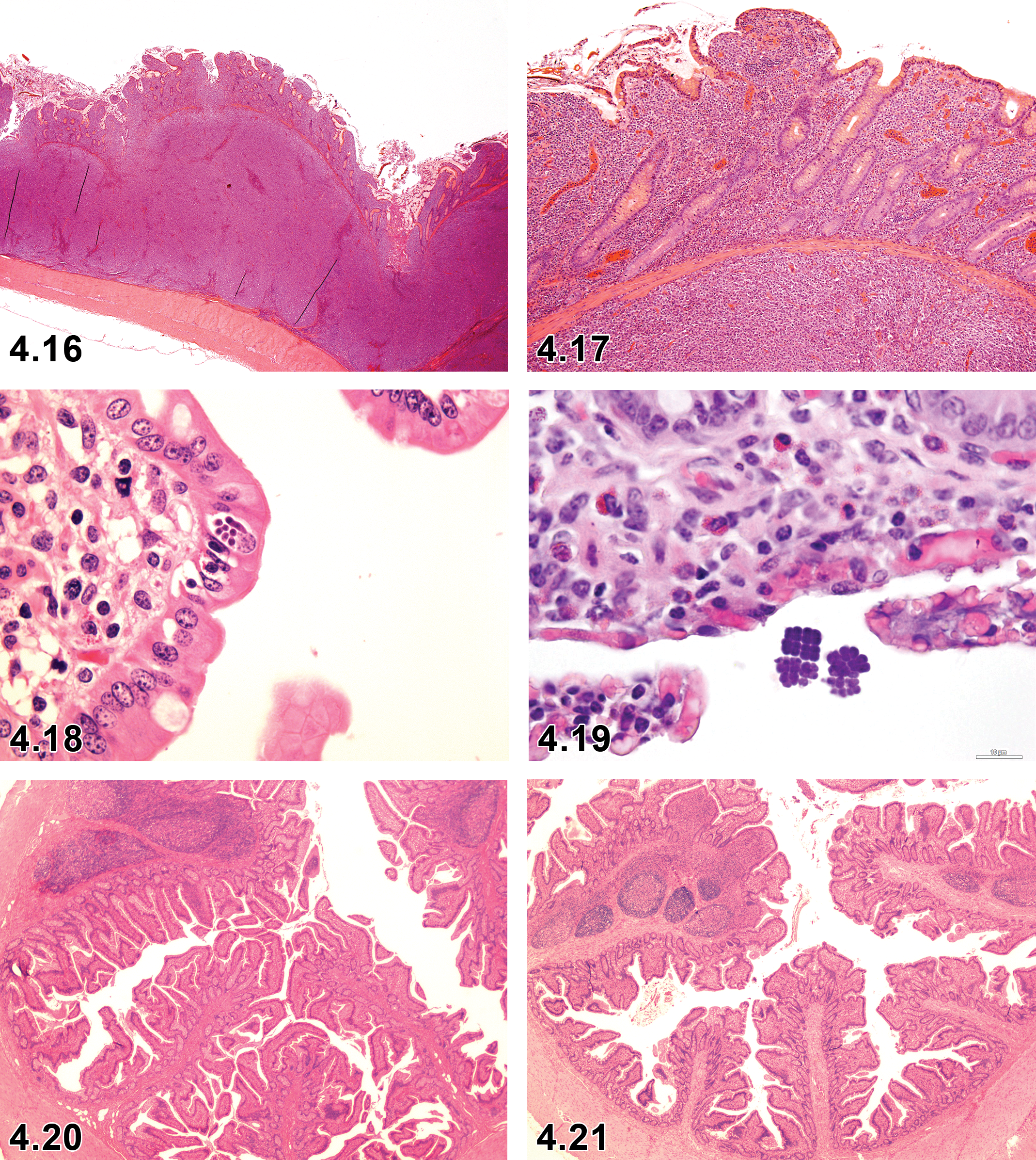
Small intestine, ileum, lymphoma, ×2.
Comment
Lymphoma has been diagnosed in the ileum (see Hematolymphoid section) (McKeag, unpublished data).
Necrosis, mucosa, small and large intestine
Comment
Necrosis of intestinal epithelial cells is a common background finding in minipig nonclinical safety studies. Intravenous administration of diacetoxyscirpenol (anguidine, a mycotoxin) to swine at 0.5 and 1.0 mg/kg resulted in enterocyte damage (cytotoxic effect), mimicking radiation poisoning. Enterocytes in different anatomical regions of the gastrointestinal tract show differing susceptibilities to the toxic effects of anguidine. Mitotically and metabolically active tissues are predominantly affected. 53
Parasite, small intestine (Figures 4.18 and 4.19)
Comment
Eimeria spp and Isospora spp can occasionally inhabit the small intestine but may also be detected in the large intestine in nonclinical safety studies.
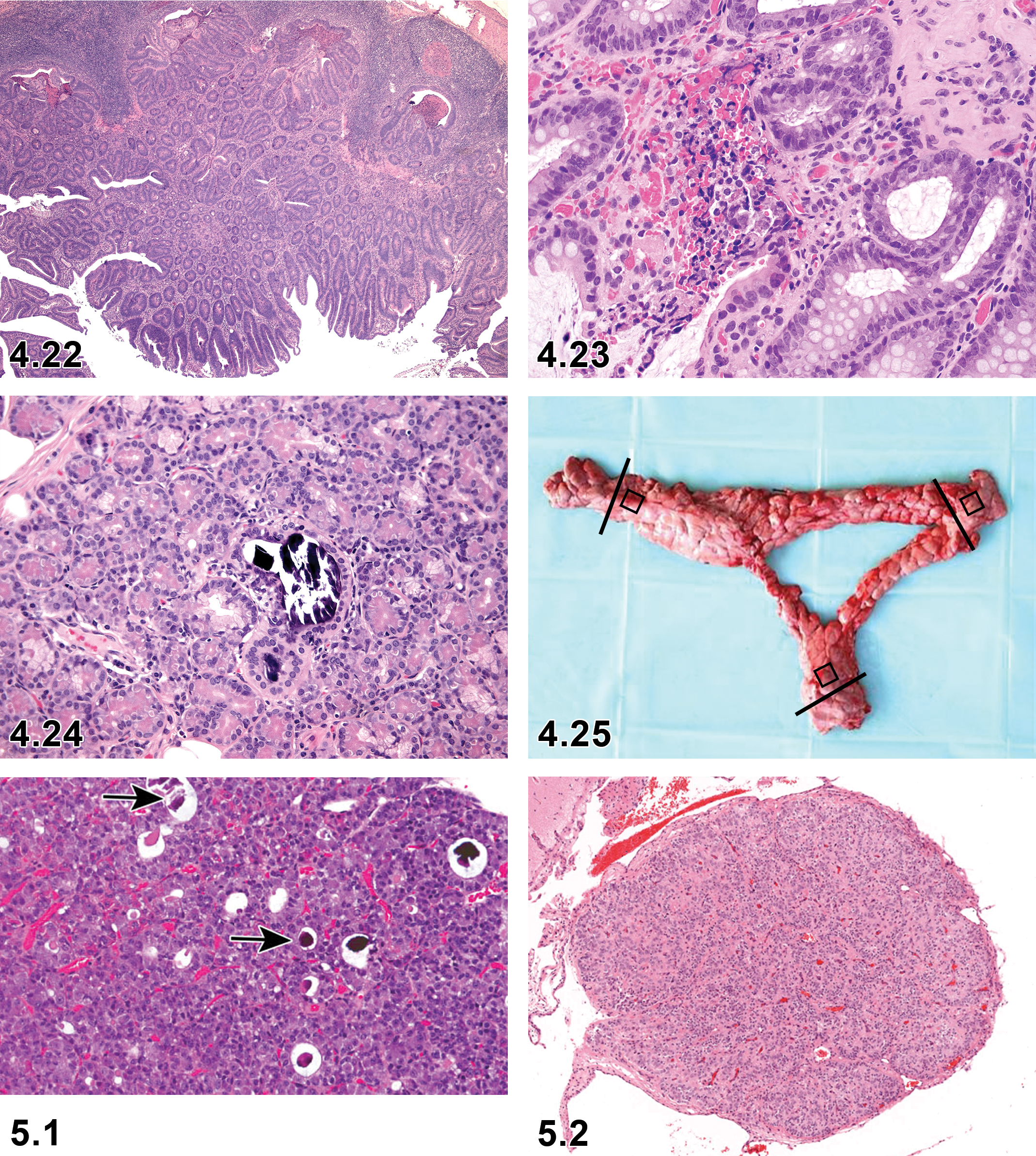
IV. Salivary Glands
Salivary glands of the pig are large and consist of paired sets of buccal (seromucous), sublingual (mucous), parotid (serous), and mandibular (seromucous) glands. 47,54
For minipig nonclinical safety studies, similar sampling and trimming as in rats and mice is recommended. The left parotid, mandibular, and sublingual glands are routinely sampled and examined microscopically. After removal of the tongue and trachea with their adjacent structures, the parotid gland is harvested. This gland is triangular, lobulated, light brown in color, and located under the skin directly caudal to the mandibular bone. Next, the mandibular gland, located medial and slightly cranial to the parotid gland, can be accessed. Sublingual gland is sampled through an incision to the left of the base of the tongue. One longitudinal section through the largest surface of the glands is examined microscopically. 26 Salivary gland background findings were rare in the Yucatan minipigs compared with Gottingen breed. 13
Microscopic Findings of Digestive System: Salivary Glands; Minipig.
a Terminology with diagnostic criteria or comments described below.
b Macroscopic lesion.
c Terminology addressed in Systemic Pathology section.
Edema, salivary glands
Pathogenesis/cell of origin
Accumulation of tissue fluid in the interstitium resulting from increased vascular permeability.
Diagnostic features
Eosinophilic, interstitial fluid within the tissue.
No involvement of inflammatory cells.
No tissue or vascular damage.
Comment
Macroscopically clear, thick, gelatinous material (edematous appearance) is occasionally noted surrounding the mandibular glands. However, this is often not visible on microscopic examination of the gland. 3 Recording the presence of edema using a macroscopic, rather than a microscopic, term is recommended in minipig nonclinical safety studies.
Hyperplasia, ductal, salivary glands
Pathogenesis/cell of origin
Ductal epithelium of the salivary glands.
Diagnostic features
Focal, multifocal.
Small ductal cells with increased basophilia.
Minimally altered ducts with basophilic cytoplasm and hyperchromatic nuclei.
Associated with interstitial fibrosis.
Differential diagnoses
Hypertrophy, adenoma, adenocarcinoma.
Comment
Isolated occurrences of focal ductular hyperplasia can be seen as a background finding in nonclinical safety studies. Focal ductular hyperplasia is associated with interstitial fibrosis. 7 Potential differential diagnoses (including hypertrophy, adenoma, adenocarcinoma) observed in other species are not observed in minipig. Therefore, criteria for hypertrophy, adenoma, and adenocarcinoma cannot be detailed.
Vacuolation, acinar cell, salivary glands
Comment
Vacuolation of the acinar cells has not been observed in Göttingen minipig. However, it has been observed in Yucatan minipigs (unpublished data).
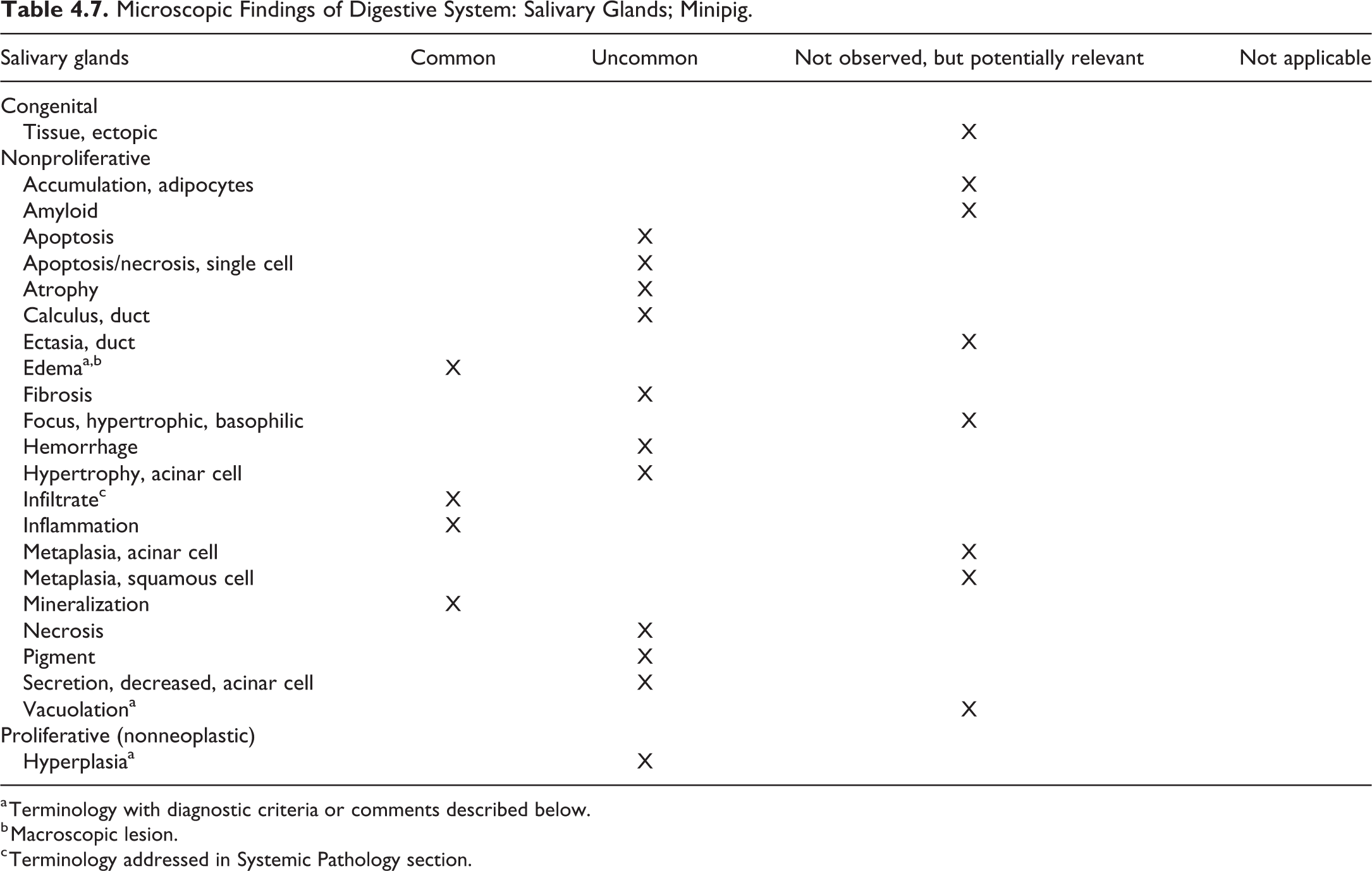
V. Pancreas (Exocrine)
The pancreas is an extensive thick gland with an irregular outline and is divided into 3 parts, the head (right lobe, duodenal portion: upper right corner in Figure 4.25), the body (including the neck: down middle in Figure 4.25), and the tail (left lobe, splenic portion: upper left corner in Figure 4.25; Figure 4.25: pancreas, indicating the locations and orientations of the histology (black lines) and molecular analyses (black rectangles), samples for investigated studies). 26 The head, which is in contact with the gastrointestinal tract from the end of the pylorus to the proximal duodenum, extends to the left and connects to the body. The body separates into 2 (anterior and posterior) portions that encompass the portal vein making the pancreas appear to be “ring-shaped.” The posterior portion extends caudally, ventral to the right kidney. The tail is located on the left of the body and extends caudosinistrally ventral to the left kidney, and it terminates near the hilus of the spleen. The 3 parts of the pancreas in minipigs can also be regarded more simplistically as two lobes, the right lobe or head (duodenal portion) and the left lobe or tail (splenic portion). 55
Small intestine, ileum, inflammation, ×2.
The pancreas should quickly be harvested after the animal’s death to prevent autolysis. Since the density of endocrine pancreatic islets is not equal across the different pancreatic lobes, it is recommended to include cross sections of the 3 parts of the pancreas (right and left lobes and body). 26 In nonclinical safety studies, usually the left lobe or tail (splenic portion) of the pancreas is sampled for microscopic examination.
For investigative studies, 26 3 samples were recommend including left and right lobes and body of the pancreas (Figure 4.25. Minipig, Pancreas).
Histologically, middle- and small-sized islets are present in all lobes of the pancreas in the porcine. The islets of Langerhans are not as clearly delimited from the adjacent exocrine tissue as in rodents. The islet cells get in the adjacent exocrine tissue and occasionally constitute a part of the acinus. The distribution pattern of α- versus β-cells in the islet is not distinctive and is basically uniform in all lobes of the pancreas regardless of the size of the islet. 26,56
Microscopic Findings of Digestive System: Pancreas (exocrine); Minipig.
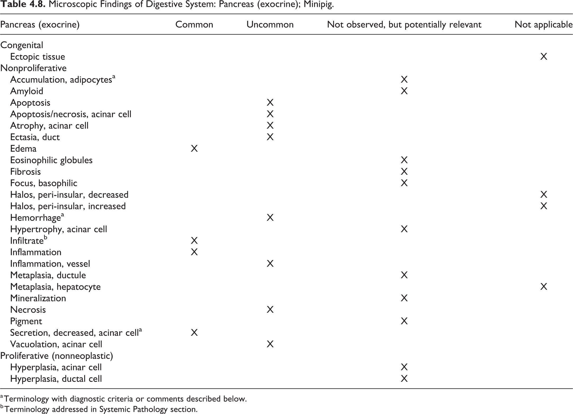
a Terminology with diagnostic criteria or comments described below.
b Terminology addressed in Systemic Pathology section.
Accumulation, adipocytes, pancreas (exocrine)
Comment
Adipocyte accumulation has not been observed in minipig nonclinical safety studies yet. However, in a minipig obesity model, adipocyte accumulation was observed. 57
Secretion, decreased, acinar cell, pancreas (exocrine)
Comment
Decreased zymogen granules in the pancreas are a commonly recorded background finding in minipig nonclinical safety studies, possibly due to a restricted diet. This can also be observed as a secondary finding in moribund and/or anorexic animals with protein deficiency. Microscopically, the acinar cells appear shrunken with increased basophilia of the cytoplasm.
Hemorrhage, pancreas (exocrine)
Comment
Focal interstitial hemorrhage around the pancreatic islets can occasionally be observed at minimal level in minipig nonclinical safety studies. 7
Chapter 5. Endocrine System
For detailed general considerations on the endocrine system, please refer to the rodent INHAND publication. 58 In this document, the pituitary, pineal, thyroid, parathyroid, adrenal glands, and endocrine pancreas will be discussed separately.
I. Pituitary Gland
The pituitary gland is structurally and functionally complex and exhibits a typical mammalian morphology in the pig. It is located immediately posterior to the optic chiasm within the bony sella turcica. The pituitary gland is harvested after removal of the brain and sagittally cut into 2 halves along its rostrocaudal axis. 26
Microscopic Findings of Endocrine System: Pituitary gland; Minipig.
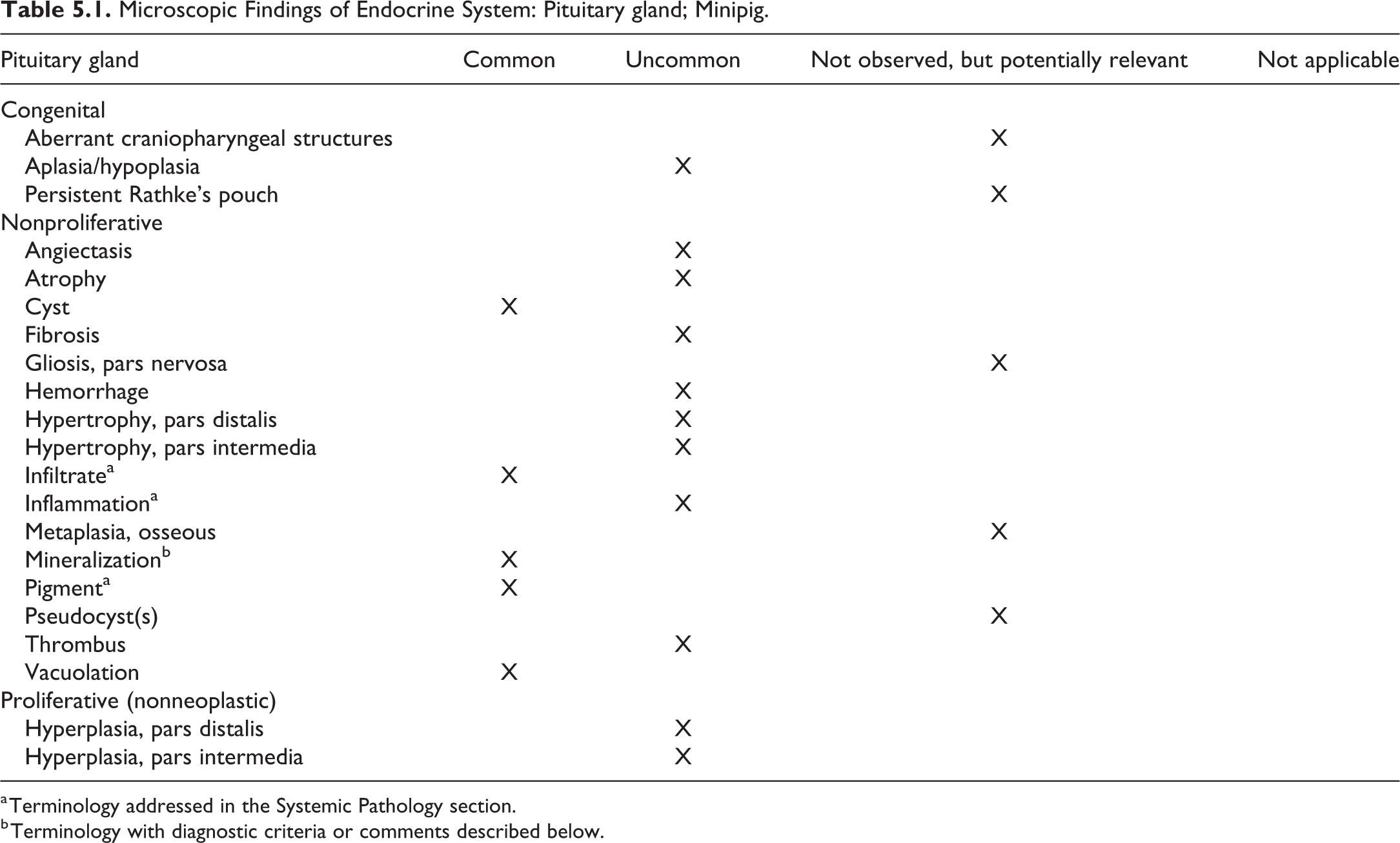
a Terminology addressed in the Systemic Pathology section.
b Terminology with diagnostic criteria or comments described below.
Mineralization, pituitary gland (Figure 5.1)
Comment
Mineralized cells, generally randomly distributed and not associated with inflammation, may occasionally occur in the pars distalis. 3,7,13,59
II. Pineal Gland
The pineal gland is not routinely examined in toxicology studies. It is one of the circumventricular organs of the brain and located on the posterior wall of the third ventricle, between the hemispheres, rostral to the cranial colliculi. It can be assessed when the brain is dissected longitudinally and it is longitudinally cut into 2 halves.
26
Dilated vessels can be quite prominent in the pineal gland of the minipig (Figures 5.2 and 5.3), and melanin pigment (melanosis; Figure 5.4) may be present. Minipig, pineal gland, range of normal vascular dilation, H&E. Microscopic Findings of Endocrine System: Pineal Gland; Minipig.
a Terminology addressed in the Systemic Pathology section.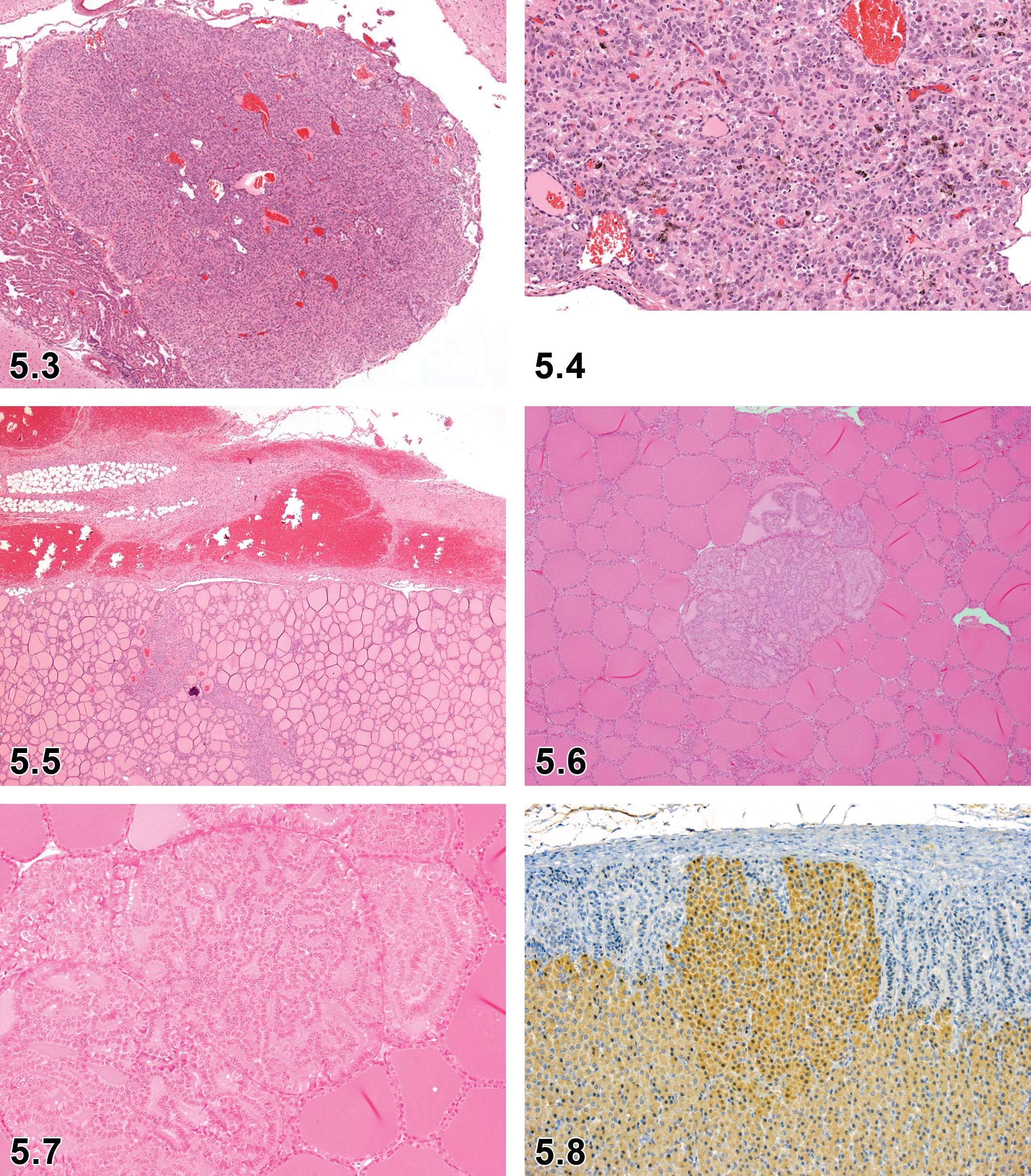
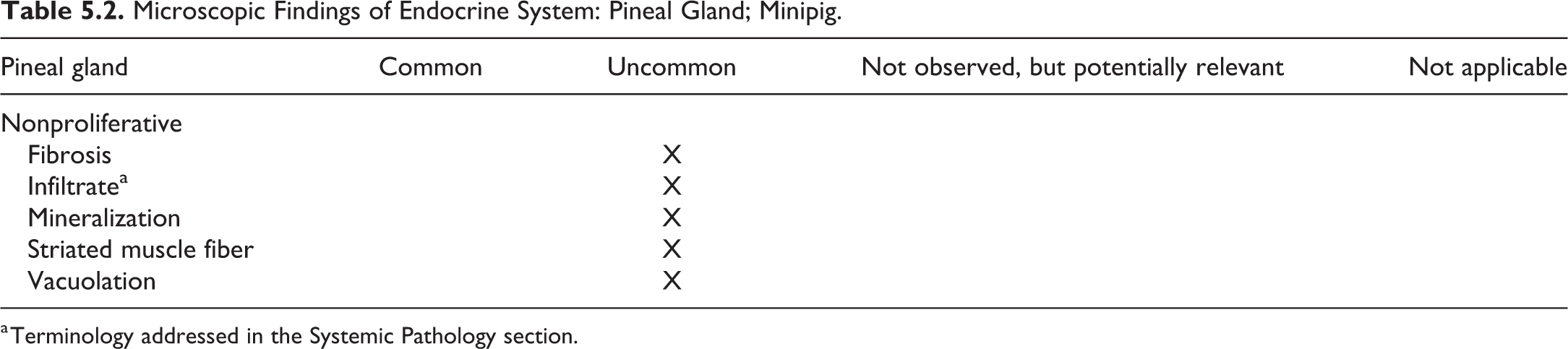
No minipig-specific terminology, criteria, or comments nor references are available.
Since the pineal gland is not routinely evaluated, there is limited experience in incidences of these microscopic findings in the minipig.
III. Thyroid Gland
In the pig, the thyroid is a bilobed organ with fusion of the lobes along the ventral trachea between trachea and thymus cranial to the thoracic inlet. The parathyroid glands are not embedded in the thyroid gland. Either a transverse or longitudinal section of the isolated thyroid gland can be prepared for histological evaluation. 26
Microscopic Findings of Endocrine System: Thyroid Gland; Minipig.
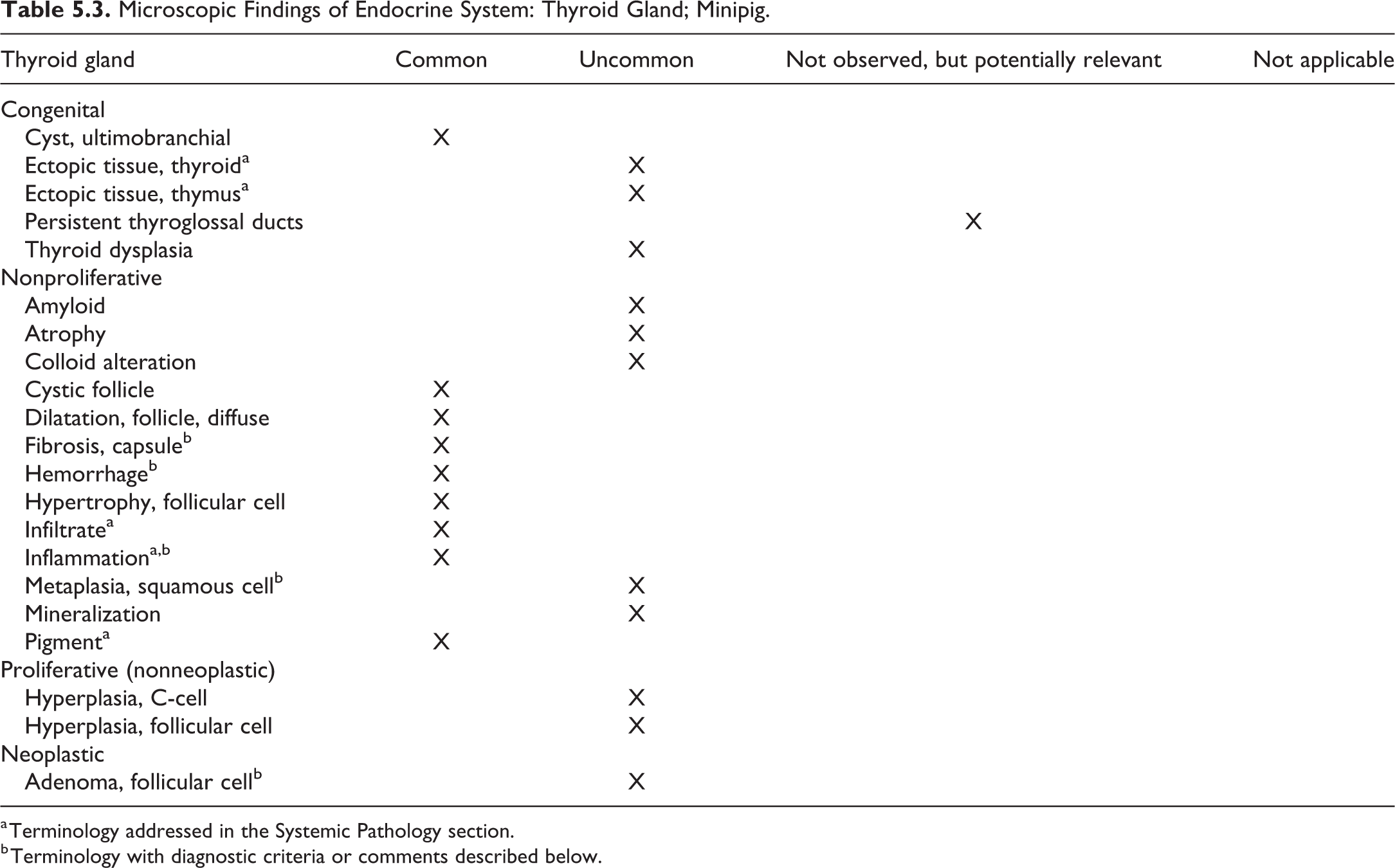
a Terminology addressed in the Systemic Pathology section.
b Terminology with diagnostic criteria or comments described below.
Fibrosis, thyroid gland (Figure 5.5)
See comment under Inflammation.
Hemorrhage, thyroid gland (Figure 5.5)
See comment under Inflammation.
Inflammation, thyroid gland (Figure 5.5)
Comment
The thyroid gland is often injured by accidental mechanical intervention due to blood sampling via the jugular vein leading to hemorrhage, inflammation, and fibrosis and, in some cases, may clinically affect thyroid hormone levels. 13,60,61
Metaplasia, squamous cell, thyroid gland
Comment
Minimal focal squamous metaplasia of follicular epithelium in the thyroid gland has been reported as spontaneous lesion. 7
Adenoma, follicular cell, thyroid gland (Figures 5.6 and 5.7)
Comment
Spontaneous thyroid follicular cell adenomas are very rare in the minipig (Jeppesen, personal communication, 2020). In other species, there appears to be a continuum from focal hyperplasia to carcinoma in induced thyroid tumors. As in rodents, the differentiation between focal hyperplasia and adenoma is not well defined. Size is not a reliable criterion. The use of neoplasm modifiers (eg, follicular, cystic, papillary, solid) is optional. 58
IV. Parathyroid Gland
In pigs, the parathyroid glands are separate from the thyroid gland and can be found near the cranial part of the cervical portion of the thymus opposed to a branch of the carotid artery at the approximate level of the larynx. The glands have a bean-like shape and are often embedded in thymic tissue. Since they are only 2 to 4 mm in diameter, they are not easy to find at necropsy but can be recognized by their firm texture and reddish color. 26,62 Collecting the parathyroid gland can be particularly difficult if there is extensive hemorrhage related to blood collection in the ventral neck region. While embryologists suggest that there are 2 pairs, only 1 pair has been observed grossly. 62,63 The parathyroid glands are small oval bodies, surrounded by a thin connective tissue capsule. For histology, they can be split into 2 halves. 26
Microscopic Findings of Endocrine System: Parathyroid Gland; Minipig.
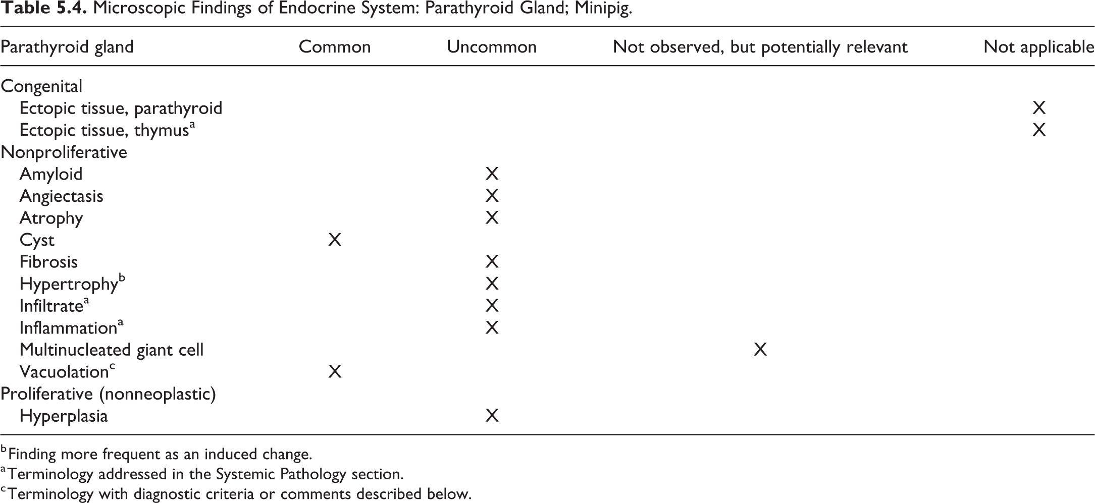
b Finding more frequent as an induced change.
a Terminology addressed in the Systemic Pathology section.
c Terminology with diagnostic criteria or comments described below.
Vacuolation, parathyroid gland
Comment
Cytoplasmic vacuolation of the parathyroid cells has been described for the Göttingen minipigs. 7
V. Adrenal Gland
In the adrenal cortex, the zona glomerulosa lies immediately beneath the capsule. However, parts of the underlying zona fasciculata, the largest of the 3 zones of the cortex, are occasionally present directly beneath the capsule (Figure 5.8). For histology, one horizontal section from the middle portion of one adrenal and one cross section of the middle portion of the other adrenal including both cortex and medulla are recommended. 26 In the medulla, there is some variation in the degree of vacuolation.
Microscopic Findings of Endocrine System: Adrenal Gland; Minipig.
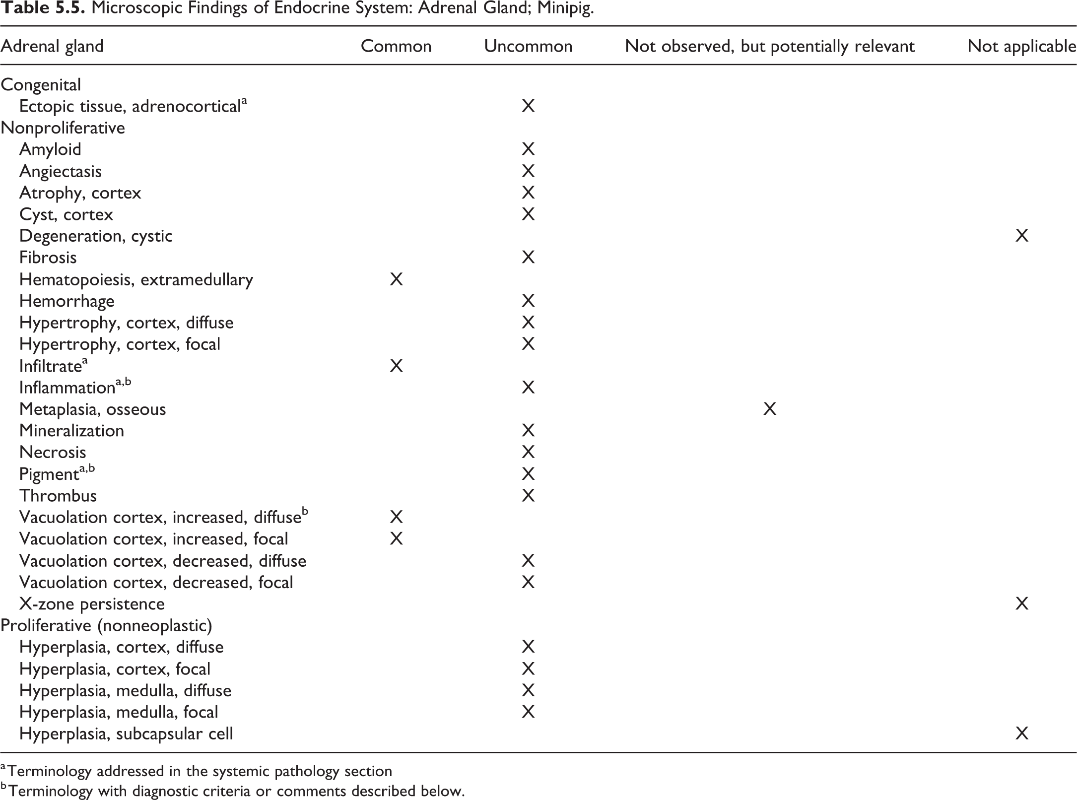
a Terminology addressed in the systemic pathology section
b Terminology with diagnostic criteria or comments described below.
Inflammation, adrenal gland
Comment
Inflammation of the adrenal has been occasionally been observed in the minipig in association with systemic disease or as an extension of peritonitis. 10,64
Pigment, adrenal gland (Figure 5.9)
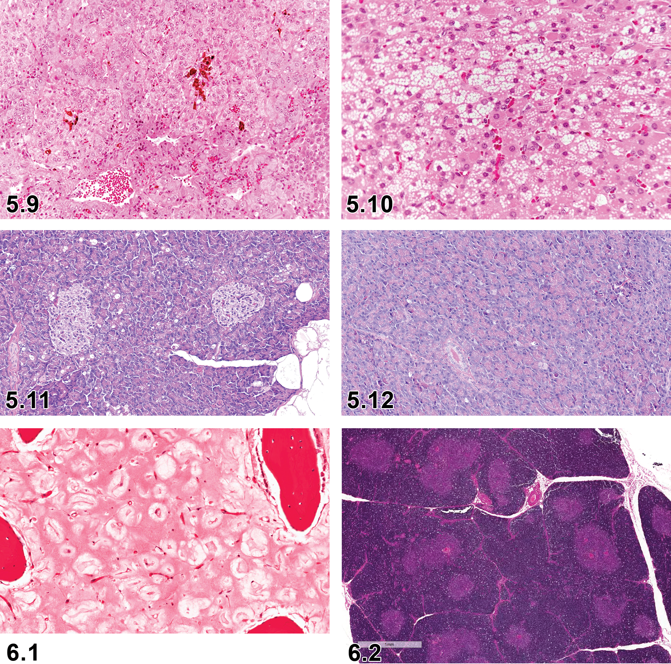
—Minipig, adrenal gland, pigment, H&E.
Comment
Small amounts of lipofuscin are commonly observed in aged minipigs. However, pigment in young animals may be indicative of increased cellular organelle turnover or impaired cell metabolism (inhibition of steroid synthesis). Lipofuscin pigmentation may be associated with severe hormone-induced atrophy. Dietary deficiency of antioxidants such as vitamin E enhances the production and storage of lipofuscin pigment. Its severity can be enhanced by the administration of estrogens and adrenocorticosteroids (personal communication).
Vacuolation, cortex, increased diffuse, adrenal gland (Figure 5.10)
Comment
The amount of vacuolation caused by lipid droplets in all 3 zones can be variable even in normal minipigs. 3,7
VI. Endocrine Pancreas: Islets of Langerhans
The endocrine pancreas consists of discrete aggregates of cells throughout the exocrine pancreas (islets of Langerhans). The islets are composed of pale staining polygonal cells of variable size and are scattered throughout the exocrine pancreas; however, the density is not equal across the different pancreatic lobes. 26 The islets are composed of a number of different endocrine cell types responsible for the production of different hormones. Alpha cells secrete glucagon; beta cells secrete insulin; delta cells secrete somatostatin; gamma cells secrete pancreatic polypeptide (thereby also referred as PP cells); and enterochromaffin cells secrete substance P. Application of immunohistochemical staining for the different hormones or the use of electron microscopy is necessary to differentiate the structure of the secretory granules in each cell type. In the pig, beta cells are found as single cells or grouped together to form the core of the islets at all ages. Some alpha cells are located in the center of islets, but most reside in the periphery, along with a smaller number of delta cells and an even smaller number of PP cells. The number of delta cells and PP-cells in islets decreases as animals’ age. PP cells are more abundant in the head, as opposed to the tail area of the pancreas. Though the number of larger islets increases with gestational age, the percentage volume density of beta cells does not. In pigs at 8 months of age, there are differences in the cellular composition of islets from different regions of the pancreas with islets in the head being rich in PP cells and poor in alpha cells, while islets in the tail are rich in alpha cells and poor in PP cells. 65 Since the density of endocrine pancreatic islets is not equal across the different pancreatic lobes, it is recommended to include cross sections of the 3 parts of the pancreas (right and left lobes and body). 26 In nonclinical safety studies, usually the left lobe or tail (splenic portion) of the pancreas is sampled for microscopic examination.
Microscopic Findings of Endocrine System: Endocrine Pancreas; Minipig.
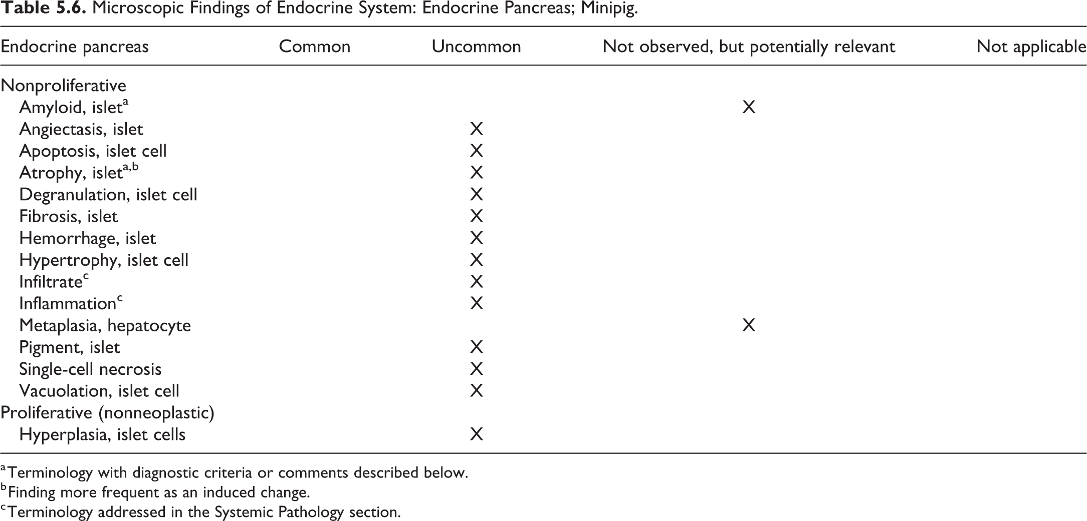
a Terminology with diagnostic criteria or comments described below.
b Finding more frequent as an induced change.
c Terminology addressed in the Systemic Pathology section.
Amyloid, islet, pancreas, endocrine
Comment
In pigs, islet amyloid polypeptide (IAPP1) is expressed mostly in beta cells but also in some alpha and delta cells. The sequence of IAPP in the amyloidogenic domain is dissimilar in pigs and humans, so that pigs are not prone to formation of pancreatic amyloid whereas humans are. 57 Amyloid deposits can be observed in humanized IAPP mutant pigs. 66
Atrophy, islet, pancreas, endocrine (Figures 5.11 and 5.12)
Comment
Göttingen minipigs are used as type 2 diabetes model induced by streptozotocin with or without combination with nicotinamide leading to reduction in beta cell mass and atrophy of the islets. 37,67 Similarly, the Wuzhishan miniature pigs show islet atrophy after alloxan administration. 68
Chapter 6. Hematopoietic and Lymphoid System
This document follows a similar anatomical approach to that used in the rodent INHAND document; however, descriptions will focus on microscopic findings that differ from the rodent in the minipig. Acknowledging that a tiered approach to findings is used in the rodent INHAND document, due to the smaller number of animals in general toxicology studies using minipigs, the authors of this document recommend use of descriptive terms (which in most cases are comparable to enhanced terminology in the rodent). Where lesions in the minipig and rodent are similar they will be tabulated with an indication of their prevalence or applicability to the minipig, and the reader is referred to the rodent manuscript for a full description of the lesion.
This article deals with a standardized nomenclature for classifying microscopic findings observed in the hematolymphoid system of minipigs, namely bone marrow, thymus, lymph node, spleen, mucosal-associated lymphoid tissue (MALT), tertiary lymphoid structures, and a section on general hematolymphoid changes. The minimum recommendation for examination of the hematolymphoid system is to examine thymus, spleen, draining lymph nodes/lymph nodes local to parenteral or topical administration sites, bone marrow in situ, and any gross lesions of a lymphoid organ. 69
General
As in other toxicology species, focal interstitial infiltrates of inflammatory cells, mostly comprised of lymphocytes with plasma cells and macrophages, are the most frequently observed lesion in minipigs. Infiltrating eosinophils can be observed, particularly in the mesenteric lymph node. 3,13
Microscopic Findings of Hematopoietic and Lymphoid System: General; Minipig.
a Terminology with diagnostic criteria or comments described below.
b Terminology addressed in the Systemic Pathology section.
c Finding more frequent as an induced change.
Extramedullary hematopoiesis
Comment
Extramedullary hematopoiesis is generally greatest at around 14 days postpartum, is decreased or absent by 35 days, and is absent after 63 days 5 or 6 weeks. 6,7 In older pigs, extramedullary hematopoiesis may be seen in response to severe anemia. The INHAND term for rodents is “extramedullary hematopoiesis, increased”; however, in minipigs over 6 weeks old, as this is not considered a normal feature of the spleen, the modifier “increased” has been dropped.
Inflammation
Comment
Inflammation can be seen in and around the thymus, thyroid, trachea, esophagus, and mandibular lymph nodes due to blood sampling procedures. 3,7
Phospholipidosis
Other term(s)
Vacuolation, macrophage
Comment
Vacuoles positive for LAMP2 or characteristic appearance on electron microscopy. Similar to the other laboratory species, the minipig is susceptible to phospholipidosis. 15
Pigment, macrophage
Comment
Pigment within macrophages can be seen in piglets due to prophylactic postnatal iron injection. 3,13
Bone Marrow
Bone marrow is usually examined in formalin-fixed, paraffin-embedded, decalcified 5 or 3 µm sections of femur and sternum (and occasionally vertebrae) stained with H&E. It is good practice to prepare contemporaneous bone marrow smears at necropsy, as unequivocal identification of cell lines on H&E-stained sections is difficult. Should changes be detected on H&E, Romanowsky-stained bone marrow smears can be examined. 70
Microscopic Findings of Hematopoietic and Lymphoid System: Bone Marrow; Minipig.
a Terminology addressed in the Systemic Pathology section.
b Terminology with diagnostic criteria or comments described below.
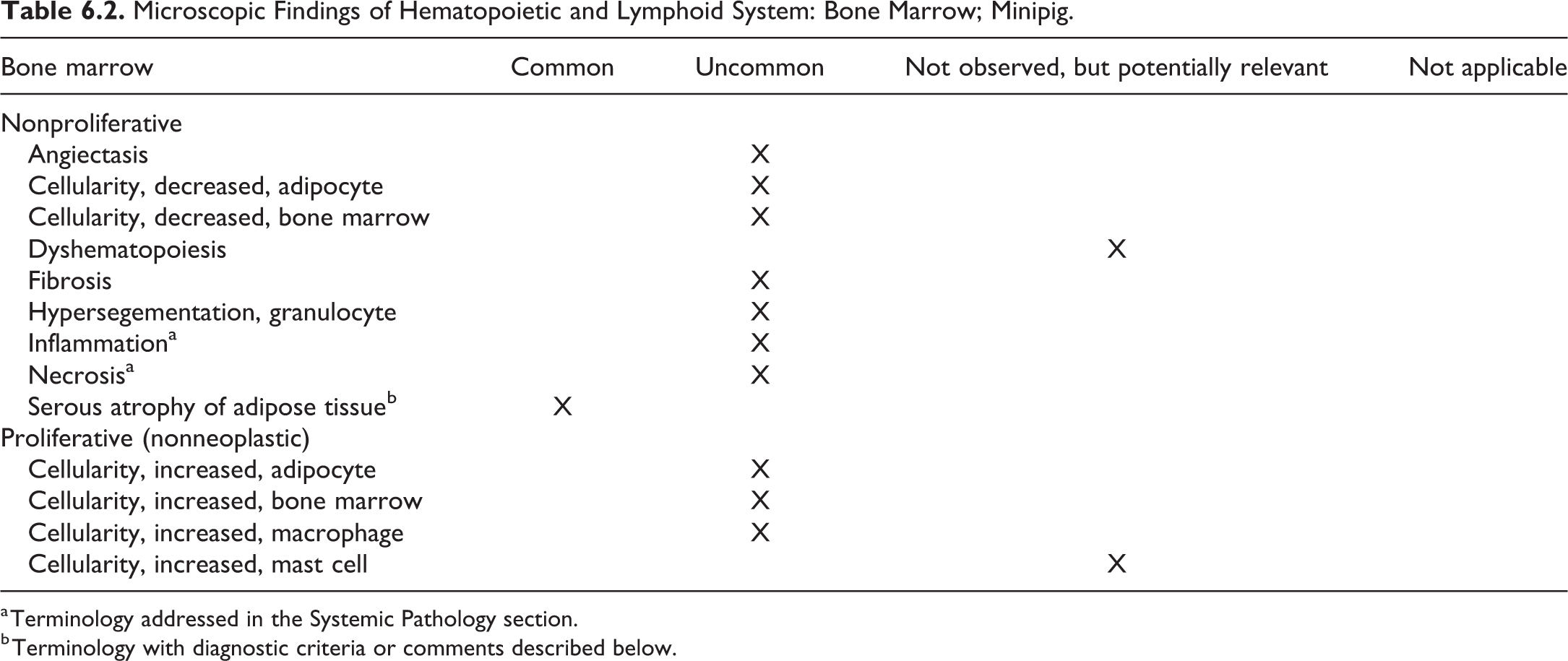
Serous atrophy of adipose tissue, bone marrow (Figure 6.1)
Other term(s)
Gelatinous transformation, serous atrophy. Please refer to the Systemic Chapter for a detailed description.
Diagnostic features
Focal or diffuse depletion of atrophied or degenerated adipocytes, often starting in the epiphysis. If only the epiphysis is affected, it is graded as minimal or slight. With increasing severity, the metaphysis and diaphysis are affected and the assigned grade would be moderate or marked.
Reduced cellularity of the hematopoietic cells.
Interstitial accumulation or total replacement of adipose tissue by homogenous eosinophilic gelatinous tissue (hyaluronic acid, mucopolysaccharides).
Differential diagnoses
Adipocyte atrophy in the absence of eosinophilic gelatinous tissue.
Decreased hemopoietic cells.
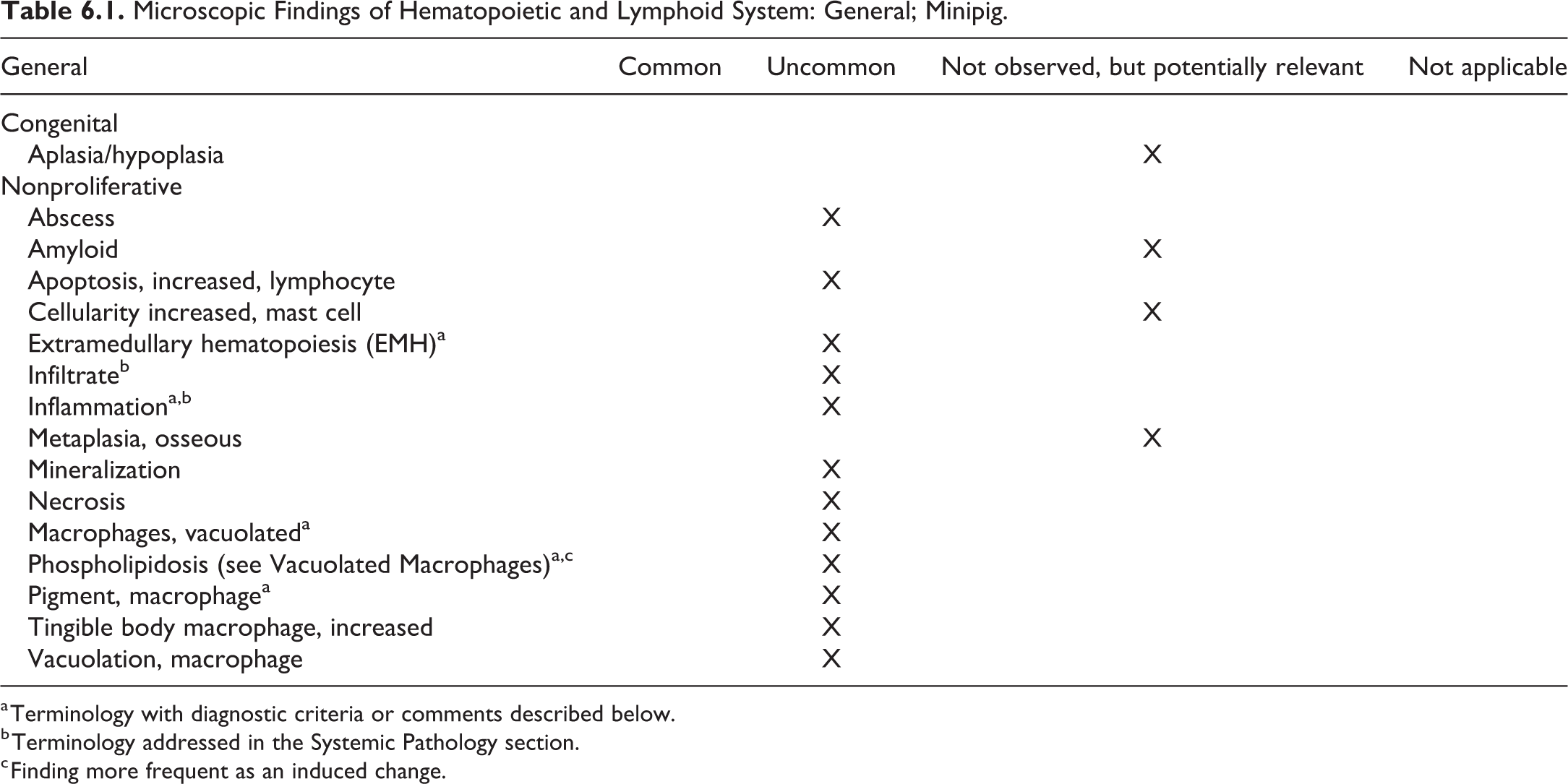
Thymus
The thymus consists of cervical and thoracic lobes, with the pig having a well-developed cervical lobe with a cranial and caudal portion. The cervical thymus runs along the jugular grooves up to the pharyngeal region, while the thoracic portion extends from the pericardium into the thoracic inlet. 71 Keratinization of Hassall’s corpuscles is prominent in minipigs as in young dogs and nonhuman primates. The minipig thymus does not involute to the same extent as that of dogs and nonhuman primates, at least not in the course of routine toxicology studies. 71 In the experience of the authors, the thymus is relatively resistant to stress-induced atrophy. 7 When sampling, both parts should be weighed and histological analysis performed. Sampling of the thoracic portion may avoid venipuncture artifact. Albl et al suggest that the caudal cervical portion should be examined; however, sampling sites vary between laboratories. 26,69 The position of the parathyroid gland is variable and may be found embedded in the thymus or in the connective tissue adjacent to the thymus. As such, ectopic parathyroid gland is not considered to be a meaningful diagnosis in this species. 3
Microscopic Findings of Hematopoietic and Lymphoid System: Thymus; Minipig.
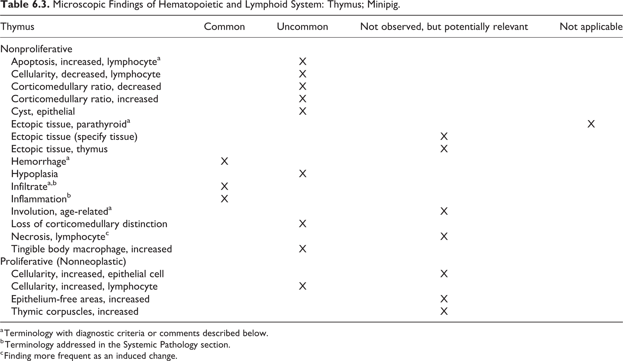
a Terminology with diagnostic criteria or comments described below.
b Terminology addressed in the Systemic Pathology section.
c Finding more frequent as an induced change.
Apoptosis, increased, lymphocyte, thymus (Figure 6.2)
Ectopic tissue, parathyroid, thymus
Comment
As the parathyroid gland is not associated with the thyroid glands in the minipig, and as the pig has a cervical thymus, parathyroid gland is commonly found within the thymus and is not considered to be ectopic.
Hemorrhage, thymus (Figure 6.3)
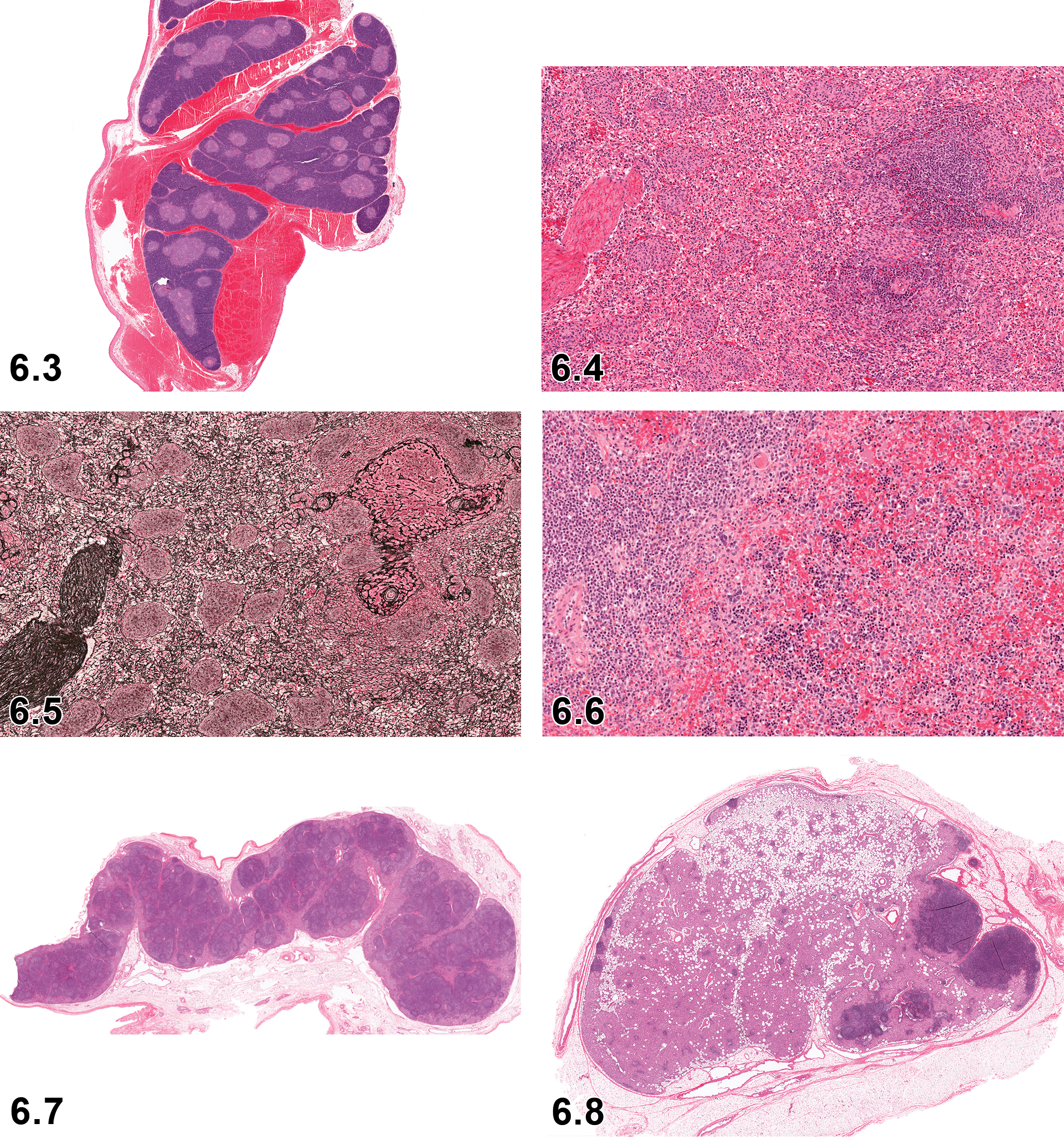
Thymus hemorrhage, H&E, subgross Iatrogenic hemorrhage due to bleeding procedures and the structure of the blood vessels of the neck and cranial thorax.
Pathogenesis/cell of origin
Extravasated erythrocytes in the vicinity of the thymus and thyroid. Blood sampling procedures are the most common cause of hemorrhage in and around the thymus.
Diagnostic features
This lesion may vary in appearance from acute to chronic. When acute, extravasated blood is present around the thymus. As the lesion ages, extravasated blood is gradually replaced by inflammatory cells, fibrous tissue, and pigmented macrophages.
Differential diagnoses
Hemorrhagic syndrome
Comment
Due to the proximity of the thymus to the major vessels of the neck used in venipuncture procedures, and the difficulty sometimes experienced while taking blood samples from pigs, hemorrhage in the vicinity of the thymus is not uncommon. Also see inflammation and/or infiltrate, inflammatory cells in the “General” section and below.
Infiltrate, inflammatory cell and inflammation, thymus
Comment
Due to the proximity of the thymus to the major vessels of the neck used in venipuncture procedures, and the difficulty sometimes experienced while taking blood samples from pigs, inflammatory cell infiltrate or inflammation, usually in conjunction with hemorrhage, in the vicinity of the thymus is not uncommon.
Involution, age-related, thymus
Thymic involution is thought to begin at puberty, with changes visible at 180 days 72 or 18 months, 73 depending on which paper is considered. Distinguishing between age-related involution and test article-related thymic lymphocyte depletion can be challenging. Prominent necrotic and/or apoptotic lymphocytes would be considered unusual in involution; however, fewer lymphocytes in cortex and medulla with a lack of definition between these two compartments, an increase in perivascular spaces, more Hassall’s corpuscles with keratinization and degeneration, and adipocyte infiltration in capsule and interlobular connective tissue may be seen. Careful comparison between control and treated animals with reference to the age of animals should be made.
Spleen
The role of the spleen in humans and rodents is primarily defensive, and in dogs, it is primarily a storage organ; however, in minipigs, the spleen shares characteristics of both. The minipig spleen is nonsinusoidal. 71 There is a thick capsule of smooth muscle and elastic fibers and similarly structured trabeculae. Lymphoid follicles are less prominent than in rats, but the periarteriolar lymphoid sheath is well developed. The red pulp contains pale eosinophilic, contractile, phagocytic structures surrounding splenic capillaries, which are known as periarteriolar macrophage sheaths, splenic ellipsoids, or Schweigger-Seidel sheaths, which can be quite prominent. 29,71 Their function is clearance of blood-borne particles. Extramedullary hematopoiesis may be seen in juveniles up to 6 weeks old or as a response to systemic disease such as hemorrhagic syndrome. 7,29 Accessory spleens may be found in the gastrosplenic ligament. 71 A cross-section through the thickest part of the spleen should be examined histologically (Figures 6.4 and 6.5).
Microscopic Findings of Hematopoietic and Lymphoid System: Spleen; Minipig.
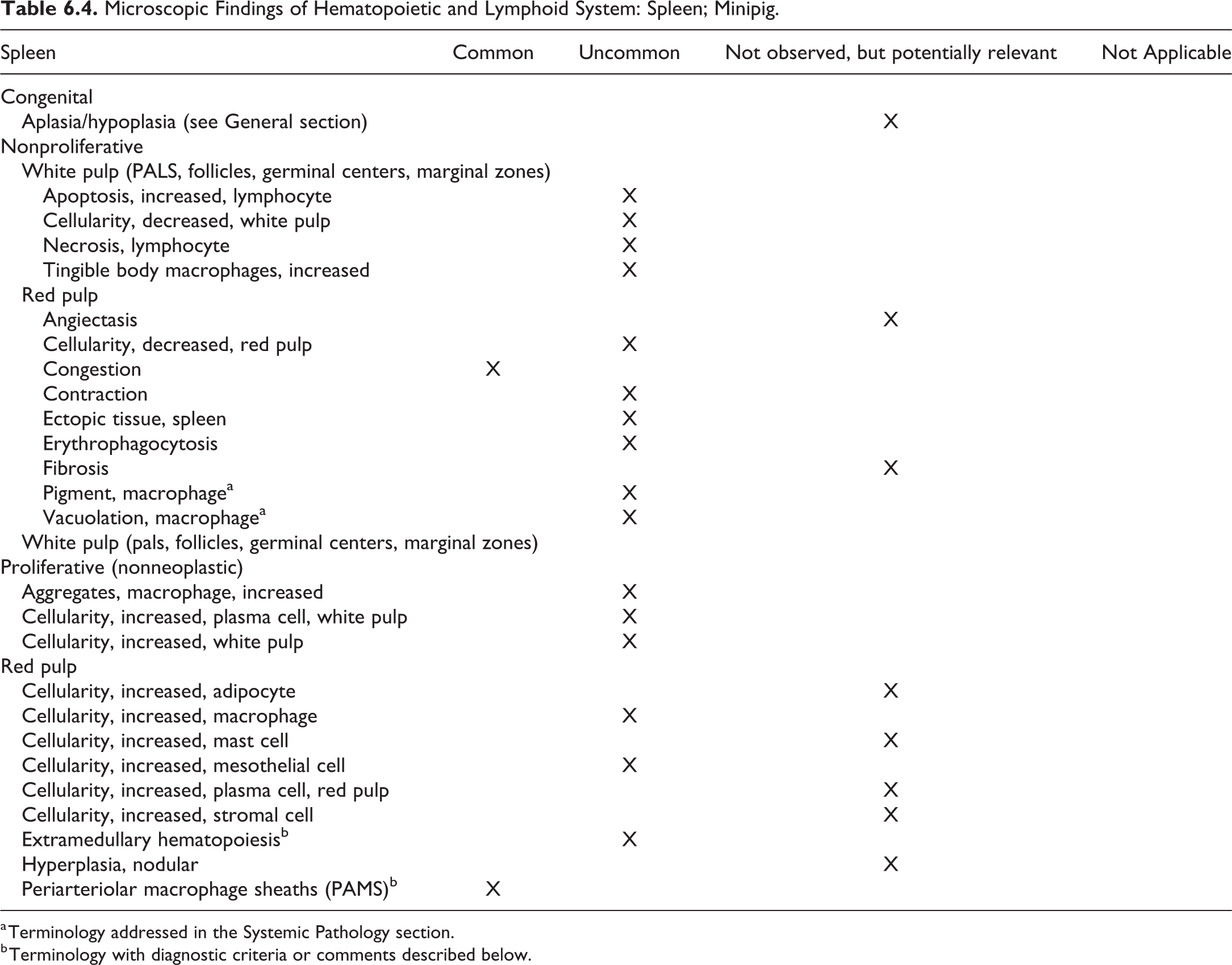
a Terminology addressed in the Systemic Pathology section.
b Terminology with diagnostic criteria or comments described below.
Extramedullary hematopoiesis, spleen (Figure 6.6)
Comment
Extramedullary hematopoiesis is generally greatest at around 14 days old, is decreased or absent by 35 days, and is absent after 63 days 5 or 6 weeks. 6,7 In older pigs, extramedullary hematopoiesis may be seen in response to severe anemia. The INHAND term for rodents is “extramedullary hemopoiesis, increased”; however, in minipigs over 6 weeks old, as this is not considered a normal feature of the spleen, the modifier “increased” has been dropped.
Periarteriolar macrophage sheaths, Spleen
Other term(s)
Ellipsoids, Schweigger-Seidel sheaths
Pathogenesis/cell of origin
These are normal structures formed from phagocytic cells and reticular fibers.
Diagnostic features
Generally rounded, pale eosinophilic accumulations of cells around capillaries or small arterioles in the red pulp.
Comment
Periarteriolar lymphoid sheaths are prominent in the Göttingen minipig. Their prominence varies between animals. Their function is to filter blood, retain circulating particulate matter, and trap immune complexes. 6,71,74
Lymph Node
Histologically, lymph nodes of pigs have a unique structure, with centrally located cortical tissue and germinal centers and peripheral medullary sinuses and cords that appear inside out when compared to other species. The follicles are arranged centrally and not peripherally, and the lymph flow is reversed with lymph entering centrally at the hilus and leaving through high endothelial veins at the capsular surface. 71 This results in few lymphocytes in the efferent lymph ducts. These differences in structure do not appear to result in significant functional changes. The appearance of lymph nodes can vary considerably depending on anatomical location, region drained, age, and plane of section (Figures 6.7, 6.8). There are varying numbers of eosinophils normally present in the porcine lymph node sinuses. 7 The mesenteric lymph node is large and is located at the root of the mesentery. In addition, the mesenteric lymph nodes are supplied by a prominent perimesenteric plexus. 29 This lymph node should be sampled in all studies as it can reflect the inflammatory state of the gastrointestinal tract. 69 It is common practice for the mandibular lymph node to be examined as a representative peripheral lymph node. For enhanced histopathology of the lymph nodes, the most proximal regional lymphoid tissues that drain the application/ administration site should be examined microscopically. 75
Microscopic Findings of Hematopoietic and Lymphoid System: Lymph Node; Minipig.
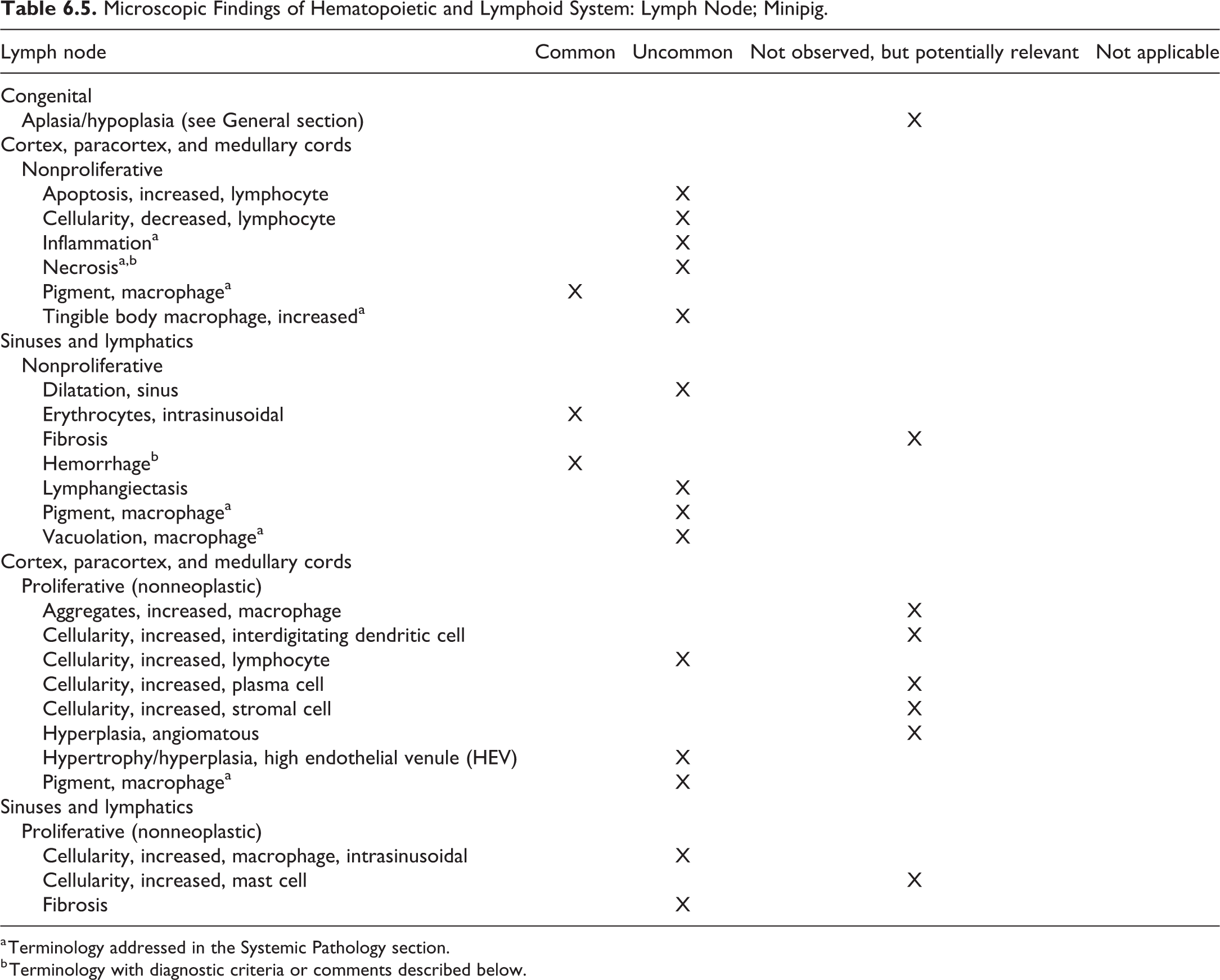
a Terminology addressed in the Systemic Pathology section.
b Terminology with diagnostic criteria or comments described below.
Hemorrhage, lymph node
Pathogenesis/cell of origin
Extravasated erythrocytes in and/or around the mandibular lymph node. Blood sampling procedures are the most common cause of hemorrhage in and around this lymph node, although agonal changes may be involved in some cases. 3
Diagnostic features
This lesion may vary in appearance from acute to chronic. When acute, extravasated blood is present around the lymph node, and/or within the sinuses. As the lesion ages, blood is gradually replaced by inflammatory cells, fibrous tissue, and pigmented macrophages.
Differential diagnoses
Hemorrhagic Syndrome
Comment
Due to the proximity of the mandibular lymph node to the major vessels of the neck used in venipuncture procedures, and the difficulty sometimes experienced while taking blood samples from minipigs, hemorrhage in the vicinity of this lymph node is not uncommon. Also see inflammation in the “General” section.
Necrosis, mesenteric lymph node (Figure 6.9)
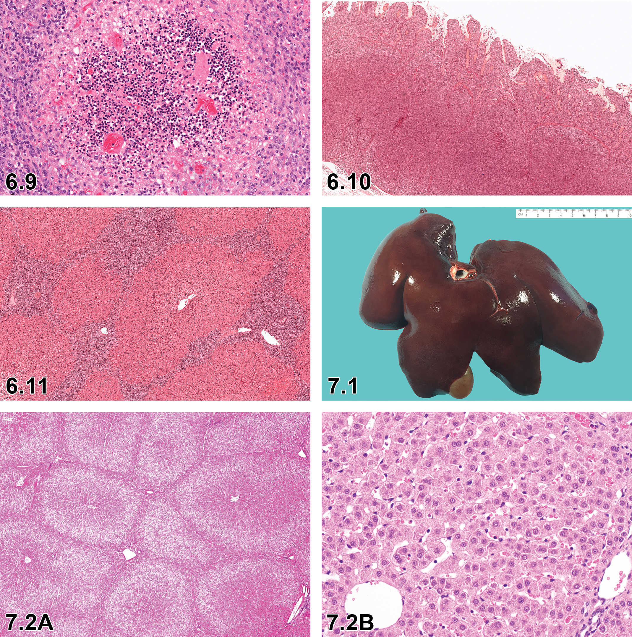
Mesenteric lymph node: Necrosis, focal, lymphocyte: H&E ×20. Showing pyknotic nuclei and cell debris.
Mucosa-associated lymphoid tissue
Mucosa-associated lymphoid tissues (MALT) include gut-associated lymphoid tissue, nasopharynx-associated lymphoid tissue (NALT), and bronchus-associated lymphoid tissue (BALT). Gut-associated lymphoid tissue should be examined in studies where the test article is administered orally and is usually examined in a routine section of jejunum or ileum that contains Peyer’s patches. Minipigs have 2 types of Peyer’s patches: discrete nodules in the jejunum and proximal ileum and linear continuous structures in the distal ileum. Antigenic stimulation is not required for growth of Peyer’s patches in the minipig, which increase in number and size with age. 71
Nasopharynx-associated lymphoid tissue and BALT should be examined in studies where the test article is administered intranasally or by inhalation. Nasopharynx-associated lymphoid tissue may be examined in routine nasal cavity sections, and BALT in routine lung sections, generally at bronchial/bronchiolar bifurcations. 76,77
Microscopic Findings of Hematopoietic and Lymphoid System: MALT; Minipig.
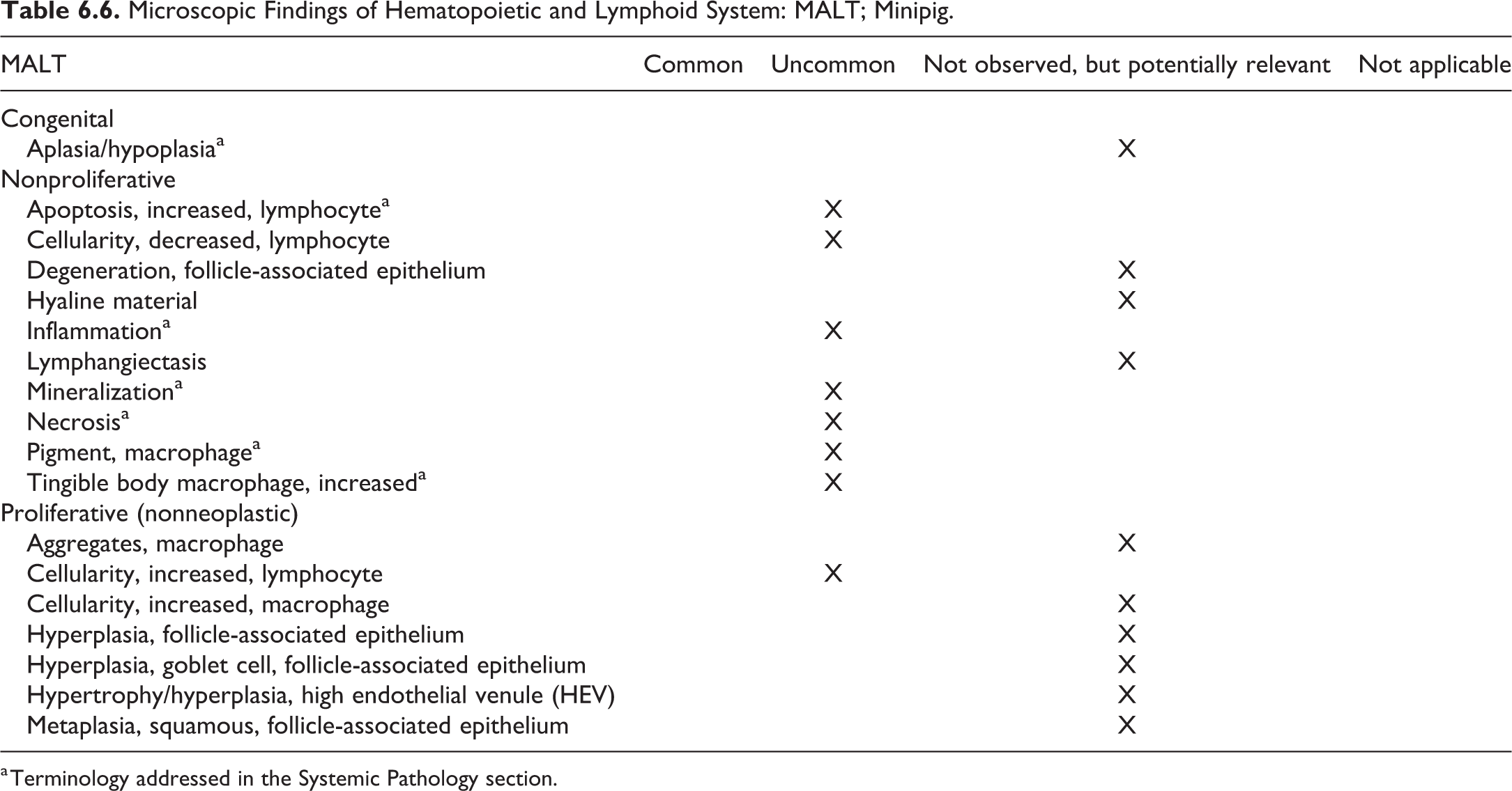
a Terminology addressed in the Systemic Pathology section.
Microscopic Findings of Hematopoietic and Lymphoid System: Other Lymphoid Tissues; Minipig.
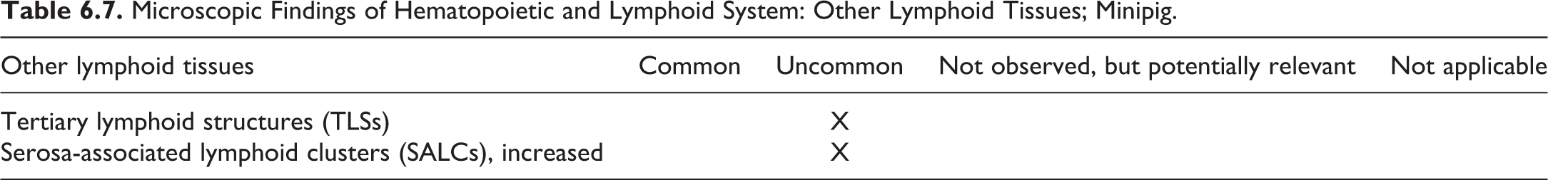
Microscopic Findings of Hematopoietic and Lymphoid System: Hematolymphoid Neoplasms; Minipig.
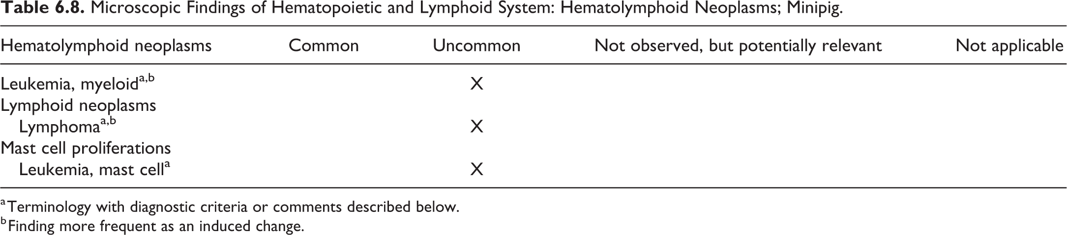
a Terminology with diagnostic criteria or comments described below.
b Finding more frequent as an induced change.
Leukemia, myeloid
Comment
Myeloid leukemias have been induced in Pitman-Moore miniature pigs by administration of strontium 90. 78
Lymphoma (Figures 6.10 and 6.11)
Comment
Lymphoma has not, to our knowledge, been described in the literature in Göttingen minipigs. However, 2 lymphomas have been diagnosed but not reported in Göttingen minipigs. One was diagnosed in an 8-month-old male and one in a 3-month-old female. Neoplastic lymphocytes were present in intestine, liver, and spleen; however, limited tissues were available for examination. Neither pig was on study (McKeag, unpublished data). 79 –81 Lymphoid neoplasms have been induced in miniature pigs by oral administration of Strontium 90. 78
Leukemia, mast cell
Comment
Mast cell leukemia has been diagnosed in a 3-year-old male pet Göttingen minipig 82 and a 9-year-old female Sinclair miniature pig. 83
Chapter 7. Hepatobiliary System
For detailed description of the hepatobiliary system lesions, refer to the rodent publication. 84 While the type and classification of pathological findings of the hepatobiliary system are identical or very similar across species, their frequency in untreated individuals may differ substantially among various species.
I. Liver
The minipig liver consists of 6 lobes. It has 4 main lobes (right lateral, right medial, left medial, and left lateral) and 2 lesser lobes (caudate and quadrate). The right lateral lobe is divided dorsally to form the caudate lobe, which in turn may be divided by a fissure into 2 parts. The quadrate lobe is ventral to the portal fissure and to the left of the gall bladder. In contrast to the other laboratory species and humans, the porcine liver displays prominent lobulation that is macroscopically visible. The lobules are demarcated by white stromal septa which become apparent in animals at approximately 4 weeks of age. The liver has 2 surfaces. The diaphragmatic surface lies in close proximity to the diaphragm and is markedly convex. The visceral surface is concave and closely associated with the abdominal viscera. The margins of the normal liver are sharp and not rounded 26,85 (Figure 7.1.).
For nonclinical safety studies, left lateral and right median lobes are sampled longitudinally in relation to the edge of the lobes, ensuring 2 peripheral surfaces (diaphragmatic and visceral) are available for microscopic examination.
Histologically, unlike rodents, normal minipig liver has markedly defined lobulation (well-defined acinar structures with well-delineated portal triads; Figures 7.2a and 7.2b). Vast amounts of interlobular and subcapsular connective tissue can give disorganized appearance of the surrounding hepatic parenchyma. 7,54 Although an increased amount of interlobular connective tissue is present, there does not appear to be any increased tendency of the porcine liver to develop cirrhosis after toxic injury compared with other nonhuman species. 44
Spontaneous changes in the porcine liver have been reported with very low incidence. They include minimal to slight focal parenchymal necrosis, minimal focal single cell necrosis, minimal to slight focal infiltration of mononuclear cells or mixed inflammatory cells, minimal to moderate vacuolation of the hepatocytes, minimal to slight focal extramedullary hematopoiesis, and minimal to moderate focal subcapsular/interlobular fibrosis. 7
Toxicologic lesions in the porcine liver are similar to those noted in other species, including rodents and dogs, and the minipig is considered predictive for human toxic injury. 44
All the main metabolic activities typical for human cytochromes P450 (CYPs) are found in liver microsomes of pigs and minipigs. Unlike other routinely used animals, minipigs possess a CYP ortholog to the main human liver enzyme of drug biotransformation, CYP3A, both in similar amounts and activities, as well as orthologs with similar sequences to human CYP2A, 2C, 3A. Some porcine CYP enzymes have been characterized including substrate specificity, inhibition, and regulation. Porcine (conventional, minipigs, and micropigs) CYP1A, 2A, and 3A metabolize the same test substrates as human enzymes, whereas porcine 2B, 2D, and 2E are different from the corresponding human enzymes regarding metabolism of the well-known substrates. Porcine CYPs that metabolize human test substrates are approximately 75% identical in N-terminal cDNA sequences. cDNA sequences of porcine (conventional and minipig) CYP2A, CYP2C, and CYP3A are similar to human counterparts. Although there is sequence homology, substrate specificity and activity may be altered in comparison with man. 31,86,87
Microscopic Findings of Hepatobiliary System: Liver; Minipig.
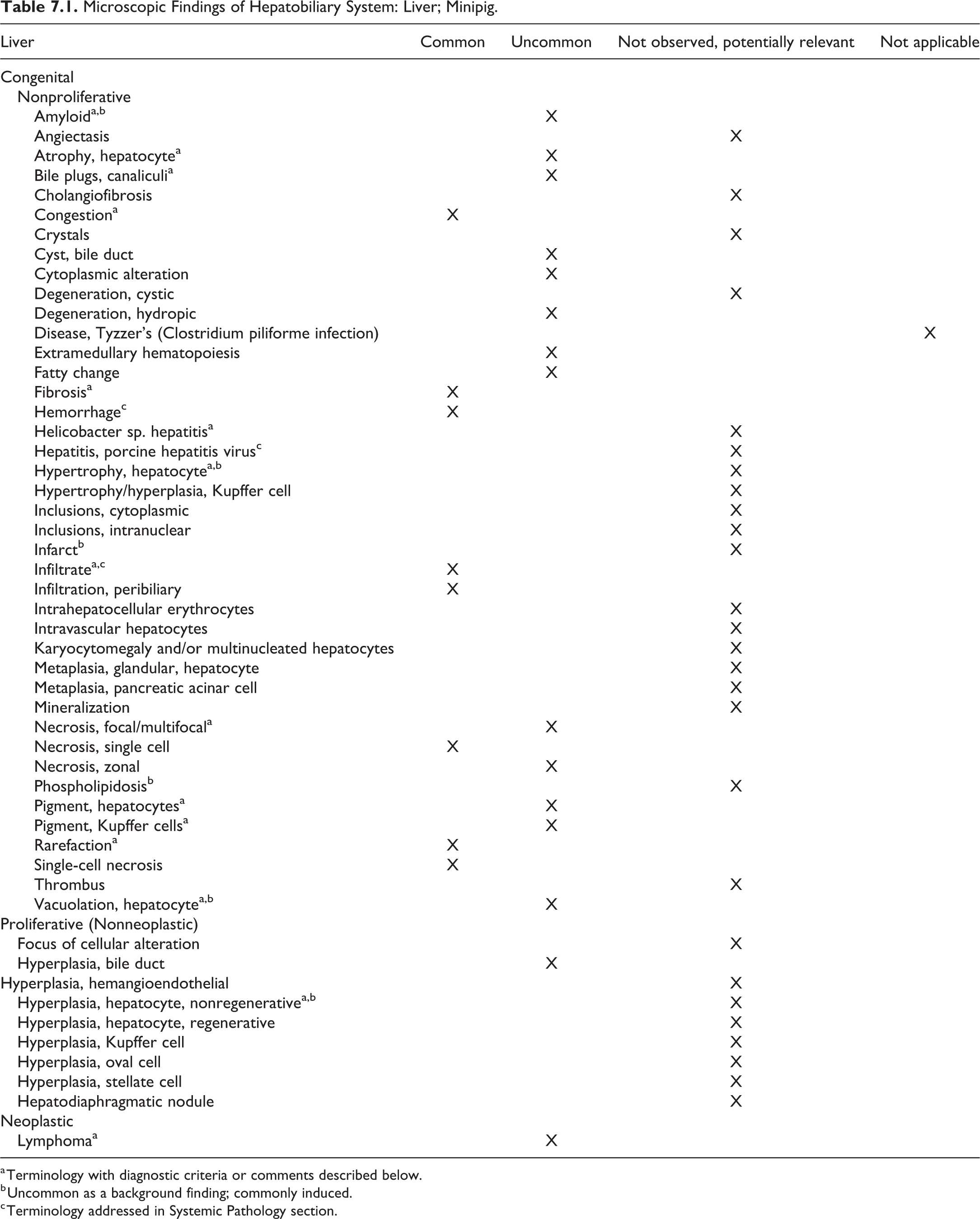
a Terminology with diagnostic criteria or comments described below.
b Uncommon as a background finding; commonly induced.
c Terminology addressed in Systemic Pathology section.
Amyloid, liver
Comment
Amyloidosis may be induced during acute infections in pigs. 88 Serum amyloid A genes are both hepatically and extrahepatically upregulated in pigs infected with Actinobacillus pleuropneumoniae or Staphylococcus aureus which is important for the use of the pig as a model for human inflammatory responses, for example, sepsis, cancer, and obesity research. 88
Atrophy, hepatocyte, liver
Comment
Diffuse hepatocellular atrophy may occur in starvation, fibrosis, portosystemic shunts, or congenital hepatic hypoplasia in minipigs. Stores of hepatocyte glycogen, lipid and protein, are utilized in starvation, resulting in a smaller liver. 85 Although hepatocellular atrophy is uncommon, minimal to mild hepatocyte atrophy can be observed in association with fibrosis during nonclinical safety studies when xenobiotics/test articles result in poor clinical condition of a minipig.
Bile plugs, canaliculi, liver
Comment
With cholestasis, bilirubin may be encountered in dilated canaliculi or hepatocytes and appears as a yellow or olive green pigment. Bile pigment stains green with Hall’s method.
Fibrosis, liver (Figure 7.3)
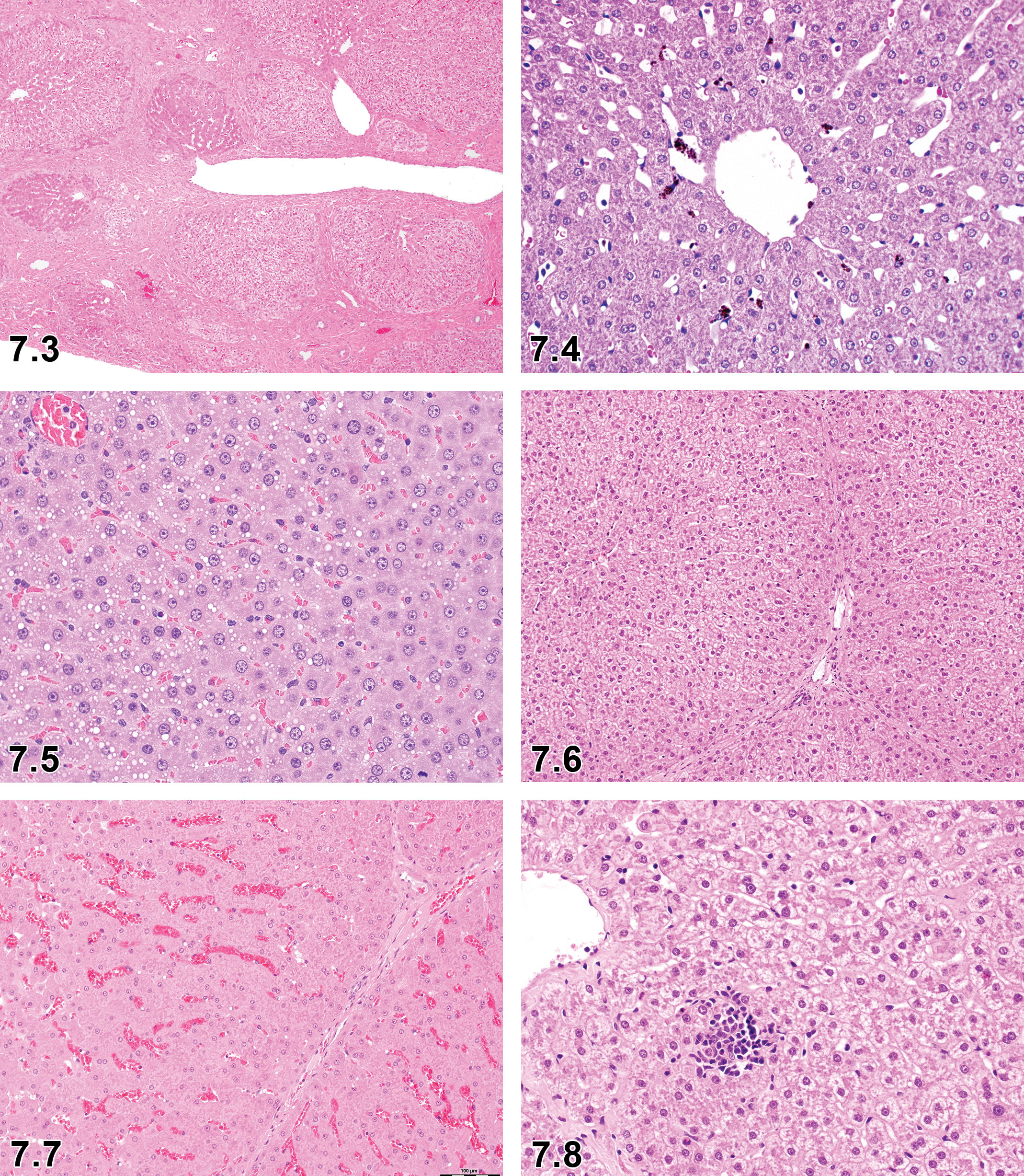
Liver, fibrosis, ×4.
Comment
In normal minipig liver, considerable interlobular and subcapsular fibrosis are present. This is a characteristic histological feature for the minipig and pig compared with other species. 7 Therefore, drug-induced fibrosis in the liver should carefully be assessed in the nonclinical safety studies. Additionally, there does not appear to be any increased tendency of the porcine liver to become cirrhotic after toxicologic injury compared with other nonhuman species. 44 Adhesions and its associated fibrosis of the hepatic capsule are common in Hanfords rather than Göttingens and Yucatans. 13
Hepatitis, Helicobacter sp, liver
Comment
Helicobacter pylori and other Helicobacter species have been isolated from porcine livers 89 ; however, Helicobacter infections have not been observed or demonstrated in minipig nonclinical safety studies. 3
Hypertrophy, hepatocyte, liver
Comment
Liver enlargement in response to hepatic enzyme induction is typically associated with hepatocellular hypertrophy and can be initiated by a wide range of stimuli including pregnancy and lactation, hormonal fluctuations, dietary constituents, infections associated with acute-phase proteins, and responses to exposure to xenobiotics such as androstane receptor (CAR), the peroxisome proliferator-activated receptor, the aryl hydrocarbon receptor, and the pregnane-X-receptor. The hypertrophy may show considerable species, strain, and sex differences in susceptibility and severity. 90 Hepatocellular hypertrophy as a result of hepatic enzyme induction to xenobiotics has also been observed in minipigs (Jeppesen, personal communication).
Phenobarbital, a barbiturate sedative known for its liver enzyme-inducing effects, has been shown to induce liver CYP2B, CYP3A, and UGT enzymes in rodents, leading to increased thyroid hormone clearance followed by increased thyroid-stimulating hormone and subsequent stimulation of cell proliferation in the thyroid. This pathway has also been studied in minipigs by Forster et al, and similar pattern of changes were also observed in the minipig as observed in the rodents (ie, increases in the liver weights associated with diffuse hepatocellular hypertrophy, reduction in plasma concentrations of T3 and T4, and increases in enzymatic activities of CYP3A429, CYP4A24, CYP1A2, and CYP2B22).
Portal vein embolization induces segmental hypertrophy of the nonembolized lobe in the liver of the minipig. 91 However, portal reperfusion by collateral vessels may limit the occurrence of hypertrophy.
Hyperplasia, hepatocyte, nonregenerative, liver
Comment
Spontaneous intralobular hepatocellular hyperplasia was seen in 5 out of a total of 51 female Yucatan minipigs ranging from 4 to 7 months of age but not observed in males. It was characterized by randomly scattered, discrete, intralobular nodules composed of hepatocytes that lacked lobular organization with an apparent compression of adjacent hepatocytes. Mitotic figures within these foci were negligible. Liver enzymes were within expected ranges for these animals. 92 Intralobular hepatocellular hyperplasia have not been reported in other strains of pigs.
Infarct, liver
Comment
Dearterialization by a combination of distal occlusion of the hepatic artery with Ivalon (polyvinyl alcohol foam) particles of 150 to 250 µm and proximal occlusion of the common hepatic artery with Gianturco coils can result in necrosis/infarction of up to 90% of the liver. 93
Lymphoma, liver
Comment
Swine multicentric lymphomas can occur in young pigs before the onset of maturity. The liver can be enlarged with multifocal, randomly distributed, small to large raised white masses throughout the parenchyma. Histologically densely packed large lymphoid cell infiltrates can be seen in the liver 94 (see Hematopoietic and Lymphoid System chapter, McKeag, unpublished data).
Pigment, liver (Figure 7.4)
Comment
Pigment is seen frequently in hepatocytes and Kupffer cells in juvenile minipigs and occasionally in 4-week nonclinical safety studies due to iron injection. Hemosiderin can be seen in the liver associated with hemorrhages or generalized intraphagocytic hemolysis. With cholestasis, bilirubin may be encountered in dilated canaliculi or in hepatocytes and appears as a yellow or olive green pigment.
Vacuolation, liver (Figure 7.5)
Comment
Hepatocyte cytoplasmic vacuolation due to lipid accumulation can occasionally be observed. These focal or multifocal, clear, sharply demarcated, empty vacuoles usually have no specific zonal distribution. 3 These are generally of minimal to slight severity. Fatty liver is a common finding in the streptozotocin diabetic pig model, as it is in other porcine models of hypertriglyceridemia. 44 Presence of lipid can be demonstrated with Oil Red O and osmium staining.
Rarefaction, hepatocyte (glycogen), liver (Figure 7.6)
Sinusoidal congestion, liver (Figure 7.7)
Infiltrate, mixed cell, liver (Figure 7.8)
Hepatocyte necrosis and congestion, liver (Figure 7.9)
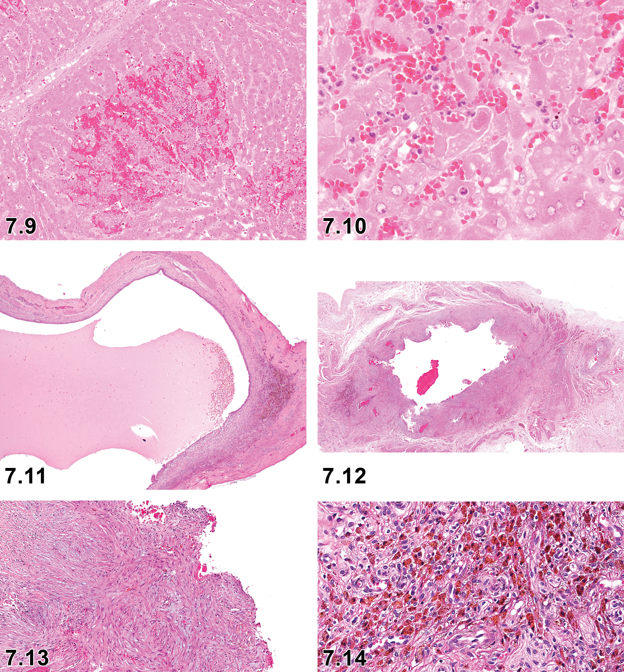
Liver, hepatocyte necrosis with congestion, ×10.
Hepatocyte necrosis with neutrophilic cell infiltrate, liver (Figure 7.10)
II. Gall Bladder
The gall bladder is located in a fossa on the visceral surface of the right medial lobe and is pear-shaped. Its serosal surface is white, and the mucosal surface is light green. 85
At necropsy, upon opening the abdominal cavity, the gall bladder should be examined and sampled immediately to prevent autolysis. A transverse section from the central region of the gall bladder is examined in nonclinical safety studies.
Some of the commonly observed background changes in the Göttingen minipig are necrotizing cholecystitis, and hypoplasia or aplasia of the gall bladder which are findings unique to the Göttingen minipig. 3,7
Microscopic Findings of Hepatobiliary system: Gall Bladder; Minipig.
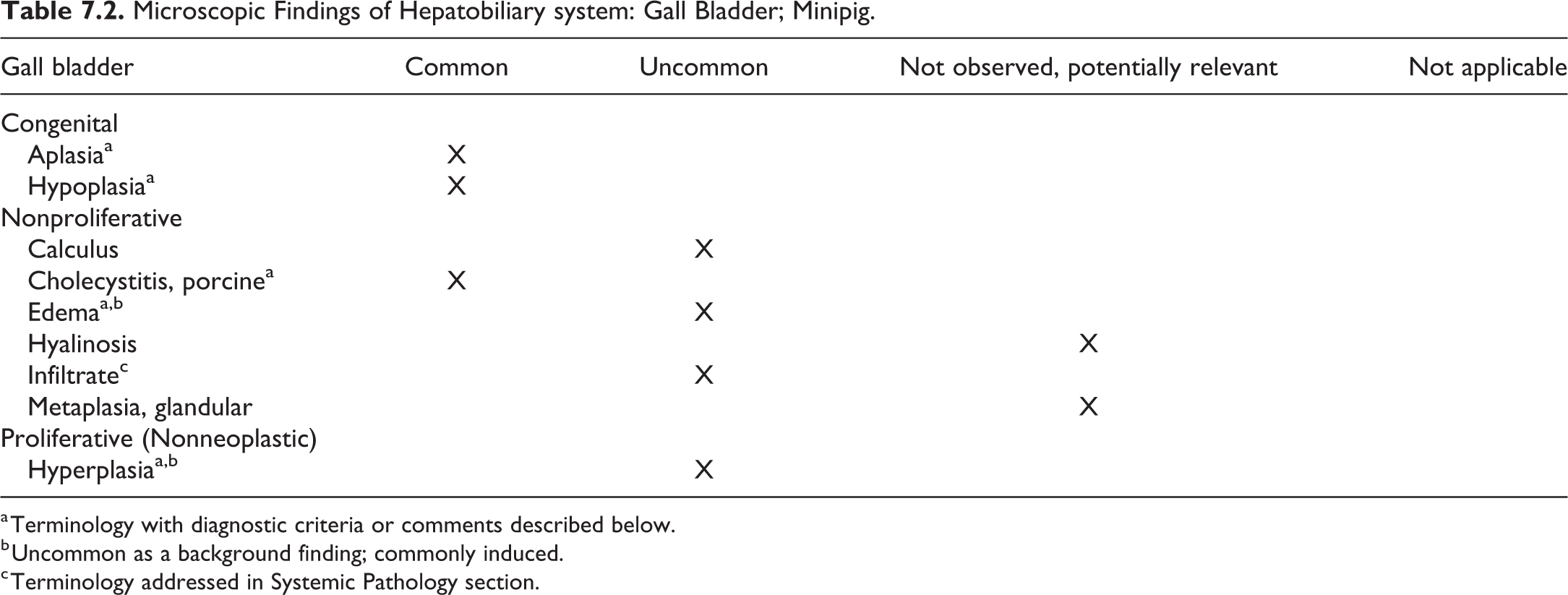
a Terminology with diagnostic criteria or comments described below.
b Uncommon as a background finding; commonly induced.
c Terminology addressed in Systemic Pathology section.
Aplasia, gall bladder
Pathogenesis
Unknown, possibly congenital.
Diagnostic features
Macroscopic appearance; gall bladder is gray to beige, small, and difficult to discern at necropsy.
Residual tissue consisting mainly of connective tissue elements observed microscopically.
Differential diagnoses
Atrophy, hypoplasia.
Comment
Aplasia of the gall bladder is occasionally observed in minipig nonclinical safety studies, particularly in Gottingen minipigs. There is no trace of bile macroscopically or microscopically in the gall bladder. In addition, there are no associated clinical signs or changes in the clinical pathology parameters routinely measured. 3 Aplasia of the gall bladder has not been reported in Hanford and Yucatan minipigs. 13
Cholecystitis, porcine, gall bladder (Figures 7.11-7.14)
Other term(s)
Necrotizing cholecystitis.
Pathogenesis
Unknown.
Diagnostic features
Macroscopically, slightly decreased in size with thickened walls.
Bile is absent or if present is inspissated.
Described as acute, subacute, chronic, or chronic active with a severity ranging from moderate to marked.
Acute cases often characterized as necrotizing.
Microscopically diffuse necrotic, hemorrhagic mucosal layer associated with granulomatous inflammation extending into the muscular layer.
Differential diagnoses
Hypoplasia.
Comment
Cholecystitis is one of the commonly observed background changes unique to the Göttingen minipig. Chronic necrotizing cholecystitis is the most common form. No clinical signs or changes in clinical pathology parameters are observed in animals diagnosed with cholecystitis in nonclinical safety studies. While this is a background finding, bile is important in drug metabolism and this entity may affect toxicological outcomes. 3,7
Edema, gall bladder (Figures 7.15 and 7.16)
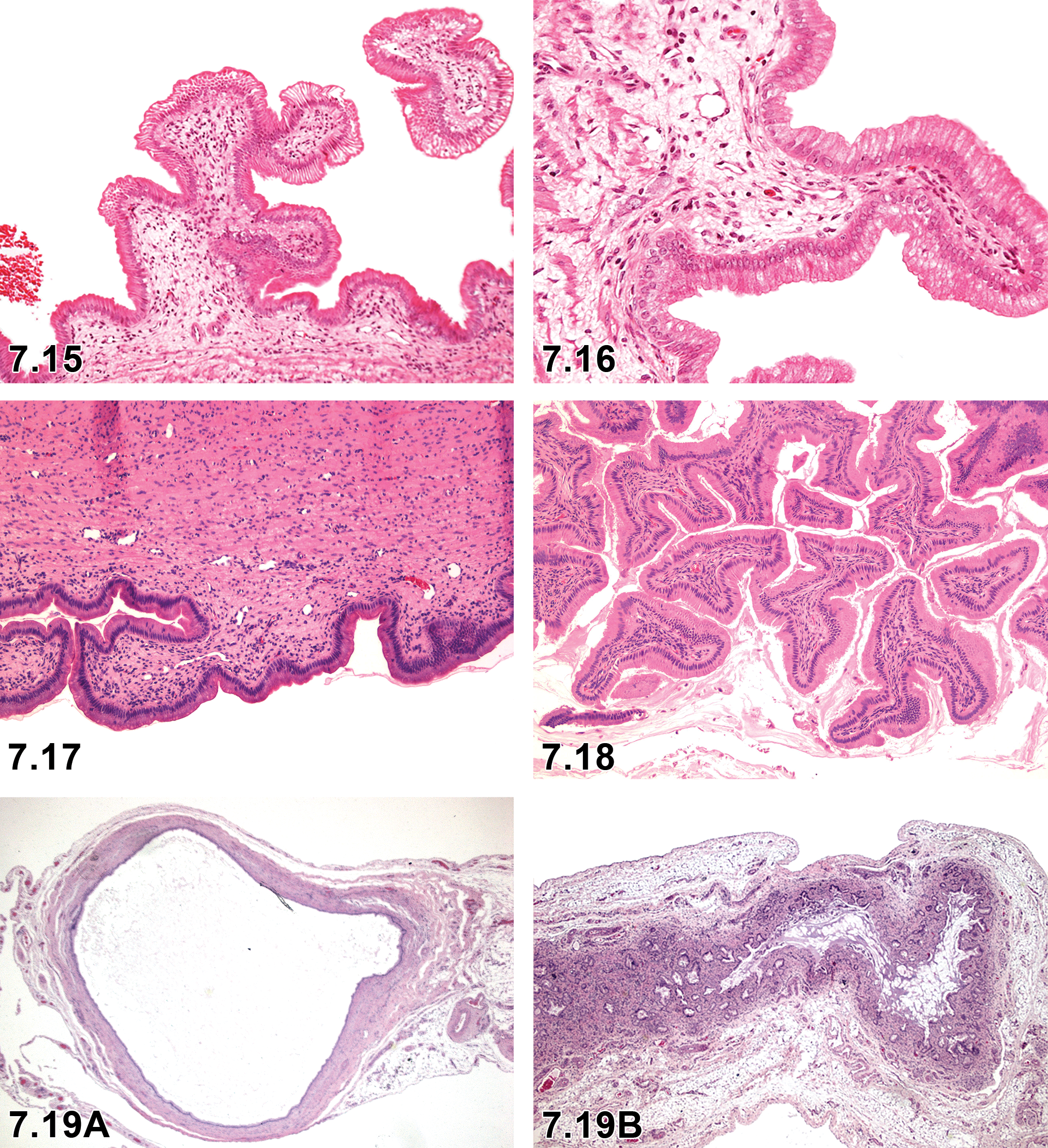
Gall bladder, mucosal edema, ×4.
Comment
Edema in the gall bladder can occasionally be observed in minipig nonclinical safety studies. Microscopically edema appears as a pale eosinophilic amorphous accumulation with minimal or no inflammatory cells in the mucosa/submucosa.
Hyperplasia, gall bladder (Figure 7.18)
Comment
In juvenile minipigs, treatment with Gattex (teduglutide, a 33 amino acid recombinant analog of the human GLP-2) can result in microscopic changes in the gall bladder (cystic mucosal hyperplasia) and extrahepatic bile duct (cystic mucosal hyperplasia) as observed in mice and monkeys. 52
Hypoplasia, gall bladder (Figures 7.19a and 7.19b)
Pathogenesis
Unknown, possibly congenital.
Diagnostic features
Dark gray and small at macroscopic examination.
Microscopically flattened or folded mucosal epithelium (disappearance of mucosal crypts), absence of glands, and underlying minimal to slight amounts of loose connective tissue rich in blood vessels.
Absence of bile macroscopically and microscopically.
Differential diagnoses
Atrophy, aplasia.
Comment
Hypoplasia of the gall bladder is occasionally observed in minipig nonclinical safety studies. There are no associated clinical signs or changes in the clinical pathology parameters routinely measured. 3 Hypoplasia of the gall bladder has only been observed in Gottingen minipigs and has not been reported in Hanfords and Yucatans yet. 13
Gallstone in an aged microminipig, gall bladder (Figure 7.20)
Gall bladder, gallstone in an aged microminipig. 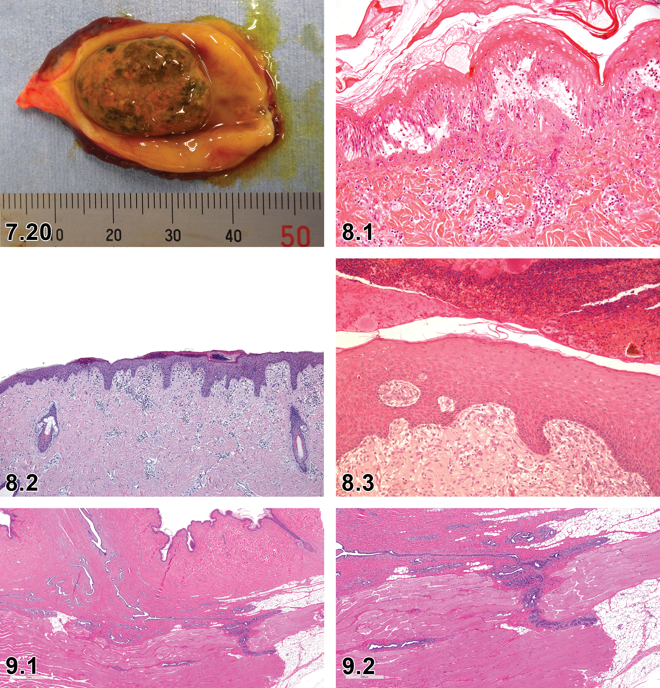
Chapter 8. Integumentary System
The anatomy of minipig skin is comparable to that of other mammals. Therefore, for detailed general considerations on the integument, please refer to the INHAND publication on rodent integument. 95 In general, pigs (domestic and minipigs) have thicker skin with less hair than most animals, especially compared to rodents. Although different minipig strains, such as Göttingen, Sinclair, Yucatan, and Hanford, exhibit differences in cutaneous pigmentation, hair coats, and growth patterns of their skin, they share many similarities in terms of general morphology, epidermal thickness, cellular composition, immunological reactivity, and cellular turnover to human skin. 96 –100 The minipig epidermal thickness varies only minimally between birth and adulthood, whereas the dermal and especially the subcutaneous thicknesses increases over time. 101 Although the epidermis generally consists of 5 to 7 cell layers with only minor regional variations, the thickness of porcine epidermis varies from 30 to 140 µm (compared to 50-120 µm in human skin) and is thinner in the abdominal region and thicker at the neck, particularly in males. Similarly, the dermis and subcutis are thicker on the dorsum and thinner at the abdomen and inner thighs. Similar to humans, pig skin has well-developed rete ridges and pars papillaris. Both pig and human skin have abundant subcutaneous fat tissue, which is divided in a superficial and a profound layer. This may lead to differences in compound diffusion depending on the depth of the injection. In general, the fat tissue layer is thicker in pigs and the skin is less vascular compared to humans. Pigs have extensive apocrine sweat glands, which play a limited role in thermoregulation and sweating. Porcine eccrine glands are only located on the snout and in the carpal area. 102 Seasonal change from apocrine to eccrine glands occurs in some pig strains. The pH of minipig skin depends slightly on the body region and is between 6 and 7, which is slightly higher than in human skin (approximately pH 5).
The similarities of porcine and human skin makes the minipig an attractive animal model in dermatotoxicology, in particular for investigating wound healing. 103,104 Regional variations in the porcine skin such as thickness of epidermis, dermis, and subcutis, as well as occurrence of apocrine and eccrine glands, and differences in the hair coat have to be considered during the planning of cutaneous toxicology studies.
This chapter does not cover hooves. Lesions in the mammary gland are covered in Chapter 9. Dermal lesions, which are covered in the soft tissue, nervous system, or cardiovascular system INHAND manuscripts, are not discussed here.
Nonproliferative skin lesions are commonly encountered as spontaneous background findings in young minipigs used in nonclinical toxicity studies. In contrast, proliferative skin lesions are observed rarely as background findings due to the young age of animals at study initiation and the short duration of toxicity studies. In domestic pigs, squamous cell papillomas and carcinomas have been reported to occur rarely in white skinned strains. 105 –107 Melanomas have been described in darkly colored pigs such as Duroc and Hampshire breeds. 108 There are certain specific minipig strains, namely Sinclair, 109 MeLiM (Melanoma-bearing Libechov Minipigs) also known as Libechov strains 110 and Munchener Miniaturschwein (Troll) minipig strains 111 that frequently develop regressing cutaneous melanomas at a very young age.
Minipigs are susceptible to many of the same skin diseases that affect domestic pigs. The term “Dippity Pig Syndrome” is commonly used to describe a skin condition in various strains of domestic pigs, as well as in minipigs. 12 The condition is recognized in Göttingen minipigs in all age groups. It is a cutaneous neurological syndrome characterized by skin sores (“weeping blisters”) and sensitivity to touch. Other clinical manifestations include assuming “hunkered-down” stance when touched, pain, weakness and/or inability to use the hind legs, falling down, and other signs of distress. The etiology is unknown, but the condition may be precipitated by stress. The cutaneous manifestations are consistent with erythema multiforme. The lesions are mainly seen in the lumbar region. Histopathologic appearance includes increased dermal congestion, edema, and perivascular lymphocytic infiltration (Figure 8.1). Although this syndrome is self-limiting, with spontaneous recovery typically within 1 to 2 weeks, the occurrence within a toxicity study in minipigs may confound interpretation of findings, particularly in the safety evaluation of topical agents. There is no known treatment; however, palliative veterinary treatment for pain (eg, oral meloxicam) may be beneficial. Intermittent withdraw of topical application of the test article may be needed in a toxicity study to allow healing of the cutaneous lesions associated with “Dippity Pig Syndrome” before reinitiating application of the test article.
Other skin diseases such as porcine juvenile pustular psoriasiform dermatitis (Pityriasis rosea), which occurs in young domestic white pig breeds, has also been found in young white minipigs. However, there are some skin diseases that are reported to occur specifically in minipigs. One such example is a thrombocytopenic purpura-like syndrome also called the hemorrhagic syndrome described in mature male and female Göttingen minipigs, characterized by extensive subcutaneous hemorrhage accompanied by marked thrombocytopenia, anemia, and membranoproliferative glomerular lesions, 18 which is discussed in the systemic pathology chapter. Another example is bullous pemphigoid reported in Yucatan minipigs characterized by subepidermal vesicles with many intact and degranulated eosinophils and exhibiting linear immunoglobulin (specifically IgGs) deposits at the dermoepidermal junction. 29,112 With advances in animal husbandry in minipig facilities, infectious skin diseases are likely to be a rare occurrence.
Trimming
For organ sampling and trimming in rats and mice, the revised guides 113 recommend the collection of skin samples from the inguinal region. Sampling of minipig skin differs between laboratories with some using the abdomen and others the dorsum. For standardization and comparison of the samples, it is important to collect the skin samples consistently. To represent the large skin surface of pigs with different morphology at different body locations, at least 2 samples from different locations may be necessary.
In dermal studies, 3 to 4 treated skin samples are routinely examined in addition to an untreated sample from a similar anatomical location. The treated skin samples should be subjected to a pooled evaluation.
Nomenclature
The incidence data are based on Göttingen, Hanford, Sinclair, and Yucatan minipigs routinely used in toxicity studies. In general, inflammatory lesions and associated reactions were the most commonly observed findings in the skin. 13
Microscopic Lesions of the Integument: Skin; Minipig.
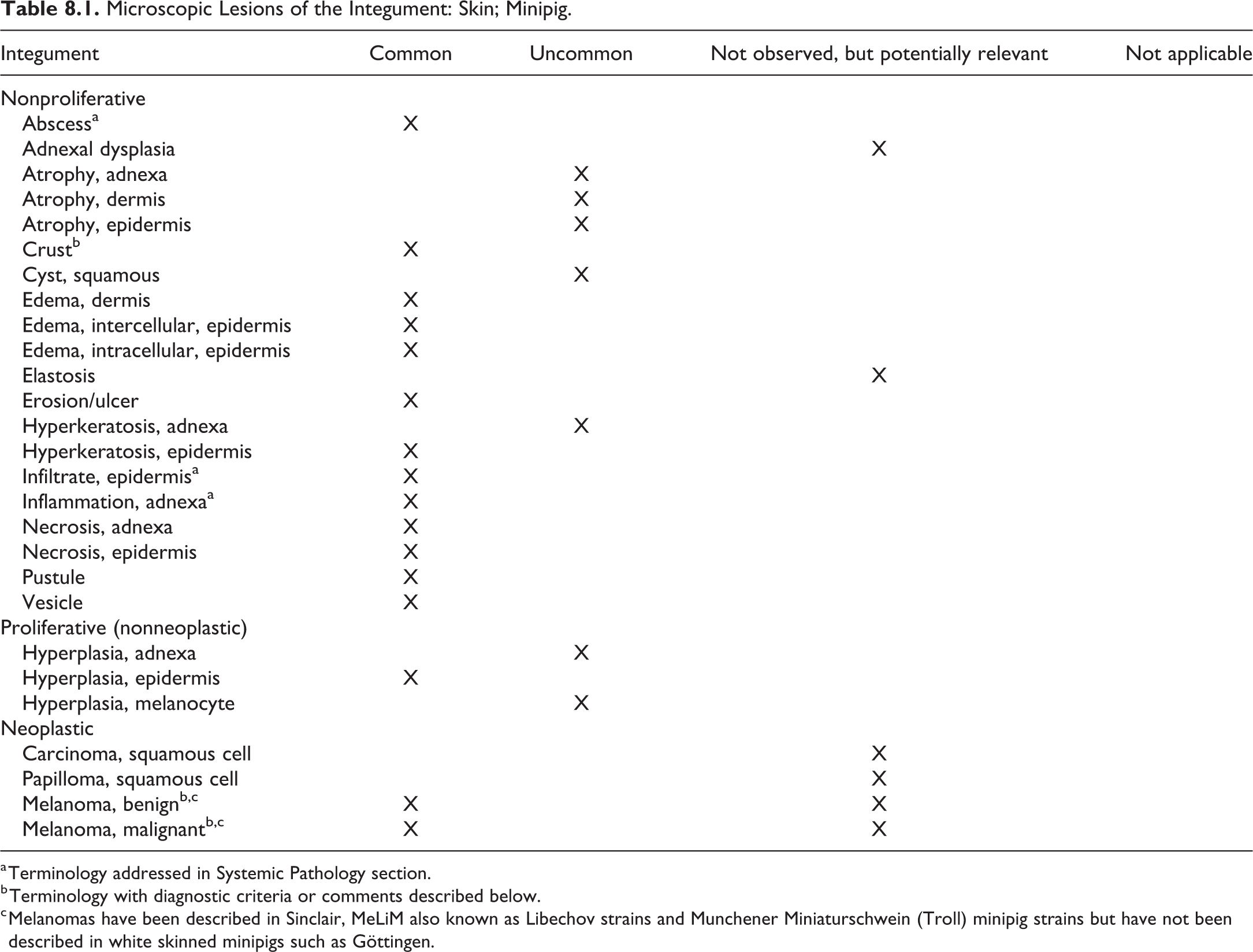
a Terminology addressed in Systemic Pathology section.
b Terminology with diagnostic criteria or comments described below.
c Melanomas have been described in Sinclair, MeLiM also known as Libechov strains and Munchener Miniaturschwein (Troll) minipig strains but have not been described in white skinned minipigs such as Göttingen.
Crust, Skin (Figures 8.2 and 8.3)
Sublocation
Epidermis.
Other term(s)
Crust, serocellular; exudate, serocellular.
Modifier(s)
Serous, hemorrhagic, cellular, serocellular.
Pathogenesis/cell of origin
Crusts are indicative of a previous exudative process. They are formed by inflammatory cells and cellular debris within the stratum corneum of the epidermis. Crusts can be formed through aging of pustules.
Diagnostic features
Desiccated accumulation of inflammatory cells, pustular debris, erythrocytes, epithelial squames, clotted plasma proteins, or microorganisms on or within the stratum corneum of the epidermis.
Inflammation
Inflammation of the skin is not confined to the stratum corneum level of the epidermis.
Comment
Crust are by far the most common microscopic finding in the skin of minipigs. 7 The crusts are usually focal in nature and of a low severity. The crusts commonly have a traumatic etiology (eg, due to rubbing against fixed items) but might be test article-induced in toxicology studies, especially when the dermal route of administration is used. In general, crusts are composed of a serocellular exudate but might also include other components such as dressing material in dermal studies. In wound healing studies, crusts covering the surface of the wound should be carefully characterized with respect to size/severity as well as the individual constituents, since these parameters change when the wound healing process proceeds toward complete healing.
Melanoma, benign, skin
Comment
The Sinclair, Libechov, and Munchener Miniaturschwein (Troll) minipig strains show a high incidence of spontaneous cutaneous melanomas in newborns, which continue to develop after birth. The benign melanomas can further develop in malignant melanomas and can also undergo regression. Histologically, tumor regression is characterized by melanomacrophages, infiltrates of lymphocytes, fibrosis, and free melanin replacing the tumor. 114
Melanoma, malignant, skin
Comment
The Sinclair, Libechov, and Munchener Miniaturschwein (Troll) minipig strains show a high incidence of spontaneous cutaneous malignant melanomas in newborns, which continue to develop after birth. The tumors may invade muscle fascia and have high metastatic activity. Metastatic spread to the draining lymph nodes is frequently observed, whereas metastases to other organs is less frequent but not uncommon. 115 Spontaneous regression of primary and metastatic lesions has been observed in Sinclair minipigs within the first year of life. Tumor regression is accompanied by depigmentation, which is not limited to the tumor itself but may range from a localized depigmentation of adjacent hair and skin to a generalized depigmentation including the iris of the eye. 109 Histologically, tumor regression is characterized by melanomacrophages, infiltrates of lymphocytes, fibrosis, and free melanin replacing the tumor. 114 . Though many melanomas in these minipig strains have been classified as malignant, they may regress, which should be considered during evaluation of these tumors.
Chapter 9. Mammary Gland
Standard INHAND nomenclature for nonproliferative and proliferative mammary gland findings in rats and mice has previously been published 116 and will be used for the minipig as appropriate following the anatomical approach used for rodent species, with a focus on those findings that are likely to be encountered frequently and those that might be considered unique to the minipig. The mammary gland is a milk-producing exocrine gland characteristic of all female mammals and present in a rudimentary and generally nonfunctional form in males. Mammary glands are regulated by the endocrine system. Minipigs have 6 to 7 pairs of mammary glands on the ventral abdomen. At necropsy, usually one of the inguinal mammary glands is sampled for histology (Figures 9.1-9.6).
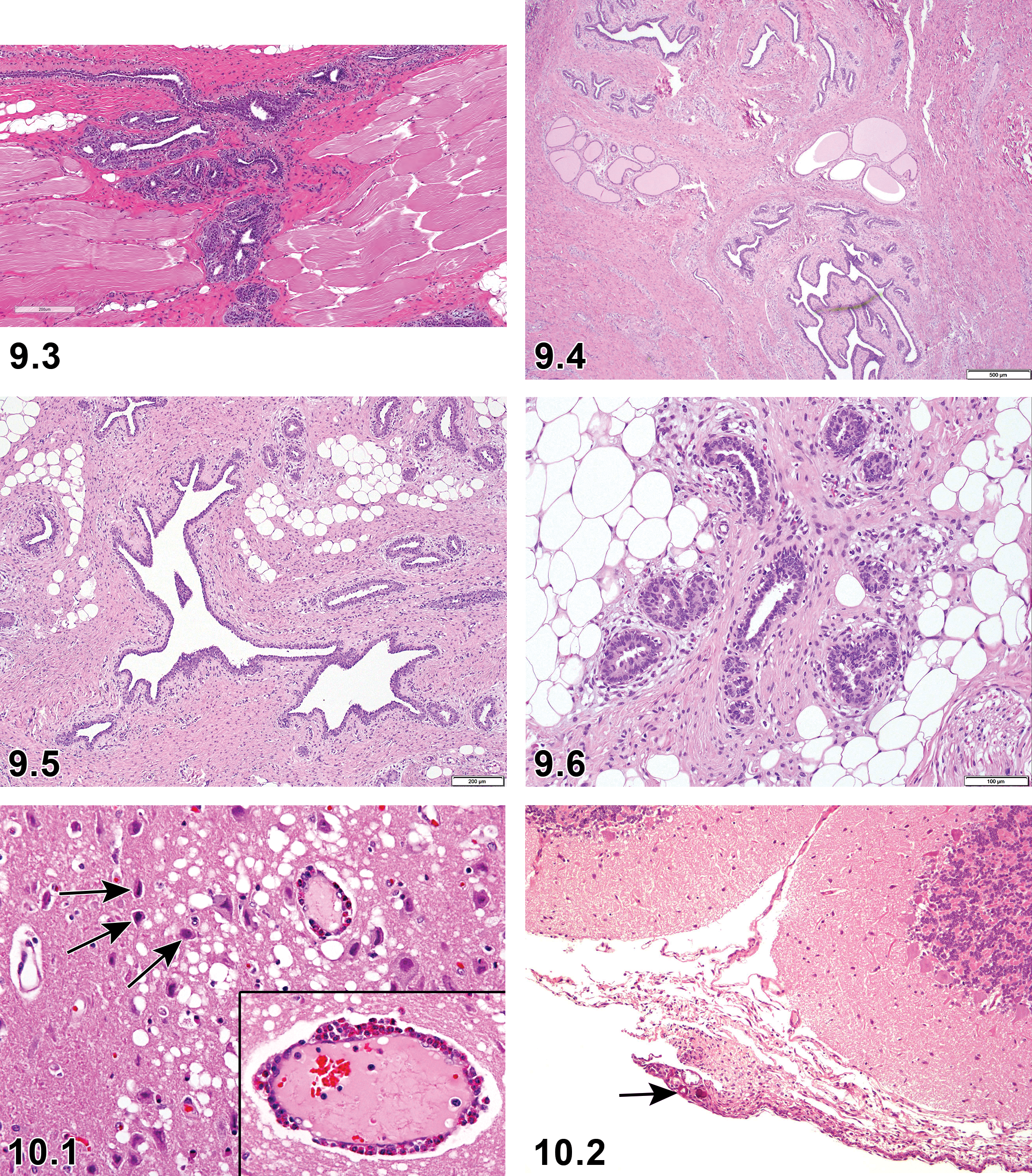
Minipig, mammary gland, normal tissue, right below the nipple, male, H&E ×100.
In immature female minipigs, there is very little mammary gland development with no secretion. There are few ducts which are small and scattered. Alveoli, where present, are lined by epithelial cells that are either quiescent (small and single layered) or show early development (larger, multilayered). In mature female minipigs, mammary glandular tissue is moderately to markedly developed, with numerous alveoli. Hyperplastic glandular cells are visible in about one-third of the cases with markedly developed mammary glands, and alveoli and canals are in many cases filled with eosinophilic material. 117 The mammary gland of female minipigs does not reflect the stage of the estrus cycle as it does for example in dogs. 118
Pathologic changes in the mammary gland of minipigs are rare in preclinical toxicity studies. 7 Proliferative lesions in laboratory-maintained minipigs may theoretically arise from genotoxic test items, infectious agents, and changes in hormonal homeostasis (eg, as part of the aging process). However, the reports on proliferative changes in the mammary gland of minipigs are sparse and is most likely due to the young age of the animals used for routine preclinical toxicity testing.
Microscopic Findings of Mammary Gland; Minipig.
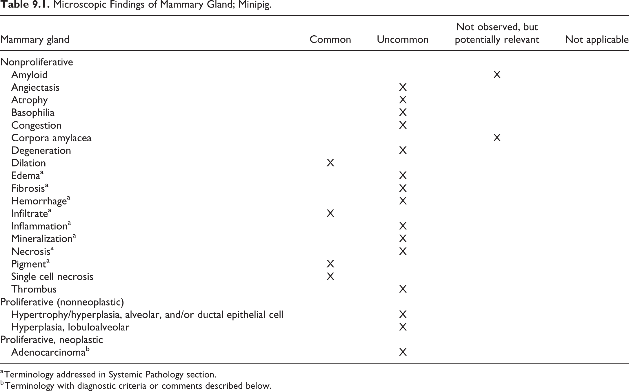
a Terminology addressed in Systemic Pathology section.
b Terminology with diagnostic criteria or comments described below.
Adenocarcinoma, Mammary Gland, Acinus
Comment
A case of a mammary gland adenocarcinoma has been reported in a 10-year-old female Göttingen minipig. 119
Chapter 10. Nervous System
For detailed general considerations on the nervous system, please refer to the INHAND publication on rodent nervous system. 120,121 The cellular composition of minipig nervous system is comparable to that of other mammals.
Minipigs are susceptible to many of the same nervous system diseases that affect domestic pigs. However, with advances in animal husbandry in minipig facilities, infectious diseases are likely to be a rare occurrence.
Trimming
The brain is typically trimmed for examination of 7 sections as recommended by Bolon et al, but a more extended trimming procedure of the minipig brain has been presented, with 14 sections examined. 122,123 Sections from 3 divisions of the spinal cord are collected including cervical, thoracic, and lumbar sections. 26 For general toxicity testing, the sciatic nerve is representative of the peripheral nervous system and is trimmed in cross and longitudinal section. 26 For studies in which neurotoxicity is expected or known, sampling of ganglia and nerves should be more extensive. 123
Nomenclature
The incidence data in the following tables are based on Göttingen, Hanford, Sinclair, and Yucatan minipigs routinely used in toxicity studies. In general, inflammatory cell infiltration and mineralization of the meninges were the most commonly observed findings in the nervous system. 13
Microscopic Lesions of the Central Nervous System: Brain and Spinal Cord; Minipig.
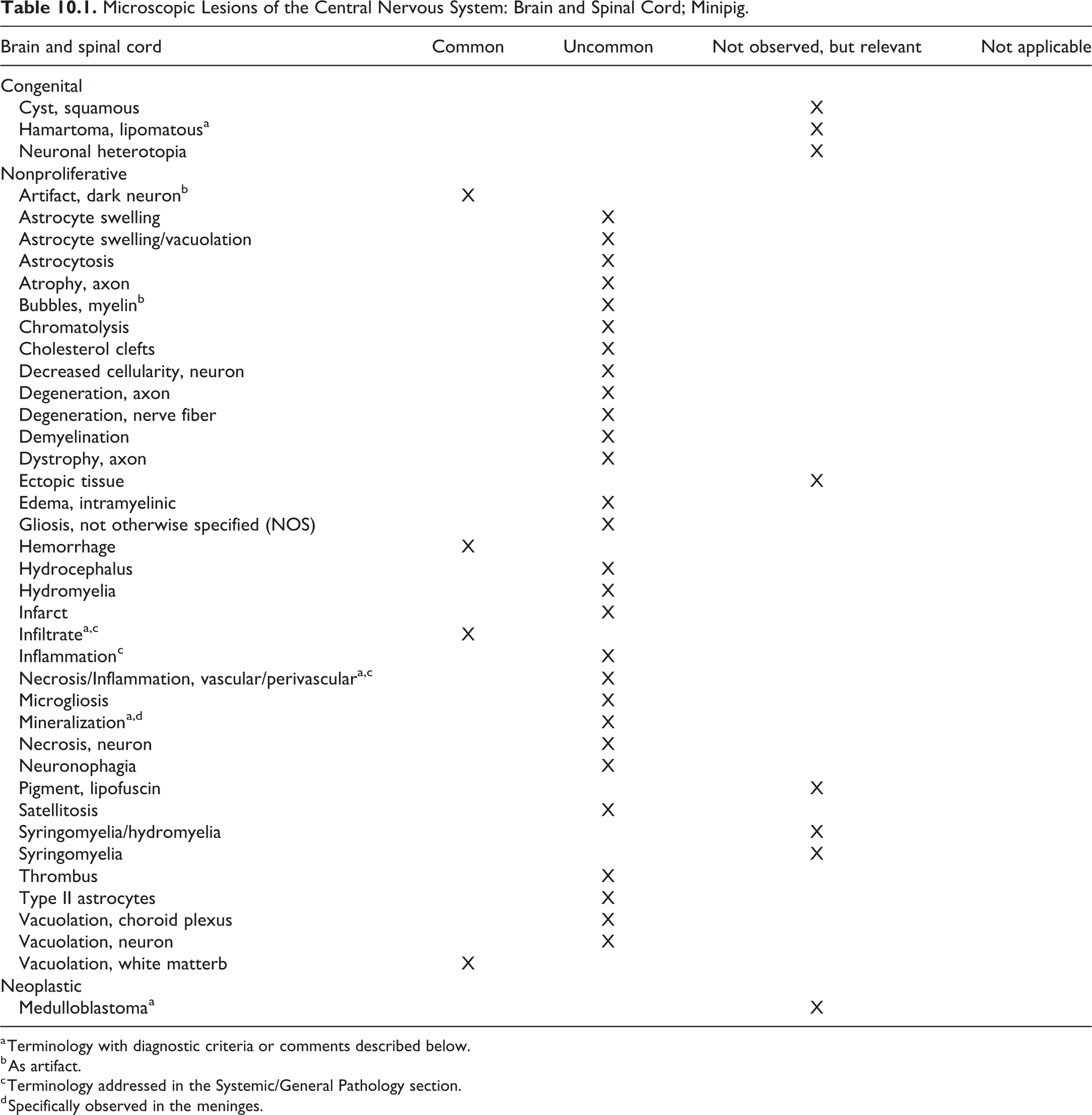
a Terminology with diagnostic criteria or comments described below.
b As artifact.
c Terminology addressed in the Systemic/General Pathology section.
d Specifically observed in the meninges.
Hamartoma, lipomatous, brain and spinal cord
Comment
Though they have not yet been reported in minipigs, lipomatous hamartomas have been described in domestic pigs. 124 These are occasionally described as lipomas.
Necrosis/Inflammation, media or wall, artery, Brain and Spinal Cord
Comment
A generalized arteritis or periarteritis in the minipig may also affect the nervous system. For detailed information about this condition please refer to the cardiovascular chapter.
Infiltrate, eosinophilic, brain and spinal cord (Figure 10.1)
Comment
Meningeal and perivascular cortical infiltration of eosinophilic granulocytes has been described in pigs in several disease states, including leukomalacia of vitamin E/selenium deficiency and various encephalitides but is specifically seen in salt toxicity (inadequate water intake followed by adequate or excess water intake). Combined with the laminar loss of cortical neurons, this infiltrate is pathognomonic for salt toxicity. 124,125
Medulloblastoma, brain and spinal cord
Comment
Blastomas of the nervous system such as medulloblastomas have been reported to occur rarely in young domestic pigs 124,126 and might therefore also be found in the minipig.
Microscopic Lesions of the Central Nervous System: Meninges; Minipig.
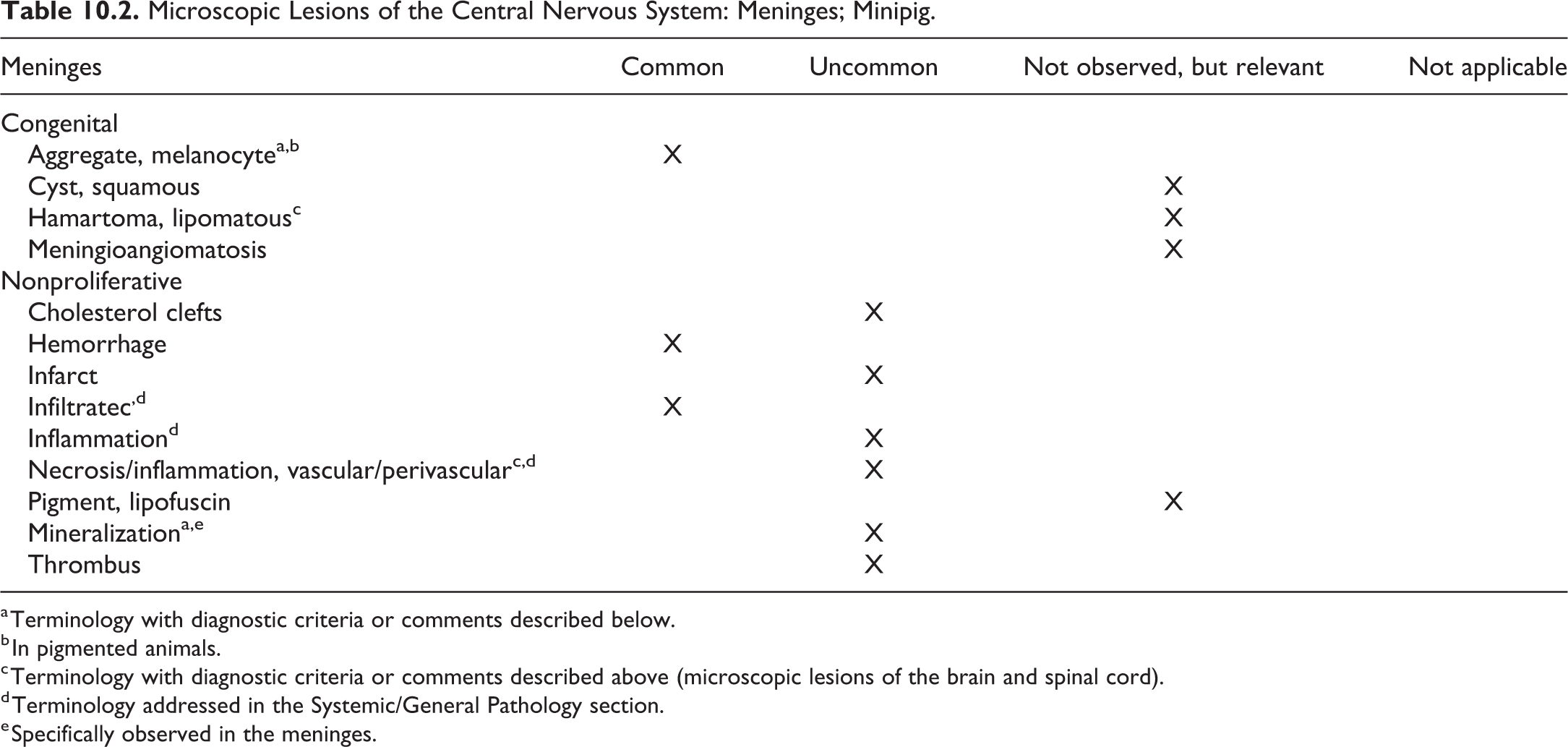
a Terminology with diagnostic criteria or comments described below.
b In pigmented animals.
c Terminology with diagnostic criteria or comments described above (microscopic lesions of the brain and spinal cord).
d Terminology addressed in the Systemic/General Pathology section.
e Specifically observed in the meninges.
Aggregates, melanocyte
Other term(s)
Meningeal melanosis; melanosis.
Pathogenesis/cell of origin
Neuroectodermal origin.
Diagnostic features
Aggregates of polygonal melanocytes filled with pigment. No cellular atypia. No displacement of normal structures.
Comment
The leptomeninges of pigmented pigs can have melanin with a high variation from animal to animal concerning the extent and degree of pigmented deposition. Congenital meningeal melanosis produces no clinical impairment in affected animals. 126
Mineralization (Figure 10.2)
Comment
Mineralization as a focal minimal lesion is often observed in the meninges of minipigs. 7,13,41
Microscopic Lesions of Nervous System: Peripheral Nervous System; Minipig.
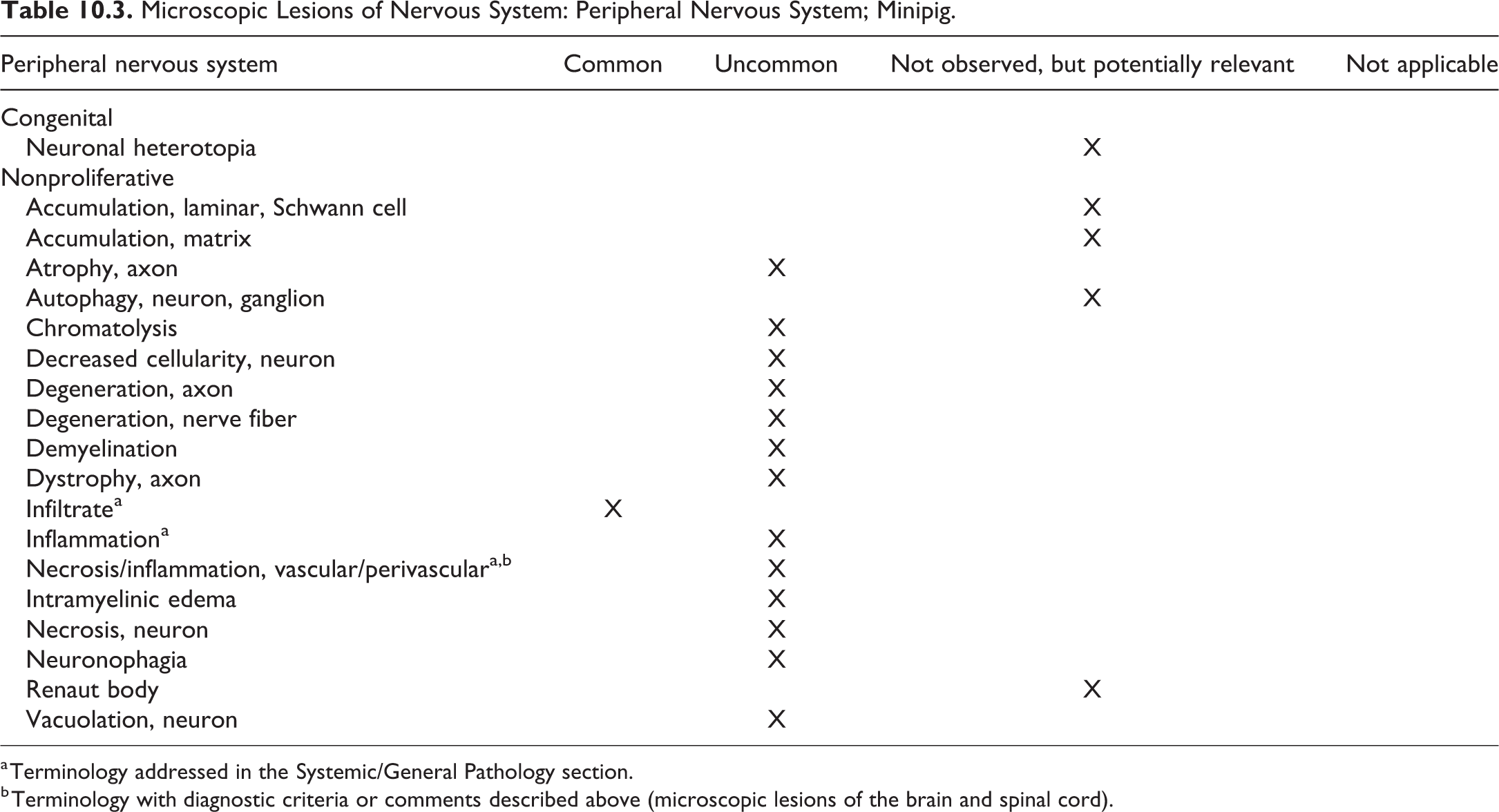
a Terminology addressed in the Systemic/General Pathology section.
b Terminology with diagnostic criteria or comments described above (microscopic lesions of the brain and spinal cord).
Chapter 11. Reproductive System—Female
Standard INHAND nomenclature for nonproliferative and proliferative female reproductive system findings in rats and mice has previously been published 127 and will be used for the minipig as appropriate following the anatomical approach used for rodent species, with a focus on those findings that are likely to be encountered frequently and those that might be considered unique to the minipig. Generally very few spontaneously or treatment induced microscopic changes are seen in the female reproductive tract of minipigs. If published references were located for experimentally induced changes, they have been indicated in the reference section and included in the comments for the changes.
Discussion of histological features of the female reproductive system with regard to sexual maturity status as well as estrus cycle specific changes is included to enable familiarization with normal variations and proper recording of alterations from normal. Discussion of maturity status in this chapter is strictly related to the degree of maturity of the reproductive tract organs and does not include consideration of skeletal maturity.
Female Minipig Reproductive Anatomy
The female reproductive tract in the minipig is similar to domestic swine, consisting of a bicornuate uterus with tortuous fallopian tubes. In adult domestic pigs, the fallopian tubes are similar in diameter to the human but longer. 128 The uterus consists of 3 functional zones comparable to the macaque endometrium including the luminal epithelium (zone I), glandular layer subjacent to the luminal epithelium (zone II), and the basal glandular layer above the myometrium (zone III). The minipig is similar to humans with regard to uterine histology and estrus cyclicity but unlike humans and cynomolgus monkeys does not menstruate. A characteristic feature of the porcine cervix not observed in humans is the pulvini cervicales, 129 which consists of numerous interdigitating prominent solid mucosal folds and protrusions throughout the length of the cervix. In pigs, more than 90% of the cervix may have a vaginal type of epithelium with stratified squamous epithelium that undergoes cyclic alterations. In minipigs, the urethra opens on the ventral surface of the vagina, creating a urogenital sinus that opens to the outside through the common urogenital orifice. Ovaries are generally bean-shaped and may weigh as little as 2 g, even in the adult. The ovarian stroma is composed of tightly packed spindle-shaped fibroblast-like cells, often arranged in a whirling pattern, and may contain lipid droplets. Under hormonal influence, the stromal cells may accumulate more lipid (luteinization).
Tissue Collection, Fixation, and Trimming
Albl et al have thoroughly detailed the proper collection, fixation, and trimming schemes for the female reproductive tract. 26 Briefly, for routine general toxicology studies, the sections examined include one cross section of the right and left oviduct (uterine tube), one mid-sagittal section of each ovary, one cross section of the right and left uterine horns (from the middle of the organ), one cross section of the cervix, and one cross section of the vagina. For advanced studies, trimming guidelines for the ovary, oviduct, and uterus described in 26 may be followed. 26
Sexual Maturity in the Female Minipig
Repeat-dose toxicity studies conducted using sexually mature animals provide important information on potential effects of the test article on the reproductive tract. The International Council for Harmonization Safety Guidance Document S5 R3 lists histopathology of reproductive tract organs from repeat-dose toxicity studies as a sensitive method for detecting effects on male and female fertility, provided animals are sexually mature at the termination of the study. 130 However, reliable interpretation of changes in the female reproductive tract in routine toxicology studies is often confounded by the wide window of sexual maturity of female minipigs. Female minpigs, based on available reports 103,117,131 as well as personal experience of the authors, generally mature between 4 and 8 months of age. Younger age (<5-6 months) at study initiation, small group size, and a wide window of sexual maturity can result in uneven distribution of immature minipigs in various treatment groups, particularly in studies of 3 months duration or less. It is therefore recommended that studies be initiated using minipigs of appropriate age, taking into consideration the study duration and the wide maturation window of this species, to ensure that adequate numbers of animals would be sexually mature in each treatment group at the end of study.
While reproductive tract organs exhibiting typical immature phenotype histologically should be considered and recorded by pathologists as within normal limits, it is prudent to capture information regarding the sexual maturity status for the study animals. Pathologists should therefore be familiar with the normal variation in the histological features of the reproductive tract related to sexual maturity status. Sexual maturity status in female minipigs can be divided into 2 major categories, immature and mature, with an intervening transitional period when the histologic appearance of the reproductive tract organs can be categorized as partially mature or peripubertal.
At the onset of sexual maturity, the female should have successfully ovulated, with progression of the ruptured follicle to formation of a corpus luteum. Progesterone levels increase following ovulation, and therefore, analysis of progesterone levels is considered a reliable method to detect the onset of sexual maturity. The first cycle of progesterone release can be considered as an indicator of the onset of sexual maturity. There have been conflicting reports on the correlation of body weight with age of onset of sexual maturity. Tortereau et al reported significant correlation between body weight and uterine weight but not ovarian weight with maturity status from an evaluation of 39 females. 117 However, de Rijk et al (2014) reported that body weight did not correlate with age of sexual maturity. 118 The histological appearance of the reproductive tract, especially changes in the ovary, enables identification of the sexual maturity status 117,131,132 and is briefly summarized below. Histological features of the uterus and vagina correlate well with ovarian changes. 117
Immature Stage
The ovary contains primordial and small to medium-sized secondary follicles but lacks tertiary follicles and corpus lutea. In the uterus, the endometrial glands are few, simple and tubular without luminal secretions. The uterine lumen tends to be narrow. The vaginal epithelium is generally regular and uniform, varying from 4 to 18 cells in thickness, with no evidence of estrus cycle activity (Figure 11.1)
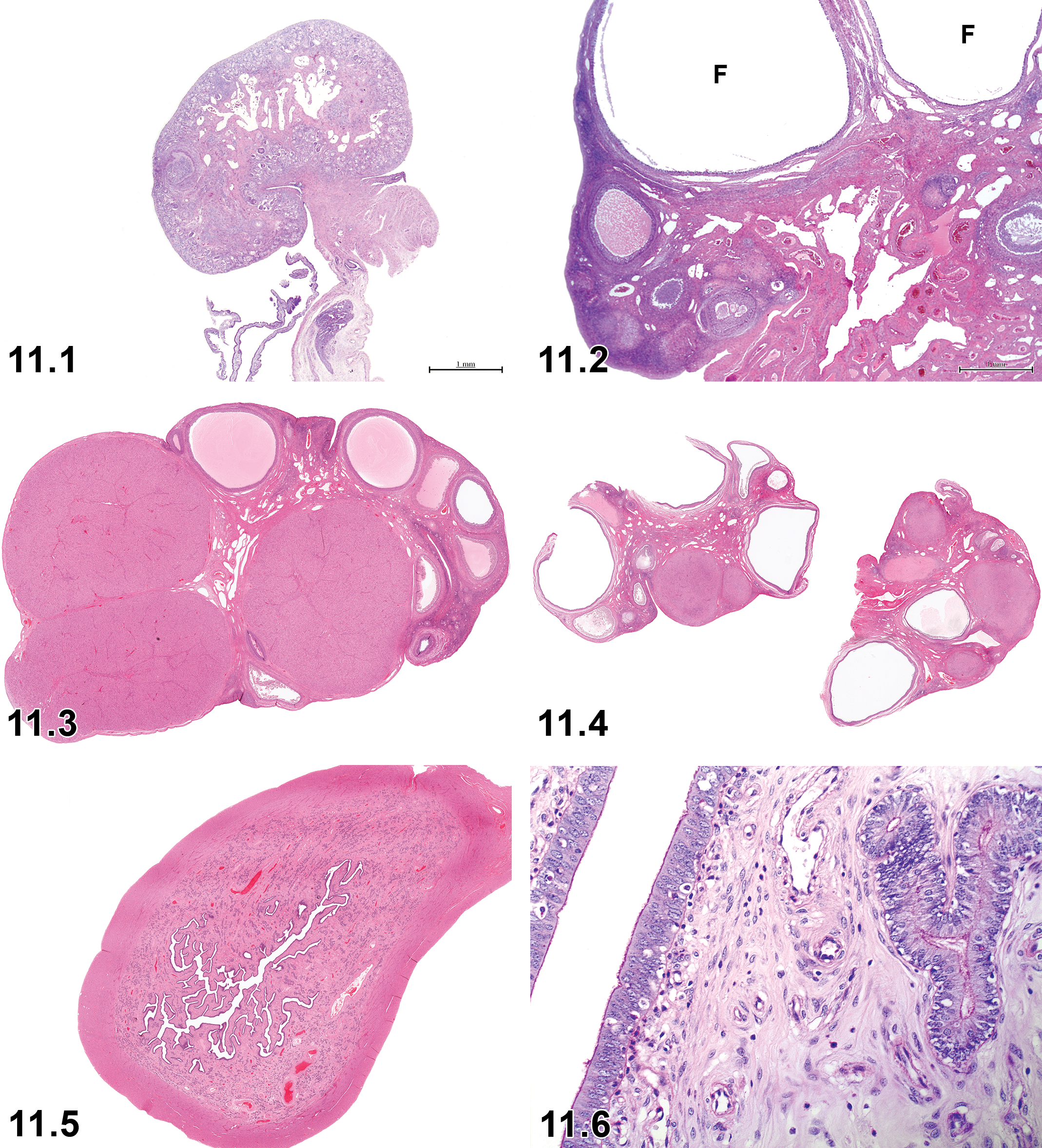
Immature ovary lacking tertiary follicles and corpus luteum. Subgross H&E (From “Onset of puberty and normal histological appearances of the reproductive organs in peripubertal female Gottingen minipigs”. Toxicol Pathol. 2013;41(8):1116-1125, with permission).
Partially Mature Stage (Peripubertal Stage)
Female minipigs are considered to be partially mature or in a peripubertal stage when the ovaries contain primordial, secondary, and tertiary follicles but lack corpora lutea (CL). The uterus appears similar to the immature stage, with endometrial glands moderately developed, but may contain frequent apoptotic bodies in the glandular and superficial epithelial cells (Figure 11.2).
Mature Stage
Ovaries contain primordial, secondary, and tertiary follicles and one or more CL. The presence of a corpus luteum in any stage is considered confirmation of sexual maturity. The uterus contains well-developed, often branched and coiled, endometrial glands, often with mucinous secretions. The uterine lumen is also moderately developed with papillary surface endometrium. Histology of the vaginal epithelium in mature females generally varies according to the stage of estrus cycle (Figure 11.3).
Estrus Cycle
Recommendations for evaluation of the female reproductive system and estrus cycle staging are similar to that for rodents. 127 While it is not necessary to record the stage of estrus cycle in routine toxicity studies, 127 it is critical that the pathologist be aware of the normal estrus cycle phase-specific histologic characteristics of the reproductive tract. Generally, a high degree of synchronicity is seen in the histologic changes in the various segments of the reproductive tract (ovaries, uterus, and vagina) during the various phases of the estrus cycle. Familiarity with the normal estrus cycle associated histology will therefore permit identification of changes, which could be direct or indirect effects of xenobiotic administration.
Unlike rodents, which have a very short estrus cycle of 4 to 5 days, the duration of estrus cycle in the mature minipig is longer, ranging from 20 to 22 days, and is more comparable to the duration of the menstrual cycle in the cynomolgus macaque (approximately 30 days). Estrus cycle-related systemic hormonal fluctuations have been well characterized and include a preovulatory luteinizing hormone surge followed by ovulation with subsequent increase in progesterone levels. 118 Progesterone levels increase for about 5 to 6 days following ovulation (estrus), stay high for another 6 to 7 days (metestrus), and finally fall during the last 3 to 4 days of the estrus cycle (end of diestrus). Staging of the estrus cycle and identification of perturbations in the cycle, based on morphologic features of the reproductive tract, is best achieved when the histologic features of the ovary, uterus, and vagina are taken into consideration together rather than relying on any one of them individually. The histologic distinction between phases, however, is not as clear as in rodents, rabbits, dogs, or monkeys. 117 The estrus cycle and the associated histological changes in the reproductive tract in the minipig have been described in detail 117,118 and are briefly described below. Estrus cycle-associated changes seen in the reproductive tract have been categorized as “common” in the tables below. These changes are, however, not routinely recorded as separate diagnoses.
Proestrus (Follicular/Proliferative Phase)
Ovary: The primary feature includes the presence of large tertiary (Graafian) follicles. Regressing CLs from previous cycle/s may also be seen (Figure 11.4).
Uterus: The uterus generally has a small lumen with few invaginations of the surface epithelium. Surface epithelium is generally cuboidal and contains many apoptotic and mitotic cells. The luminal surface is lined by a thin layer of periodic acid–Schiff (PAS)-positive material. Endometrial glands in zones 2 and 3 are small and contain few apoptotic cells (Figures 11.5 and 11.6).
Vagina: The vaginal epithelium is characterized by the presence of a layer of stratum germinativum, up to 10 intermediary layers, and a superficial layer of cuboidal to ovoid cells with pale cytoplasm (Figure 11.7).
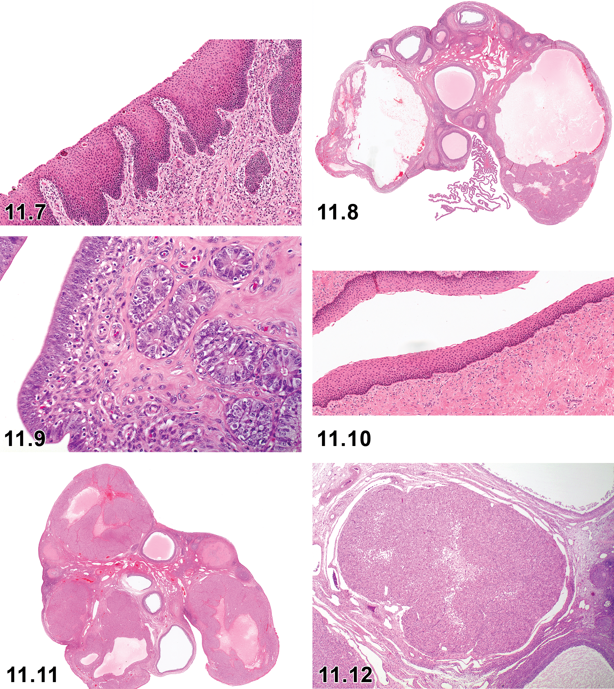
Proestrus vagina.
Estrus (Ovulatory Phase)
Ovary: The ovary contains a collapsed Graafian follicle indicative of ovulation (Figure 11.8). Regressing CLs from pervious cycles may be present.
Uterus: In the uterus, very few distinct changes from the proestrus stage are noted. Apoptotic bodies tend to disappear toward the later part of estrus and a pseudostratified appearance of nuclei is seen in the luminal epithelium (Figure 11.9).
Vagina: Unlike rodents, cornification of the superficial epithelium is not a feature in the vagina. A layer of basal stratum germinativum is overlain by intermediate layers and several layers of superficial flattened cells (Figure 11.10).
Metestrus (Early Secretory/Mid-Secretory Phase)
Ovary: The ovary contains large, newly formed CLs with a central space during the early phase, which progresses to fully mature CLs filled with luteinized cells (Figures 11.11 and 11.12). Tertiary follicles may be present at this stage. Hyalinized regressed CLs from previous cycles may be present. Other CL-like structures with central cavities may be present which are considered to represent either CLs that have not yet completely filled or Graafian follicles that have failed to rupture and have undergone luteinization (luteinized unruptured follicles [LUFs]).
Uterus: The luminal epithelium of the uterus is tallest (columnar) at this stage (Figure 11.13), and the epithelium has a maximal number of invaginations. Luminal epithelial cells no longer have a pseudostratified appearance. Endometrial glands in zones 2 and 3 are developed with abundant luminal secretion. Glandular stroma may contain lymphocytic infiltration. Few apoptotic bodies and mitotic cells may be noted during early metestrus but are generally not present during late metestrus. Metestrus uterus with tall columnar luminal epithelium (Courtesy de Rijk, Eveline). 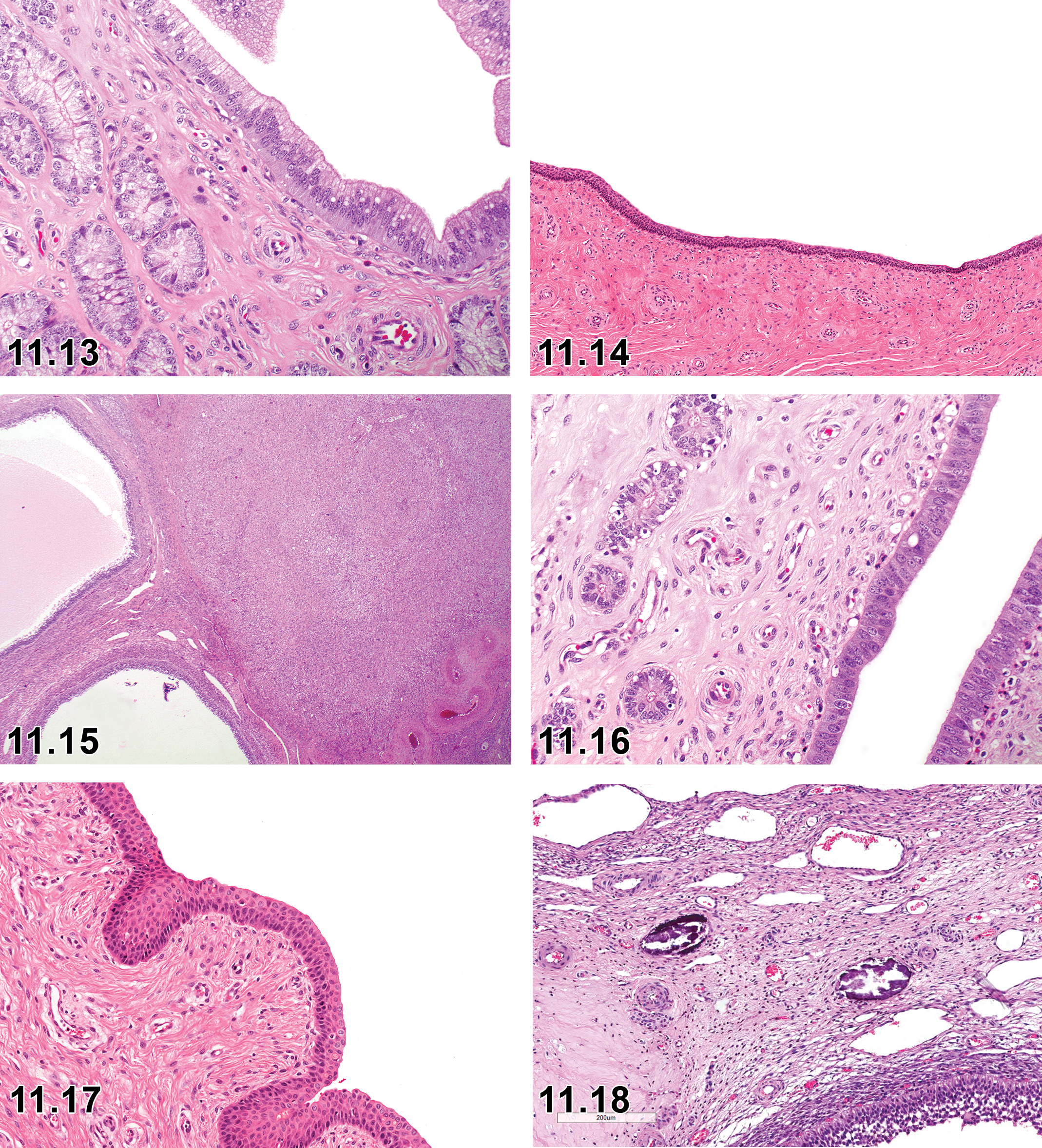
Vagina: Vaginal epithelium is characterized by 4 to 8 layers of cuboidal to ovoid cells with polymorphonuclear cell infiltration of the epithelium and lamina propria (Figure 11.14).
Diestrus (Late Luteal/Late Secretory Phase)
Ovary: Degeneration of CLs is noted with cells losing eosinophilic character and medium-sized to large tertiary follicles are present (Figure 11.15).
Uterus: Endometrial glands in zone 3 are lined by flattened epithelium and contain inspissated secretions. Luminal epithelium is comparable to proestrus with many apoptotic cells. Mitotic activity is present in the luminal epithelium but not in zone 2 and 3 glandular epithelium. Periodic acid–Schiff-positive material is present on the luminal surface (Figure 11.16).
Vagina: Epithelium generally is similar to that of metestrus with the exception that polymorphonuclear cell infiltration is minimal or absent (Figure 11.17).
Microscopic Findings of the Female Reproductive System: Ovary; Minipig.
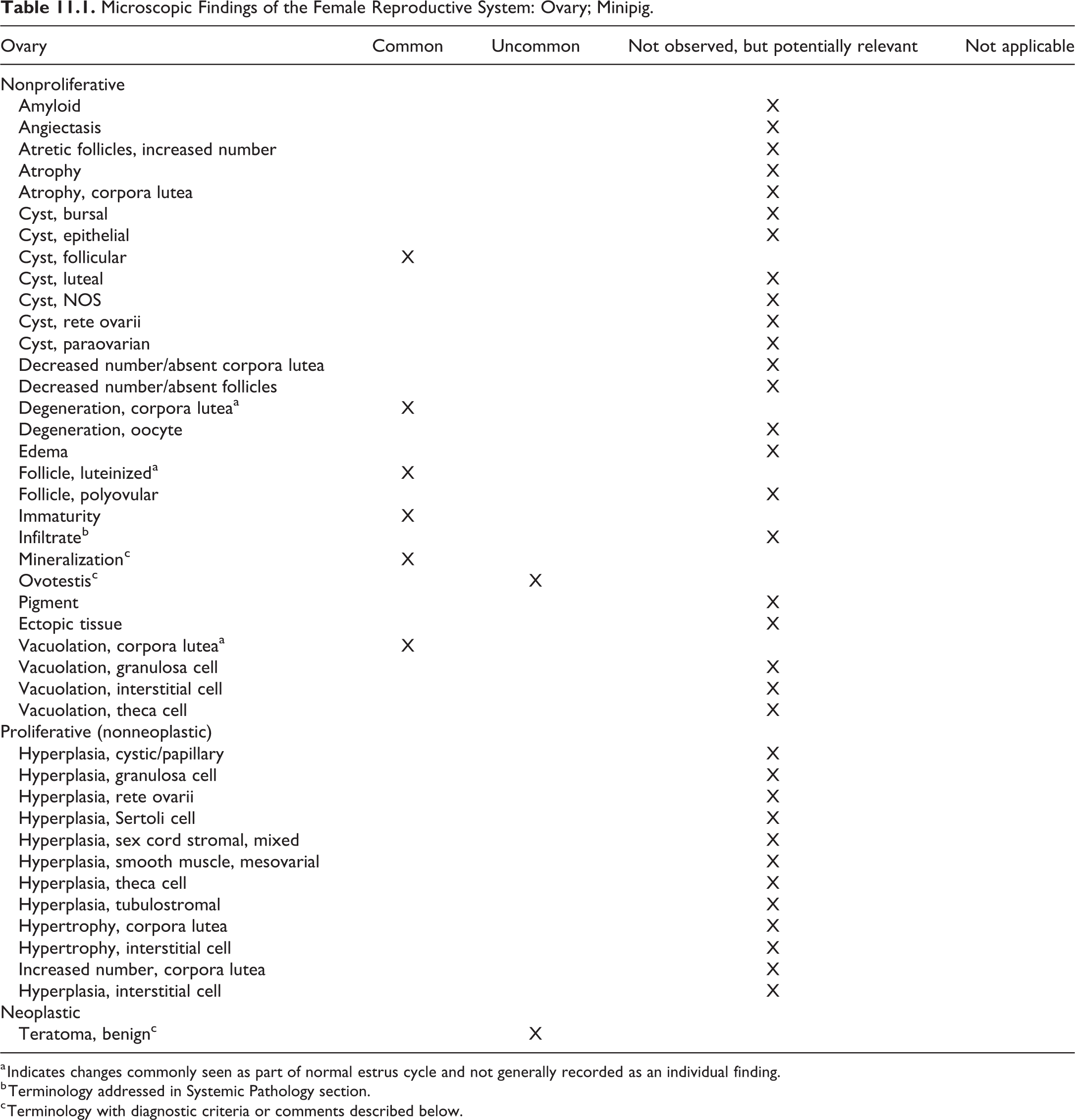
a Indicates changes commonly seen as part of normal estrus cycle and not generally recorded as an individual finding.
b Terminology addressed in Systemic Pathology section.
c Terminology with diagnostic criteria or comments described below.
Follicle, Luteinized
Comment
Graafian follicles that fail to rupture undergo atresia resulting in atretic follicles. Occasionally CL-like structures with prominent central cavities lined by luteinized epithelium can be seen in the ovaries and may represent unovulated Graafian follicles that have luteinized. 118 Luteinized unruptured follicles should be differentiated from normal nonclosed corpus lutea. Presence of oocyte is confirmative of LUF. Similar LUFs have been described as a background finding in beagle dogs. 133 Hormonal manipulation can induce LUFs in rats and monkeys. 134
Mineralization (Figure 11.18)
Comment
Mineralization can be seen in the ovaries and is often associated with atretic follicles. 7
Ovotestis
Comment
Ovotestis has been seen rarely (unpublished data) and, similar to rodents, is characterized by gonads containing both ovarian and testicular tissue.
Teratoma
Comment
Neoplastic changes in the ovary are rare. One instance of a benign teratoma was noted in a female Göttingen minipig. 7 Benign teratomas are generally encapsulated, expansile masses composed of varying proportion of tissues derived from the endoderm, mesoderm, and ectoderm.
Microscopic Findings of the Female Reproductive System: Oviduct; Minipig.
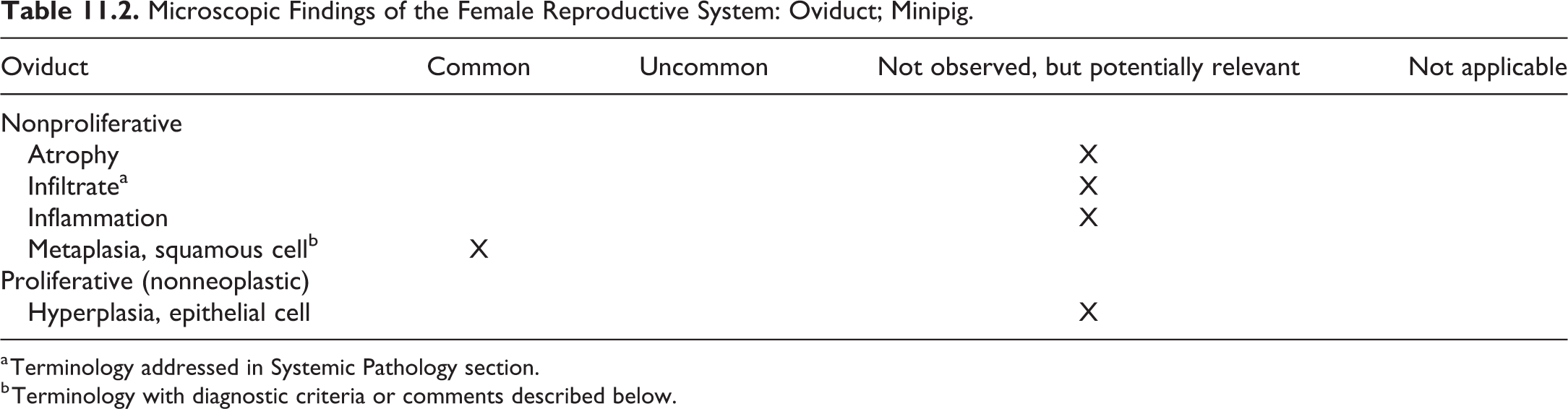
a Terminology addressed in Systemic Pathology section.
b Terminology with diagnostic criteria or comments described below.
Metaplasia, squamous cell (Figure 11.19)
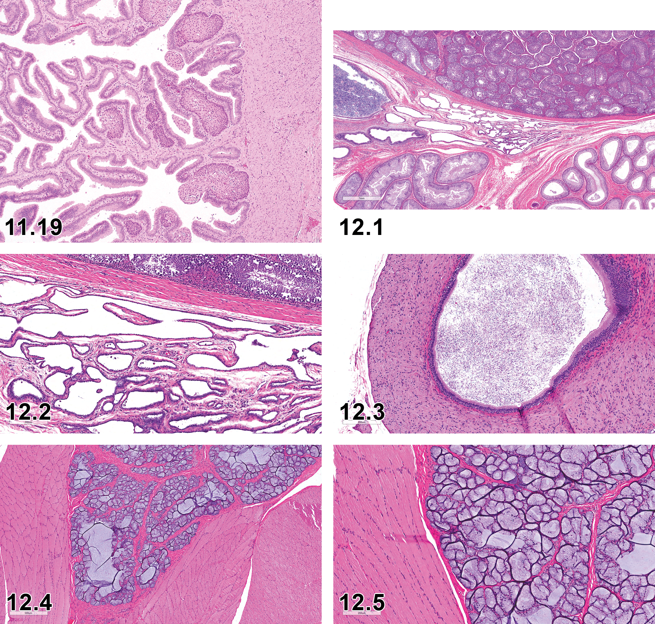
Oviduct squamous cell metaplasia (Courtesy de Rijk, Eveline).
Comment
Squamous cell metaplasia can be commonly found in the oviduct 7 and is characterized by the presence of keratinizing or nonkeratinizing squamous cells replacing normal cuboidal or columnar epithelium of the oviduct.
Microscopic Findings of the Female Reproductive System: Uterus; Minipig.
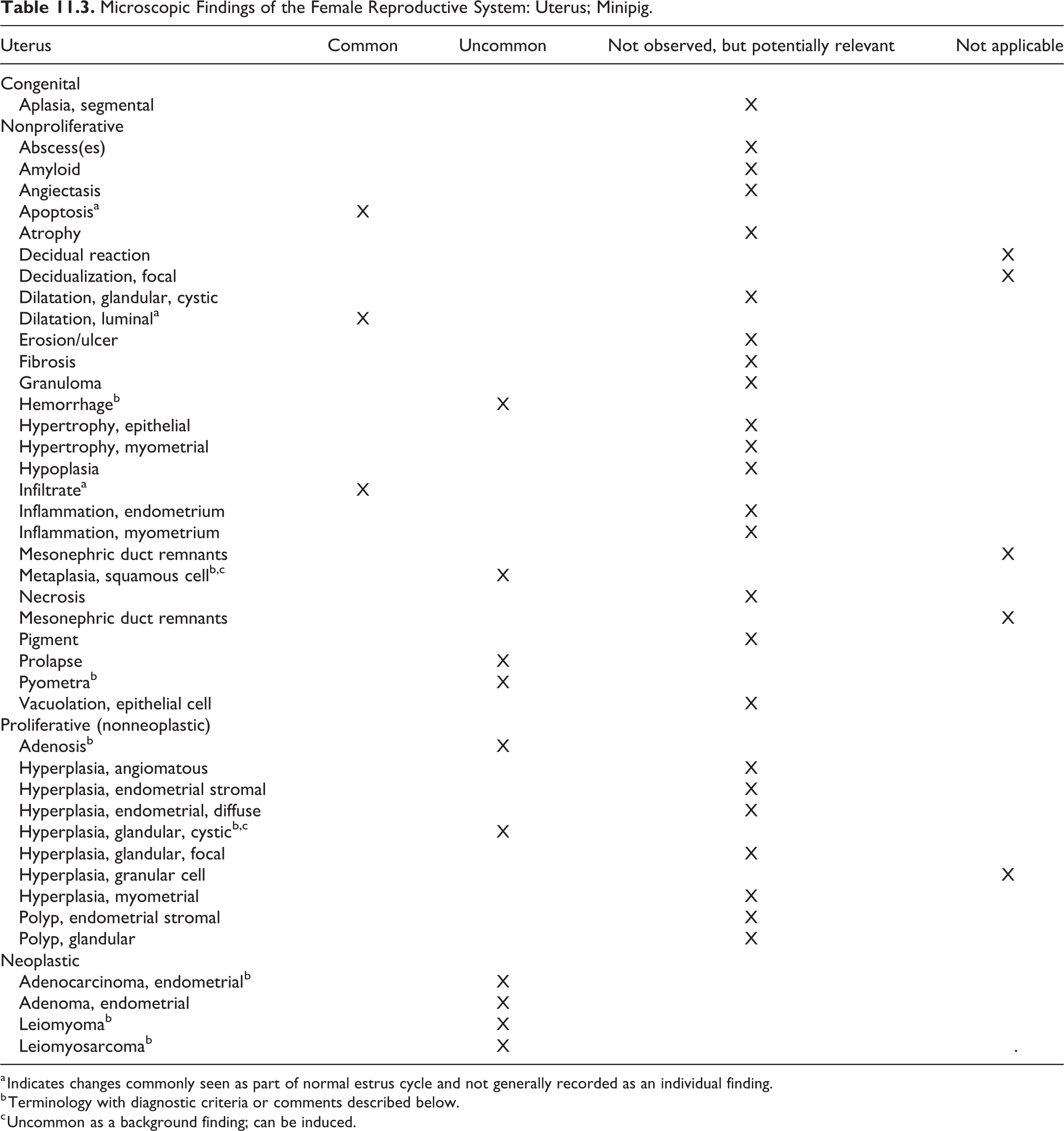
a Indicates changes commonly seen as part of normal estrus cycle and not generally recorded as an individual finding.
b Terminology with diagnostic criteria or comments described below.
c Uncommon as a background finding; can be induced.
Hemorrhage
Comment
Focal hemorrhage of minimal severity has been noted with a very low incidence in the uterus of the minipig. 7
Hyperplasia, glandular, cystic
Comment
Cystic glandular hyperplasia, characterized by dilated and cystic glands, has been reported in female miniature pigs as a background finding. 135 Cystic glandular hyperplasia has also been induced by exposure to zearalenone 136 and methoxychlor. 137
Metaplasia, squamous cell
Comment
Squamous metaplasia, characterized by focal or multifocal replacement of the surface and/or glandular columnar epithelium by keratinizing or nonkeratinizing epithelium, is an uncommon finding. However, cystic dilatation of endometrial gland with squamous metaplasia has been reported following experimental administration of zearalenone to pigs. 136
Pyometra
Comment
Pyometra characterized by suppurative (purulent) luminal exudate, which can be a sequel to inflammation of the endometrium, has been reported infrequently in female miniature pigs. 135
Adenosis
Comment
Adenosis is an uncommon change reported to occur in the uterus of female miniature pigs 135 and may be observed macroscopically as nodular lesions. Diagnostic features are similar to those reported for rodents and include the presence of endometrial glandular and stromal elements that do not exhibit atypical features within the myometrium. Spread of endometrial elements (glands and stroma) to the peritoneum has not been reported, and therefore, unlike human and nonhuman primates, adenomyosis in pigs should not be equated with endometriosis. Adenosis should be differentiated from endometrial adenoma and adenocarcinoma.
Neoplastic changes
Comment
Neoplastic changes are generally rarely observed in the female reproductive system of minipigs. A few sporadic reports of neoplastic changes include endometrial adenocarcinoma in an 8-year-old mixed breed (Spotted Poland China × Chapel Hill) gilt 138 and adenocarcinoma, leiomyoma, and leiomyosarcoma in female potbellied pigs and miniature pet pigs. 135,139 Most of these cases involved older pigs, and therefore, the low incidence of neoplasms observed could potentially be related to the fact that the vast majority of minipigs used in safety assessment programs are relatively young. Studies involving pigs of longer durations (≥12 months) are rare.
Microscopic Findings of the Female Reproductive System: Uterine Cervix; Minipig.
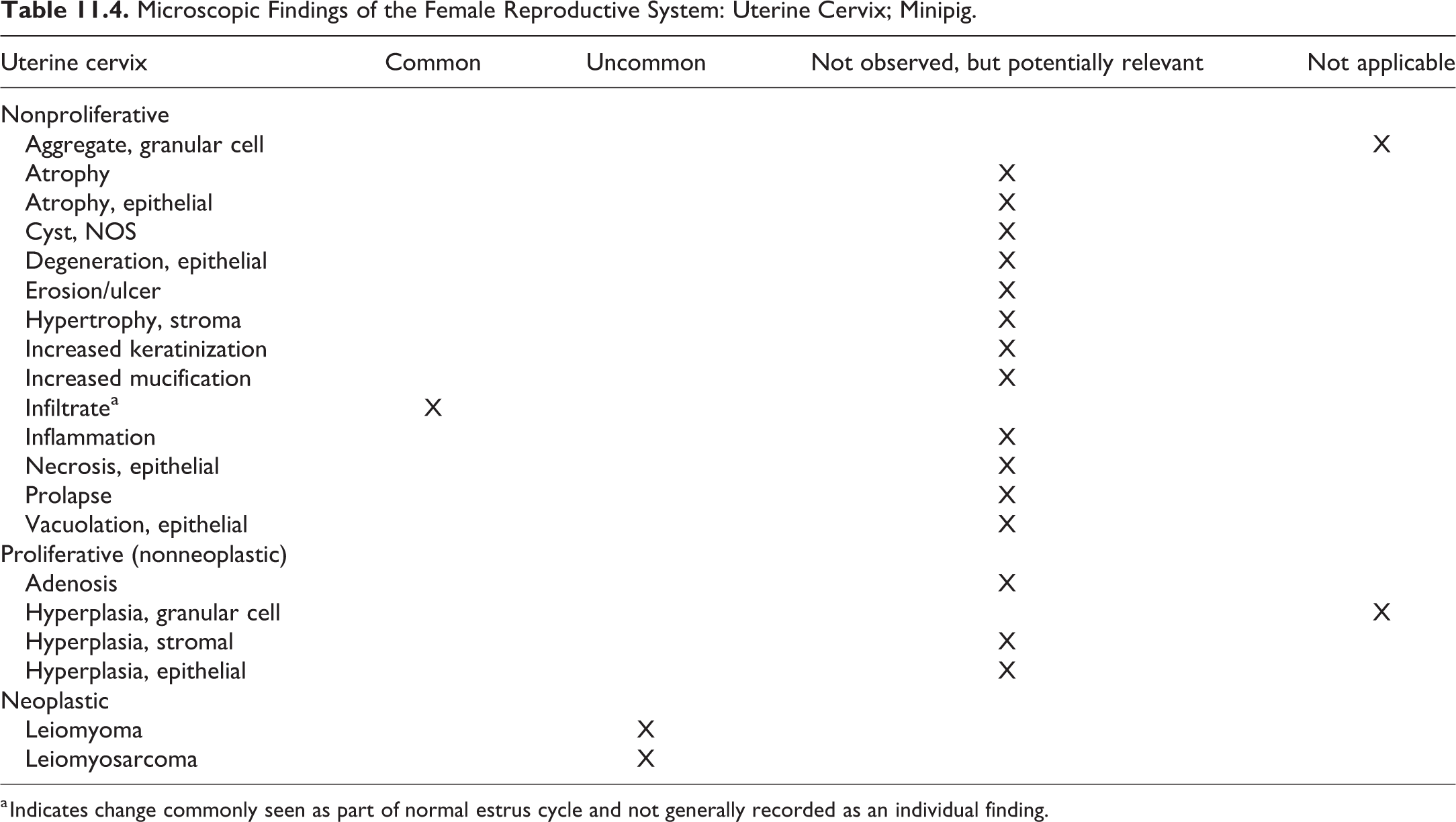
a Indicates change commonly seen as part of normal estrus cycle and not generally recorded as an individual finding.
Microscopic Findings of the Female Reproductive System: Vagina; Minipig.
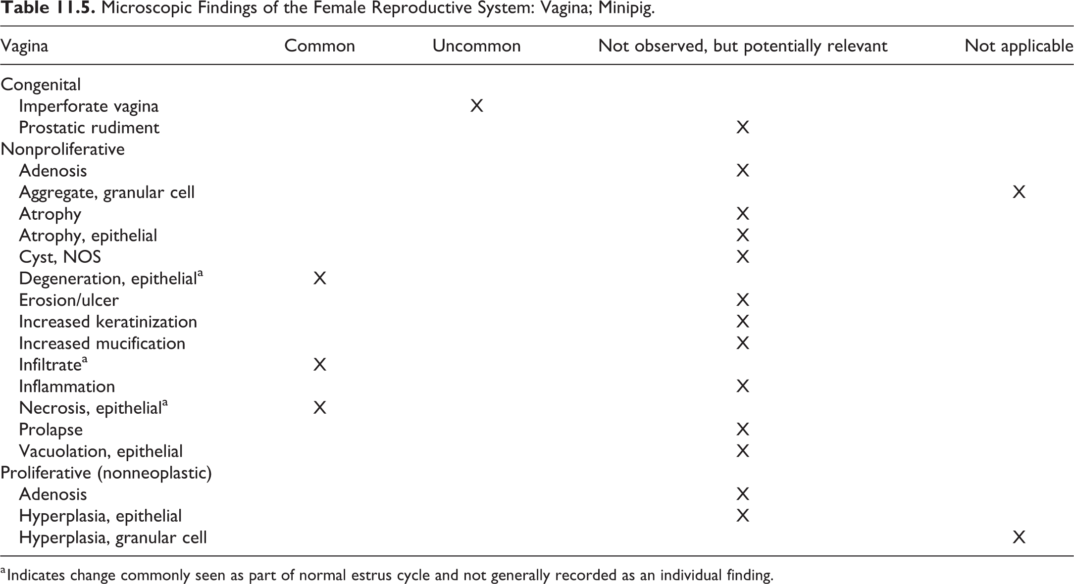
a Indicates change commonly seen as part of normal estrus cycle and not generally recorded as an individual finding.
Chapter 12. Reproductive System—Male
Standard INHAND nomenclature for proliferative and nonproliferative male reproductive tract findings in rats and mice has previously been published 140 and will be followed for the minipig as appropriate. There are some specific anatomical features of the porcine male reproductive tract that will be addressed here, with the remainder of the document following the anatomical approach used for rodent species, with a focus on those findings unique or applicable to the minipig. Specifics of anatomical and histological differences of the minipig from the rodent as well as the suggested examination, fixation, and sampling procedures will be addressed.
This chapter deals with a standardized nomenclature for classifying microscopic lesions observed in the reproductive system of male minipigs, including the testes, epididymides, seminal vesicles (or vesicular glands), prostate, efferent ducts, spermatic cord, bulbourethral glands, penis, prepuce, and dorsal preputial diverticulum. Spontaneous lesions, as well as lesions induced by exposure to test article, are included.
The reproductive system is susceptible to injury or change caused by a wide range of xenobiotics/test articles and should be routinely assessed in general toxicity studies. However, a comprehensive evaluation of the male reproductive system is best performed on fully mature animals or animals of known maturation status as a specialized study emphasizing reproductive tract collection, preservation, sampling, and proper staining for enhanced evaluation of any microscopic changes. As minipigs are more widely used in general toxicity studies and as dermal administration of systemically acting compounds becomes more common, an increased emphasis on proper evaluation of reproductive tract changes may be expected.
Nonproliferative lesions in general may be associated with experimental perturbation or are a result of degenerative changes frequently associated with aging. Modern laboratory animal management practices within minipig facilities are such that spontaneous infectious processes should be infrequently encountered; thus, the lesions related to infectious reproductive tract diseases are not described in detail in this document. Proliferative lesions in the male reproductive tract are rarely seen as background findings but may be due to toxicity, infectious agents, or background lesions. This may be seen, for example, in instances of experimental perturbation of the normal hormonal balance. Minipigs are not used in long-term carcinogenicity studies; thus, there is a paucity of data on neoplastic lesions.
Male Minipig Reproductive Anatomy, Histology, and Examination (Figures 12.1 -12.12)
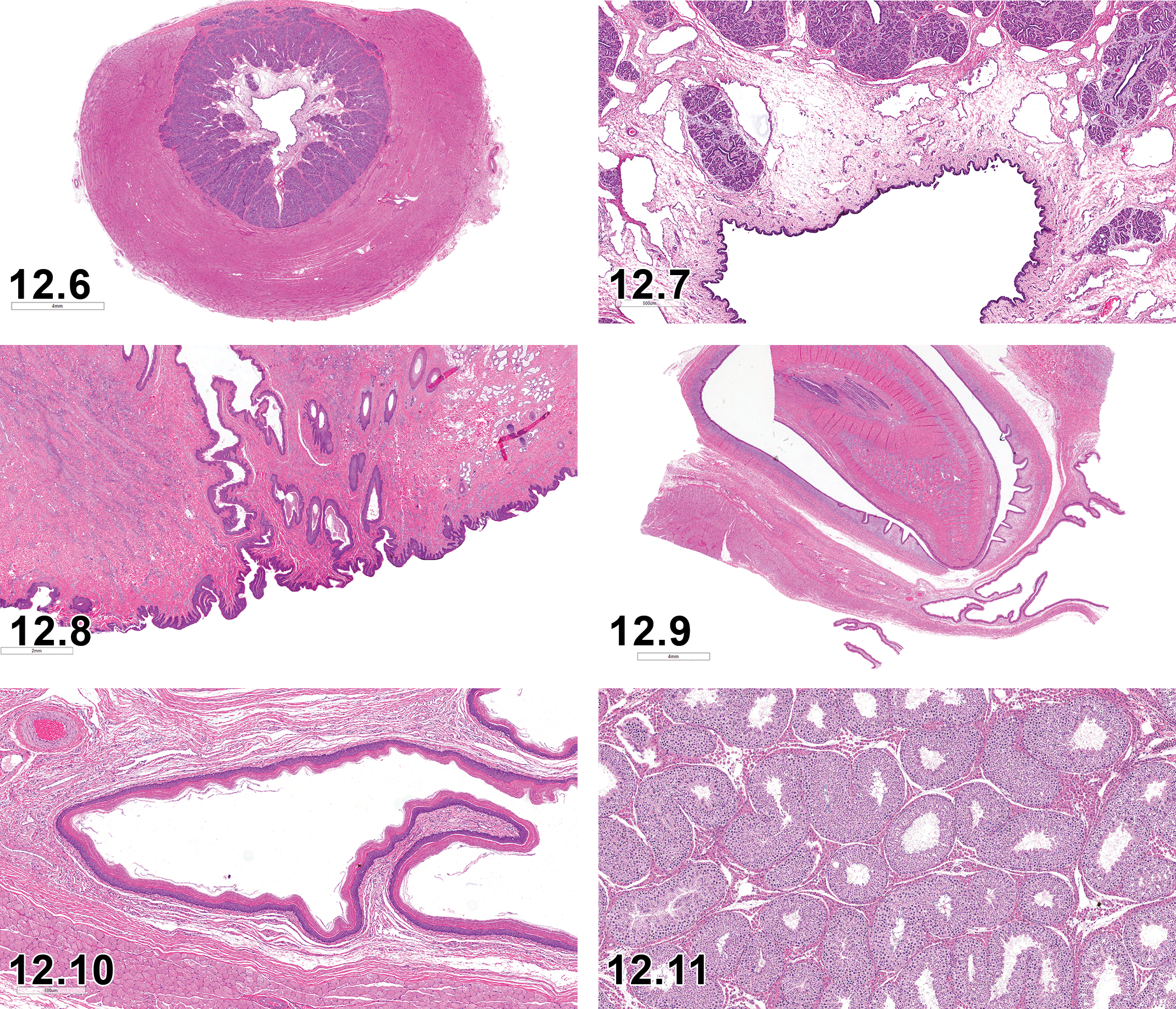
Normal minipig penis, cross-section. H&E x6.
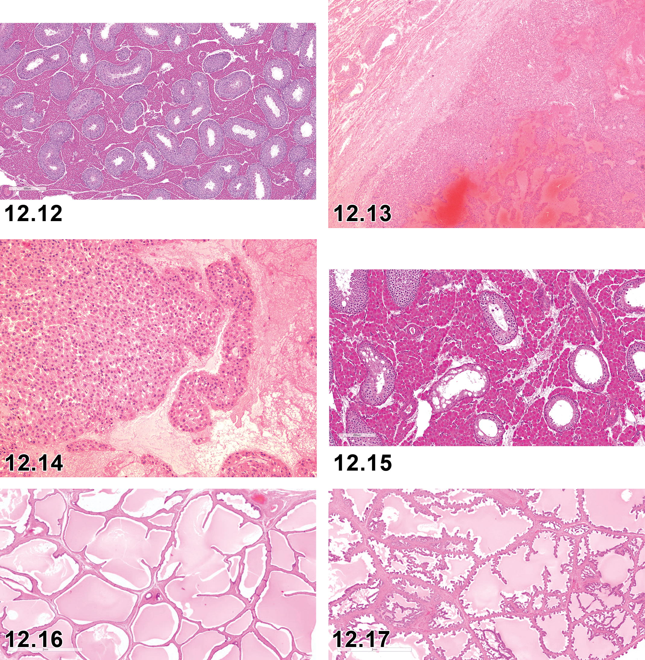
Testis: Control mature male (11 month old) Göttingen minipig testis with high numbers of Leydig cells. H&E x100.
The anatomy of the male minipig reproductive tract is the same as that of domestic swine but differs from the rodent anatomy in several features. Notably, coagulating glands and preputial glands are not prominent or easily sampled and the bulbourethral glands are more prominent and readily accessible in swine as compared to rodents. The suggested set of tissues for routine evaluation in the boar includes testes, epididymides, seminal vesicles (or vesicular glands), and prostate. 26 Sections of testis for routine histopathological evaluation should include the capsule, parenchyma, and the rete testis. It is recommended that the head, tail, and body of the epididymides are examined. Additional tissues may be added for specific studies but are not generally sampled in routine toxicity studies. These secondary tissues would include efferent ducts, spermatic cord, bulbourethral glands, penis, prepuce, and dorsal preputial diverticulum. Albl et al have thoroughly detailed a collection and trimming scheme for the male reproductive tract, often significantly more in depth than needed for routine toxicologic pathology assessment. 26 Representative photomicrographs of the normal microscopic anatomy of these nonstandard tissues are presented for reference in this article (Figures 12.1 -12.12).
Testes in the minipig are characterized by much the same features as those seen in the rat or mouse. Leydig (interstitial) cell density, size, and numbers may vary widely in the minipig, both as a function of maturation status and between individual animals (Figures 12.11 and 12.12 demonstrate age-matched mature adult male control animals). Leydig cells sustain intratesticular androgen levels and may be affected by direct toxic effects on steroidogenesis as well as indirectly by toxic effects on gonadotropin release. However, in light of the extensive histological variation in Leydig cell numbers, size, and appearance in normal mature control boars, evaluation of toxic effects on Leydig cells must be carefully considered.
Stage-aware evaluation of the minipig testis: At the present time, there are limited published guidelines available for stage-aware evaluation of the Göttingen minipig testis. Extant methods for other swine breeds often use periodic acid–Schiff (PAS) stain for increased resolution and improved evaluation of spermatogonia substructures rather than a more general H&E stain. An 8-stage method (PAS and H&E) to assess spermatogenesis in the Göttingen minipig has been proposed, 141,142 and Kangawa et al have published a staging method in the microminipig using an 11-stage method (PAS). 143 Some further guidelines may be inferred from literature detailing reproductive maturation of the testes of various breeds of domestic swine, but at this time, stage-aware evaluation is not routinely performed for this species and there are no clear, comprehensive published guidelines for performance of this evaluation in the Göttingen minipig.
Sexual Maturation in the Male Minipig
Particularly for the porcine testis, but also for other reproductive tract tissues, a comprehensive knowledge of normal cellular morphology, spermatogenesis, and maturation processes is important for detecting any abnormalities. In many short-term studies in minipigs, the male reproductive system is undergoing maturation over the course of the study. Thus, there may be notable differences in histoanatomical features within the reproductive tract as a result of normal maturation variance in the population. These differences must be separated from histopathologic findings of either background pathology or toxicity.
Taberner et al reported onset of histological puberty at 2 months of age in a group of 6 Göttingen minipigs, although there was no information regarding the onset of full reproductive maturation. 144 There are reports in the literature asserting histological reproductive maturation in the Yucatan minipig and microminipig at ∼4.5 months of age. 145,146 However, these studies have small sample sizes and may or may not represent a consistent level of maturation suitable for histological evaluation of potential reproductive toxicity in large-scale research facilities. Reports from breeding facilities suggest that mature sexual behavior begins at 3 to 4 months of age in male Göttingen minipigs (Ellegaard), although this may not directly correlate with complete histomorphological reproductive maturity. In the experience of the INHAND Minipig group, groups of animals less than 4 to 5 months of age will have some number of males that are immature and it is not until animals are of 5 to 6 months of age that we may be assured of their overall reproductive histomorphological maturity and full progression past pubertal status. While this is not always vital in individual toxicity studies, ability to assess effects of test compounds on the reproductive tract of mature individuals is important, particularly in instances where there is a potential reproductive effect, which cannot always be predicted. Thus, we recommend routinely using animals of 5 to 6 months of age at the start of the study period to mitigate potential confounding effects. A more definitive study of maturation status at various ages should be undertaken, as full histomorphological maturation of the reproductive organs is important for accurate assessment of toxic effects.
As in many nonrodent species, maturation in the male minipig may occur over a range of age, so individual animals of the same age may be at very different points in their reproductive maturation. It is suggested that sexual maturity be routinely recorded, using the immature, peripubertal, and mature categories. This will allow for more rigorous evaluation of potential toxicity affecting maturation status or sperm development. Brief definitions of the 3 separate stages of sexual maturation are given below:
Immature: Undeveloped seminiferous tubules lacking spermatozoa. Undistended seminiferous tubules are lined only by Sertoli cells and gonocytes (lacking mature spermatids) with rare luminal germ cell debris and multinucleated cells. No mature spermatids and rare to absent germ cell debris in epididymides. Secondary sex organs are undeveloped.
Peripubertal: Seminiferous tubules in varying stages of development. Mature spermatozoa may be present in some seminiferous tubules, with variable numbers of seminiferous tubules containing intraluminal spermatids and/or occasionally germ cell debris and multinucleated cells. Small amounts of mature sperm and/or germ cell debris may be seen in the ductular lumen of the epididymides, with cell population consisting primarily of giant cells and spermatids, with rare to no mature spermatozoa. Secondary sex organs development may be variable.
Mature: Mature spermatozoa predominate in all/most seminiferous tubules and epididymides. Seminiferous tubules are fully developed with mature spermatids/spermatozoa and all 4 generations of germ cells (spermatogonia, spermatocytes, round spermatids, and elongating spermatids). Epididymides contain abundant spermatozoa in the ductular lumen with rare to absent rounded germ cells. Secondary sex organs development may be variable.
Neoplasia
Although there are no published reports of neoplasia within Göttingen minipigs or other minipig breeds used in toxicologic pathology, a single incidence of Leydig cell adenoma has been noted (Figures 12.13 and 12.14) 147 and should be considered as an uncommon finding in the Göttingen minipig. Additionally, tumors of the male reproductive tract have been rarely reported in the literature in domestic swine. These include Leydig cell (interstitial cell) tumors of the testis, 148 -150 Sertoli tumors of the testis, 149,151 hemangiomas and hemangiosarcomas of the scrotum and testis, 148,151 intratubular germ cell tumors of the testis, 151,152 and papillomas of the penis. 106 These neoplasms may be seen at a very low incidence in the minipig and should be considered as possible findings, although they have not yet been reported.
Microscopic Findings of the Male Reproductive System: Testis; Minipig.
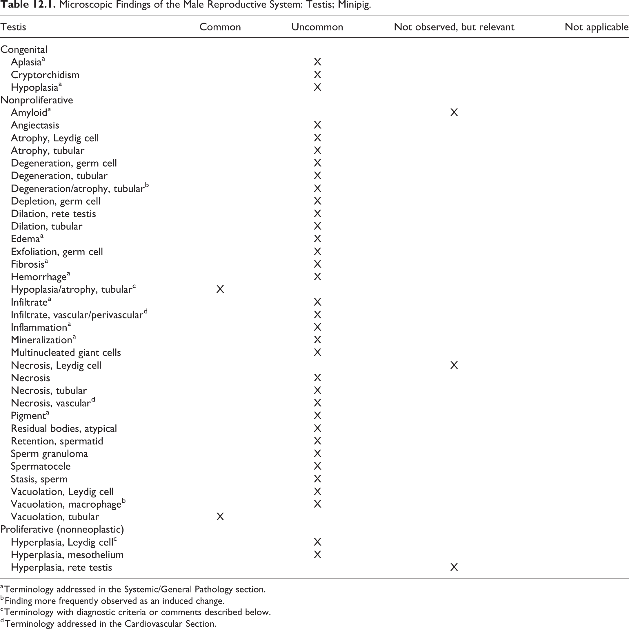
a Terminology addressed in the Systemic/General Pathology section.
b Finding more frequently observed as an induced change.
c Terminology with diagnostic criteria or comments described below.
d Terminology addressed in the Cardiovascular Section.
Nomenclature
Hypoplasia/Atrophy, Tubular, Testis, Epididymis (Figure 12.15)
Other term(s)
Hypospermatogenesis, hypoplasia
Diagnostic features
A continuum of microscopic changes, including one or more of the following, multifocally affecting a number of tubules:
Decreased numbers of spermatocytes and/or spermatids (hypospermatogenesis) to a complete absence of germ cells, leaving only Sertoli cells lining the affected tubules.
Vacuolation of seminiferous tubule epithelium with decreased spermatogonia.
Increased numbers of multinucleated or rounded germ cells in the lumen of seminiferous tubules.
Changes seen in tubules immediately surrounding the rete (tubuli recti) are considered normal and do not represent tubular hypoplasia/atrophy.
Affected tubules are clumped multifocally and adjacent to normal tubules.
This finding may be accompanied by decreased luminal sperm in the epididymides, along with increased numbers of round and multinucleated germ cells.
Number of Leydig cells might seem more prominent when the severity of hypoplasia/atrophy is high.
Differential diagnoses
Tubular degeneration/atrophy.
The presence of degenerate and apoptotic germ cells is one potential differentiator, with apoptotic cells being a feature of this finding and not noted in the more common background finding of tubular hypoplasia/atrophy. Tubular degeneration/atrophy may be seen in alcohol intoxication or folate deficiency, with tubular epithelial vacuolation and germ cell degeneration. 153
Comments
Tubular hypoplasia/atrophy is a testicular finding that can be present with variable severity in a majority of Göttingen minipig males. It may be a developmental lesion but does not seem to have an age-associated variance in incidence or severity. This finding is commonly seen in Göttingen minipig males, with reported incidences ranging from 70% 154 to 25%, 7,13 and can affect over 50% of the testis in some animals. 154 There does not appear to be an age-related progression or regression of the lesion. 7,154 Hypospermatogenesis is one facet of this continuum of findings, as is tubular atrophy, and care should be taken in differentiating this finding from possible test article-related decreased spermatogenesis or tubular degeneration/atrophy.
Microscopic Findings of the Male Reproductive System: Efferent Ducts, Epididymis, and Spermatic Cord; Minipig.
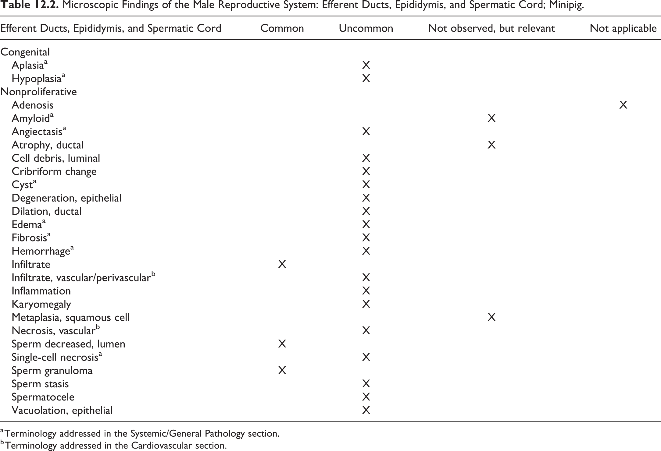
a Terminology addressed in the Systemic/General Pathology section.
b Terminology addressed in the Cardiovascular section.
Microscopic Findings of the Male Reproductive System: Bulbourethral Gland, Seminal Vesicles, and Prostate; Minipig.
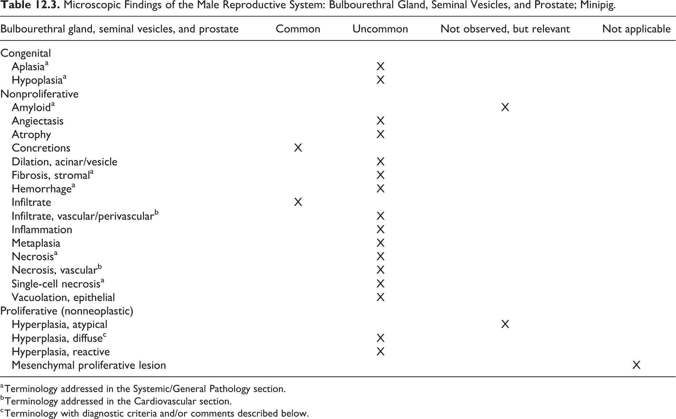
a Terminology addressed in the Systemic/General Pathology section.
b Terminology addressed in the Cardiovascular section.
c Terminology with diagnostic criteria and/or comments described below.
Hyperplasia, diffuse, prostate, seminal vesicles (Figures 12.16 and 12.17)
Other terms
Hyperplasia, functional; hyperplasia, adaptive
Pathogenesis
Specific pathogenesis is not always known in the swine but may, as in other mammals, represent effects of increased steroidal compounds or steroidogenic compounds producing increased testosterone, stimulating growth of glandular epithelium of the accessory sex organs.
Diagnostic features
Thickening of the vesicle epithelium, with increased glandular epithelial cell numbers.
May include formation of small papillary fronds of proliferating cells.
Differential diagnoses
N/A.
Comments
Functional hyperplasia of accessory sex organs is steroid dependent in other species, such as the canine, and may be so in the minipig as well.
Chapter 13. Respiratory System
The respiratory system is susceptible to injury caused by inhaled and blood-borne (including dermally absorbed) xenobiotics/test articles and thus should be routinely examined in all toxicity studies. This is especially important since inhaled xenobiotics are the most frequent cause of toxicity and the minipig is beginning to be used for short-term inhalation studies. In addition, the minipig is being used to generate animal models of respiratory disease, for example, inhaled lipopolysaccharide (LPS) has been used as a model for chronic obstructive pulmonary disease, 155 –157 one of the leading causes of human mortality and morbidity worldwide. More porcine inhalation studies and models of disease may be anticipated given the similarity of the porcine respiratory system to that of humans.
Lung toxicity in the pig may be mediated by similar mechanisms as in other species including biotransformation, via cytochrome P450 and phase II enzymes. 158 Metabolizing enzymes, several of which are inducible, have been identified in the porcine lung. 159 In addition to injury of pulmonary epithelial and endothelial cells, toxicological insults in the pig may cause injury to resident pulmonary intravascular macrophages (PIMs). 158,160 Unlike rodents and humans, PIMs that adhere to endothelium are a significant resident cell population in the pig lung. Pulmonary intravascular macrophages increase in number with age and function to remove particulate matter including bacteria from the blood. 161,162 Injection of microspheres has been reported to lead to severe pulmonary vasoconstriction in the pig due to release of thromboxane A2 from PIMs. 160 In rodents and humans, PIMs are not normally present but may be induced in cases of liver dysfunction. 163
Respiratory tract lesions can be treatment-related, spontaneous background changes or due to infection, environmental factors, or aging. Modern laboratory animal management practices within minipig facilities are such that spontaneous infectious processes should be infrequently encountered; thus, lesions caused by infectious respiratory tract diseases are not described in this document. Excellent reviews have been published on the effects of infectious disease, diet, and environmental factors on the pig respiratory tract. 164 –166 It has recently been proposed that the microbiome plays a role in the pulmonary response to infection and by extrapolation may have effects on toxicological responses. 164
While the focus is on the minipig, it should be noted that most lesions described in the domestic pig could occur in the minipig since both are Sus scrofa. Reports of neoplastic lesions in the respiratory tract of pigs are rare, and to the best of our knowledge, none have been reported in minipigs.
Systematic sampling and careful fixation by airway or intravascular perfusion are prerequisites to identifying lung lesions; however, this is not as easily done in larger animals as it is in rodents. The entire larynx, trachea, or lung lobe cannot be examined histologically. Thus, a uniform method must be developed within laboratories which includes consistent sections from each of these tissues (see Albl et al for recommendations). 26 Larynx should be sampled, although there is variation in sampling procedures among laboratories. Trachea should be sampled at 1 to 2 sites; either longitudinal or transverse sections are acceptable, and again, consistency within laboratories is important. Both left and right lung lobes should be sampled, with sections containing main stem bronchi, secondary bronchi, and lung margins. The primary goal is to assess all anatomical features. Lung sections should be carefully inflated with fixative, 26 but again there is variation in techniques used among laboratories.
I. Nasal Cavity
Swine sinonasal anatomy is largely similar to human as demonstrated by computed tomography of domestic and Yucatan pig heads. 167 The middle and inferior turbinates arise from a single uniturbinate and the superior turbinates contain large concha bullosa. Unlike human, swine nasal septum consists of bone anteriorly and cartilage posteriorly. 167 The nasal cavity is structurally complex, reflecting the diverse physiological functions associated with this anatomical site. Kuper et al 168 provide an overview of normal nasal passages of Gottingen minipigs from the neonatal period to young adult. Several additional articles describe the anatomy and histology of porcine nasal cavity tissues. 167,169 In pigs, a caudal diverticulum of the pharynx lies immediately dorsal to the esophagus. In young pigs, foreign materials occasionally lodge in this diverticulum, causing pharyngitis and cellulitis that may lead to dysphagia and death from starvation.
There are few reports of inhalation studies or chemically induced upper respiratory lesions in pigs or minipigs. 170 –173 The majority of reported inhalation studies do not include morphologic evaluation of the nasal cavity and the nasal cavity is not routinely examined in most noninhalation toxicology studies. Therefore, the frequency of individual microscopic findings, as provided in the Tables 13.1 and 13.2, may need revision in the future.
Albl et al 26 recommend that in routine noninhalation studies, one sample each of the nasal septum, turbinates (frontal section at level of first premolar), and ethmoidal labyrinth (parasagittal section) be taken to examine the transitional, respiratory, and olfactory epithelia. For inhalation studies, Albl et al recommend 2 additional frontal sections of the nasal septum and turbinates (caudal to the first incisors and caudal end of turbinates) to examine squamous and transitional epithelium. Kuper et al 168 provide additional guidance for a standard method to sample the minipig nasal passages, including a section of the nasopharynx to examine NALT. The importance of consistent sampling cannot be overemphasized in evaluation of the nasal cavity since normal changes in epithelial type with location may be mistaken for xenobiotic-induced lesions. It is recommended that the nasal cavity be first flushed with 10% neutral-buffered formalin. Appropriate samples of the turbinates and ethmoidal labyrinth should be fixed in formalin and then decalcified before embedding in paraffin. A standardized approach for recording histologic lesion distribution is essential and nasal diagrams, such as provided by Yang et al, 169 can provide assistance in mapping lesions. 168
Microscopic Findings of Respiratory System: Nasal Cavity; Minipig.
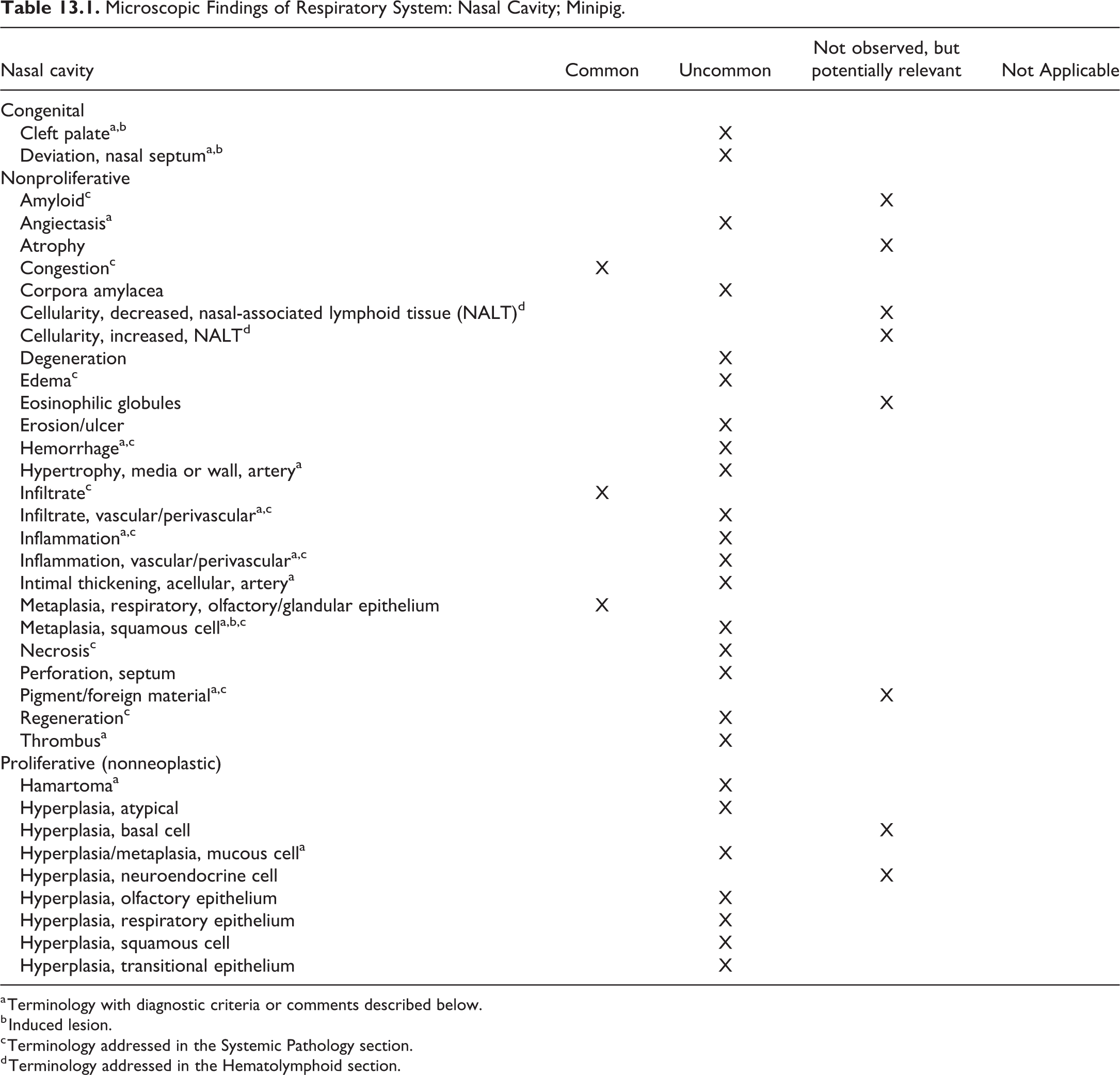
a Terminology with diagnostic criteria or comments described below.
b Induced lesion.
c Terminology addressed in the Systemic Pathology section.
d Terminology addressed in the Hematolymphoid section.
Microscopic Findings of Respiratory System: Nasopharynx, Paranasal Sinuses; Minipig.
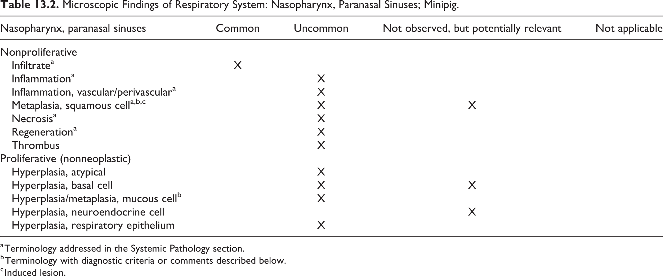
a Terminology addressed in the Systemic Pathology section.
b Terminology with diagnostic criteria or comments described below.
c Induced lesion.
Angiectasis: Nasal cavity
Comment
Unlike rodents, mononuclear cell leukemia has not been reported in pigs, so angiectasis of the nasal cavity is not associated with this lesion.
Cleft palate: Nasal cavity
Comment
Cleft palate (palatoschisis) has been reported as a background finding in the Gottingen minipig 174 and induced in domestic pigs by feeding poison hemlock (Conium maculatum), tobacco plants (Nicotiana glauca and N tabacum), and Crotalaria retusa seed during pregnancy. 175 -177 The toxic principle in poison hemlock is a piperidine alkaloid. The mechanism of cleft palate formation by poison hemlock and tobacco is believed to be the lack of fetal movement during the critical stage of gestation with arthrogryposis also frequently present. Feeding vitamin A depleted domestic sows a vitamin A-free ration during the first month of pregnancy also resulted in cleft palate formation. 178
Deviation, nasal septum: Nasal cavity
Comment
Deviation of the nasal septum as a congenital lesion should be differentiated from that due to atrophic rhinitis.
Hamartoma: Nasal cavity
Pathogenesis/cell of origin
Defective development of blood vessels resulting in disordered proliferation of mature blood vessels.
Differential Diagnosis
Hemangioma or hemangiosarcoma, however these have not been reported.
Diagnostic features
Tumor-like mass consisting of primitive to well-formed blood vessels.
Comment
A vascular hamartoma has been reported in the minipig nasal cavity. 13
Hemorrhage: Nasal cavity
Comment
Hemorrhage in the nasal cavity is most likely due to trauma or foreign material. Unlike rodents, antemortem blood collection via the retroorbital plexus in the medial canthus of the eye is not done in pigs, and therefore, this is not a cause of hemorrhage in the minipig.
Hypertrophy, media or wall, artery, nasal cavity
Comment
See cardiovascular section. Medial hypertrophy of the vessels in the nasal cavity has been reported in the minipig. 13
Hyperplasia/metaplasia, mucous cell: nasal cavity, nasopharynx, paranasal sinus
Comment
Mucous cell hyperplasia has been reported in domestic pigs following chronic aerogenous exposure to ammonia. 179
Infiltrate, vascular/perivascular: Nasal cavity
Comment
See systemic section. Perivascular mononuclear cell infiltrates in the nasal cavity have been reported in the minipig. 13
Inflammation (granulomatous): Nasal cavity
Comment
Foci of granulomatous inflammation with or without mineralization have been reported in the propria/submucosa of the nasal cavity and trachea in pigs. They are believed to be due to impacted particles of inhaled calcium salts from the feed. 180
Inflammation, vascular/ perivascular: Nasal cavity
Other terms
Arteritis, periarteritis.
Comment
See systemic and cardiovascular sections. Inflammation of the vasculature (arteritis) has been reported in the minipig nasal cavity. 10
Intimal thickening, acellular, artery: Nasal cavity
Comment
See cardiovascular section. Intimal proliferation of the vessels in the nasal cavity has been reported in the minipig. 13
Metaplasia, squamous cell: Nasal cavity, nasopharynx, paranasal sinus
Comment
In the domestic pig, squamous metaplasia has been observed following chronic exposure to corn dust and SO2 181 as well as with experimental infection with porcine reproductive and respiratory syndrome virus. 182
Pigment/foreign material: Nasal cavity
Other terms
Foreign body
Pathogenesis/cell of origin
Foreign material may be seen in the respiratory system of the minipig and may be associated with porcine rooting behavior. 59
Comment
This term includes foreign body. Although not reported specifically in the nasal passages, this is likely due to the fact that there is limited information available on this portion of the respiratory tract.
Thrombus: Nasal cavity
Comment
Unlike rodents, mononuclear cell leukemia has not been reported in pigs so thrombosis of the nasal cavity is not associated with this lesion. 13 See Cardiovascular section for additional information.
II. Larynx, Trachea, Bronchi, and Bronchioles
The mucosal epithelium of the airway conducting system in the pig larynx, trachea, and bronchi is similar to other species in that it is less complex than that lining the nasal airways. The pig has a large larynx. The supraglottis and glottis are lined by stratified squamous epithelium and the subglottis by pseudostratified columnar epithelium. 183,184 Sampling of the right aryepiglottic fold is recommended by Albl et al. 26
Unlike other species, there is a bronchial branch emanating from the trachea cranial to the carina that supplies the right cranial lung lobe. 185 Similarly to humans, but unlike the rodent, the pig has cartilage and submucosal glands in intrapulmonary bronchi. Bronchial glands are similar in size and secretion to those in humans, which increase in number as the animal ages. 186 Cartilage and submucosal glands are not present at the level of the bronchioles. 187 Bronchial-associated lymphoid tissue (BALT) only develops when animals are antigenically stimulated and therefore is not present in germ-free pigs. 188,189 When BALT is present, it manifests as a single follicle found at bronchial bifurcations and protrudes into the lumen. 190
Microscopic Findings of Respiratory System: Larynx; Minipig.
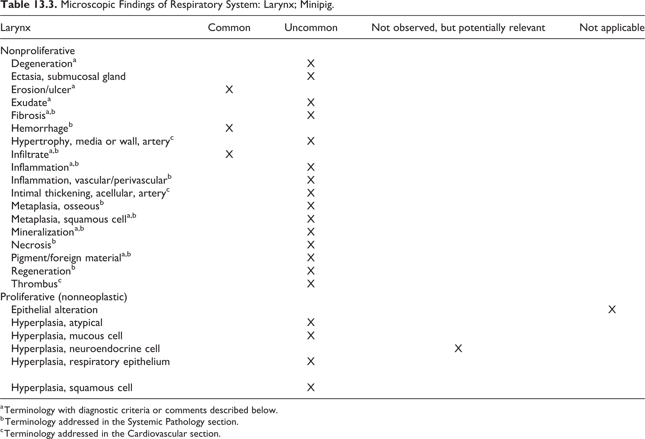
a Terminology with diagnostic criteria or comments described below.
b Terminology addressed in the Systemic Pathology section.
c Terminology addressed in the Cardiovascular section.
Microscopic Findings of Respiratory System: Trachea: Minipig.

a Terminology with diagnostic criteria or comments described below.
b Terminology addressed in the Systemic Pathology section.
c Terminology addressed in the Cardiovascular section.
Microscopic Findings of Respiratory System: Bronchi: Minipig.
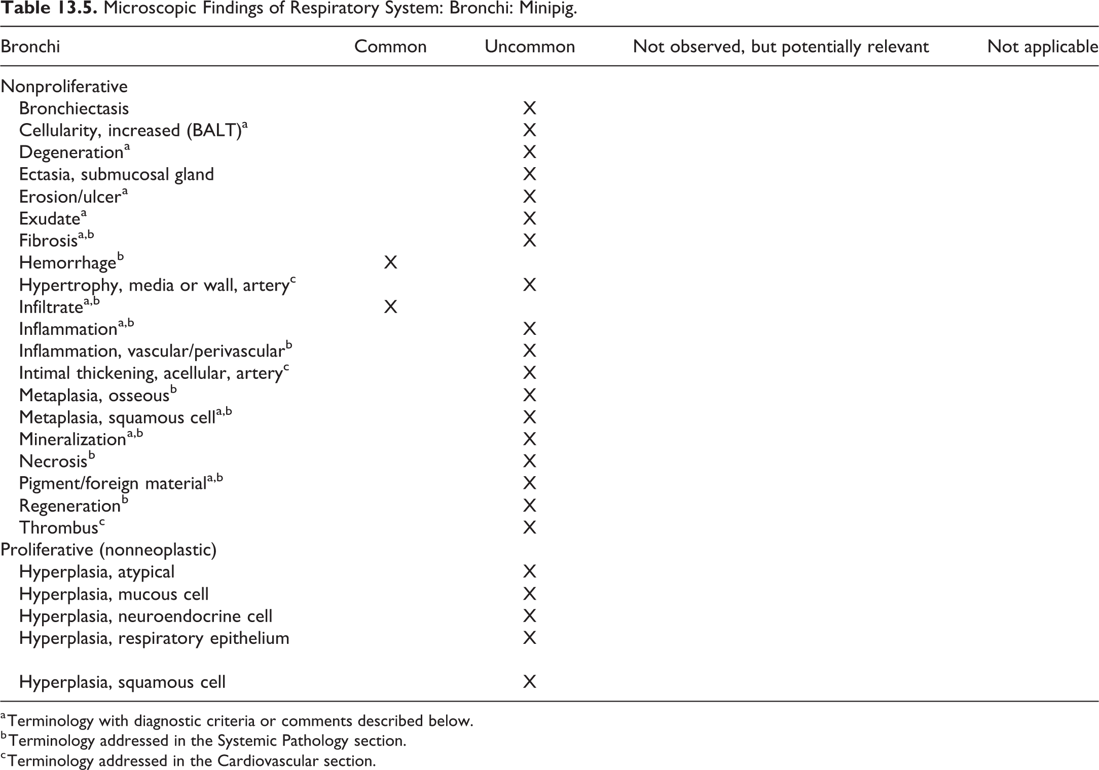
a Terminology with diagnostic criteria or comments described below.
b Terminology addressed in the Systemic Pathology section.
c Terminology addressed in the Cardiovascular section.
Microscopic Findings of Respiratory System: Lung: Bronchioles: Minipig.
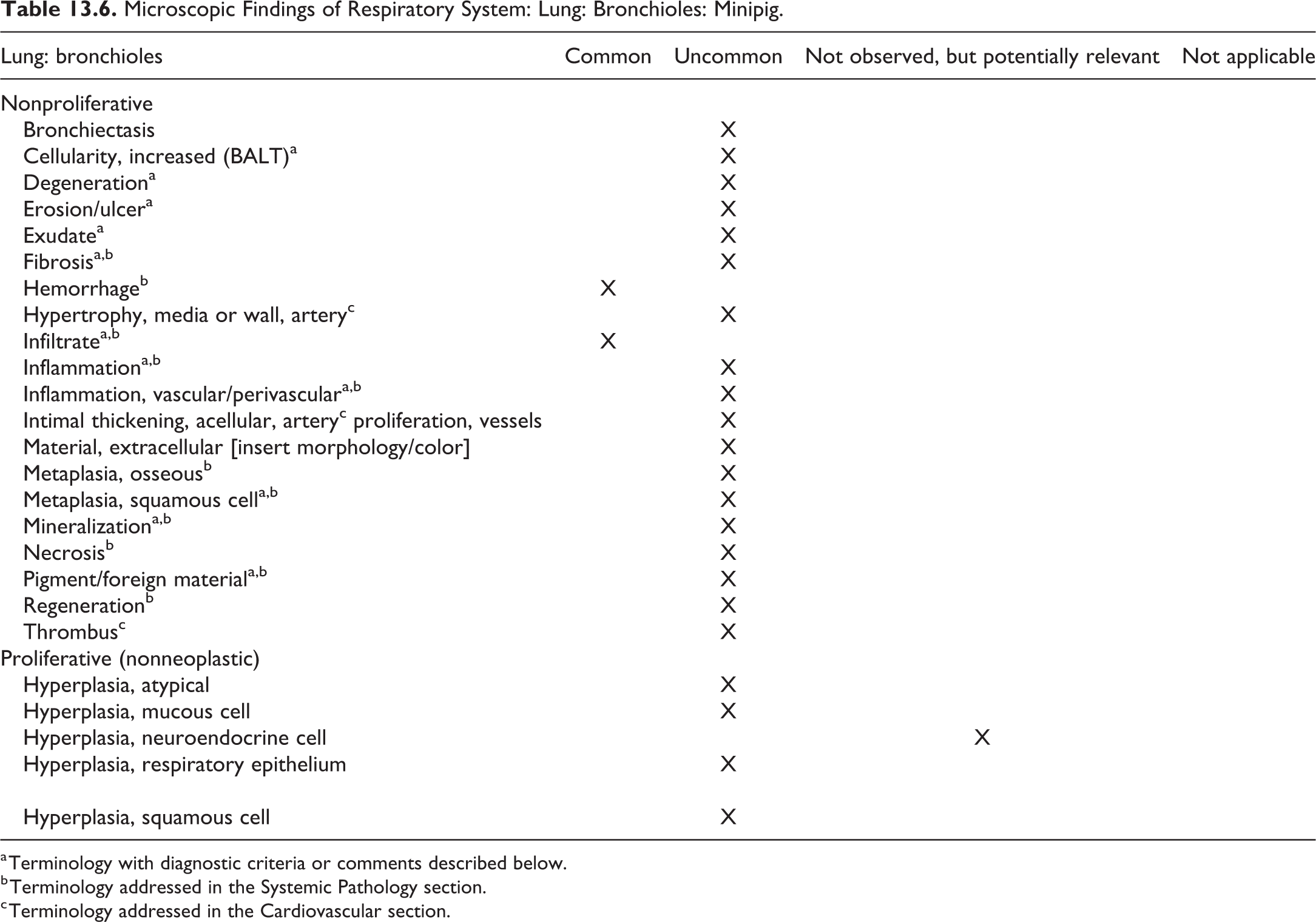
a Terminology with diagnostic criteria or comments described below.
b Terminology addressed in the Systemic Pathology section.
c Terminology addressed in the Cardiovascular section.
Cellularity, increased; BALT: Bronchi, bronchioles
Pathogenesis/cell of origin
BALT is only present in pigs that are antigenically stimulated. It normally consists of a single follicle-like structure at the bifurcation of bronchi. 188 -190 When antigenic stimulation is excessive, it can expand in both size and location to encompass other regions of the bronchi or be multifollicular.
Differential diagnosis
Inflammation, acute, chronic, or granulomatous.
Comment
See hematolymphoid system for additional information.
Degeneration: Larynx, trachea, bronchi, bronchioles
Comment
This lesion has been reported in the minipig. 13
Erosion/ulcer: Larynx, trachea, bronchi, bronchioles
Comment
Erosions/ulcerations of the vocal folds are hypothesized to be related to excessive vocalizations. Erosion and ulceration in the trachea may be a spontaneous lesion 13 but may also be due to injury from an endotracheal tube if the animal was anesthetized (as in device studies).
Exudate: Larynx, trachea, bronchi, bronchioles
Pathogenesis/cell of origin
Cell accumulations are typically primarily neutrophils and macrophages but can be composed of any inflammatory cell type (lymphocytes, neutrophils, eosinophils, and macrophages). Exudate may be related to inhalation of foreign material. 13
Diagnostic features
The cells accumulate within the lumen of the tissue but without damage to surrounding tissue which is typical of inflammation. This may be due to translocation by the mucociliary apparatus from inflammation in the lower respiratory tract which may or may not be associated with foreign particles.
Differential diagnosis
Inflammation, acute, chronic, or granulomatous.
Fibrosis: Trachea, bronchi, bronchioles
Pathogenesis/cell of origin
Blood sampling in the pig is often performed in the cervical region and can result in lesions within surrounding tissues. If the trachea or surrounding tissue is damaged during sampling, peritracheal hemorrhage/inflammation and fibrosis may result. 13 Mild peritracheal fibrosis can also be found in the Hanford minipig. 191
Infiltrate: Larynx, trachea, bronchi, bronchioles
Comment
Cell infiltrates are common in the pig in all tissues and should not be confused with inflammation. 7,13,59 See Systemic section.
Inflammation (acute, chronic, granulomatous): Larynx, trachea, bronchi, bronchioles
Comment
Acute inflammation is often seen in pigs as a background finding secondary to inhaled particles. Inflammation secondary to foreign particles needs to be carefully differentiated from inflammation due to an inhalant or test article. 10,13,59 When foreign material becomes trapped in respiratory tissues, it can lead to chronic inflammation. 10,13,59 Many instances of granulomatous inflammation in the pig respiratory system are due to the propensity for rooting behavior leading to inhaled foreign material. This foreign material often gets trapped in respiratory tissues leading to granulomatous inflammation. 10,13,59 Inflammation of glands in larynx, trachea, and bronchi has been reported in the minipig. 13
Metaplasia, squamous cell: Larynx, trachea, bronchi, bronchioles
Comment
Tiny islands of squamous metaplasia within bronchi have been reported in domestic pigs. 192 Squamous metaplasia has also been noted to occur in the ducts of the bronchial glands of domestic pigs. 193
Mineralization: Larynx, trachea, bronchi, bronchioles
Pathogenesis/cell of origin
It is believed that multifocal mineralization in the lamina propria of respiratory mucosa is due in part to inhaled calcium salts from dry and pelleted feed. 180
Diagnostic features
If related to inhalation of particles, the mineralization will be primarily along the dorsal wall of the trachea. 180
Comment
This change has also been reported as an incidental finding. 13
Pigment/foreign material: Larynx, trachea, bronchi, bronchioles
Other term
Foreign body.
Pathogenesis/cell of origin
Foreign bodies, generally either food particles or bedding material, may be found in the respiratory tract. Normal porcine behavior, such as rooting, can result in foreign bodies and an associated inflammatory response being encountered in minipigs. 7,13 Foreign bodies initially elicit a suppurative inflammatory response, accompanied or followed by influx of macrophages. If the foreign body persists, the inflammatory response will gradually evolve to chronic inflammation dominated by macrophages, with varying numbers of other cell types; fibrosis may also be present. Foreign material may be seen in the respiratory system of the minipig and is often associated with porcine rooting behavior. 59
III. Terminal Bronchioles, Alveoli, and Pleura
Unlike rodents, the pig lung is lobulated meaning that there is division of the lung into lobules by connective tissue septa. 194 Humans have incomplete lobulation. Lobulation limits movement and transport of cells, molecules, and potentially toxic substances between adjacent lobules. 194 Interlobular septa and a thickened pleura are prominent in the pig. Similarly to rodents, but unlike humans, pigs have few to no respiratory bronchioles. 195,196 It is difficult to impossible to distinguish between respiratory bronchi and alveolar ducts in the porcine lung. 197 Similarly to humans and cattle, the visceral pleura and interstitial connective tissue of porcine lungs are relatively thick as compared to other domestic and laboratory species. 195,198,199
Microscopic Findings of Respiratory System: Lung: Terminal Bronchioles; Minipig.
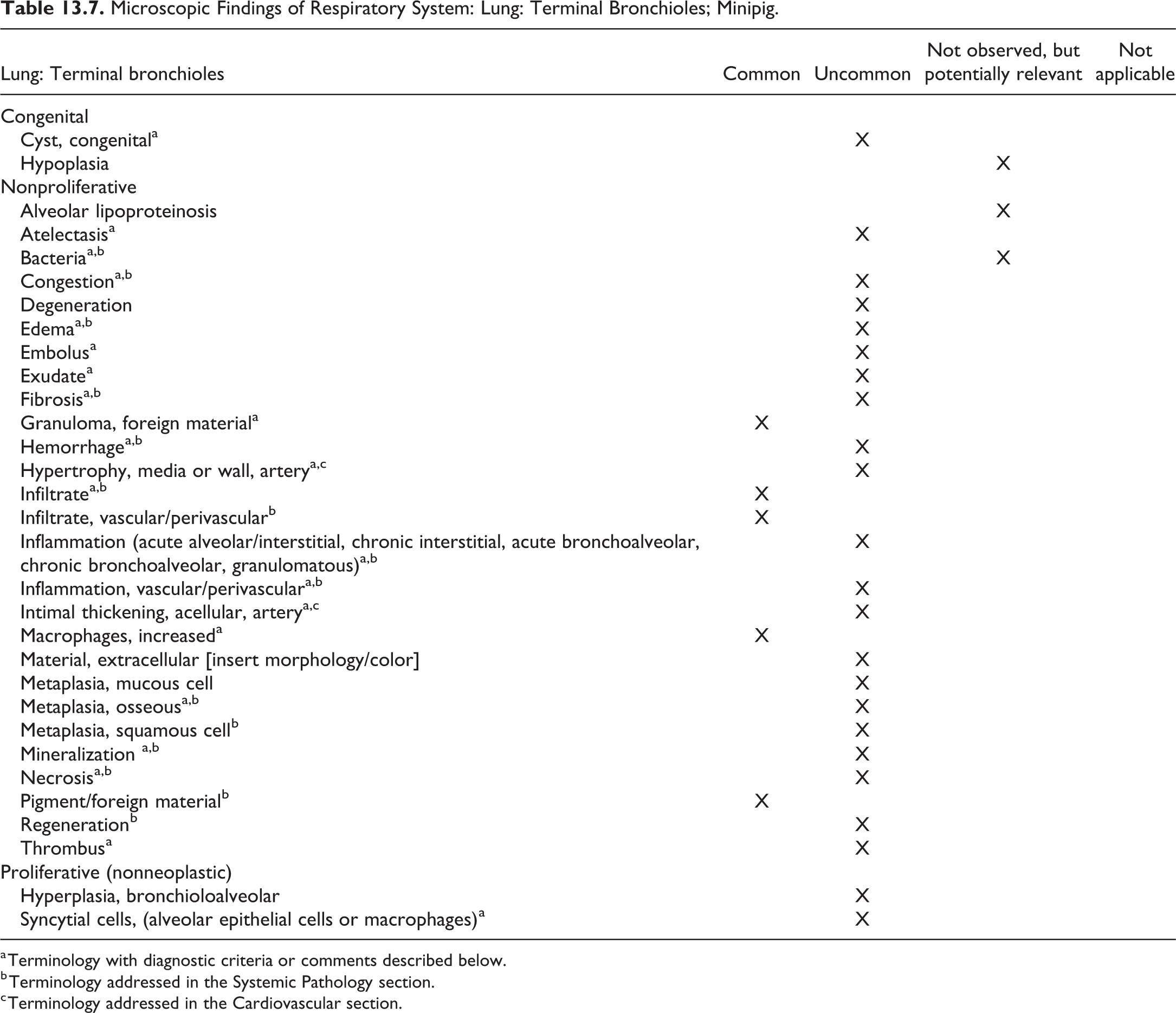
a Terminology with diagnostic criteria or comments described below.
b Terminology addressed in the Systemic Pathology section.
c Terminology addressed in the Cardiovascular section.
Microscopic Findings of Respiratory System: Lung: Alveoli: Minipig.
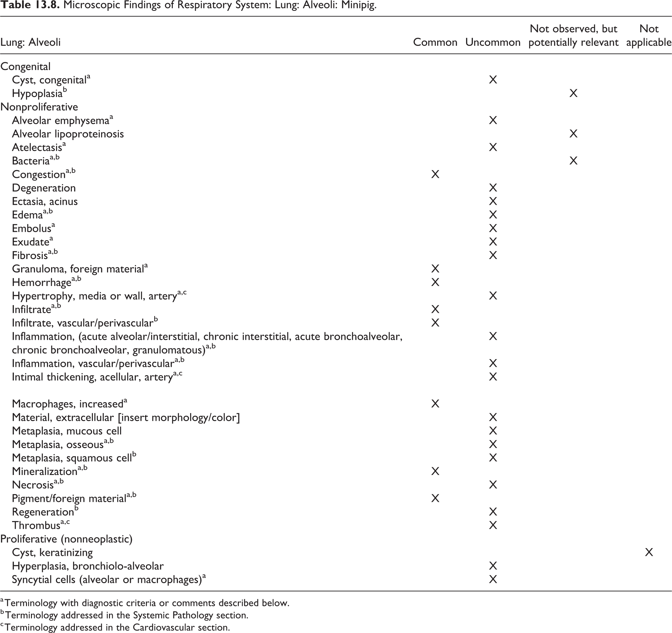
a Terminology with diagnostic criteria or comments described below.
b Terminology addressed in the Systemic Pathology section.
c Terminology addressed in the Cardiovascular section.
Microscopic Findings of Respiratory System: Pleura: Minipig.
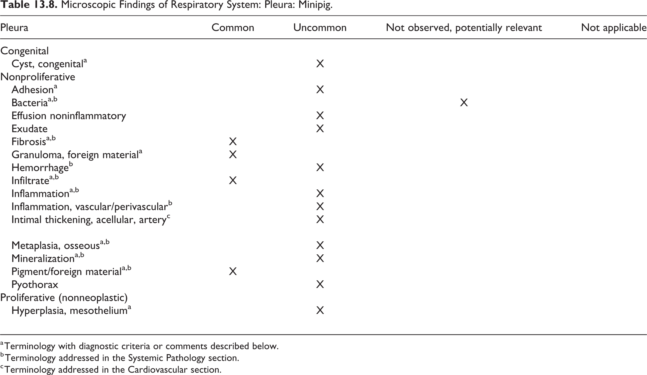
a Terminology with diagnostic criteria or comments described below.
b Terminology addressed in the Systemic Pathology section.
c Terminology addressed in the Cardiovascular section.
Adhesion: Pleura (Figure 13.1)
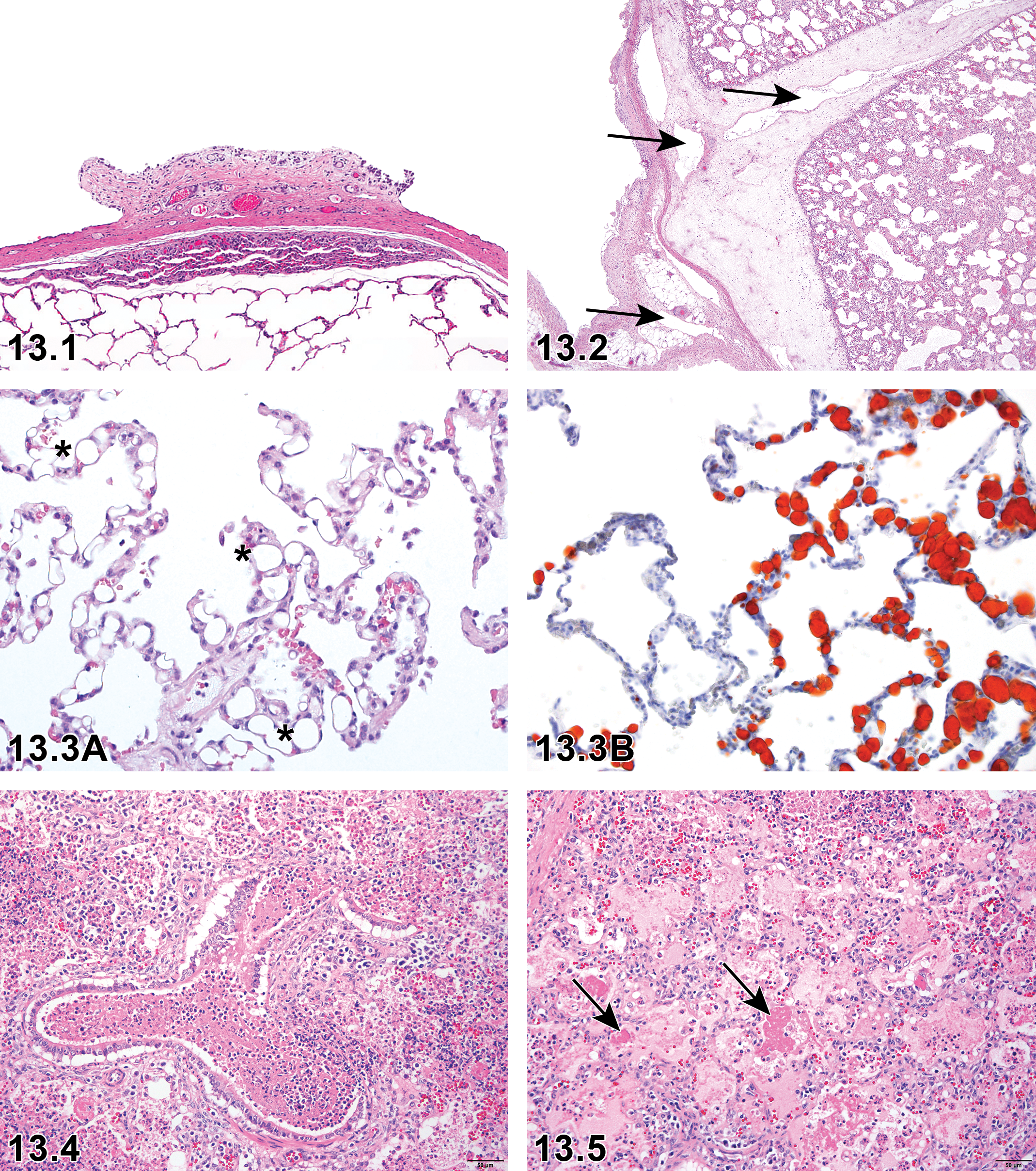
Pleura, adhesion. Pleural adhesion with subpleural inflammation (H&E).
Other terms
None.
Pathogenesis/Cell of origin
Adhesions can follow damage or inflammation of the visceral or thoracic pleura. These are frequently encountered in animals that have had parasites or respiratory infections. They also occur in SPF animals and have been infrequently observed in Hanford minipigs. 191
Diagnostic features
This lesion is typically diagnosed grossly, but irregularities of the pulmonary margin can be identified histologically. The adhesions present on the surface of the lung may contain abundant collagen (fibrosis) or inflammatory cells.
Comment
Adhesions are commonly seen following thoracic surgery (device models).
Alveolar emphysema: Alveoli
Comment
This lesion can be induced by mechanical ventilation, which may be relevant in medical device studies. Emphysema may also be induced in minipigs using inhaled LPS. 157
Atelectasis: Terminal bronchioles, alveoli
Comment
This lesion has been reported in the minipig. 13
Bacteria: Terminal bronchioles, alveoli, pleura
Comment
The rooting behavior of the pig results in inhalation of foreign materials, some of which may contain bacteria. 13
Cyst, congenital: Bronchioles, alveoli, pleura
Comment
This lesion has been reported in the minipig. 13
Congestion: Terminal bronchioles, alveoli
Comment
Congestion is common in alveoli, uncommon in terminal bronchioles and pleura, but may be found in all locations. 3,10,13,59 See Systemic section.
Edema: Terminal bronchioles, alveoli, pleura (Figure 13.2)
Comment
In rodents, edema is primarily a finding in bronchioles and alveoli, but in the pig, it also occurs in pleura and interlobular septa. 13,29,59 Lymphatics in interlobular septa and pleura can be widely dilated leading to misinterpretation of the lesion.
Embolus: Terminal Bronchioles, Alveoli (Figures 13.3a and 13.3b)
Comment
Several types of emboli may be found in the porcine lung: lipid, 40 thromboembolus, or bacterial. These are described more fully in the Cardiovascular section. Lipid may be used as a modifier for embolus in the pig. Embolus has been reported in the minipig. 7,13,59
Exudate: Terminal Bronchioles, Alveoli, Pleura
Comment
Small foci of exudate have been reported in the smaller airways of the minipig. 13,59
Fibrosis: Terminal Bronchioles, Alveoli, Pleura (Figures 13.4-13.6)
Comment
Fibrosis is uncommon in both the terminal bronchioles and alveoli but is common in pleura. 3,7,13,59 Fibrosis of the interlobular septa has been induced with radiation exposure in the minipig. 200
Granuloma, foreign material: Terminal bronchioles, alveoli (Figure 13.7)
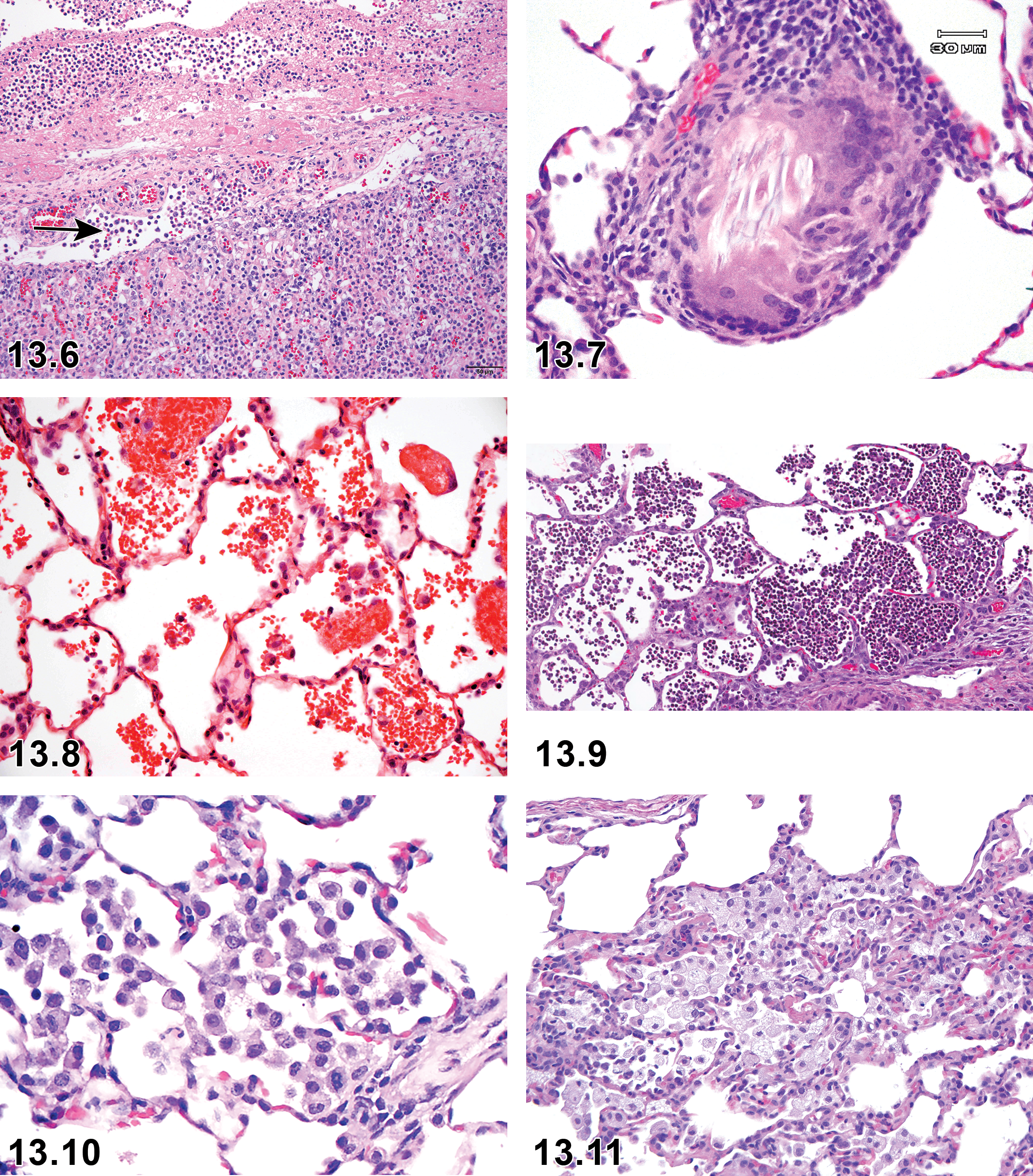
Lung, alveoli, granuloma, foreign material. There is foreign material surrounded by macrophages including multinucleated giant cells.
Pathogenesis/cell of origin
As above in section II. Often associated with inhaled foreign material or potentially parasites. 7,13,59
Hemorrhage: Terminal Bronchioles, Alveoli, Pleura (Figure 13.8)
Comment
Pulmonary hemorrhage is common in alveoli but uncommon in terminal bronchioles and pleura. 3,10,13,59 Hemorrhage in the lung may also be seen with thrombocytopenic purpura. See Systemic section.
Hypertrophy, media or wall, artery: Terminal bronchioles, alveoli
Comment
See Cardiovascular section. This lesion has been reported in the minipig. 7,13
Hyperplasia, mesothelium: pleura
Comment
Infiltrate: Terminal bronchioles, alveoli, pleura (Figure 13.9)
Comment
Inflammation, acute bronchioloalveolar: Terminal bronchioles, alveoli, pleura (Figure 13.10)
Comment
Inflammation, acute alveolar/interstitial: Alveoli
Comment
Inflammation, chronic bronchioloalveolar: Terminal bronchioles, alveoli, pleura
Comment
Inflammation, chronic interstitial: Terminal bronchioles, alveoli, pleura
Comment
Inflammation, granulomatous: Terminal bronchioles, alveoli, pleura
Comment
Inflammation, vascular/perivascular: Terminal bronchioles, alveoli, pleura
Comment
Other terms are arteritis/periarteritis, see Systemic and Cardiovascular sections. 10
Intimal thickening, acellular, artery: Terminal bronchioles, alveoli, pleura
Comment
See Cardiovascular section. This lesion has been reported in the minipig. 13
Macrophages, increased: Terminal bronchioles, alveoli (Figures 13.11 and 13.12)
Comment
Accumulations of alveolar macrophages have been reported to be associated with inhaled feedstuffs. 201 This is commonly seen as an incidental finding in pigs. 3,7,10,13,29,59,64 Accumulation of alveolar macrophages is also common in Hanford, Sinclair, and Yucatan breeds. Pigmented breeds such as the Yucatan contain pigment within some of their cells, and therefore, accumulated macrophages may be pigmented. 13 Occasionally, the macrophages are vacuolated and may be used with the modifier (vacuolated).
Metaplasia, osseous: Terminal bronchioles, alveoli, pleura
Comment
Mineralization: Terminal bronchioles, alveoli, pleura
Comment
This lesion has been reported in the minipig. 3,7,10,13,59 Also see Systemic section.
Necrosis: Terminal bronchioles, alveoli
Comment
This lesion has been reported in the minipig. 13
Pigment/foreign material: Terminal bronchioles, alveoli, pleura
Comment
Pigment is common in alveoli but uncommon in terminal bronchioles and pleura. Pigs often root and inhale dusts and inert materials. 3,13,59,201
Syncytial cells (alveolar epithelial cells or macrophages): Alveoli
Other terms
Giant cells, multinucleate cells.
Pathogenesis/cell of origin
Syncytial cells in the porcine lung may be formed from alveolar epithelial cells (typically viral in origin) but are more commonly formed from macrophages. Multiple activated macrophages may fuse to form syncytia.
Diagnostic features
Within alveoli, a single cell outline can be identified with multiple nuclei. Occasionally, foreign material may be found within or in close proximity to the cells.
Comments
This lesion has been reported in the minipig. 13
Thrombus: Terminal bronchioles, alveoli
Comments
This is uncommon as a spontaneous lesion but often seen following intravenous continuous infusion studies using vascular access ports. 7 See also Cardiovascular section.
Chapter 14. Skeletal System
Standard INHAND nomenclature for nonproliferative and proliferative skeletal system findings in rats and mice has previously been published 202 and will be used for the minipig as appropriate following the anatomical approach used for rodent species, with a focus on those findings that are likely to be encountered frequently and those that might be considered unique to the minipig. While the type and classification of bone and joint lesions is identical or very similar across species, their frequency in untreated individuals may differ substantially in the various species. The discussion herein is focused on spontaneous lesions observed in young minipigs (up to 2 years of age) used in nonclinical toxicity studies.
Skeletal tissues covered in this chapter are bones and joints. Teeth are not routinely examined in nonclinical toxicity studies and have thus been excluded from this chapter.
The standard bone samples examined during a toxicity study are distal femur and proximal tibia including the femorotibial joint. Additionally, the sternum is examined with the main purpose of evaluating the bone marrow. The preparation of the bones for optimal H&E sections is a time-consuming procedure in the nonrodent species. Although several attempts have been made to optimize the decalcification procedure, the traditional method often provides the best results.
Fractures of the bones are very rarely observed and joint lesions, often seen in conventional pigs, are uncommon in minipigs.
I. Bone
Lesions in the bones are uncommon in minipigs and only 2 lesions not described for rodents will be described herein: (1) serous atrophy of the adipose tissue in the bone marrow in Göttingen minipigs and (2) closed/partially closed/open growth plate.
Serous atrophy of the bone marrow does not specifically affect the bones but affects adipocytes and the hematopoietic tissue located in the bone marrow. This change is described in greater detail in the section on systemic conditions of minipigs. Serous atrophy of the adipocytes in the bone marrow occurs with a variable incidence and geographical variation, being more common in Europe than in North America. 13 Although the reason for this difference is uncertain, it may be related to different feeding regimes. Serous atrophy of the adipocytes in the bone marrow has not been described in Hanford or Yucatan minipigs. 13
Unlike the rodent, the physis of minipigs closes when the animal reaches maturity and growth ceases. The recording of physis closure may become necessary if a treatment causes or is considered to possibly cause a delay in physis closure or premature physis closure. In the untreated, healthy female Göttingen minipig, growth plate closure of femur starts at the age of 25 months and is complete by 42 months. 55 Another paper describes closure of the physis to be complete by 16 to 24 months. 203
Microscopic Findings of Skeletal System: Bone; Minipig.
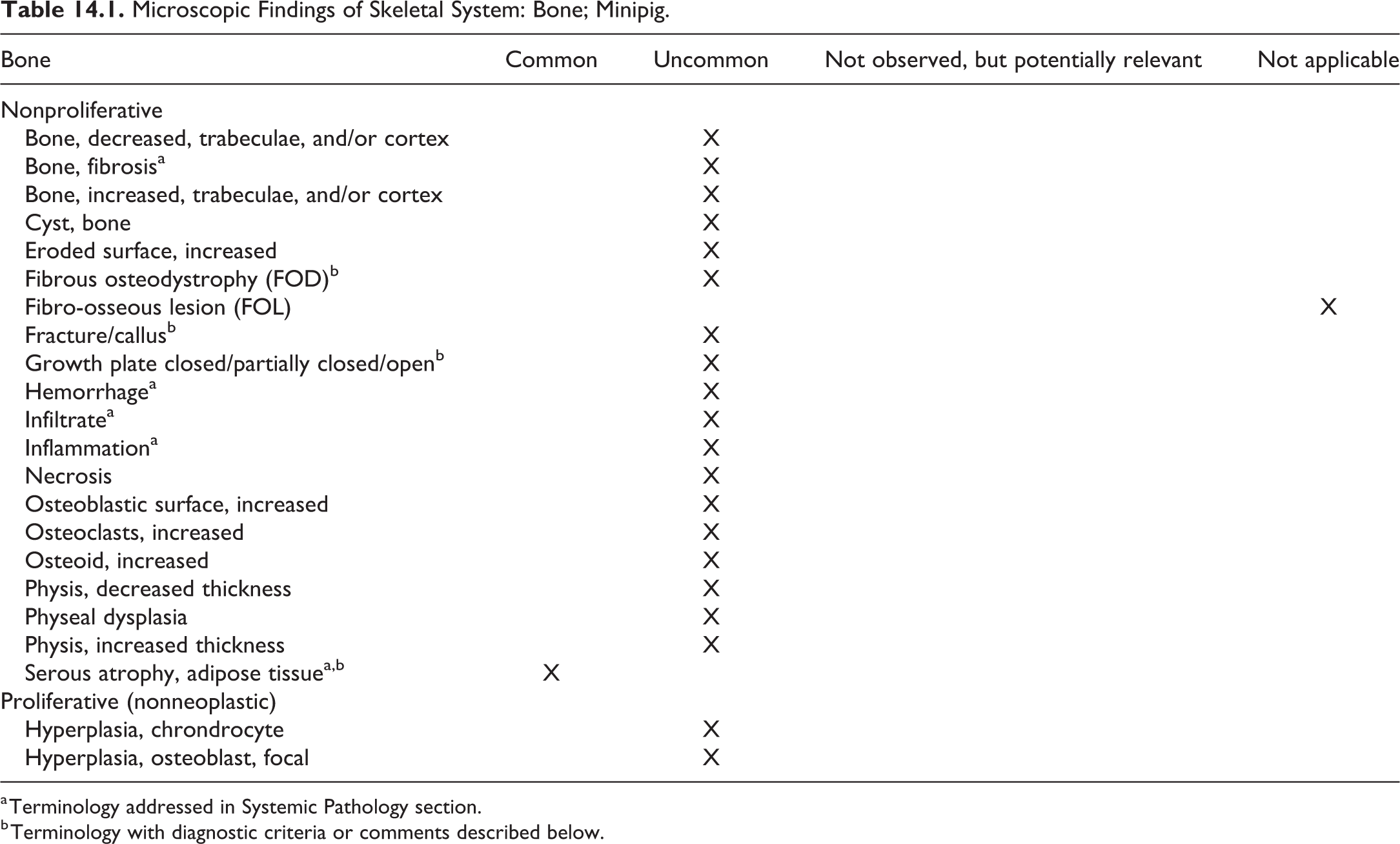
a Terminology addressed in Systemic Pathology section.
b Terminology with diagnostic criteria or comments described below.
Fibrous osteodystrophy; bone (Figures 14.2-14.6)
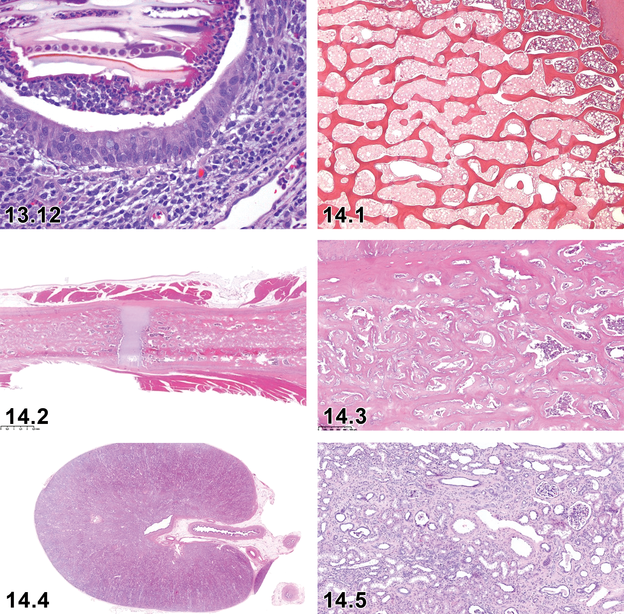
Minipig, bone marrow, serous atrophy of adipose tissue, H&E ×40.
Other term(s)
Renal osteodystrophy
Comment
A case of renal osteodystrophy in sternum and femur of an 8-month-old female Göttingen minipig has been described. 204 The bones exhibited high bone turnover associated with increased cortical porosity, higher trabecular thickness, and abnormal collagen texture (woven versus lamellar osteoid and bone). The pathogenesis in humans is described in the following reference. 205
Fracture/Callus
Comment
Fractures in minipigs are rare and most often associated with callus formation which can be used as a modifier.
Growth plate closed, partially closed, open; bone
Other term(s)
Physis closed, partially closed, open
Pathogenesis/cell of origin
Ossification of the growth plate
Diagnostic features
Thickness of the growth plate (physis) depends on the rate of growth.
When growth slows, the layers of the growth plate become narrower.
At the end of the process of bone growth, the cartilage of the growth plate is replaced by a bony scar, which is remodeled into trabecular bone.
Comment
Unlike the rodent, the growth plate (physis) of minipigs closes when the animal reaches maturity and growth ceases. The recording of growth plate closure may become necessary if a treatment causes or is considered to possibly cause a delay in growth plate closure or premature growth plate closure. In the untreated, healthy female Göttingen minipig, growth plate closure of femur starts at the age of 25 months and is complete by 42 months. 55 Another paper describes closure of the physis to be complete by 16 to 24 months. 203
Serous atrophy of adipose tissue (bone marrow) (Figure 14.1)
Other term(s)
Gelatinous bone marrow transformation.
Comment
The spontaneous lesion most commonly observed in the femur and tibia of control Göttingen minipigs is minimal to marked serous atrophy of the adipose tissue of the bone marrow which can occur as a physiological response to a normal degradation of adipose tissue under conditions where the minipig needs additional energy and the diet is insufficient to meet those needs. 3,7,13,31,41
Serous atrophy of the adipose tissue in the bone marrow is described in greater detail in the section on systemic conditions of minipigs.
II. Joint
Spontaneous lesions of the joints and synovium are rare in minipigs used in toxicity studies and just one change, degenerative joint disease (DJD), is described herein. The relative lack of changes in the joints of the minipigs is most likely related to the young age of the animals used in nonclinical toxicity studies.
Microscopic Findings of Skeletal System: Joint and Synovium; Minipig.

a Terminology with diagnostic criteria or comments described below
b Terminology addressed in Systemic Pathology section.
Degenerative joint disease; joint
Other term(s)
Osteochondrosis (OC); Osteochondritis dissecans, osteoarthritis
Pathogenesis/cell of origin
Degenerative joint disease is a noninfectious, degenerative condition involving the major weight bearing joints of the limbs, characterized by a gradual loss of articular cartilage leading to secondary bone changes. Degenerative joint disease is an uncommon observation in the minipig, and when it occurs, it is usually monoarticular. Osteoarthritis is the preferred term in humans, but as this term incorrectly implies an inflammatory origin, DJD is the more appropriate term based on the putative noninflammatory pathogenesis. Degenerative joint disease may be classified as primary, where no predisposing factors are present (mainly occurring in older animals) or secondary, associated with an underlying abnormality (malformation or deformation) in the joint or supporting structures leading to premature degeneration of the joint cartilage. 200,206 -208
Chondrocyte dysfunction leads to aberrant maintenance of the matrix in articular cartilage. The resulting alterations in the shape and continuity of cartilage surfaces permits the release of pro-inflammatory factors and promotes joint instability, thereby leading to secondary reactions in other joint components. A case of osteochondritis in the tarsal joint of a 3-month-old male minipig exhibiting lameness has recently been described in the literature. 29 The histopathology of this lesion included cartilage necrosis and cartilage flap formation.
Diagnostic features
Macroscopic findings may be absent.
Irregular contours of articular surfaces.
Thickening of articular cartilage.
Tinctorial changes in articular cartilage matrix (eg, focal loss of basophilia in H&E-stained sections or metachromasia in toluidine blue-stained sections).
Fibrillation of cartilage matrix; erosion/ulceration of cartilage.
Chondrocytes arranged in closely packed clusters/nests or “clones” (ie, proliferative response).
Chondrocyte degeneration and necrosis.
Microfissures in articular cartilage.
Gradual progression of the primary cartilage lesion through the entire articular surface leading to involvement of other joint structures and the following secondary morphologic changes:
Subchondral bone resorption.
Cyst/pseudocyst formation.
Hypertrophy/hyperplasia of synovial lining cells.
Fragments of degenerate cartilage lodged within the synovium often eliciting an inflammatory reaction.
Thickening and ossification of the joint capsule.
Differential diagnoses
Chondromucinous degeneration: An incidental, typically focal change affecting articular cartilage that does not progress to DJD.
Chapter 15. Soft Tissue
Standard INHAND nomenclature for nonproliferative and proliferative soft tissue findings in rats and mice has previously been published 209 and will be used for the minipig as appropriate following the anatomical approach used for rodent species, with a focus on those findings that are likely to be encountered frequently and those that might be considered unique to the minipig. The discussion herein is focused on spontaneous lesions observed in young minipigs (up to 2 years of age) used in nonclinical toxicity studies.
This chapter includes lesions of the soft tissues, adipose tissue, mesothelium, skeletal and smooth muscle, and mesothelium. Lesions of the synovium are described in the chapter on the skeletal system of minipigs. Proliferative neoplastic lesions are rarely observed as spontaneous background or test article-related findings in routine toxicity studies in minipigs due to the young age of animals and the short duration of toxicity studies relative to the minipig’s natural life span.
Soft tissue lesions observed in minipigs are generally identical to those observed in rodents and other domestic animals, as well as in humans.
Microscopic Findings of Soft Tissue, Adipose Tissue, and Mesothelium; Minipig.
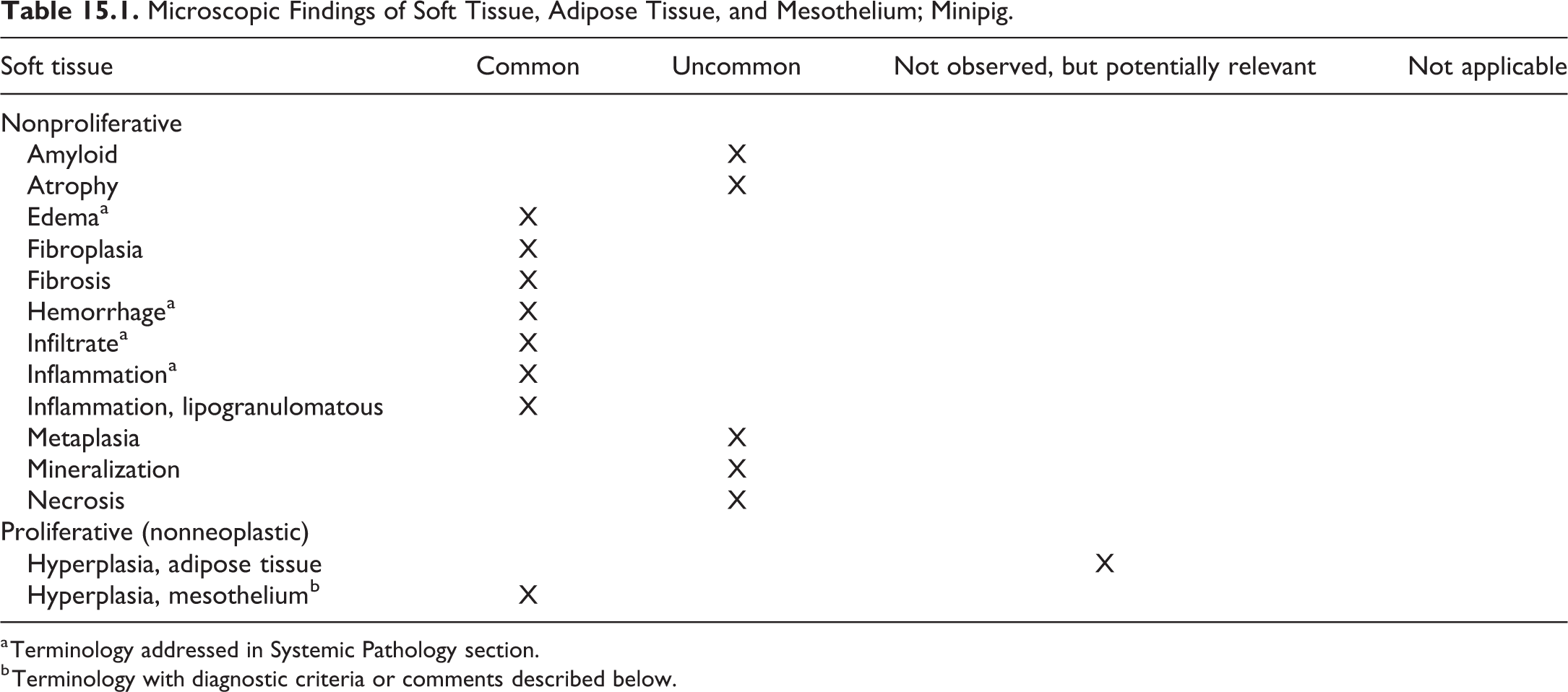
a Terminology addressed in Systemic Pathology section.
b Terminology with diagnostic criteria or comments described below.
I. Soft Tissue
Hyperplasia, mesothelium; mesothelium (Figures 15.1 and 15.2)
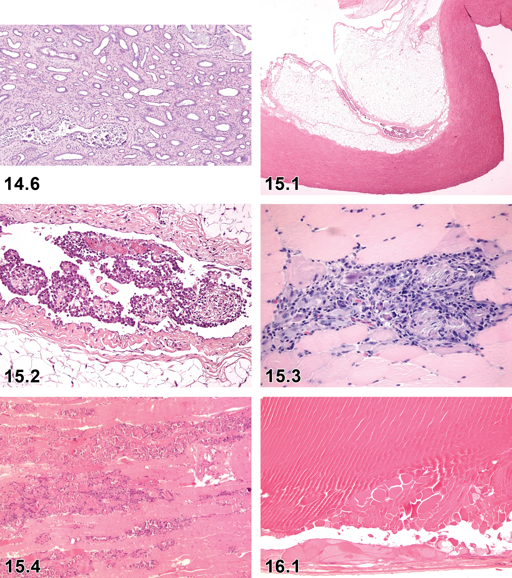
Minipig, kidney, mineralization, tubular, H&E ×10 (Courtesy Rosa Anna Manno, ERBC srl).
Other term(s)
Proliferation, mesothelial; proliferation, serosal
Pathogenesis/cell of origin
Hyperplasia of mesothelial cells of the pleura, pericardium, peritoneum, or tunica vaginalis. In the Göttingen minipig, the finding is most often minimal and focal, primarily observed in relation to the arch of the aorta or the atria of the heart. 3,7,13,31,41
Diagnostic features
Usually localized but may be diffuse.
Focal thickening or villous projections covered by relatively uniform cuboidal cells with little or no stratification, usually limited to 1 to 2 cells in thickness.
Lacks evidence of significant mitotic activity.
Little or no cellular atypia.
May possess a small fibrovascular core or stalk.
In the minipig, fibrosis and inflammation rarely accompany the changes.
Differential diagnoses
Mesothelioma, malignant: Is highly cellular and pleomorphic and shows extensive spread within the body cavities or infiltrates adjacent tissues.
Epithelioid mesothelioma, malignant: Exhibits poorly formed glandular structures or ill-formed glands. Sarcomatoid malignant mesothelioma consists of spindle-shaped cells.
Comment
In the Göttingen minipig, focal mesothelial proliferation is a common incidental finding thought to arise due to friction at the predilection sites (mainly the aortic arch and the atria of the heart).
Microscopic Findings of Skeletal and Smooth Muscle; Minipig.
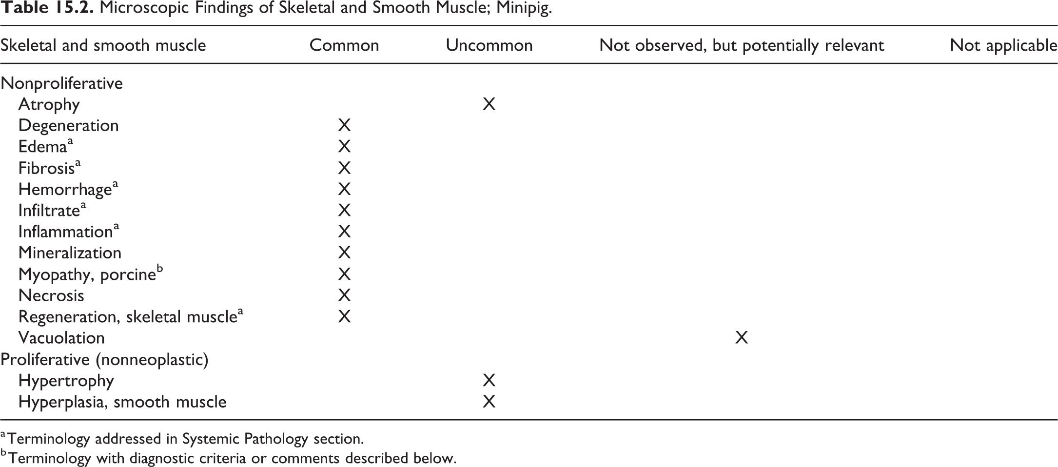
a Terminology addressed in Systemic Pathology section.
b Terminology with diagnostic criteria or comments described below.
II. Skeletal and Smooth Muscle
Myopathy, porcine; skeletal muscle (Figures 15.3 and 15.4)
Other term(s)
Myositis; degeneration/regeneration/necrosis/inflammation of myofibers; myopathic changes; cytopathic changes.
Pathogenesis/cell of origin
Myofibers of the skeletal muscle
Diagnostic features
Signs of degeneration, including vacuolation of fibers, split fibers, and hyaline changes.
Myofiber necrosis may be present.
Hemorrhage and edema may be observed.
Inflammatory cell infiltrates are often present. Macrophages are often a prominent feature of infiltrates.
Regenerating basophilic myofibers might occur.
Mineralization may be present.
Differential diagnoses
A variety of different pathological processes are embraced by this term. Clear distinction often requires special staining techniques to clarify the precise nature of the degenerative alterations.
Comment
In the minipig, “porcine myopathy” occurs as a common incidental background finding. The finding ranges from a focal minimal lesion to a multifocal severe lesion leading to the prescheduled euthanasia of the affected animal due to adverse clinical signs. The term covers the entire span of diagnostic features ranging from acute lesions dominated by necrosis, hemorrhage, edema, and mixed inflammatory cell infiltrates to the more chronic lesions characterized by basophilic regenerating myofibers, mineralization, and occasionally fibrosis. Some prefer to use the term “myositis” to describe this entity; however, as the inflammatory component is not always a prominent feature, the term “myopathy, porcine” is after long discussions in the working group considered the most appropriate. 3,7,13,31,41,210
Chapter 16. Special Senses
Standard INHAND nomenclature for nonproliferative and proliferative special sense findings in rats and mice has previously been published 211 and will be used for the minipig as appropriate following the anatomical approach used for rodent species, with a focus on those findings that are likely to be encountered frequently and those that might be considered unique to the minipig. This chapter covers the ocular system. The otic (ear) and olfactory systems are not routinely evaluated in toxicity studies in the minipig. The ocular system is subdivided into the eye and the surrounding glands. At necropsy, Davidson’s fixative is usually the fixative of choice for the eyes.
For a thorough examination of the ocular system, H&E-stained slides of samples from the eyes, eyelids, lacrimal glands (glandulae lacrimales), third eyelids/nictitating membranes with superficial accessory lacrimal glands (glandulae superficiales, nictitating glands), Harderian glands (glandulae profundae palpebrarum tertiarum), and optic nerves should be prepared. 26 However, for most routine toxicity purposes, it is sufficient to evaluate sagittal sections of the eyes as well as the optic nerves and not examine the glands. The lacrimal gland is located dorsal and caudal to the eye in the orbital cavity. The nictitating gland is closely associated with the third eyelid. Both the lacrimal and nictitating glands are thin, pale structures that are difficult to distinguish from the surrounding connective tissue. The Harderian gland is located medio-rostro-ventral to the eye in the orbital cavity, is pale brown in color, and has a lobulated structure.
The recommended eye section for a routine toxicity study is a superior–inferior sagittal section, passing through the optic nerve head, with proper orientation and free of artifacts. The cornea should be free of clefts or folds, and the corneal endothelial cells should not be vacuolated. Shattering or vacuolation of the lens should be avoided, and the lens should be correctly oriented in the globe, with the epithelium facing the cornea. Artifactual retinal separation or vacuolation is a common problem, and evaluation of photoreceptors demands sections no greater than 5 µm in thickness. Specialized ocular studies may require a different sectioning protocol, depending on the route of administration, the nature of the test article (aqueous solution, viscous depot, slow-release capsule, stem cells, subretinal device), or as a result of unusual ophthalmoscopic findings. Pathologists should be involved in determining the best protocol for a particular study.
The genesis of a good ocular section begins at necropsy. Rough handling of the eye at enucleation can induce retinal separation and optic nerve artifacts. The optic nerve should be transected at the level of the orbit to maximize the available nerve tissue. Extraocular tissues, including glands, should be trimmed off the globe prior to fixation to optimize the fixation of the retina and avoid separation; this also allows better visualization of the landmarks for subsequent trimming. Incision of the globe prior to fixation will compromise the architecture of the retina due to the reduced pressure inside the globe. Similarly, injection of fixative into the globe is not recommended. If orientation is critical, consider using tissue marking fluid or a suture to identify landmarks or the 12-o’clock position at the time of collection, as landmarks are more difficult to see in a fixed globe. Left and right eyes should be clearly differentiated to allow correlation with clinical findings.
A variety of fixation methods may be used. Since perfusion fixation frequently results in artifactual spaces in the retina, immersion fixation is probably the best option. Ensure that the eye is immersed in a sufficiently large volume of fixative (at least 10× the volume of the eye) as rapidly as possible to prevent autolytic change in the retina. Submersion in 10% formalin is frequently used in toxicology studies, but retinal preservation is often compromised.
Davidson’s solution gives better retinal fixation than 10% formalin, but prolonged exposure will result in artifacts associated with hardening of the lens and clefting and pseudoedematous changes in the cornea. For best results, the eyes should be transferred directly to ethanol in the tissue processor; consider washing and transferring to ethanol if a short delay (up to 10 days) is anticipated, but longer term archival of eyes warrants transfer to 10% formalin. Since Davidson’s fixation is associated with artifactual vacuolation in the optic nerve due to the ethanol content, a small section of optic nerve should be collected for fixation in 10% formalin for cross-section examination. Davidson’s fixation is compatible with immunohistochemistry techniques for many antigens and morphology is superior to that obtained with formalin fixation, but it is not suitable for electron microscopy evaluation.
Fixation with solutions containing glutaraldehyde (eg, Karnovsky’s solution) is suitable if electron microscopy is planned. To improve results, submerge the globe in the fixative for 2 hours to allow initial firming of the globe and then cut a small window in one side of the globe before continuing submersion for another 2 days. This fixative tends to cause distortion of the globe due to osmolality effects but generally gives good corneal, lens, and retinal morphology. Possible artifacts include fissuring of the lens, clefting of the cornea, and vacuolation of photoreceptors.
Trimming of globes requires a very sharp blade and should be performed with a single cut; a sawing action will result in retinal separation.
I. Eye
In general, spontaneous or treatment-related changes in the ocular system of the minipig are rare. The most commonly observed, albeit rare, finding of degeneration of the lens fibers is presented below.
Apart from degeneration of lens fibers, inflammatory cell infiltrates or inflammation are the most common (but with a low incidence) findings in the eyes of the minipigs. These are most commonly observed in the uvea, conjunctiva, and cornea. The diagnostic features, pathogenesis, and differential diagnosis for the minipig are similar to those described in the INHAND rodent manuscript for the special senses.
Microscopic Findings of Special Senses: Eye; Minipig.
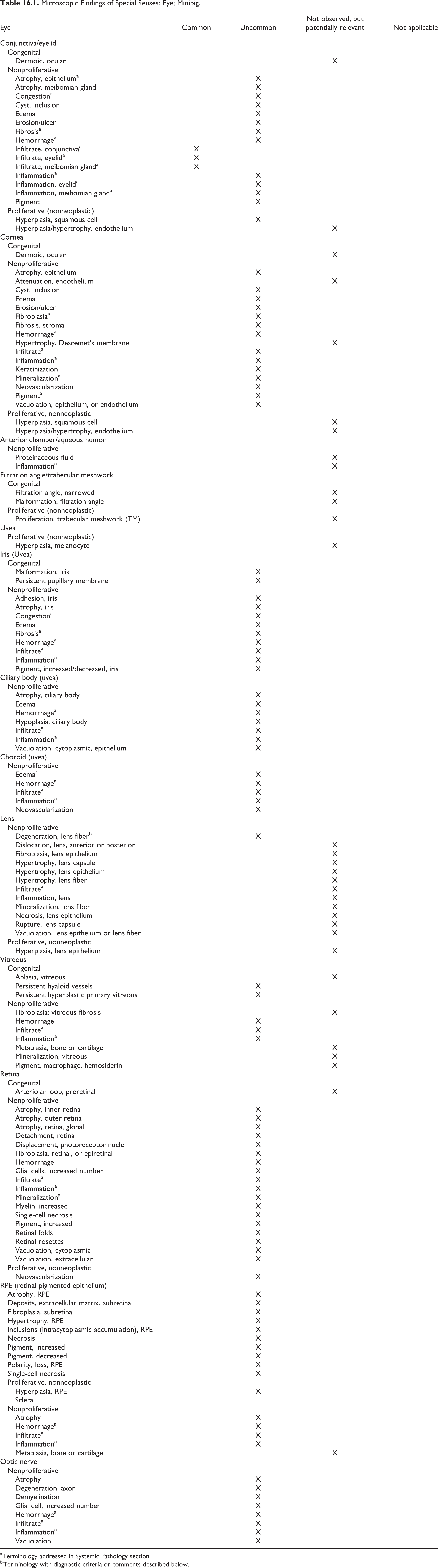
a Terminology addressed in Systemic Pathology section.
b Terminology with diagnostic criteria or comments described below.
Degeneration, fiber (lens) (Figures 16.1 and 16.2)
In the minipig, the lesion is most commonly observed in the posterior segment of the lens. 3,7,13,41
Microscopic Findings of Special Senses: Glands of the Eye; Minipig.
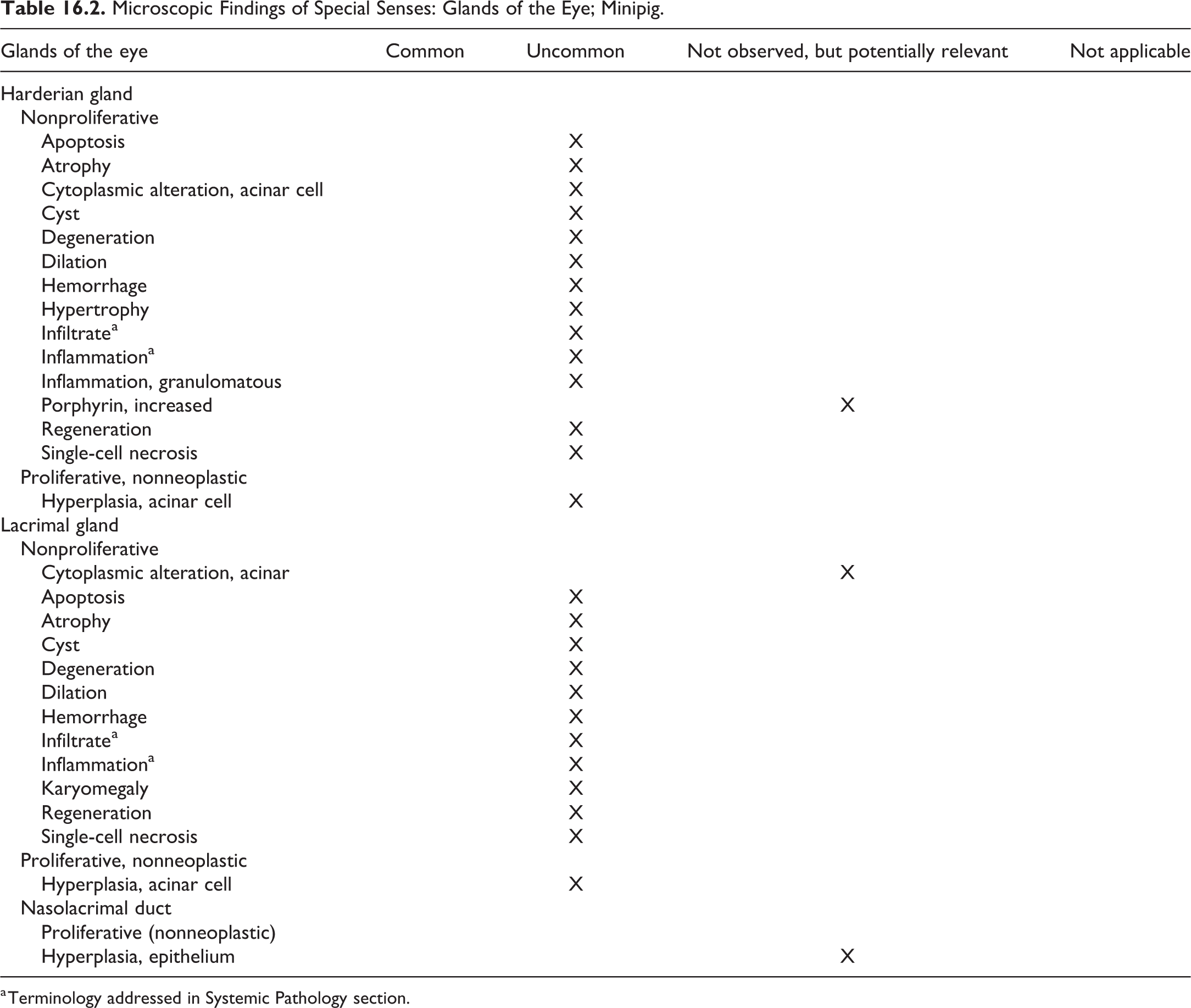
a Terminology addressed in Systemic Pathology section.
Chapter 17. Urinary System
For detailed general considerations on the urinary system, please refer to the rodent publication. 212 While the type and classification of kidney lesions is identical or very similar across species in the renal pelvis, ureter, urethra, and urinary bladder, the frequency may differ substantially.
Porcine kidneys have anatomical (multireniculate and multipapillate) and physiological characteristics similar to humans. 3
At necropsy, the kidneys are removed from the abdominal cavity after the adrenal glands and the gastrointestinal tract. The renal capsule should be removed and both kidneys weighed and measured. The renal cortex, outer zone of medulla, and inner zone of medulla (including the papilla) should be sampled from both kidneys. The urinary bladder should be incised and a tissue block of bladder wall that includes mucosa, muscle layer, and serosa should be prepared. If possible, include ureters or proximal urethra in the tissue block with bladder. 26
Microscopic Findings of Urinary System: Kidneys; Minipig.
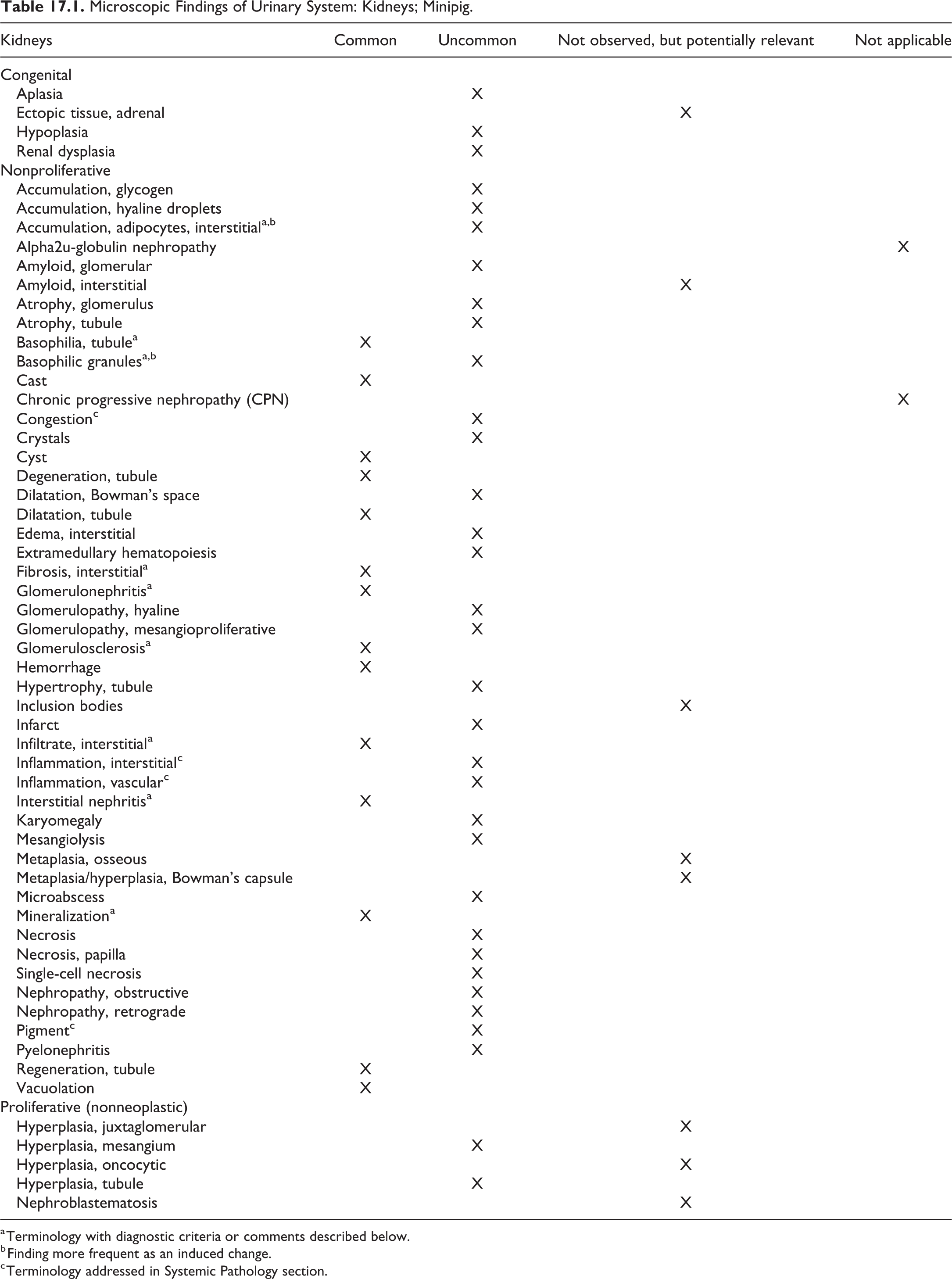
a Terminology with diagnostic criteria or comments described below.
b Finding more frequent as an induced change.
c Terminology addressed in Systemic Pathology section.
I. Kidney
Accumulation, adipocytes, interstitial, kidney (Figure 17.1)
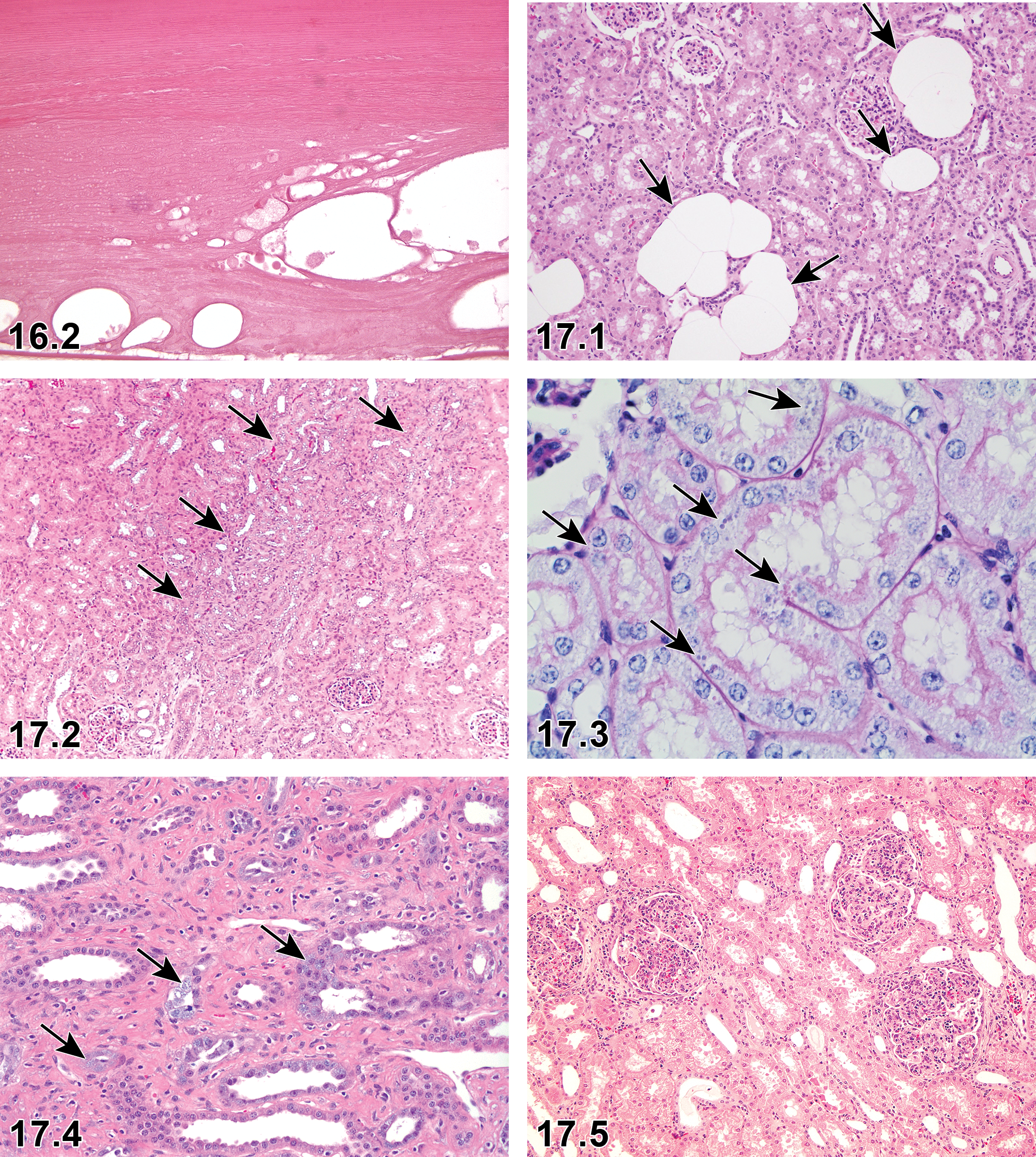
—Minipig: Eye, degeneration, fiber (lens), H&E ×40 (From Haschek and Rousseaux’s Handbook of Toxicologic Pathology. Haschek WM, Rousseaux CG, and Wallig MA, eds. 3rd ed. Elsevier; 2013. Figure 13.19, p 473, with permission).
Species
Minipig.
Comment
Interstitial accumulation of adipocytes was observed in obese ovariectomized Göttingen minipigs. 213
Basophilia, tubule, kidney (Figure 17.2)
Species
Minipig.
Comment
Tubule basophilia is an extremely common manifestation of mild nephrotoxicity in the minipig kidney and often occurs without vacuolation, single cell necrosis, or cell sloughing which are prominent with degeneration in other laboratory species having a similar degree of tubule injury. 13 Tubule basophilia may be accompanied by mitoses or cellular hypertrophy, which in other species would suggest regeneration. However, in minipigs, this is a stereotypic response to tubule injury and “basophilia, tubule” should be the preferred terminology with a diagnosis of “regeneration, tubule” reserved for lesions associated in off-dosing phases where resolution is inferred. With further renal injury, basophilia often progresses to tubule dilation and/or necrosis. Tubule basophilia can also occur as a spontaneous aging lesion in minipigs, which may involve thickened basement membranes or peritubule fibrosis, akin to chronic progressive nephropathy in rodent kidneys. This lesion is most commonly encountered in the corticomedullary junction, but its pathogenesis remains uncertain. 214
Basophilic granules, kidney (Figure 17.3)
Species
Minipig.
Comment
Basophilic granules may be present in the proximal tubular cells of minipigs exposed to or treated with antisense oligonucleotides. 215
Fibrosis, interstitial, kidney (Figure 17.4)
Species
Minipig.
Diagnostic features
Comment
These focal and mild lesions can be attributed to inflammation or tubular defects of resorption or secretion. To see these changes at moderate or marked levels is quite rare. 3 The lesion may be observed with hemorrhagic syndrome, which is further described in Chapter 2, Systemic Pathology. 19
Glomerulonephritis, Kidney (Figure 17.5)
Species
Minipig.
Diagnostic features
In spontaneous cases of glomerulonephritis, macroscopic enlargement, hyperemia, and adhesion may be observed grossly. On microscopic examination, the reported types of glomerulonephritis in minipigs are commonly membranous or membranoproliferative/mesangioproliferative. Tubular basophilia, hyaline casts, and interstitial hemorrhage, fibrosis, and cell infiltration have also been observed in association with glomerulonephritis. 7,13,216
Comment
In the streptozotocin (STZ)-induced diabetic model in minipigs, the glomeruli showed a widening of the mesangium with cell proliferation and increased matrix. In some glomeruli, hyaline droplets were observed in the parietal layer of Bowman’s capsule. Arterioles with hyaline thickening of the walls were present at the vascular pole. 57
The lesion may be observed with hemorrhagic syndrome, which is further described in Chapter 2, Systemic Pathology. 13,18,19,29,39
Glomerulosclerosis, kidney (Figures 17.6 and 17.7)
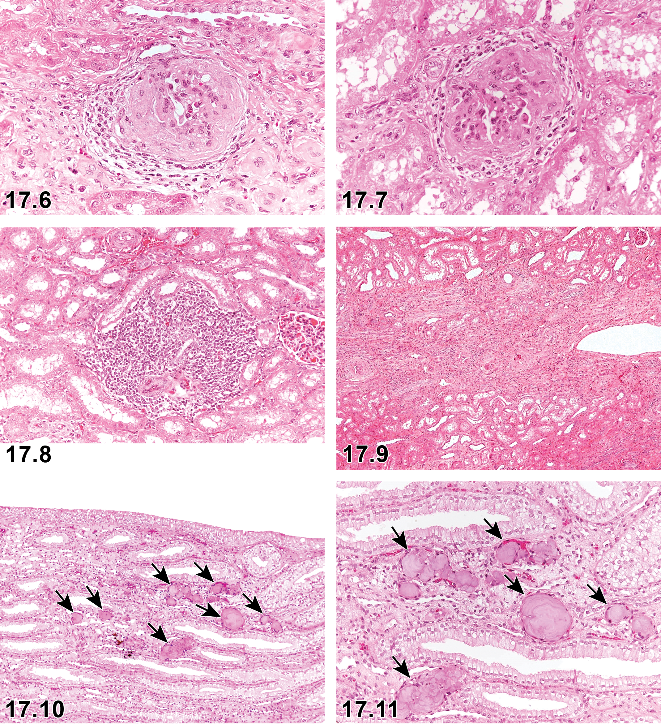
Kidney: Glomerulosclerosis. H&E ×40.
Species
Minipig.
Diagnostic features
Glomeruli undergo sclerotic changes, with loss of capillaries substituted by mesangial matrix.
Comment
Glomerulosclerosis in minipigs can occasionally be seen in a few glomeruli at the corticomedullary junction. 3,7,216
The lesion may be observed with hemorrhagic syndrome, which is further described in Chapter 2, Systemic Pathology. 13
Infiltrate, interstitial, kidney (Figure 17.8)
Species
Minipig.
Other term(s)
Infiltration interstitium; infiltrate, inflammatory cell, interstitium; cell infiltration, interstitium.
Modifier(s)
Type of inflammatory cell that represents the predominant cell type in the infiltrate: lymphocyte; plasma cell; mast cell; monocyte/macrophage; mononuclear; neutrophil; eosinophil; basophil; mixed.
Diagnostic features
Minimal to mild multifocal interstitial mononuclear cell infiltrates (predominantly lymphocytes) have been observed.
Comment
This lesion has been observed in Göttingen, Hanford, and Yucatan minipigs and is more commonly observed in females than in males in all breeds. Mononuclear cell and/or mixed inflammatory cell infiltrates are quite common findings. Focal infiltration of eosinophilic granulocytes has been observed in the pelvic region in a few cases. Lymphoplasmacytic and/or neutrophilic infiltrate was also reported in a Göttingen minipig with spontaneous glomerulonephritis. 3,7,10,13,31,41,216
Interstitial nephritis, kidney (Figure 17.9)
Species
Minipig.
Comment
Focal mild interstitial inflammation associated with fibrosis (interstitial nephritis), usually located in the renal cortex, can be seen occasionally. 3,7,13,31,216
These spontaneous changes are rarely reported at moderate or marked levels. 3
The lesion may be observed with hemorrhagic syndrome, which is further described in Chapter 2, Systemic Pathology. 3,19
Mineralization, kidney (Figures 17.10 and 17.11)
Species
Minipig.
Special techniques for diagnostics
Mineralization can be demonstrated with Alizarin Red S or Von Kossa stains.
Comment
Small foci of mineral deposits observed as amorphous deposits in H&E-stained sections, mostly in the papilla, are commonly seen in Göttingen minipig. 3,7,10,13,31,41
II. Renal Pelvis
Microscopic Findings of Urinary System: Renal Pelvis; Minipig.
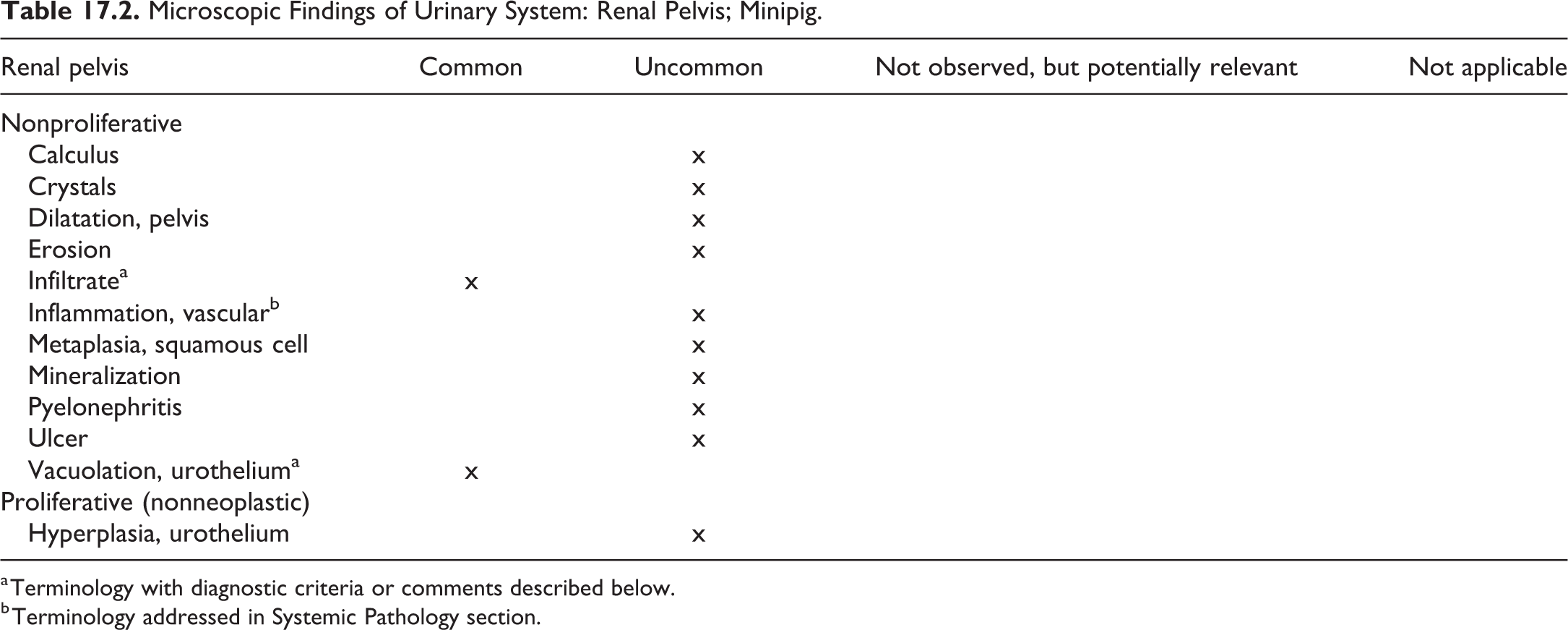
a Terminology with diagnostic criteria or comments described below.
b Terminology addressed in Systemic Pathology section.
Infiltrate, Renal pelvis, (Figures 17.12 and 17.13)

Renal pelvis: Infiltrate. H&E ×5.
Species
Minipig.
Other term(s)
Infiltration; infiltrate, inflammatory cell; cell infiltration.
Modifier(s)
Type of inflammatory cell that represents the predominant cell type in the infiltrate: lymphocyte; plasma cell; mast cell; monocyte/macrophage; mononuclear; neutrophil; eosinophil; basophil; mixed.
Comment
Focal infiltration of eosinophilic granulocytes has been reported in the pelvic region of Göttingen minipigs in a few spontaneous cases. 7
Vacuolation, urothelium, renal pelvis (Figures 17.14 and 17.15)
Species
Minipig.
Comment
Vacuolation of the urothelium in the kidney has been reported in Göttingen minipigs. This finding is very common and most often not recorded. 13
III. Ureter
Microscopic Findings of Urinary System: Ureter; Minipig.
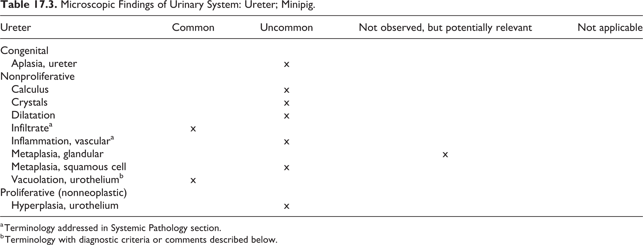
a Terminology addressed in Systemic Pathology section.
b Terminology with diagnostic criteria or comments described below.
Vacuolation, Urothelium, Ureter (Figure 17.16)
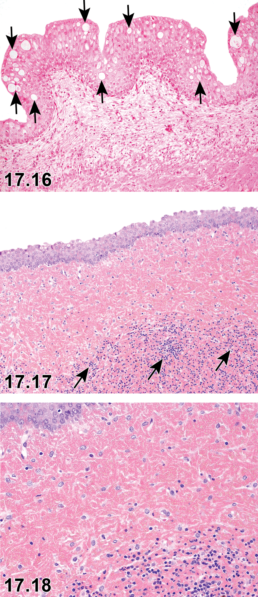
Ureter: Vacuolation, urothelium of ureter (arrows). H&E ×20.
Species
Minipig.
Comment
Vacuolation of the urothelium of the ureter has been reported in Göttingen minipigs. This finding is very common and most often not recorded. 13
IV. Urethra
Microscopic Findings of Urinary System: Urethra; Minipig.
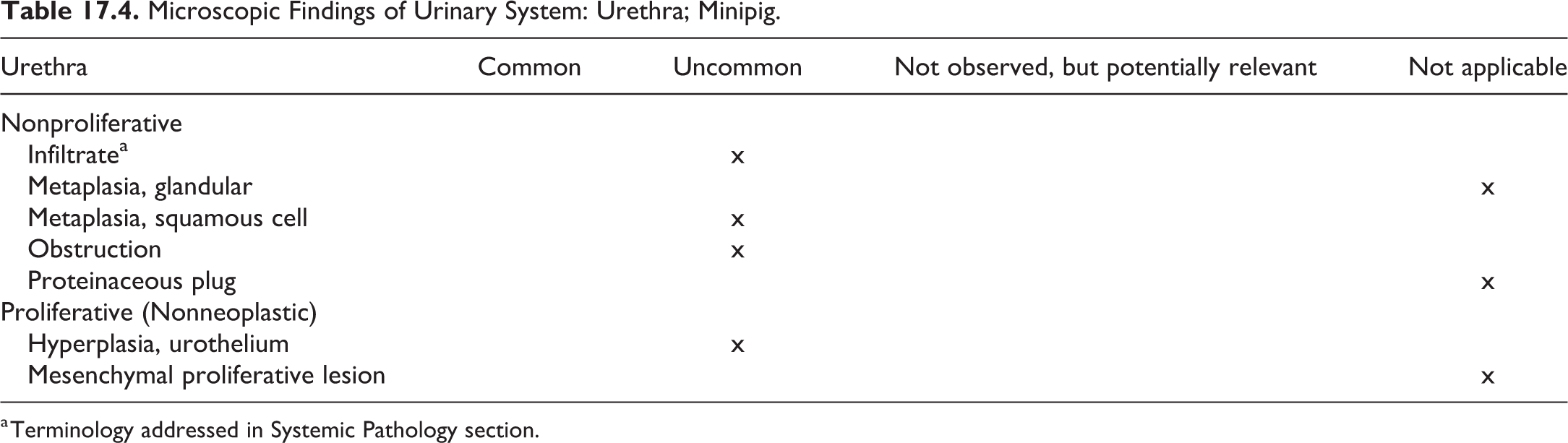
a Terminology addressed in Systemic Pathology section.
V. Urinary Bladder
Microscopic Findings of Urinary System: Urinary Bladder; Minipig.
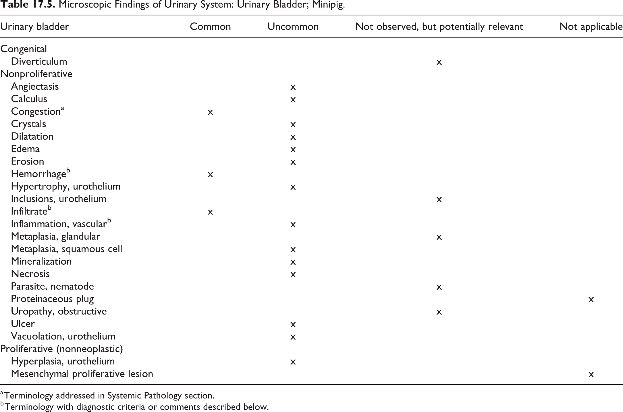
a Terminology addressed in Systemic Pathology section.
b Terminology with diagnostic criteria or comments described below.
Hemorrhage, Urinary bladder (Figures 17.17 and 17.18)
Species
Minipig.
Comment
The lesion may be observed with hemorrhagic syndrome, which is further described in Chapter 2, Systemic Pathology. 3,13,18,19,29
Infiltrate, urinary bladder (Figure 17.17)
Species
Minipig.
Other term(s)
Infiltration; infiltrate, inflammatory cell; cell infiltration.
Modifier(s)
Type of inflammatory cell that represents the predominant cell type in the infiltrate: lymphocyte; plasma cell; mast cell; monocyte/macrophage; mononuclear; neutrophil; eosinophil; basophil; mixed.
Comment
Small foci of lymphoid aggregates may occasionally be observed in the mucosa and submucosa, often in association with minor hemorrhage, which is often described as reddened mucosa at necropsy. 3,7,13,31
Inflammation of the urinary bladder can be observed on occasion, secondary to kidney pathology. Spontaneous infiltration of inflammatory cells has been reported in Göttingen and Yucatan minipigs. 13,31
Inflammation, vascular, Urinary bladder
Species
Minipig.
Comment
The lesion may be observed with hemorrhagic syndrome, which is further described in Chapter 2, Systemic Pathology. 3,7
Footnotes
Authors' Note
All procedures used to prepare macroscopic and microscopic images of animal specimens for this article were performed in accordance with regulations and established guidelines for humane treatment of research animals and were reviewed and approved in advance by an Institutional Animal Care and Use Committee.
Acknowledgments
The authors wish to express their thanks to the BSTP, ESTP, JSTP, and STP membership for the comprehensive reviews, excellent comments, and helpful edits. The authors also thank Rupert Kellner for manuscript review and Beth Mahler for image editing.
Declaration of Conflicting Interests
The author(s) declared no potential, real, or perceived conflicts of interest with respect to the research, authorship, and/or publication of this article.
Funding
The author(s) received no financial support for the research, authorship, and/or publication of this article.