
Abstract
The use of three-dimensional (3-D) in vitro culture systems (spheroids, organoids) in biomolecular and drug discovery research has become increasingly popular. The popularity is due, in part, to a diminished reliance on animal bioassays and a desire to develop physiologically relevant cell culture systems that simulate the in vivo tissue microenvironment. Most evaluations of 3-D cultures are by confocal microscopy and high-content imaging; however, these technologies do not allow for detailed cellular morphologic assessments or permit basic hematoxylin and eosin histologic evaluations. There are few studies that have reported detailed processes for preparing 3-D cultures for paraffin embedding and subsequent use for histochemical or immunohistochemical staining. In an attempt to do so, we have developed a protocol to paraffin-embed human liver spheroids that can be sectioned with a microtome and mounted onto glass slides for routine histochemical and immunohistochemical staining and light microscopic evaluations.
Keywords
In an effort to address the replacement, reduction, and refinement effectively in toxicology and toxicologic testing, there has been a push toward decreasing the number of animals used in testing and research programs. The refinement of end points to derive maximum information under ethical and humane conditions has moved toxicologic assessments toward replacement of animal bioassays with lower species or in vitro systems for testing (MacArthur Clark 2017). To this end, there has been a gradual transition from methods predominantly involving mammalian screens toward in vitro and nonmammalian models. The use of higher-throughput methods for predicting the toxicological impacts of environmental agents is a major effort of the Tox21 testing program, a U.S. federal collaboration that involves the National Toxicology Program at the National Institute of Environmental Health Sciences, National Institutes of Health (NIH), and other federal agencies. In addition, a better understanding of human health effects and disease by using human cell lines and three-dimensional (3-D) organoid or spheroid cultures is underway as part of phase III of the Tox21 testing program (Tox21 2018). Differentiated HepaRG spheroids are used as an in vitro model system as part of a National Toxicology Program effort to evaluate the usefulness of 3-D human liver cultures in predicting chemical-induced toxicity on hepatic molecular pathways and to evaluate their ability to maintain function, metabolic enzyme capabilities, and cellular structures in culture.
Two-dimensional (2-D) culture systems have been used for years, and some studies have shown that 2-D cultures are limited in their representation of a cell’s physiological capabilities and in vivo microenvironment (Edmondson et al. 2014). As a result, 3-D culture systems (spheroids, organoids) are being explored and have become increasingly valuable tools in predictive toxicology research, mechanistic studies, and drug development and discovery (Lovitt, Shelper, and Avery 2014; Nath and Devi 2016; Ramaiahgari et al. 2017). Many of the 3-D culture systems are composed of heterogeneous cell populations and have a structural complexity that shows physiologically relevant cell–cell or cell–matrix interactions and gene expression or cell signaling events similar to that of the in vivo microenvironment (Nath and Devi 2016). The use of human and rodent 3-D culture systems has increased in the analysis of therapeutic agents and other pharmaceutical molecules in many drug discovery research environments. In laboratories worldwide, 3-D culture use ranges from the study of cancer progression to the evaluation of drug metabolism and efficacies (Ware et al. 2016). In cancer research, 3-D culture systems allow investigators to replicate the morphological structure of in vivo tumors and the biological interactions within complex tissues. Some features, such as gene expression, spatial architecture, secretion of soluble mediators, drug-resistant mechanisms, and physiological responses of solid tumors, are mimicked in 3-D cultures (Costa et al. 2016).
Despite the advantages of 3-D cultures in research, there is a need for the development of histologic techniques that can be easily followed and applied in a routine histology or pathology laboratory, which allows for detailed microscopic visualization and evaluation of complex 3-D structures. There are few documented protocols in the literature that provide comprehensive information on methods to fix and prepare spheroids or organoids for paraffin embedding and sectioning (de Hoogt et al. 2017; Pinto, Jacobsen, and Horwitz 2011; Stock et al. 2016). The technique proposed in our article is novel in that it is quick and straightforward and does not require the use of a matrigel/histogel “sandwich” technique often used for paraffin embedding of 3-D cultures (Stock et al. 2016; Pinto, Jacobsen, and Horwitz 2011). Our technique does not involve mechanical scraping of Matrigel/cells from chamber slides that could damage the morphology of the spheroids or prestaining of samples before processing and is amenable to collection of samples from 96- or 384-well plates used for high-throughput screening (HTS) platforms. To address the issue of optimal histologic visualization of 3-D cultures, without the use of several steps of Matrigel/cell and histogel layering or modifications of the “sandwiching” technique for HTS platforms (Stock et al. 2016), we have developed a detailed, yet simple protocol for fixing, processing, and embedding 3-D liver cultures for histochemical and immunohistochemical staining.
Material and Method
3-D Cultures
Differentiated HepaRG cells (Triangle Research Labs, Durham NC, recently acquired by Lonza, Walkersville, MD) were thawed and seeded at 1,000 cells per well onto round bottom, ultra-low attachment plates (Corning Inc., New York, NY, catalog no. 4520/96-well) in 50 µl Williams E medium (ThermoFisher, Waltham, MA) supplemented with induction additive MHPIT (Media - HepaRGTM - Pre-Induction & Tox Additive; Triangle Research Labs). Some spheroids were grown on Matrigel® (Corning, New York, NY) prepared from a stock concentration of 5 mg/ml. Culture media were refreshed every 2 to 3 days by removing 30 µl (from 50 µl total medium per well) and adding fresh 30 µl medium using a Viaflo semiautomated liquid handler (Integra Biosciences, Hudson, NH). The spheroids were maintained for up to 28 days in culture incubated at 37°C with 5% CO2.
Sample Collection, Fixation, Paraffin Embedding, and Sectioning
The spheroid samples (3 in culture media alone and 3 in culture media + Matrigel) were sequentially prepared and processed as follows (Figure 1). HepaRG spheroid suspensions in media were collected from plates, placed into a microcentrifuge tube, and spun at 1,000 rpm for 2 min to form a pellet. The media supernatant was decanted, and the resultant pellet was washed in phosphate-buffered saline (PBS) and centrifuged at 1,000 rpm for 2 min to reform a pellet. The pellet was fixed in 10% neutral buffered formalin (NBF) for 10 min, followed by removal of fixative and replacement with PBS, and stored at 4°C until the time of sample preparation for processing and paraffin embedding. At processing and embedding, the spheroid samples were centrifuged at 1,000 rpm for 5 min in microcentrifuge tubes with PBS. The PBS was removed, and a 1% agarose solution (Agarose I, VWR#0710, VWR Corporation, Radnor, PA) was pipetted into the microcentrifuge tube containing the pellet, gently dispersed throughout the agar, and allowed to solidify for at least 1 hr at room temperature. The spheroid pellets in agarose were removed from the microcentrifuge tubes intact. One pellet of each type of suspension (media alone or media + Matrigel) was sliced longitudinally and placed into a tissue cassette cut side down (see Figure 1). Another pellet of each suspension was sliced transversely near the tip of the pellet. The pellets were processed on a tissue processor (Leica ASP6025, Leica Microsystems GmbH, Wetslar, Germany) and infiltrated with 70% and 80% ethanol for 10 min each, followed by 95% ethanol for 15 min and three changes of 100% ethanol for 10 min each. Next, the pellets were processed in three changes of xylene, 10 min, followed by three baths of paraffin: 15 min in the first bath and 25 min in each of the remaining two paraffin baths and then placed (embedded) in a mold with paraffin. Sections were cut at 5 µm and mounted onto hydrophilic glass slides.
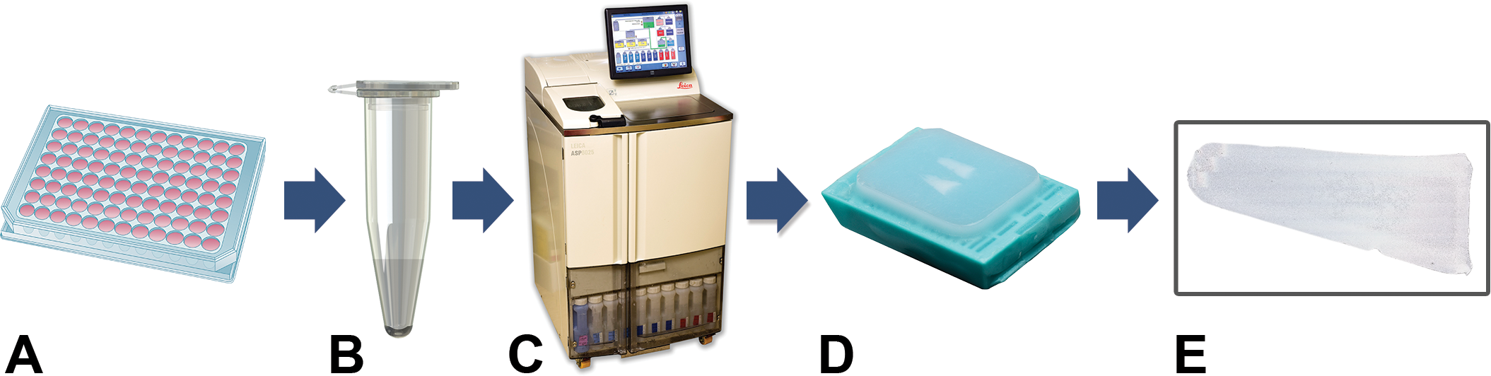
Preparation and processing of liver spheroids. (A) Spheroids in media were pipetted from a 96-well plate and placed into a microcentrifuge tube and spun to form a pellet. (B) The pellet was washed in phosphate-buffered saline (PBS), centrifuged to reform a pellet, and fixed in 10% neutral buffered formalin (NBF) followed by replacement with PBS and stored at 4°C until time of processing. At the time of processing and embedding, 1% agarose was added to the microcentrifuge tube with pellet and allowed to solidify at room temperature. (C) Pellets in agarose cast were sectioned longitudinally, and halves placed into a cassette flat side down, and processed in a tissue processor (D) followed by paraffin embedding. (E) Paraffin-embedded spheroids were sectioned with a microtome and mounted onto glass slides.
Histochemical Stains
Spheroid sections (5 μm) were deparaffinized in xylene for 2 changes at 6 min each. Next, they were hydrated through a series of 100% and 95% graded ethanol to distilled water and stained with hematoxylin and eosin (H&E), periodic acid–Schiff (PAS), or PAS after treatment with diastase to remove glycogen. Formalin-fixed paraffin-embedded human liver tissue slides were used as positive controls for histochemical stains.
Immunohistochemical Stains
Liver spheroids were immunohistochemically stained for Ki-67 (proliferation marker), cytokeratin 19 (CK19, epithelial bile duct marker), multidrug resistance–associated protein 2 (MRP2, a canalicular multispecific organic anion transporter), and carcinoembryonic antigen (CEA, a bile canaliculus marker) as outlined in Table 1. Briefly, formalin-fixed, paraffin-embedded HepaRG spheroids and control liver tissue sections were deparaffinized in xylene and hydrated through a series of 100% and 95% graded ethanol. Antigen retrieval for Ki-67 and CK-19 was performed by subjecting the slides to heat at 110°C and pressure at 6.1 psi in a Decloaker® pressure chamber (Biocare Medical, Concord, CA) using 10 mM pH 6.0 1× antigen decloaker citrate buffer, or for MRP2 using ethylenediaminetetraacetic acid, pH 8.0 buffer retrieval solutions (Biocare Medical) for 15 min. The slides were then allowed to cool at room temperature for 10 min. Antigen retrieval for CEA was with ready-to-use proteinase K for 15 s at room temperature (Dako, Santa Clara, CA). Endogenous peroxidase activity was quenched using 3% hydrogen peroxide following antigen retrieval. Nonspecific sites were blocked using normal horse (Vector Laboratories, Burlingame, CA) or donkey serum (Jackson Immunoresearch Laboratories, Inc., West Grove, PA), respective to the secondary antibody used, followed by incubation with an avidin/biotin blocking kit (Vector Laboratories) or immediate incubation with the primary antibody. Liver spheroids were incubated with the primary antibodies listed in Table 1 for 1 hr at room temperature followed by incubation with the appropriate secondary antibody for 30 min at room temperature. Negative controls were incubated with normal rabbit IgG (Sigma-Aldrich, St. Louis, MO), normal mouse IgG1 (BD Biosciences, San Jose, CA), or purified mouse IgG2a (BD Biosciences) diluted to the same concentration as the primary antibody (see Table 1). Antigen–antibody complexes were labeled using an anti-rabbit Ig ImmPRESS kit (Vector Laboratories) or an avidin–biotin affinity system (Vectastain Elite ABC kit, Ready-to-Use; Vector Laboratories) and visualized with 3,3′-diaminobenzidine chromogen (Dako). The tissue sections were counterstained with hematoxylin, dehydrated through a graded series of 95% and 100% ethanol, cleared in xylene, and cover-slipped. Formalin-fixed paraffin-embedded human liver tissue sections on slides were used as positive controls.
Immunohistochemical Stains.
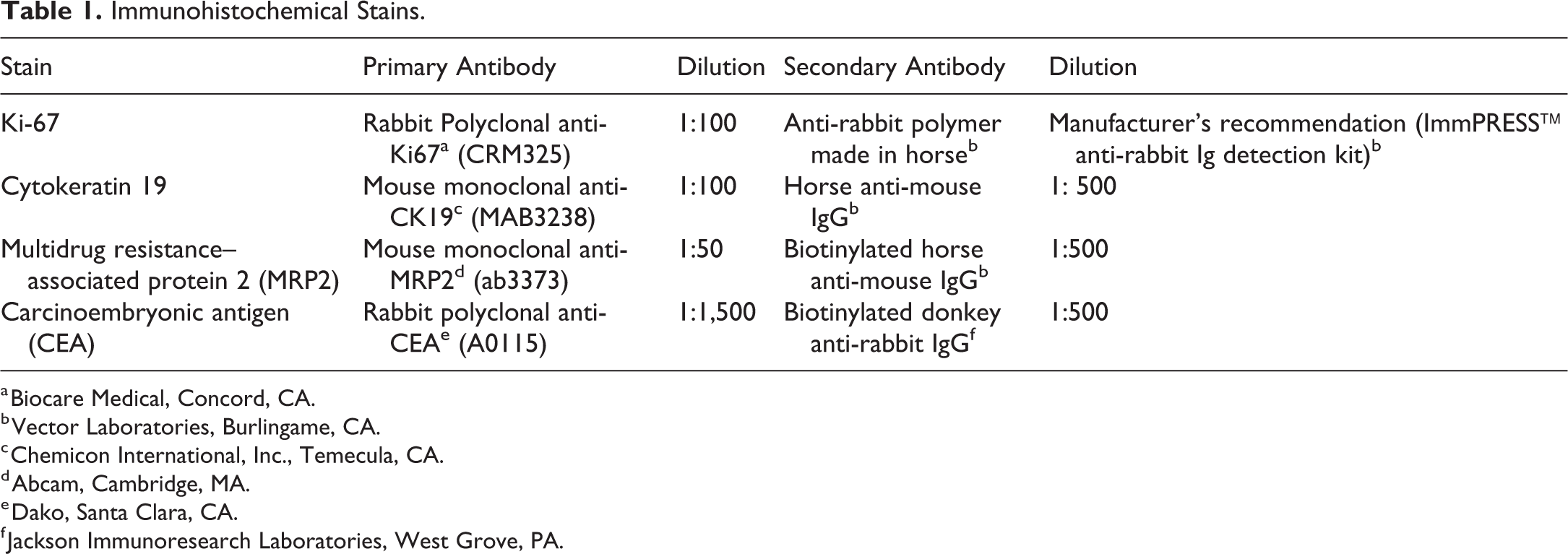
a Biocare Medical, Concord, CA.
b Vector Laboratories, Burlingame, CA.
c Chemicon International, Inc., Temecula, CA.
d Abcam, Cambridge, MA.
e Dako, Santa Clara, CA.
f Jackson Immunoresearch Laboratories, West Grove, PA.
Results
Sections and Histochemical Stains
The HepaRG spheroid pellets were successfully paraffin embedded and sliced longitudinally into 5 µm sections and mounted onto hydrophilic slides (see Figure 1). We determined that centrifugation of the spheroids or the collection of spheroids in a Matrigel matrix did not affect the ability to embed them in paraffin or alter the morphology of the spheroids, although the Matrigel could be visualized on the slide in addition to the agarose (Figure 2). We also determined that more spheroids were present on the slide when the pellet was longitudinally sectioned versus transversely sectioned near the tip of the pellet. The sections were easily mounted on hydrophilic slides and stained with H&E (Figure 3), PAS, and PAS with diastase (Figure 4).
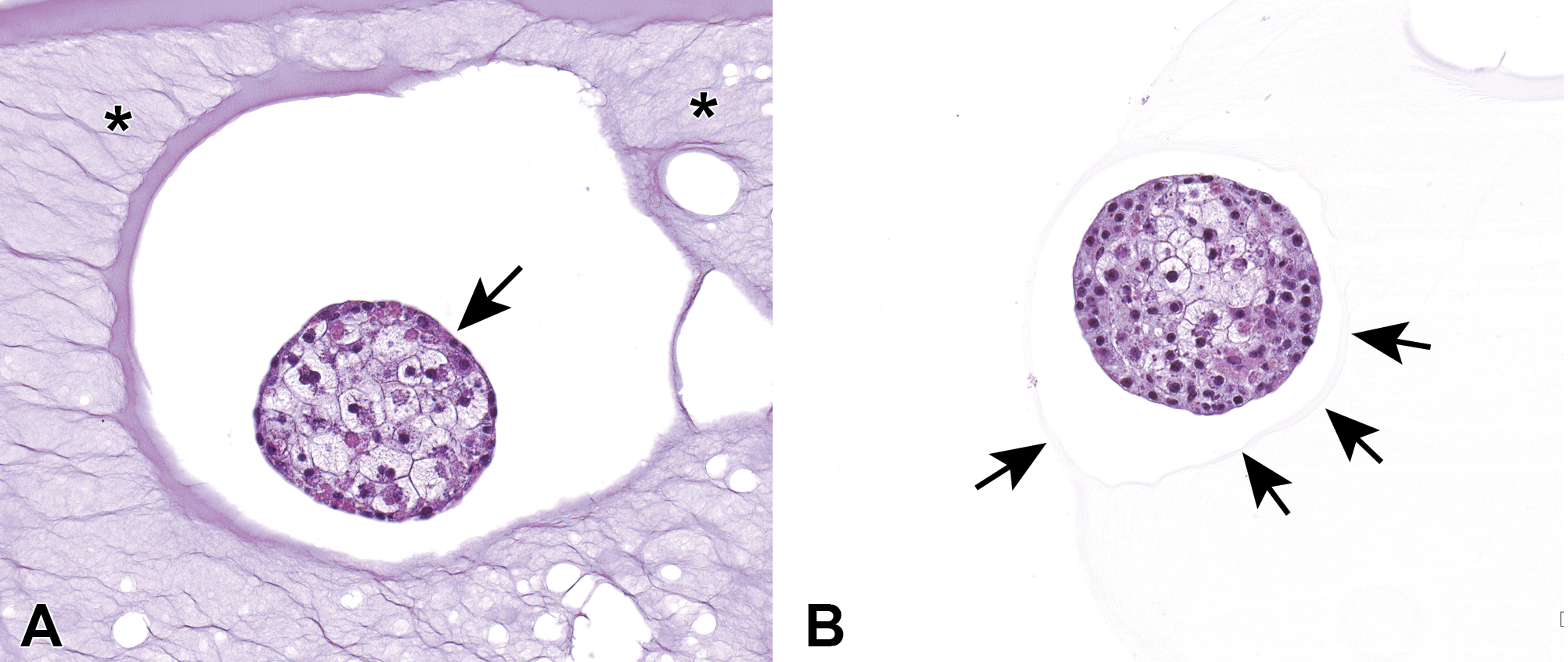
Liver spheroids with or without Matrigel. (A) Liver (HepaRG) spheroid grown in Matrigel. Matrigel (asterisks) surrounding the spheroid (arrow); however, the Matrigel did not affect the morphology of the spheroid. (B) A liver spheroid without Matrigel. See the thin layer of surrounding agarose (arrows).

Hematoxylin and eosin (H&E)-stained spheroids. (A) Liver (HepaRG) spheroids mounted on a hydrophilic slide and stained with H&E. (B) Higher magnification of four liver spheroids shown in a box in A. (C) Higher magnification of one liver spheroid shown in B.
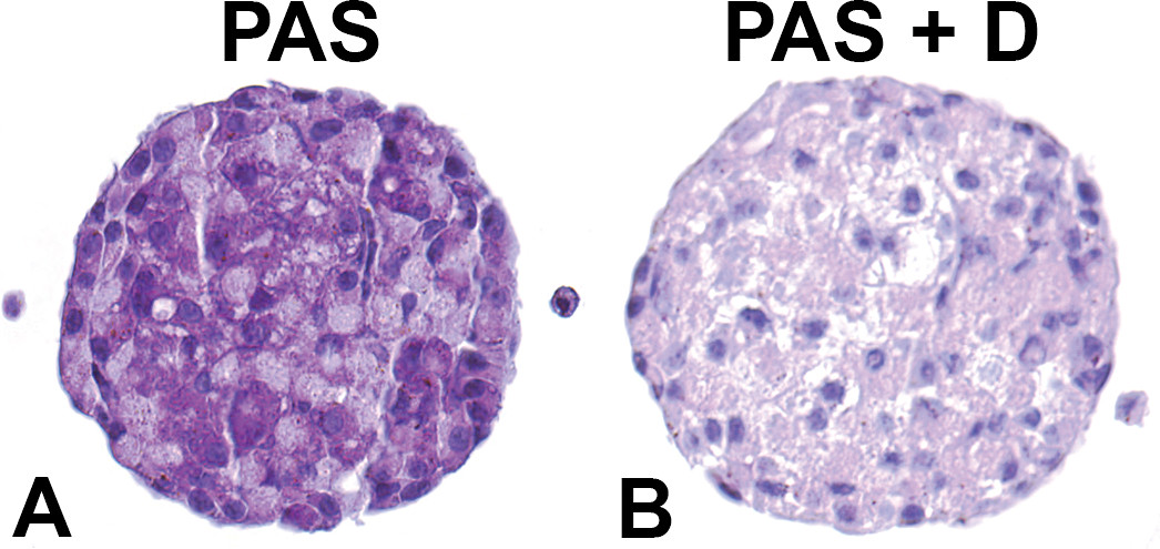
Liver (HepaRG) spheroids stained with periodic acid–Schiff (PAS) with or without diastase. (A) Liver spheroid stained with PAS. (B) Liver spheroid stained with PAS + diastase (PAS + D), which removes glycogen from the cells.
Immunohistochemical Stains
Spheroids fixed in 10% NBF used for different immunohistochemical stains showed staining comparable to control sections of human liver tissue (Figure 5). Ki-67 labeled the nuclei of cells in the liver spheroids similar to those of hepatocytes in the control human liver tissue. Comparable staining was observed in spheroids and control human liver sections when stained with liver markers such as CEA, a marker for bile canaliculi; CK19 expressed in bile duct epithelium; and MRP2, an adenosine tripohsphate–binding cassette transporter (see Figure 5).
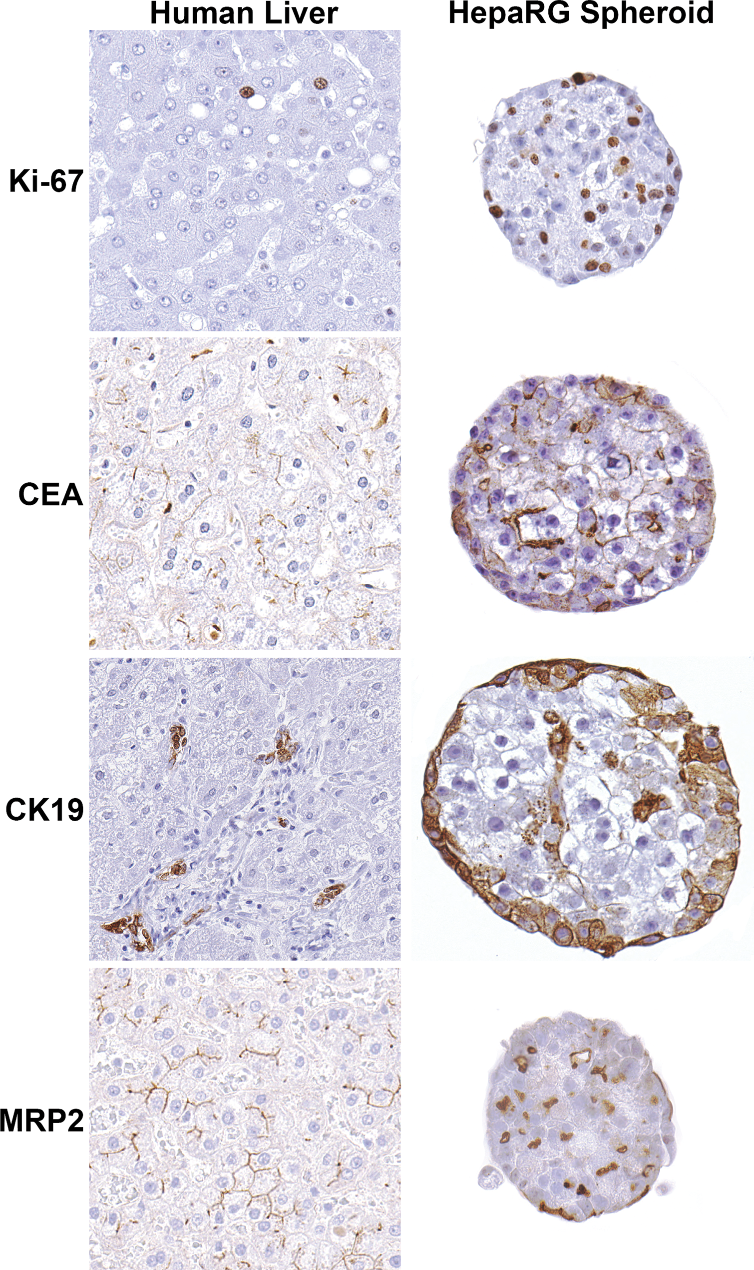
Immunohistochemical markers expressed in human liver tissue and human liver (HepaRG) spheroids. Note comparable nuclear staining in human liver and liver spheroids with Ki-67, a proliferation marker; cytoplasmic staining and cell membrane staining with carcinoembryonic antigen (CEA), a bile canaliculi marker; cytoplasmic and cell membrane staining with cytokeratin 19 (CK19), a marker of bile duct epithelium; and cell membrane staining with multidrug resistance–associated protein 2 (MRP2), a transporter protein.
Discussion
The popularity of 3-D in vitro biological systems has increased tremendously in recent years with the concept of developing cell and tissue cultures that resemble in vivo cells and tissues (Clevers 2016; Hoffman et al. 2017). Because 3-D models are relatively new in toxicologic research, there are few technologies available to successfully determine the detailed architecture and morphology of the cells growing in complex 3-D cultures. For years, formalin-fixed paraffin embedded (FFPE) samples have been used by researchers to analyze cell and tissue morphology and architecture and protein expression with the use of immunohistochemical markers (Canene-Adams 2013; Zhang et al. 2017). With the advent of high-throughput testing paradigms, FFPE samples are being explored as sources of RNA and DNA for molecular genomics and transcriptomics technologies. FFPE samples provide the benefit of storing and preserving tissues or cells for long periods of time, and samples can be used to perform a variety of staining techniques (Alturkistani, Tashkandi, and Mohammedsaleh 2015; Zhang et al. 2017). The FFPE spheroids described in this article, once stained with H&E, offered detailed morphology that allowed for observation of distinct nuclear and cytoplasmic cellular architecture. The FFPE spheroids were optimally prepared for immunohistochemical staining, and we were able to distinguish distinct cell types such as bile duct epithelia from hepatocytes using specific cell markers. In addition, the multidrug MRP2 and bile canaliculi could be observed using specific immunohistochemical stains. Our protocol effectively provided excellent preservation of cellular detail and architecture in addition to retention of protein epitopes that could be detected using immunohistochemistry. This protocol has been applied to rat liver spheroids for histochemical and immunohistochemical staining with excellent reproducibility (see Online Supplemental Figure S1).
Due to the increase in the use of 3-D spheroids in predictive toxicology research, drug development, and understanding mechanistic processes of toxicity and tumorigenesis, we have developed a protocol that is easy to follow and can be implemented in a standard histology or pathology laboratory without difficulty. The protocol produces sections that provide detailed cellular morphology and cellular microenvironment composition of 3-D cultures, far superior to that observed with confocal microscopy and high-content imaging. The technique proposed in our article is novel in that it is a quick, simple, and straightforward method that does not involve scraping Matrigel/cells from chamber slides, forming a Matrigel/cell and histogel “sandwich,” or performing a hematoxylin prestain as previously described in other protocols for embedding 3-D cultures (Pinto, Jacobsen, and Horwitz 2011; de Hoogt et al. 2017; Stock et al. 2016). We found that with our methodology, the morphology of the liver spheroids did not change if they were grown in media alone or required growth and collection in a collagen matrix, such as Matrigel, and that by using longitudinal paraffin sections rather than transverse sections, more spheroids could be sectioned onto a glass slide. Once paraffin embedded, sections of the spheroids could be stained with standard H&E, PAS, or immunohistochemical stains for proliferation, bile duct epithelium and canaliculi, or specific drug transporter markers that gave accurate cellular representation and localization of structures similar to those observed in sections of control human liver tissue. The ability to prepare 3-D cultures for embedding in paraffin blocks and sectioning onto glass slides allows for long-term storage of samples and their use in visualizing the localization and expression of protein markers using immunohistochemical and histochemical staining.
Supplemental Material
Supplemental Material, dixon_sup_fig1 - Preparation of Three-dimensional (3-D) Human Liver (HepaRG) Cultures for Histochemical and Immunohistochemical Staining and Light Microscopic Evaluation
Supplemental Material, dixon_sup_fig1 for Preparation of Three-dimensional (3-D) Human Liver (HepaRG) Cultures for Histochemical and Immunohistochemical Staining and Light Microscopic Evaluation by Natasha P. Clayton, Alanna Burwell, Heather Jensen, Barbara F. Williams, Quashana D. Brown, Pamela Ovwigho, Sreenivasa Ramaiahgari, Tonia Hermon, and Darlene Dixon in Toxicologic Pathology
Footnotes
Authors’ Note
Natasha P. Clayton and Alanna Burwell contributed equally.
Acknowledgments
The authors would like to thank Drs. Ronald Herbert and Gordon Flake for their critical review of this article. The authors greatly appreciate the expert technical assistance of Ms. Elizabeth Ney, Ms. Beth Mahler, and Mr. Paul Cacioppo with the compilation of figures and image plates.
Author Contributions
Authors contributed to conception or design (NC, AB, HJ, BW, QB, PO, SR, TH, DD); data acquisition, analysis, or interpretation (NC, AB, HJ, BW, QB, PO, SR, TH, DD); drafting the manuscript (NC, AB, TH, DD); and critically revising the manuscript (NC, AB, HJ, BW, QB, PO, SR, TH, DD). All authors gave final approval and agreed to be accountable for all aspects of work in ensuring that questions relating to the accuracy or integrity of any part of the work are appropriately investigated and resolved
Declaration of Conflicting Interests
The author(s) declared no potential conflicts of interest with respect to the research, authorship, and/or publication of this article.
Funding
The author(s) received no financial support for the research, authorship, and/or publication of this article.
Supplemental Material
Supplemental material for this article is available online.
References
Supplementary Material
Please find the following supplemental material available below.
For Open Access articles published under a Creative Commons License, all supplemental material carries the same license as the article it is associated with.
For non-Open Access articles published, all supplemental material carries a non-exclusive license, and permission requests for re-use of supplemental material or any part of supplemental material shall be sent directly to the copyright owner as specified in the copyright notice associated with the article.