
Abstract
The Göttingen minipig is often used in preclinical toxicity studies. Therefore, knowledge of spontaneously occurring pathologies is important to differentiate them from test drug–related effects. We report on a Göttingen minipig, which developed exudating widespread dermatitis during a preclinical toxicity study with a subcutaneously injected drug. The lesions were resistant to topical and oral antibacterial medications. Skin cultures were positive for Candida albicans, and treatment was changed to topical antifungal cream with quick resolution of the skin lesions. Cutaneous candidiasis in pigs has been rarely reported in the literature, and this is the first report on such condition in preclinical toxicity studies. Knowledge of this condition, which is not drug related, is important, especially in toxicity studies involving subcutaneous injections that are commonly accompanied by inflammatory skin reactions.
The minipig is being used more commonly as a translational, nonrodent animal model in regulatory toxicity and safety assessments studies (Manno et al., 2016; Ramot, Rousselle, et al., 2016, Ramot et al., 2015; Vezzali et al., 2011). It bears a high similarity to humans and can be utilized for many types of toxicity studies assessing different target organs (Bode et al., 2010; van der Laan et al., 2010). The most utilized strain is the Göttingen minipig (Ramot, Weber, et al., 2016). Therefore, it is of utmost importance to differentiate between effects that are related to the test compound and spontaneously occurring pathologies in this strain (Vezzali et al., 2011). Factors that can influence the interpretation of pathological findings include, for example, symptoms and signs related to an infectious agent.
In this brief communication, we report the symptoms and signs related to cutaneous candidiasis in a Göttingen minipig participating in a toxicity study involving subcutaneous administration of a test compound. This infection was not related to the tested drug.
Thirty-two male and 32 female Göttingen minipigs were allocated to 26- and 39-week treatment arms, with an additional 6-week recovery phase. The animals were divided into a vehicle group and 3 treatment groups (low, medium, and high doses). The interim sacrifice groups included 6 animals, the main study groups included 8 animals, and 4 animals were allocated to the recovery period (vehicle and high dose only). The animals were maintained in accordance with the guide for the care and use of laboratory animals (Institute for Laboratory Animal Research, 1996).
During the first week of treatment, 1 male animal from the low-dose group developed a deteriorating skin rash, composed of extensive scabs on the dorsum and around the face, neck, and ears which were gray, raised, firm, and adherent to the underlying skin, which appeared erythematous following removal (Figure 1). The animal was, however, expressing normal behavior, eating well, and showing normal weight gain. Skin swabs were taken for bacterial and fungal cultures, and the animal was treated with 0.05% chlorhexidine (Unisept®, Medlock Medical Ltd., Oldham, UK) and 0.5% fucidic acid/0.1% betamethasone valerate (Isaderm gel®, Dechra Veterinary Products A/S, Uldum, Denmark). Since no improvement was noted in the skin condition, the treatment was augmented to include amoxycillin–clavulanic acid (400: 57 mg/ml) and miconazole base 5.2%/chlorhexidine gluconate 5.9% mixture (Malaseb® Concentrate Rinse, Dechra Veterinary Products A/S, Uldum, Denmark) baths, with stabilization of the skin rash. Cultures showed a moderate growth of Escherichia coli, 2 Coliform spp., and scant growth of 3 Staphylococcus spp. Candida spp. was identified by selective fungal culture. No dermatophytes were isolated. No biopsies from the lesions were taken for histopathological evaluation.
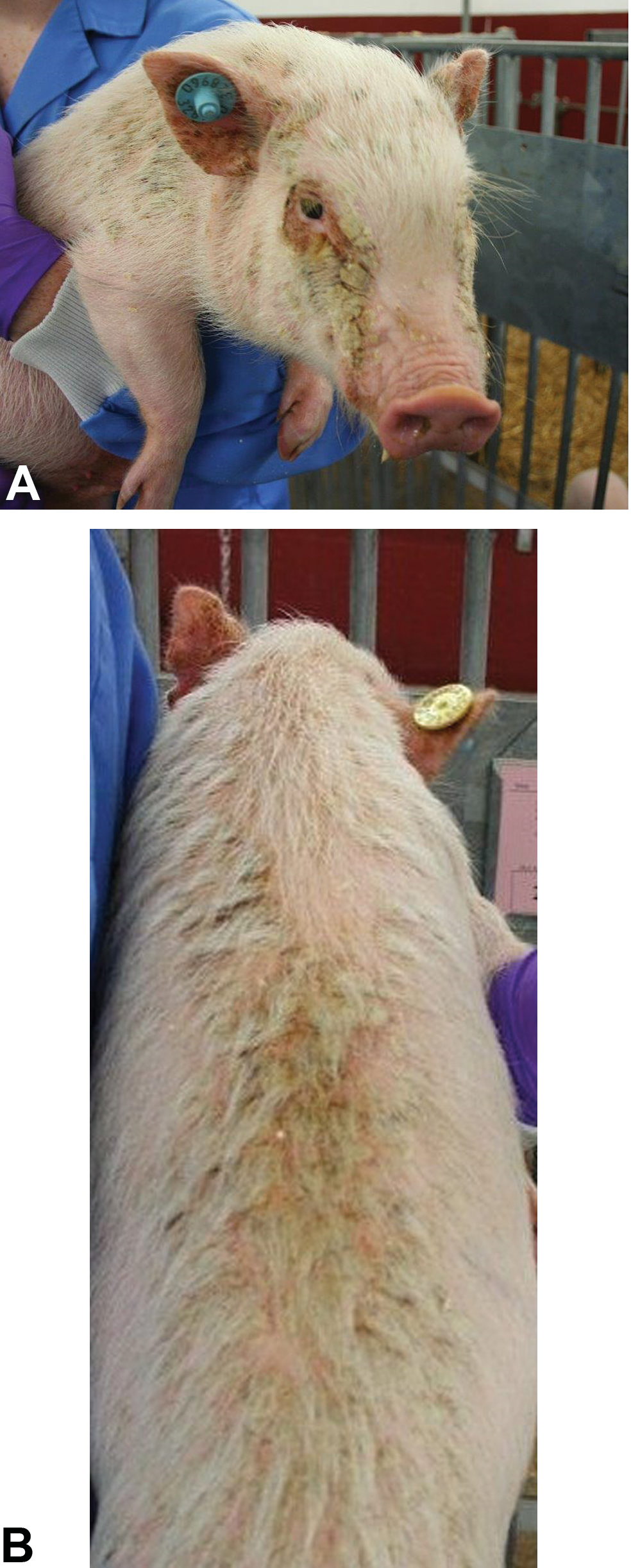
Extensive gray, raised, firm scabs involving the face, which are adherent to the underlying skin. (A) Aspect of scabs on the neck and ears. (B) Aspect of the scabs on the dorsum.
Following the results of the skin cultures, treatment was changed to clotrimazole 1% (Canesten®, Bayer plc, Newbury, Berkshire, UK) cream applied for 10 days, with marked and quick improvement of the skin rash 4 weeks after its first appearance. No similar lesions were observed in any of the other animals participating in the study, and no test article–related clinical signs or mortalities were noted, except for several injection site findings, including edema and erythema, across all dose groups and both sexes that were procedural in origin. Histopathological examination of the skin at termination of the study, including samples from the clinically affected regions, did not reveal any treatment-related findings in any of the treated animals.
Candida species are commensal in both humans and animals and are found in the upper respiratory, alimentary canal, genital mucosa, and the skin (Mueller, Bettenay, and Shipstone, 2002). Indeed, according to Ellegaard’s health monitoring information (July 2016, http://minipigs.dk/the-goettingen-minipig/health-status), C. albicans was cultured in more than 40% of the tested Göttingen minipigs. It has also been suggested to be involved in the formation of hyperkeratosis in Göttingen minipigs (Bollen et al., 1998), although such findings are not common in these species (Helke et al., 2016; Jeppesen and Skydsgaard, 2015).
Candida infection can become pathogenic and result in skin signs when there is a disturbance in the physical, chemical, or immune mechanisms. Often, this is due to mechanical stress and inflammation of the skin leading to damage to the epidermal barrier. Alternatively, Candida species can proliferate excessively following changes in the normal skin flora after antibacterial treatment or due to corticosteroid treatment resulting in reduced immunity against fungal infections (Mueller, Bettenay, and Shipstone, 2002). In the present case, the trigger for the extensive cutaneous candidiasis is not entirely clear; however, the fact that the lesions were localized periorificially points to a mechanical trigger. The fact that the animal was treated with antibacterial and corticosteroids presuming a bacterial infection may have predisposed the animal to the exacerbation and spread of the skin condition.
When encountered with exudating and exfoliating skin lesions in a young pig, the diagnosis of exudative epidermitis, which is a common skin condition in the pig, should be considered (Park et al., 2013). This disease has multifactorial predisposition causes that include damage to skin, stress, and changes in the environment and housing conditions. These causes will facilitate the bacterial overgrowth on the skin and the production and release of toxins by Staphylococcus hyicus and usually resolves following antibacterial treatment. Exudative epidermitis was also the working diagnosis in the present case. However, based on the clinical signs, the ineffectiveness of the antibacterial treatments, and growth of Candida in the culture media, the diagnosis was changed to cutaneous candidiasis. The rapid response to antifungal treatment further established this diagnosis.
Cutaneous candidiasis is a common condition in humans; however, it is much less prevalent in animals (Shokri and Khosravi, 2016). Skin infections owing to Candida species have been reported in several animals, including guinea pigs, dogs, rodents, birds, and rabbits (Gupta et al., 2010; Moretti et al., 2004; Shokri and Khosravi, 2016). In pigs, spontaneous cutaneous candidiasis leading to chronic dermatitis has been reported only rarely in the literature (Reynolds, Miner, and Smith, 1968) and never as a skin condition during preclinical in vivo studies. Knowledge of this clinical condition is important when evaluating preclinical toxicity studies and especially when assessing studies involving subcutaneous injections, which can often result in inflammatory skin changes (Shaltiel-Karyo et al., 2017; Ramot et al., 2012).
Footnotes
Author Contribution
Authors contributed to conception or design (AN, AO, AM); data acquisition, analysis, or interpretation (YR, AN, AO, AM); drafting the manuscript (YR, AN); and critically revising the manuscript (YR, AN, AN, AM). All authors gave final approval and agreed to be accountable for all aspects of work in ensuring that questions relating to the accuracy or integrity of any part of the work are appropriately investigated and resolved.
Declaration of Conflicting Interests
The author(s) declared no potential conflicts of interest with respect to the research, authorship, and/or publication of this article.
Funding
The author(s) received no financial support for the research, authorship, and/or publication of this article.