
Abstract
This review analyzes the published data on cases of pulmonary alveolar proteinosis (PAP) in workers inhaling crystalline aluminum, indium, silicon, and titanium particles and possible sequelae, that is, inflammation and fibrosis, and compares these findings with those from animal experiments. In inhalation studies in rodents using crystalline indium and gallium compounds, pronounced PAP followed by inflammation and fibrosis down to very low concentration ranges have been reported. Crystalline aluminum, silicon, and titanium compounds also induced comparable pulmonary changes in animals, though at higher exposure levels. Laboratory animal species appear to react to the induction of PAP with varying degrees of sensitivity. The sensitivity of humans to environmental causes of PAP seems to be relatively low. Up to now, no cases of PAP, or other pulmonary diseases in humans, have been described for gallium compounds. However, a hazard potential can be assumed based on the results of animal studies. Specific particle properties, responsible for the induction of PAP and its sequelae, have not been identified. This review provides indications that, both in animal studies and in humans, PAP is not often recognized due to the absence of properly directed investigation or is concealed behind other forms of lung pathology.
Keywords
Introduction
Pulmonary alveolar proteinosis (PAP) is a relatively rare lung disease in humans, which was first described by Rosen, Castleman, and Liebow (1958). It is characterized by the accumulation of large quantities of mostly acellular, lipoproteinaceous material in the alveoli; this material is periodic acid Schiff (PAS) positive. Its water-insoluble part is rich in phospholipids and mainly consists of dipalmitoyl phosphatidyl choline; the water-soluble part is mainly composed of serum proteins (Agassandian and Mallampalli 2013; Onodera et al. 1983; Postle, Heeley, and Wilton 2001). It is presently taken as certain that surfactant is a major component of the PAP material, which is produced by overproduction by alveolar type II epithelial cells and/or a disturbed removal process by type II cells and alveolar macrophages (Seymour and Presneill 2002). Electron microscopy has also revealed, apart from amorphous material, accumulation of myelin-like, multilamellar bodies in PAP (Costello et al. 1975; Gilmore, Talley, and Hook 1988; Maygarden et al. 2001; Wasserman and Mason 1994; Yi et al. 2012). Depending on the degree of severity, this accumulated material leads to an impairment of gas exchange (Briens et al. 2002; Inoue et al. 2008).
Three different classes or categories of PAP are recognized in human pathology:
autoimmune PAP = primary PAP also designated as idiopathic or acquired PAP, secondary PAP, and congenital PAP.
(a) Autoimmune PAP is characterized by high levels of granulocyte–macrophage colony-stimulating factor (GM-CSF) neutralizing autoantibodies. It is supposed to account for more than 90% of all human PAP diseases. A series of GM-CSF neutralizing autoantibodies have been recently characterized (Wang et al. 2013). Particle exposure is generally not considered to be a cause. However, in larger studies involving a thorough recording of patient histories, a relatively high (up to 54%) percentage of patients with autoimmune PAP had a mostly occupational exposure to dusts and vapors (Bonella et al. 2011; Briens et al. 2002; Inoue et al. 2008). (b) In the case of secondary PAP, GM-CSF neutralizing autoantibodies are not involved. Secondary PAP has either been preceded by malignant hematological diseases (according to Ishii et al. 2011 in more than 75%) or high exposure to poorly soluble particles from mineral or metallic/metalloid substances. This means that the latter represents only a very small percentage of all human PAP cases described (Briens et al. 2002; Inoue et al. 2008; Ishii et al. 2011). (c) In rare cases, inherited genetic defects are present as a result of recessive mutations in the receptors of the GM-CSF effects (Suzuki et al. 2011).
Recent reports of serious lung lesions (PAP, fibrosis) in workers of the indium producing and processing industry (Cummings et al. 2012) together with results from animal experiments with indium and gallium compounds demonstrating a high pneumotoxic potential, starting with PAP and followed by inflammation and fibrosis (Nagano, Gotoh, et al. 2011; Nagano, Nishizawa, Eitaki, et al. 2011; National Toxicology Program [NTP] 2000, 2001), indicate a possible hazard potential from inhalation at the workplace also for particulate gallium compounds.
Already there are several reviews on PAP. They deal with the different aspects of its clinical manifestations, epidemiology, pathophysiological mechanisms, different forms and their causes, treatment methods, morphology, and so on (e.g., Borie et al. 2011; Carey and Trapnell 2010; Hook 1991; Khan and Agarwal 2011; Patel et al. 2012; Seymour and Presneill 2002; Wang et al. 2012). The importance of (crystalline) particles in its formation and the comparison of human data with the corresponding animal toxicological results as well as the question of concomitant and/or sequel conditions in the lungs have, up to now, not been dealt with.
This review aims to describe the cases of secondary PAP (and accompanying lung pathology) in humans and experimental animals exposed to (crystalline) particles of aluminum, gallium (no human cases reported), indium, silicon, and titanium compounds and how the histopathological diagnosis was made, that is, with or without PAS reaction. An attempt is made to determine if specific particle properties or experimental conditions exist, which favor the development, incidence, and strength of PAP induction. Where possible, the sequence of events in the lung lesion formation is evaluated, if there are differences in the sensitivity of PAP induction between species, and whether a possible hazard to humans can be predicted for particle-induced PAP (and sequels) from animal experiments.
Human Cases of Particle-induced PAP
The majority of human cases of PAP induced by inhalation of particles has been reported in workers exposed to silica/quartz, with only very few cases in workers exposed to aluminum, indium, and titanium dioxide.
Aluminum
In the literature, only one study is found that clearly indicates aluminum as an inducer of PAP (Miller et al. 1984). In the case of a rail grinder who had been working for 6 yr at a very dusty work site, histological examination and electron microscopy of lung biopsies provided clear evidence for a PAP caused by aluminum particles; PAS-stained lung slides were positive. Minimal interstitial inflammation or fibrous thickening was also observed.
Indium
Three cases of PAP from indium compounds have been described in the literature (Cummings et al. 2010, 2012; Xiao et al. 2010). In all 3 cases, PAP occurred within 1–2 yr after initial exposure. PAS reaction on lung biopsies was positive in all 3 cases. In an interdisciplinary workshop, all 10 cases of the so-called indium lung disease known worldwide up to that time (7 with interstitial lung disease and 3 with PAP) were subjected to an accurate comparative analysis (Cummings et al. 2010; T. Homma et al. 2003; S. Homma et al. 2005; Nakano et al. 2007; Taguchi and Chonan 2006; Takeuchi 2008; Xiao et al. 2010). Histopathological features included intra-alveolar exudate typical of PAP (n = 9), cholesterol clefts and granulomas (n = 10), and fibrosis (n = 9). This lung disease progressed following cessation of exposure in most patients and was fatal in 2 patients. The radiographic data, using different methods, showed that in 2 patients with PAP a subsequent fibrosis developed. The authors suggested that this disease may begin with PAP and progress to include fibrosis (Cummings et al. 2012).
Silicon Dioxide/Silica/Quartz
A pathological condition, considered to be PAP, occurred after a relatively short-term and high-level exposure to dusts containing silicon described by Buechner and Ansari (1969). This was first reported in 4 young men who had been involved in sand-blasting work and described as silicoproteinosis. All workers died on average 7.5 months after the occurrence of the first symptoms. Histologically, the material in the alveoli was PAS positive. Silicotic nodules and interstitial fibrosis were additional lung findings. Other terms for this disease are silicotic alveolar proteinosis or alveolar lipoproteinosis (Silicosis and Silicate Disease Committee 1988). In contrast to silicosis, in which inflammation associated with subsequent fibrosis are caused by relatively low concentrations over long periods (≥10 yr), silicoproteinosis occurs after periods lasting between weeks and a few years. The development of silicoproteinosis is progressive, and its prognosis is highly unfavorable. A series of case reports on silicoproteinosis are available (e.g., Abraham and McEuen 1986; Arakawa et al. 2005; Banks et al. 1983; Silicosis and Silicate Disease Committee 1988; Suratt et al. 1977; Xipell et al. 1977). In only a few cases, the particles detected histologically, or via electron microscopy, were characterized and only termed as birefringent (Abraham and McEuen 1986; Banks et al. 1983; Suratt et al. 1977; Xipell et al. 1977). Most cases were diagnosed on the basis of patient history combined with histopathology data.
Titanium
Only 1 case of titanium dioxide as inducer of PAP (Keller et al. 1995) has been identified in a painter. Biopsied material from the lungs was PAS positive. Some parts of the lungs showed a mixture of PAP with focal fibrotic alveolar septa and organizing granulation tissue.
Experimental Toxicology
There are animal studies that correlate with the examples of particle-induced PAP in humans. For the reasons given in the introduction, gallium is included. A number of animal strains have been used in these experiments. Since no strain-specific differences could be recognized in the lung reaction to the investigated particles, further details on strains are not presented. For the sake of brevity, information on the sex, dose/concentration levels, and singular data on particle properties (others than size) are also not included.
Other particulate inorganic substances, in which a PAP was demonstrated in animal experiments, have not yet been reported as causes of human PAP cases. Examples are antimony trioxide, bismuth orthovanadate, carbon black, cadmium chloride, chromium dioxide, cerium dioxide, cobalt oxide (Co3O4), cobalt sulfate, iron sulfide (pyrite), nickel compounds, and talc. Most of these substances are described as poorly soluble, but cadmium and nickel chloride as well as nickel sulfate heptahydrate are generally considered to be soluble.
The experimental methods, the details on particle properties, and the diagnoses of pathological changes in the lungs are cited as they appear in the original papers.
Aluminum
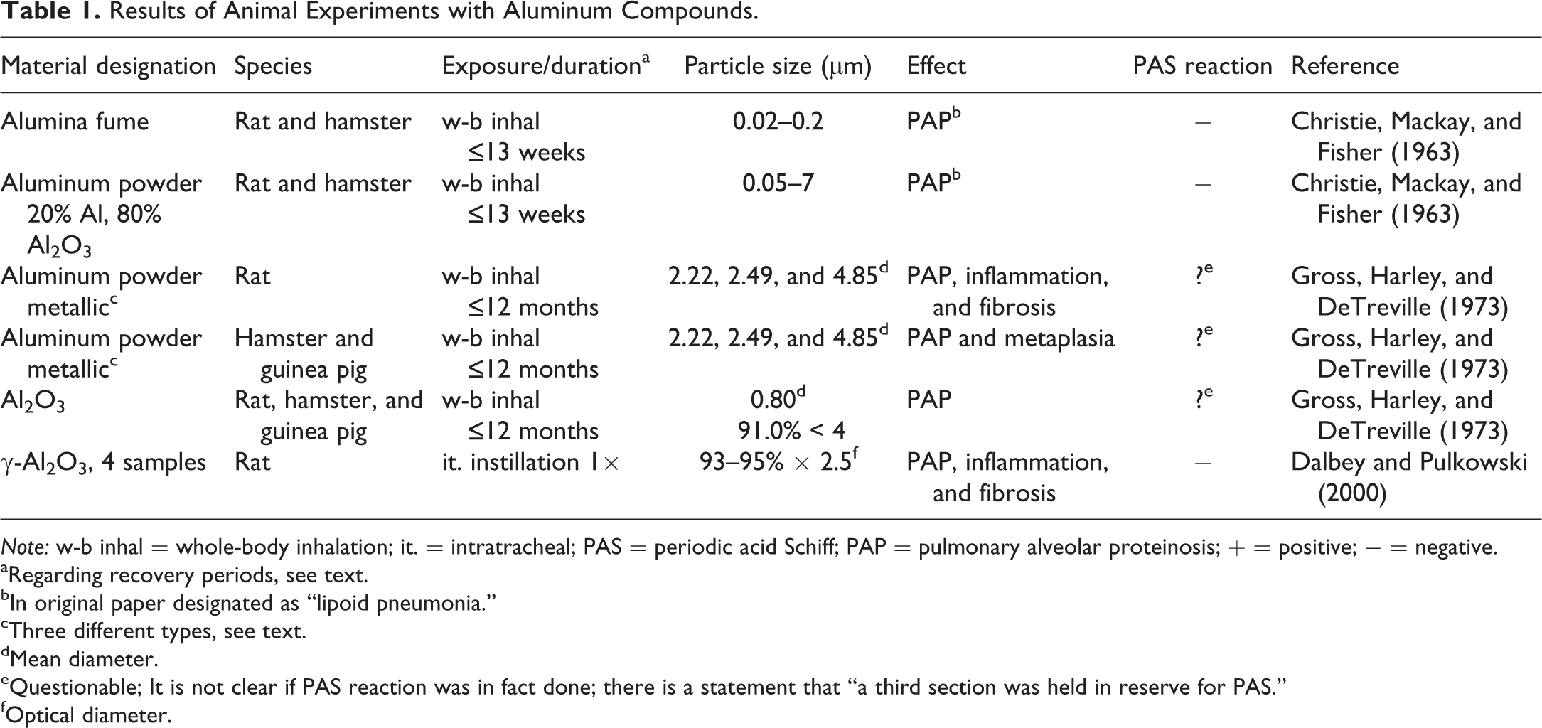
The results of animal studies with aluminum compounds are summarized in Table 1.
Results of Animal Experiments with Aluminum Compounds.
Note: w-b inhal = whole-body inhalation; it. = intratracheal; PAS = periodic acid Schiff; PAP = pulmonary alveolar proteinosis; + = positive; − = negative.
aRegarding recovery periods, see text.
bIn original paper designated as “lipoid pneumonia.”
cThree different types, see text.
dMean diameter.
eQuestionable; It is not clear if PAS reaction was in fact done; there is a statement that “a third section was held in reserve for PAS.”
fOptical diameter.
Rats and hamsters inhaling aluminum fumes or aluminum powder for up to 13 months initially produced proliferation of macrophages within the alveolar space followed by a PAP (here designated as lipoid pneumonia). In rats, PAP was also diagnosed somewhat earlier than in hamsters (Christie, Mackay, and Fisher 1963). Following inhalation (6 hr/day, 5 days/week, 6 or 12 months) of fine metallic aluminum powders or Al2O3, PAP occurred in rats, hamsters, and guinea pigs. The severity of PAP was greatest in the rats. Sequential sacrifice up to 18 months after termination of exposure revealed that PAP, especially in rats, lasted for a very long time (up to 1 yr), although with decreasing intensity, whereas it disappeared in hamsters and guinea pigs within 3 months. In some rats, subpleural foci of fibrosis associated with cholesterol needles developed, whereas epithelial metaplasia was described in hamsters and guinea pigs, both at the later stages of the experiments (Gross, Harley, and DeTreville 1973). Single intratracheal instillation of 4 samples of alumina (Bayer pseudoboehmite, Bayer γ-Al2O3, Ziegler pseudoboehmite, or Ziegler γ-Al2O3) to rats produced PAP, in addition to focal areas of granulomatous and interstitial inflammation with fibrosis in the investigation after 6 months (Dalbey and Pulkowski 2000). In summary, independently of the investigated aluminum material and in all 3 investigated species, PAP was demonstrated and was most pronounced in rats and persisted a long time after discontinuing exposure. Particle size did not seem to play a decisive role in the development of PAP. The PAS reaction was not necessary for the diagnosis of PAP.
Gallium
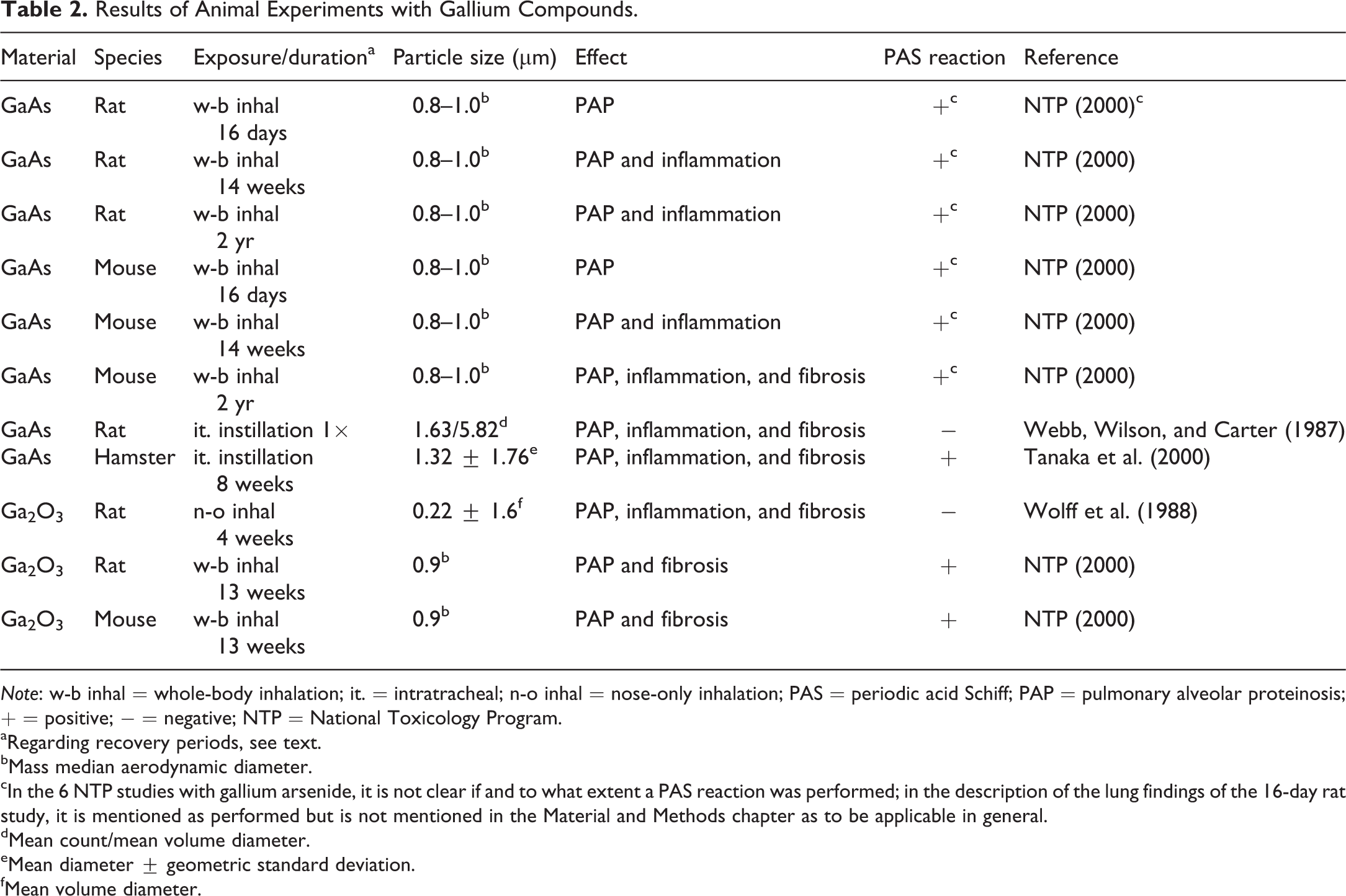
The results of studies with gallium compounds are summarized in Table 2. Sixteen-day, 14-week, and 2-yr studies (6 hr/day, 5 days/week) were carried out with gallium arsenide (GaAs) in rats and mice. A concentration-dependent PAP was found in male and female rats and mice after 16 days. The PAP increased in intensity in the subchronic and chronic studies, affecting rats at lower concentrations than mice. After 2 yr, the rat lungs showed a broad spectrum of pathological changes, that is, PAP, atypical hyperplasia, chronic active inflammation, squamous metaplasia, alveolar epithelial hyperplasia, and alveolar epithelium metaplasia. The lung lesions in mice were somewhat different, with histiocytic cellular infiltration, focal chronic and suppurative inflammation, alveolar epithelial hyperplasia, and PAP. The incidence of lung tumors was increased in female rats only at 0.1 and 1.0 mg/m3 but not at 0.01 mg/m3 (NTP 2000). After a single intratracheal instillation of GaAs to rats, there was, apart from various inflammatory and fibrotic changes, also a time-dependent increase in PAP from day 7 up to the end of the study (day 28). Other pulmonary changes (congestion/hemorrhage, generalized interstitial pneumonia, and alveolitis) were already present 1 day after treatment (Webb, Wilson, and Carter 1987). After intratracheal instillation of GaAs twice weekly to hamsters for 8 weeks, PAP, pneumonitis, and fibrotic proliferation were found (Tanaka et al. 2000).
Results of Animal Experiments with Gallium Compounds.
Note: w-b inhal = whole-body inhalation; it. = intratracheal; n-o inhal = nose-only inhalation; PAS = periodic acid Schiff; PAP = pulmonary alveolar proteinosis; + = positive; − = negative; NTP = National Toxicology Program.
aRegarding recovery periods, see text.
bMass median aerodynamic diameter.
cIn the 6 NTP studies with gallium arsenide, it is not clear if and to what extent a PAS reaction was performed; in the description of the lung findings of the 16-day rat study, it is mentioned as performed but is not mentioned in the Material and Methods chapter as to be applicable in general.
dMean count/mean volume diameter.
eMean diameter ± geometric standard deviation.
fMean volume diameter.
Male and female rats were exposed to gallium oxide Ga2O3 2 hr/day, 5 days/week for 4 weeks and investigated 1 day, 2 weeks, and 2, 6, and 12 months after the end of exposure. Histopathological lesions consisted initially of PAP, progressing in severity to large focal lesions of alveolar histiocytosis and septal fibrosis 6 and 12 months after exposure (Wolff et al. 1988). After 13-week exposure (6 hr/day, 5 days/week) of rats and mice, PAP, inflammation, type II cell hyperplasia, and fibrosis were observed. These data are only available as a secondary quotation (NTP 2000).
In summary, the studies with GaAs in rats and mice and the subacute inhalation study with Ga2O3 in rats show that PAP is present prior to the occurrence of inflammatory and fibrotic lesions, therefore these latter findings can be described as sequel conditions.
Indium
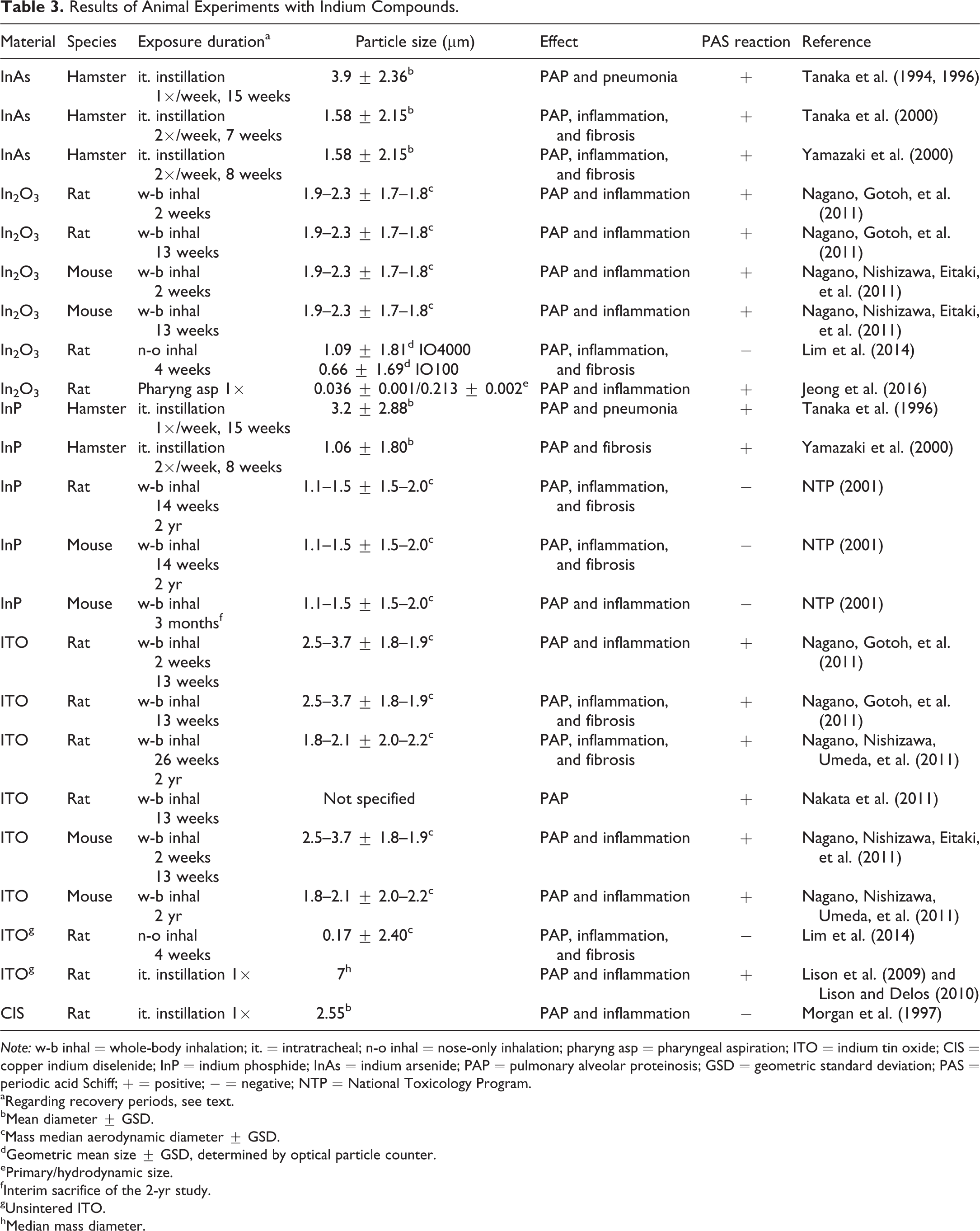
The results of animal studies with indium compounds are summarized in Table 3. In three studies with indium arsenide in hamsters and once or twice weekly intratracheal instillation for 7 (no recovery period), 8 (up to 88 weeks recovery), or 15 weeks (lifelong recovery), PAP, designated as “proteinosis-like lesions,” accompanied by inflammation (pneumonia, metaplasia/metaplastic ossification, alveolar and bronchiolar hyperplasia), fibrosis, and/or emphysema was observed (Tanaka et al. 1994, 1996, 2000; Yamazaki et al. 2000).
Results of Animal Experiments with Indium Compounds.
Note: w-b inhal = whole-body inhalation; it. = intratracheal; n-o inhal = nose-only inhalation; pharyng asp = pharyngeal aspiration; ITO = indium tin oxide; CIS = copper indium diselenide; InP = indium phosphide; InAs = indium arsenide; PAP = pulmonary alveolar proteinosis; GSD = geometric standard deviation; PAS = periodic acid Schiff; + = positive; − = negative; NTP = National Toxicology Program.
aRegarding recovery periods, see text.
bMean diameter ± GSD.
cMass median aerodynamic diameter ± GSD.
dGeometric mean size ± GSD, determined by optical particle counter.
ePrimary/hydrodynamic size.
fInterim sacrifice of the 2-yr study.
gUnsintered ITO.
hMedian mass diameter.
With indium oxide (In2O3), 2-week and 13-week inhalation studies (6 hr/day, 5 days/week) with rats and mice were carried out. The most important finding after 2 and 13 weeks was PAP in all rats and mice. Infiltrations of alveolar macrophages and inflammatory cells were the further findings. In mice PAP was somewhat less pronounced than in rats (Nagano, Gotoh, et al. 2011; Nagano, Nishizawa, Eitaki, et al. 2011). Sprague–Dawley rats were exposed to 2 different-sized In2O3 (called “IO4000” and “IO100”) by inhalation (6 hr/day, 5 days/week) for 4 weeks and observed during a 4-week recovery period. PAP was diagnosed in IO4000-treated rats at the end of exposure but not in IO100-treated animals. After the 4-week recovery, PAP was observed in both groups, more pronounced with the larger particles. Migration of alveolar macrophages, alveolar wall fibrosis, type II epithelial cell hyperplasia/hypertrophy, perivascular inflammation, and pleural thickening with prominent mesothelial cells were additional diagnoses more pronounced with IO4000 (Lim et al. 2014). In2O3 nanoparticles were administered to female rats via pharyngeal aspiration, and lung effects were evaluated 1, 3, 14, and 28 days after treatment. Neutrophilic inflammation was observed on day 1 and worsened until day 28, and severe PAP was observed on days 14 and 28 (Jeong et al. 2016).
In 2 studies with indium phosphide (InP) in hamsters and once or twice weekly intratracheal instillation for 8 (up to 88 weeks recovery) or 15 weeks (lifelong recovery), PAP accompanied by signs of inflammation (pneumonia, metaplastic ossification, alveolar, and bronchiolar hyperplasia), fibrosis, and/or emphysema were observed (Tanaka et al. 1996; Yamazaki et al. 2000). PAP and fibrosis increased up to the end of the recovery period (Yamazaki et al. 2000). InP was investigated in 14-week and 2-yr inhalation studies (6 hr/day, 5 days/week) with rats and mice. After 14-week inhalation, all treated animals had a marked PAP. In addition, chronic active inflammation, hyperplasia of the alveolar epithelium, and interstitial fibrosis were present. In the 2-yr studies, exposure to the 2 highest concentrations was discontinued after 22 (rat) or 21 (mouse) weeks because of the severity of lung lesions. In mice, PAP was much less pronounced, both in incidence and in intensity than in rats but had higher scores of chronic active inflammation and fibrosis. Nearly all treated rats had a marked PAP persisting past the end of exposure, in addition to chronic active inflammation, atypical hyperplasia, hyperplasia, and metaplasia of the alveolar epithelium as well as interstitial fibrosis. The incidence of lung tumors in rats and mice was markedly increased (NTP 2001).
Two-week and 13-week inhalation studies (exposure 6 hr/day, 5 days/week) in rats and mice were carried out with indium tin oxide (ITO). One group was observed during a 26-week recovery period. The most important finding after 2 and 13 weeks was PAP. Infiltrations of alveolar macrophages and inflammatory cells were additional findings. At the end of the recovery period, all animals had PAP and fibrosis of the alveolar walls. Rats were affected at lower concentrations (Nagano, Gotoh, et al. 2011; Nagano, Nishizawa, Eitaki, et al. 2011). Mice and rats were exposed (6 hr/day, 5 days/week) for 104 weeks. An additional group of rats was exposed for 26 weeks followed by a recovery period of 78 weeks. In rats, PAP, bronchioloalveolar hyperplasia, alveolar wall fibrosis, thickened pleura wall, and infiltrations of alveolar macrophages and inflammatory cells were observed. In mice, PAP, thickened pleura, and infiltrations of alveolar macrophages were diagnosed, though incidences and degree were lower compared to rats. Lung tumors were only increased in rats (Nagano, Nishizawa, Umeda, et al. 2011). Inhalation exposure (6 hr/day, 5 days/week) for 13 weeks produced a PAP in all rats. All rats were negative for GM-CSF autoantibodies (Nakata et al. 2011). Single intratracheal treatment of rats caused dose-dependent PAP and inflammation 3, 15, and 60 days after instillation (Lison et al. 2009; Lison and Delos 2010). Rats were exposed to nano-sized ITO by inhalation for 6 hr/day, 5 days/week, and for 4 weeks, partly followed by 4-week recovery. Histopathology after 4 weeks revealed marked PAP and less pronounced inflammatory as well as fibrotic lesions. These changes were increased in severity 4 weeks after the exposure period (Lim et al. 2014).
Single intratracheal instillation of copper indium diselenide in rats with histological examination on days 7, 14, and 28 produced interstitial inflammation and PAP from day 7, with increasing severity up to day 28 (Morgan et al. 1997).
In summary, in all studies with inhalation exposure of indium-containing molecules, in which a PAS reaction was carried out, and in all NTP studies with InP without a PAS reaction, PAP was diagnosed. In the 2-week studies with ITO and also in the 13-week studies with In2O3, it was the only relevant lung finding in rats and mice. With increasing study duration, mostly inflammatory and fibrotic changes occurred in addition and were then in the foreground. The data on particle properties are mostly restricted to the diameter, whereby in all studies, the major part was within the respirable range. The dimensions deviated by a factor of over 40, with the majority between 1.1 and 3.7 µm. From these data, no qualitative or quantitative effects on the occurrence or absence of PAP can be derived.
Silicon Dioxide/Silica/Quartz
A very large number of publications are available that have dealt with the acute to chronic changes in the lungs of laboratory rodents after inhalation or intratracheal instillation. In this context, a number of articles focusing on crystalline SiO2 species will be cited as examples.
Inhalation studies
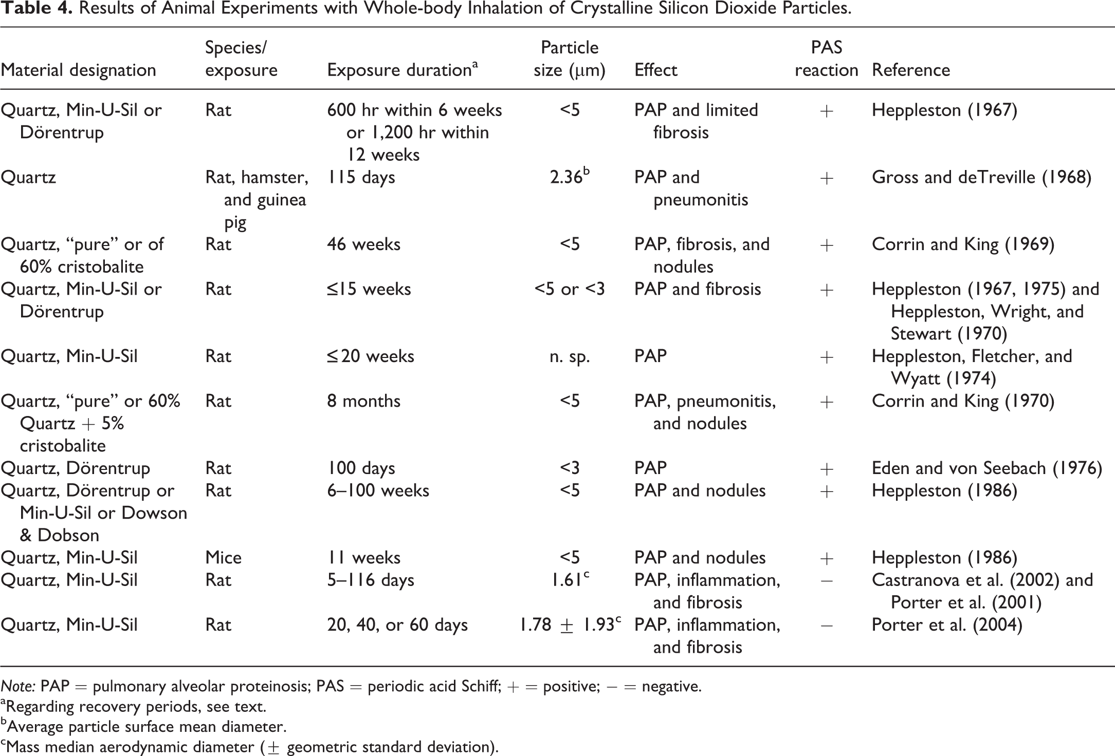
Those inhalation studies (all with whole-body exposure) where PAP has been demonstrated are compiled in Table 4. In a series of experiments with rats inhaling quartz from different sources for 6–15 weeks, histological and histochemical investigations revealed a pronounced accumulation of amorphous material, rich in phospholipids and proteins, showing a picture very similar to that of PAP in humans diagnosed as “pulmonary alveolar lipoproteinosis.” Additionally, intra-alveolar and interstitial fibrosis were described at later time points during posttreatment observation lasting from 2 to 19 months. Clear evidence was found that the cause for the accumulation of phospholipids is both increased synthesis and reduced elimination (Heppleston 1967, 1975; Heppleston, Wright, and Stewart 1970; Heppleston, Fletcher, and Wyatt 1974). Rats, hamsters, and guinea pigs were exposed to quartz dust (6 hr/day, 5 days/week) for 115 days and were examined 14 months after the first dust exposure. The outstanding lesion in rats was PAP; in addition, desquamative pneumonitis and occasional nodules were diagnosed. Hamsters and guinea pigs had extensive desquamative pneumonitis with relatively little PAP (Gross and deTreville 1968). Corrin and King (1969, 1970) described PAP in rats with 8-month exposure to pure quartz or quartz of 60% with 5% cristobalite content (necropsy at monthly intervals up to 27 months). Silicotic nodules and interstitial fibrosis were additional findings. In a study of rats exposed to quartz dust, with exposure up to 100 days, the changes in the lungs were so similar to those of PAP in humans that the authors proposed this type of study as model for this condition (Eden and von Seebach 1976). In a series of studies, rats were exposed (6, 7, 16, or 18 hr/day, 5 days/week) to different quartz samples. Mice and guinea pigs were exposed to quartz (18 hr/day, 5 days/week) for 11 weeks. PAP, nodules, and fibrosis were common findings in all experiments with rats with very little variation. Exposed mice developed a fibrous, granulomatous reaction, while PAP did not attain the degree of that of rats. Guinea pigs exhibited qualitatively different pathological features to those seen in rats and mice. Their lungs showed an extensive intra-alveolar accumulation of dust-bearing macrophages but no PAP and no fibrosis (Heppleston 1986). In male rats, the inhalation of quartz (6 hr/day, 5 days/week, and 5–116 days) produced a pronounced increase in phospholipids in the bronchoalveolar lavage fluid starting after 5 days, with an explosive rise between days 41 and 79. PAP was confirmed (PAS stain) histopathologically on days 79 and 116. At the same time points, fibrosis was diagnosed. Inflammation (alveolitis) started at day 20 (Castranova et al. 2002; Porter et al. 2001). Rats were exposed (6 hr/day, 5 days/week) to quartz for 20, 40, or 60 exposure days. Sacrifice was at the end of exposure and after 36 days’ recovery. Pulmonary inflammation and PAP increased during the recovery period in rats exposed for 40 and 60 days but not 20 days; fibrosis developed even when exposure was stopped prior to its initial development (Porter et al. 2004).
Results of Animal Experiments with Whole-body Inhalation of Crystalline Silicon Dioxide Particles.
Note: PAP = pulmonary alveolar proteinosis; PAS = periodic acid Schiff; + = positive; − = negative.
aRegarding recovery periods, see text.
bAverage particle surface mean diameter.
cMass median aerodynamic diameter (± geometric standard deviation).
Several inhalation studies in rats with various quartz molecules, and with exposure from 5 days to 13 weeks (Absher et al. 1989; Arts et al. 2007; Baggs, Ferin, and Oberdörster 1997; Driscoll et al. 1991; Henderson et al. 1995; Vallyathan et al. 1995), one 4-week inhalation study in mice (Velan, Kumar, and Cohen 1993), and the 11-week study in guinea pigs mentioned above (Heppleston 1986) did not demonstrate PAP but showed inflammation and fibrosis. With the exception of the guinea pig study, a PAS reaction was not performed. Neither mean/median particle sizes nor other particle properties (so far mentioned) gave hints for the lack of PAP induction.
Instillation studies
The results of experiments with intratracheal instillation of quartz particles are compiled in Table 5.
Results of Animal Experiments with Intratracheal Instillation of Quartz Particles.
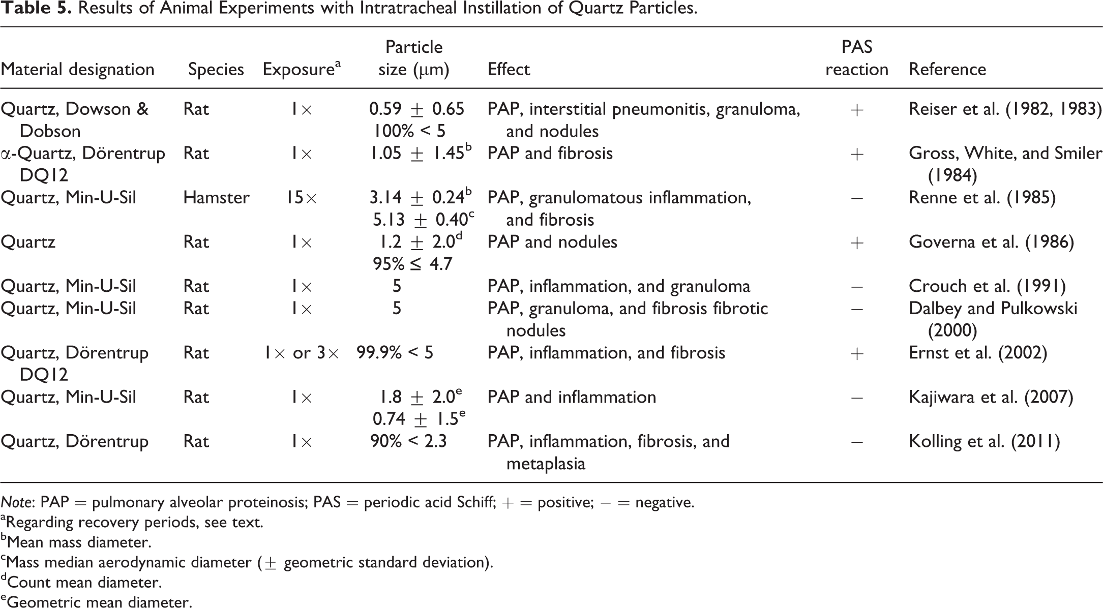
Note: PAP = pulmonary alveolar proteinosis; PAS = periodic acid Schiff; + = positive; − = negative.
aRegarding recovery periods, see text.
bMean mass diameter.
cMass median aerodynamic diameter (± geometric standard deviation).
dCount mean diameter.
eGeometric mean diameter.
Rats receiving a single instillation of quartz through a tracheostomy were observed for up to 12 months. After 1 week, typical silica-containing lung granulomas and areas of PAP associated with “interstitial pneumonitis” were present. These changes and silicotic nodules were still observed in most of the lungs 1 yr after treatment (Reiser et al. 1982, 1983). Single administration of quartz to rats produced PAP during the 24-week recovery period (necropsy after 3, 12, and 24 weeks) and fibrotic nodules (Gross, White, and Smiler 1984). Following 15 weekly quartz instillations to hamsters (posttreatment observation until 24.5 months of age), granulomatous inflammation was the most striking pulmonary lesion followed by pulmonary fibrosis. There were also indications of PAP (Renne et al. 1985). A single quartz dose administered to rats produced typical granulomas, most of them mature silicotic nodules, and areas of PAP (Governa et al. 1986). PAP (together with active inflammatory reactions and numerous granulomas) was diagnosed in rats after single instillation of quartz (Crouch et al. 1991). Single quartz treatment of rats followed by necropsy at various time points up to 6 postexposure months revealed PAP, granulomas with early fibrosis, and fibrotic nodules (Dalbey and Pulkowski 2000). Rats were administered quartz once (4-week study) or 3 times (3-month study, treatment monthly). Histopathology revealed PAP in all animals together with bronchioloalveolar hyperplasia, interstitial fibrosis and alveolar/interstitial inflammatory cell infiltration, granulomatous and fibrosing alveolitis, and fibrosing pleuritis (Ernst et al. 2002). Rats, which received a single dose of quartz of two different sizes, were sacrificed at 3 days, 1 week, and 1, 3, and 6 months after exposure. PAP and inflammation started from day 3, with increasing scores until the end of studies, the larger particles being somewhat more inflammatory than the smaller ones (Kajiwara et al. 2007). A single dose of quartz to rats with subsequent observation over 29 months produced the following pulmonary changes: bronchioloalveolar hyperplasia, inflammation, interstitial fibrosis, PAP, and goblet/squamous cell metaplasia. In addition, the incidence of lung tumors was markedly increased (about 40% vs. 0% in controls; Kolling et al. 2011).
In a large number of studies with rats, mice, and guinea pigs using single intratracheal instillation of several crystalline quartz varieties at similar particle sizes as described in Table 5, PAP was not diagnosed but inflammation and/or fibrosis was diagnosed (e.g., Dauber et al. 1980; Driscoll et al. 1990; Henderson et al. 1995; Lugano, Dauber, and Daniele 1982; Roursgaard et al. 2011; Warheit, Webb, and Reed 2007; Wiessner et al. 1988). With the exception of the study in mice by Roursgaard et al. (2011), none of the studies utilized a PAS reaction.
In summary, a PAP was diagnosed in all inhalation studies with crystalline quartz (except the study of Heppleston [1986] in guinea pigs), and in all 4 studies with intratracheal instillation of crystalline quartz, in which a PAS reaction was carried out. In 4 studies with intratracheal instillation of quartz PAP partly accompanied by pneumonitis/inflammation and/or granuloma/fibrosis were/was diagnosed also without a PAS reaction. With increasing study duration, PAP was accompanied by other lesions (inflammation, fibrosis, and silicotic nodules). In all other inhalation and instillation studies with quartz in which no PAS reaction was carried out, PAP was not described. However, based on the lesions, PAP may have been present. Due to the rather heterogeneous data on the particle properties of the quartz samples used, no statement on their capability of inducing PAP or on their potency can be made. The database on PAP induction in species other than the rat is rather limited. The data of Gross and deTreville (1968), Renne et al. (1985), and Heppleston (1986) could indicate that the rat is the most sensitive species followed by mouse, hamster, and guinea pigs.
A large number of investigations with amorphous SiO2 are available (e.g., Arts et al. 2007; Ernst et al. 2002; Hemenway et al. 1986; Kolling et al. 2011; Landsiedel et al. 2014; Lee and Kelly 1993; Reuzel et al. 1991; Rosenbruch et al. 1990). A PAS reaction in lung sections seems to have been carried out in only one of these studies (Ernst et al. 2002). Inflammatory and fibrotic pulmonary changes are reported in part. As far as exposure conditions were comparable, a considerably lower pneumotoxic potential can be recognized than with crystalline SiO2. The diagnosis PAP was not made.
Titanium Dioxide
Whole-body exposure of rats to TiO2 at three concentration levels (6 hr/day, 5/week for 24 months with interim necropsies at 3, 6, and 12 months; mass median aerodynamic diameter: 1.5–1.7 µm; about 84% respirable size; rutile structure) produced a PAP (PAS-positive reaction) apart from a series of other pulmonary changes (e.g., type II pneumocyte hyperplasia, cholesterol granulomas, fibrosis in alveolar walls, and squamous metaplasia). PAP was first diagnosed by the end of 3 months. After 24 months, 56% of animals at the intermediate and 97% of animals at the highest level were diagnosed with PAP. No PAP was found at the lowest level. A high incidence of lung tumors was diagnosed only at the highest level (Lee, Trochimowicz, and Reinhardt 1985; Lee et al. 1986).
Several inhalation studies with TiO2 mainly of rutile and/or anatase structure in rats and mice lasting 5 days up to 2 yr showed either no lung pathology or inflammatory and/or slight fibrotic changes (Baggs, Ferin, and Oberdörster 1997; Bermudez et al. 2002; Driscoll et al. 1990, 1991; Ma-Hock et al. 2009; Muhle et al. 1989, 1995; Velan, Kumar, and Cohen 1993). None of these studies included a PAS reaction.
Discussion
Diagnosis of PAP
It is clear that a positive PAS reaction helps in making the diagnosis of PAP. In the normally used hematoxylin–eosin staining, PAP can also be recognized. It appears, however, that without a PAS reaction, other pathological findings eclipse the accumulation of surfactant, so that it is either not worth mentioning or overlooked. As an example, the results of reevaluation of “indium lung diseases” by a multidisciplinary panel can be mentioned. The original diagnosis in the 10 cases was 7 cases of interstitial lung disease and 3 of PAP. The reevaluation revealed “intra-alveolar exudate typical of alveolar proteinosis” (n = 9) and “fibrosis” (n = 9; Cummings et al. 2012). At the same time, this reevaluation demonstrates the problem of varying diagnoses of the same pathological lung condition by different pathologists. In some cases of animal experiments, where the description and figures presented clearly point to a PAP (among other changes), the authors do not mention this diagnosis (e.g., Leach et al. 1961; Tanaka et al. 2010; Warheit et al. 1991).
Another problem when making a PAP diagnosis in humans can probably be found in the fact that practically all cases that had contracted PAP were characterized by severe to life-threatening symptoms requiring extensive treatment, such as whole lung lavage, and with a frequently fatal termination. The fact that milder forms are also present, involving spontaneous remission to even symptom-free development, was not realized until recently, as a result of serial examinations (Briens et al. 2002; Inoue et al. 2008). According to the experience gained from the animal experiments or also, for example, from the 2 indium-exposed PAP patients (in whom a fibrosis subsequently developed), PAP could initially have been present in a number of human cases with particle-induced fibrosis, and was likely the inducing factor for the subsequent fibrosis, based on the evidence from the experimental animal studies.
Particle Properties
From the paucity of data in humans, no firm statements can be made with regard to the question as to what particle properties trigger the development a PAP. As can be expected, the majority of the particles found in the lungs were smaller than 5 µm because otherwise they would not have reached the alveoli (Abraham and McEuen 1986; Keller et al. 1995; Miller et al. 1984; Xiao et al. 2010). In this respect, however, the particles were not different from those that were reported to induce, for example, fibrosis (De Vuyst et al. 1986; Elo et al. 1972; Gilks and Churg 1987; T. Homma et al. 2003; Määttä and Arstila 1975). The crystallinity is certainly of decisive importance. Amorphous variants of these particles obviously do not induce PAP (Ernst et al. 2002; Rosenbruch et al. 1990). The database on different crystal forms (e.g., for crystalline aluminum and silicon particles) with regard to the induction of PAP are too small for any firm conclusion. Other determinants of lung effects, such as particle surface properties and area, have not been systematically studied in the case of PAP.
Sequence of Events
In cases of human PAP, especially in cases of prolonged exposure, inflammation and fibrotic lesions have often been observed (e.g., Alper et al. 2008; Arakawa et al. 2005; Buechner and Ansari 1969; Keller et al. 1995; Kefeli et al. 2012; Xipell et al. 1977). In the 2 cases of PAP following indium exposure, fibrosis subsequently developed (Cummings et al. 2012). In those animal experiments with detailed investigation of the sequential pathological changes, the results show that PAP appeared first and/or at lower-dose levels than inflammation and fibrosis. From the results with metallic aluminum powder inhalation of rats (Gross, Harley, and DeTreville 1973), the inhalation studies with GaAs and InP, In2O3, and ITO in rats and mice (Nagano, Gotoh, et al. 2011; Nagano, Nishizawa, Eitaki, et al. 2011; NTP 2000, 2001) and the studies with intratracheal instillation of quartz in rats (Gross, White, and Smiler 1984), it may be concluded that PAP precedes inflammatory and fibrotic responses.
It may be concluded that PAP and lung fibrotic changes are not mutually exclusive but are rather a question of time; the longer the exposure, the more likely PAP will evolve into fibrosis, with the identification of PAP becoming more difficult, especially without the use of PAS stain. In most studies with intratracheal instillation, inflammatory reactions or fibrosis appeared first or at the same time as PAP or even without indications of PAP, especially those with single administration. In many cases where PAP, inflammation, and fibrosis were diagnosed, it is not possible to state which changes came first due to the absence of examinations at the relevant time points. One certain conclusion can be drawn from the animal experiments: inflammation and/or fibrosis are/is regular conditions accompanying a long-term particle-induced PAP.
Species Sensitivity
From the relatively few studies available, allowing a ranking of the sensitivity of various laboratory species as regards the induction of PAP, the following sequence can be derived: rat > mouse > hamster ≈ guinea pig (Gross and deTreville 1968; Gross, Harley, and DeTreville 1973; Heppleston 1986; Nagano, Gotoh, et al. 2011; Nagano, Nishizawa, Eitaki, et al. 2011; NTP 2000, 2001; Renne et al. 1985). On the basis of the data presented, it is difficult to answer which of these species comes closest to the sensitivity found in humans. Secondary PAP from very poorly soluble particular substances is, in humans, a very rare disease. From the very few human cases comprising aluminum, indium, and titanium dioxide, of which a large number of persons have been or still are exposed to rather high levels, it can be assumed that at least rats and mice are markedly more sensitive than humans.
Comparison between Particle-induced PAP in Animals and Humans
In earlier studies, a high level of agreement has been found regarding pathomorphological appearance and biochemical composition of surfactant accumulation (Eden and von Seebach 1976; Heppleston, Wright, and Stewart 1970). On comparison between histological (light and electron microscopy) lung findings of an experimentally induced PAP by the inhalation of quartz in rats with 2 human PAP cases, no major differences were found either at the alveolar walls or in the intra-alveolar material (Heppleston and Young 1972). In an investigation comparing the lung response of humans and rats to coal dust, silica, and talc, based on pathology materials, both similarities and differences in response to the same agents were observed. For example, a high score of centriacinar fibrosis was to be seen in human silicosis, whereas in rats this was very low. On the other hand, the score of PAP was very high in talc- and silica-exposed rats and very low in humans. Also, acute intra-alveolar inflammation and alveolar epithelial hyperplasia were both greater in rats than in humans exposed to the same agents (Green, Vallyathan, and Hahn 2007). The course of the disease in humans and animals also seems to show major differences; whereas, in humans, PAP is often a life-threatening condition, animals, especially rats, are able to tolerate even a massive PAP over long periods of time without any clinical signs (Gross, Harley, and DeTreville 1973; Nagano, Nishizawa, Umeda, et al. 2011; NTP 2000). In spite of these differences, it should be assumed that PAP demonstrated in animal experiments—under corresponding exposure conditions—should be considered relevant for humans.
In the absence of relevant data, it is not possible to make any definitive statements on the strength of PAP induction of the different materials for humans.
Footnotes
Author’s Note
The author has the sole responsibility for the writing and content of this review.
Acknowledgments
The technical assistance of Mrs. Dr. Rita Groß and Mrs. Bianca Leubner is gratefully acknowledged. The author is grateful for the support of Dr. Joel Leininger in revising this review.
Declaration of Conflicting Interests
The author(s) declared no potential conflicts of interest with respect to the research, authorship, and/or publication of this article.
Funding
The author(s) disclosed receipt of the following financial support for the research, authorship, and/or publication of this article: This review was partly sponsored by a grant of the German Federal Ministry of Education and Research.