
Abstract
The purpose of this article is to describe a case of drug-induced cutaneous toxicity observed in cynomolgus monkeys and to introduce approaches attempted in order to elucidate mechanisms. The test article was a small molecule with a ubiquitously distributed target, especially in rapidly dividing cells, and which modulated cell cycle regulation. After 7 consecutive days of oral dosing, animals developed multifocal skin lesions. The lesions were characterized clinically by vesicles and scabs formation and were distributed mainly in thin-skinned areas of the body including the cheek, chest, abdomen, and inner limbs. Microscopically, the lesions were confirmed as epidermal vesicle formation and ulceration. Immunohistochemical staining revealed that the levels within the epidermis where separation (vesicle formation) occurred were not consistent. The differential diagnoses for vesicular skin lesions and our efforts to elucidate the mechanism of toxicity using in-house database searches and immunohistochemistry are discussed. To the best of our knowledge, similar cutaneous toxicity has not been reported previously, although there are reports of other types of cutaneous toxicities. Understanding the mechanisms of the toxicity is very important when assessing human relevancy during drug development. Our investigative approach can be utilized when unusual skin toxicities are observed in the future.
Keywords
Introduction
There are many reports of drug-induced cutaneous toxicities. However, cutaneous toxicity similar to the type we have detected has not been reported previously. Therefore, a case of drug-induced cutaneous toxicity observed in a 2-week cynomolgus monkey study is described, and our investigative efforts for the case are introduced.
The Case Presentation
Material and Method
The project background
The test compound (compound-X) is a small molecule with a ubiquitously distributed target, especially in rapidly dividing cells, which modulates cell cycle regulation.
Study design (2-week monkey study)
Compound-X was planned to be administered orally via gavage for 14 consecutive days to male cynomolgus monkeys (Macaca fascicularis; Asian mainland origin) in a 2-week dose-range finding toxicity study (Table 1). A total of 9 animals (3 animals per group; vehicle control, low dose, and high dose) were assigned to the study. At the completion or termination of the study, complete necropsies were performed; selected tissues were collected and examined microscopically. The immunohistochemistry (IHC) investigations on the skin lesions were conducted using keratinocyte and leukocyte markers listed in Table 2. The skin samples we used for comparison were collected from (1) skin lesions, (2) concurrent controls that were collected from the identical area of the skin lesions, and (3) nonlesion skins from the affected animals. All steps of each of the IHC stains were performed by a Leica BOND RX autostainer (Leica, Buffalo Grove, IL) using a Leica BOND Polymer Refine Red Detection kit or a Vector ImmPRESS Anti-Mouse Ig Peroxidase Kit. Throughout the study period, all procedures involving animal use were approved by the Novartis Institutional Animal Care and Use Committee.
Study Design and Animal Allocation.
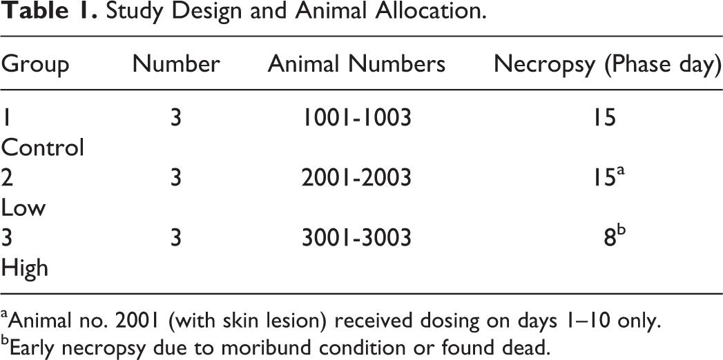
aAnimal no. 2001 (with skin lesion) received dosing on days 1–10 only.
bEarly necropsy due to moribund condition or found dead.
Keratinocyte and Leukocytes Markers for IHC Staining.
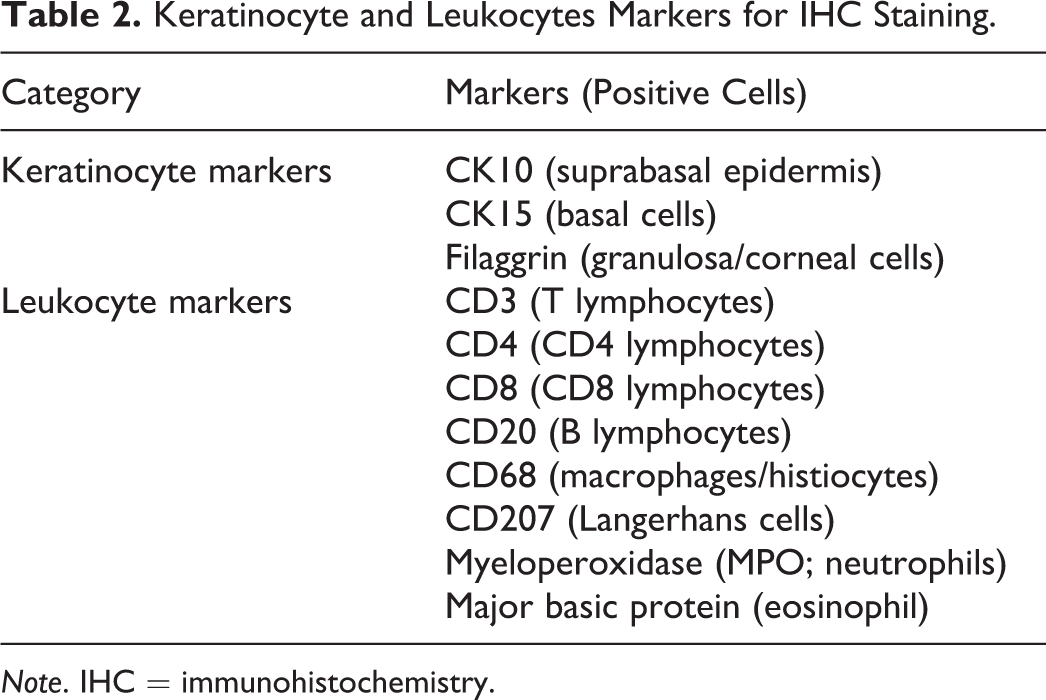
Note. IHC = immunohistochemistry.
Results
Clinical signs and mortality
There was dose-dependent onset of skin lesions (Figure 1). After 7 consecutive days of dosing (on day 7), 2 of the 3 animals in the high-dose group developed multifocal skin lesions. The skin lesions were characterized clinically by the presence of scabs formation. The lesions were distributed mainly in glabrous/thin-skinned areas of the ventral body including the cheeks, chest, abdomen, and inner limbs. In addition, 1 animal had similar changes in the buccal mucosa. All animals that developed skin lesions also had concurrent systemic clinical signs including decreased activity and body temperature. Despite the veterinary care, on day 8, all animals in the high-dose group were either found dead or euthanized due to poor general condition.
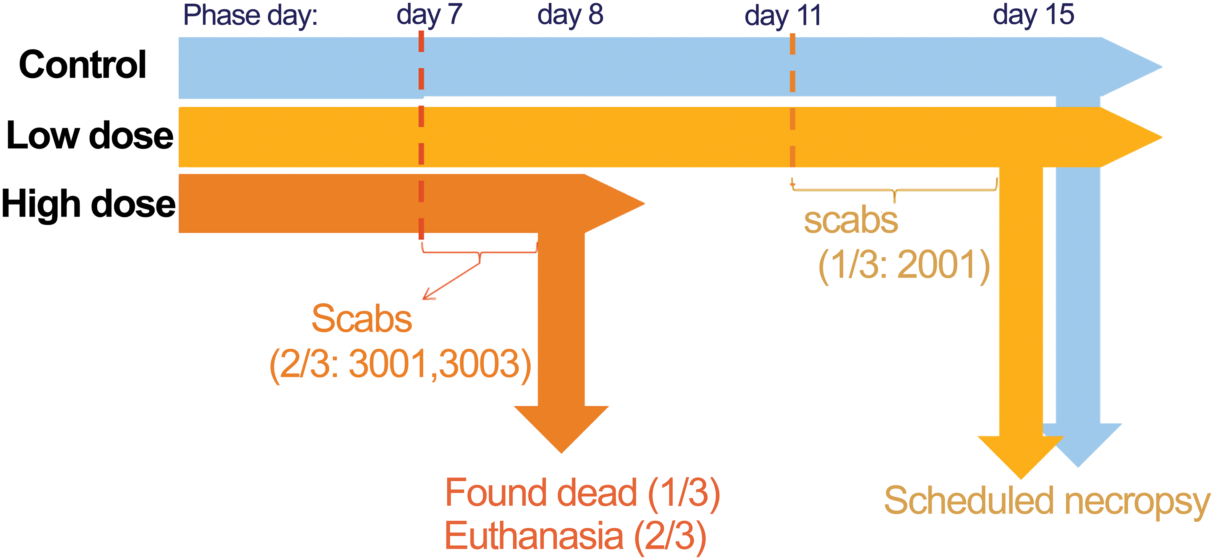
Onset of skin lesions and mortality.
For the low-dose group, 1 of the 3 animals developed skin lesions on day 11. The skin lesions in the low-dose animal were distributed similar to high-dose animals and were characterized by vesicle and scab formations. As in the high-dose animals with skin lesions, this animal also had systemic clinical signs. The subsequent dosing of the test article to this low-dose animal was withheld until the last day of study period on day 15. The remaining 2 animals in the low-dose group did not have any skin lesions or clinical signs throughout the study period.
Microscopic observation
There were various types of skin lesions mainly presented as vesicles and/or pustules formation, full thickness epidermal necrosis (Figure 2), and ulceration. Generally, there were relatively a few inflammatory cell infiltrations to the epidermis and fewer infiltrations in the dermis and subcutis unless ulcerated.
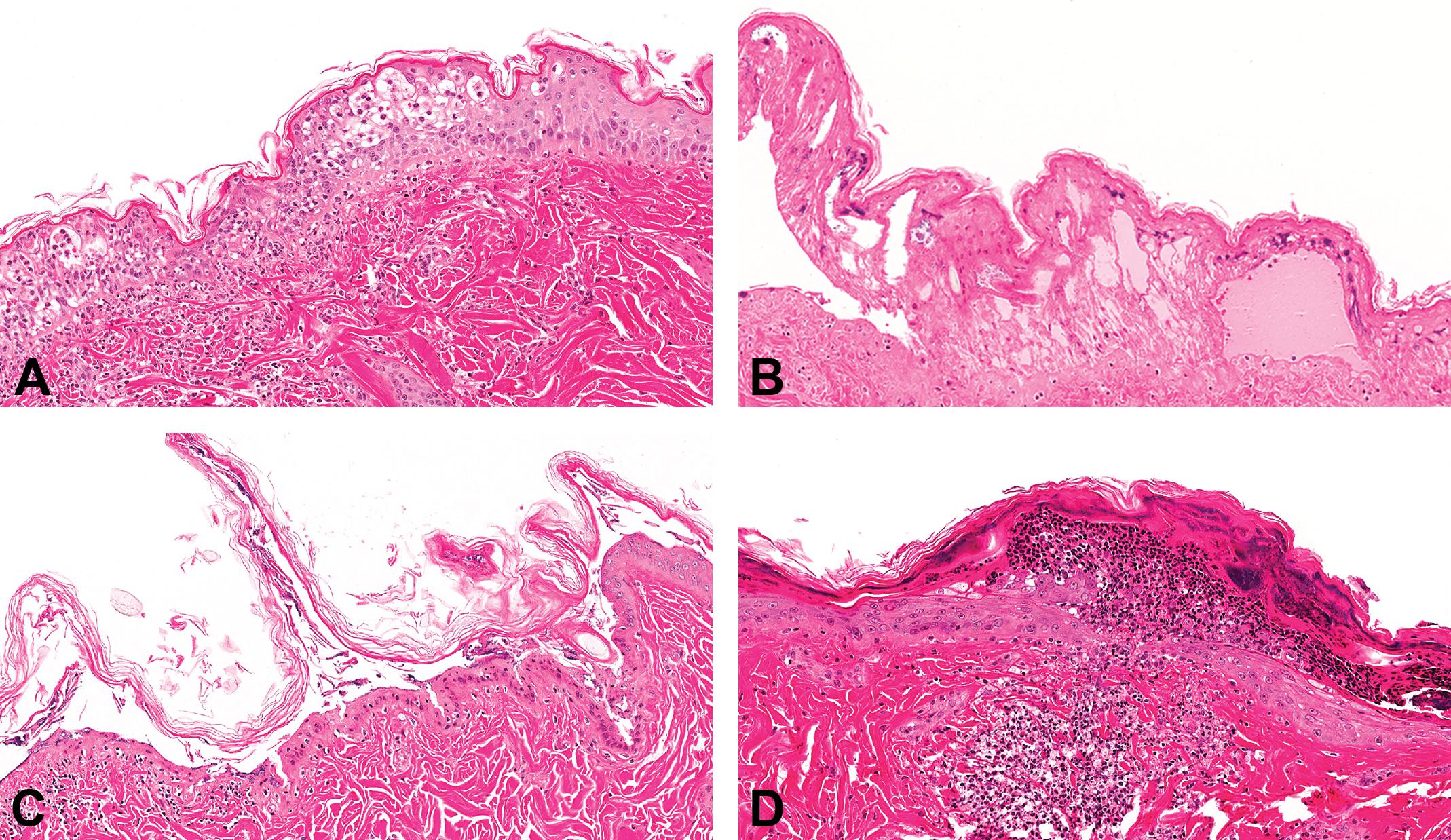
Light microscopic photographs of skin lesions by compound-X (hematoxylin and eosin stain). (A) Vesicle formation in the hyperplastic epidermal layer. Some vesicles contained inflammatory cells and cellular debris. The spongiosis and cytoplasmic vacuolation were also observed. Low-dose animal. (B) Vesicle formation in the epidermal layer with full thickness epidermal necrosis. Intralesional bacterial colonization without inflammatory cell infiltrate was observed. Low-dose animal. (C) Full thickness epidermal necrosis without epidermal hyperplasia. Detachment of epidermis occurred at the subcorneal level without inflammatory cell reactions. Loss of epidermal layers can also be observed in some areas. High-dose animal. (D) Pustule formation at the subcorneal level, covered by hyperkeratotic keratinocytes with bacterial colonization. Pustule contained neutrophils and a few sloughed epithelial cells. High-dose animal.
The vesicles formation seemed to occur at variable levels of the epidermis and occasionally contained a few inflammatory cells (predominantly neutrophils and macrophages), eosinophilic proteinaceous materials, and/or cellular debris. Where it was ulcerated or severely necrotized, the lesion also contained bacterial colonization. In the relatively less affected areas, epidermal hyperplasia, intercellular edema (spongiosis), and intracellular edema (vacuolation) were observed.
IHC
The IHC staining results with keratinocyte markers confirmed that the levels within the epidermis where separation occurred were not consistent across sites or animals/groups. The IHC staining results with leukocyte markers showed there were immunopositivities of inflammatory cells for Cluster of Differentiation (CD) 68 and myeloperoxidase (MPO; Figure 3), and there was no increased immunopositivity for the rest of markers listed in Table 2 (e.g., lymphocytes or eosinophils) when compared to control animals. Therefore, predominant inflammatory cell infiltrates were confirmed as MPO-positive neutrophils and CD68-positive macrophages without involvement of T or B lymphocytes or eosinophils.
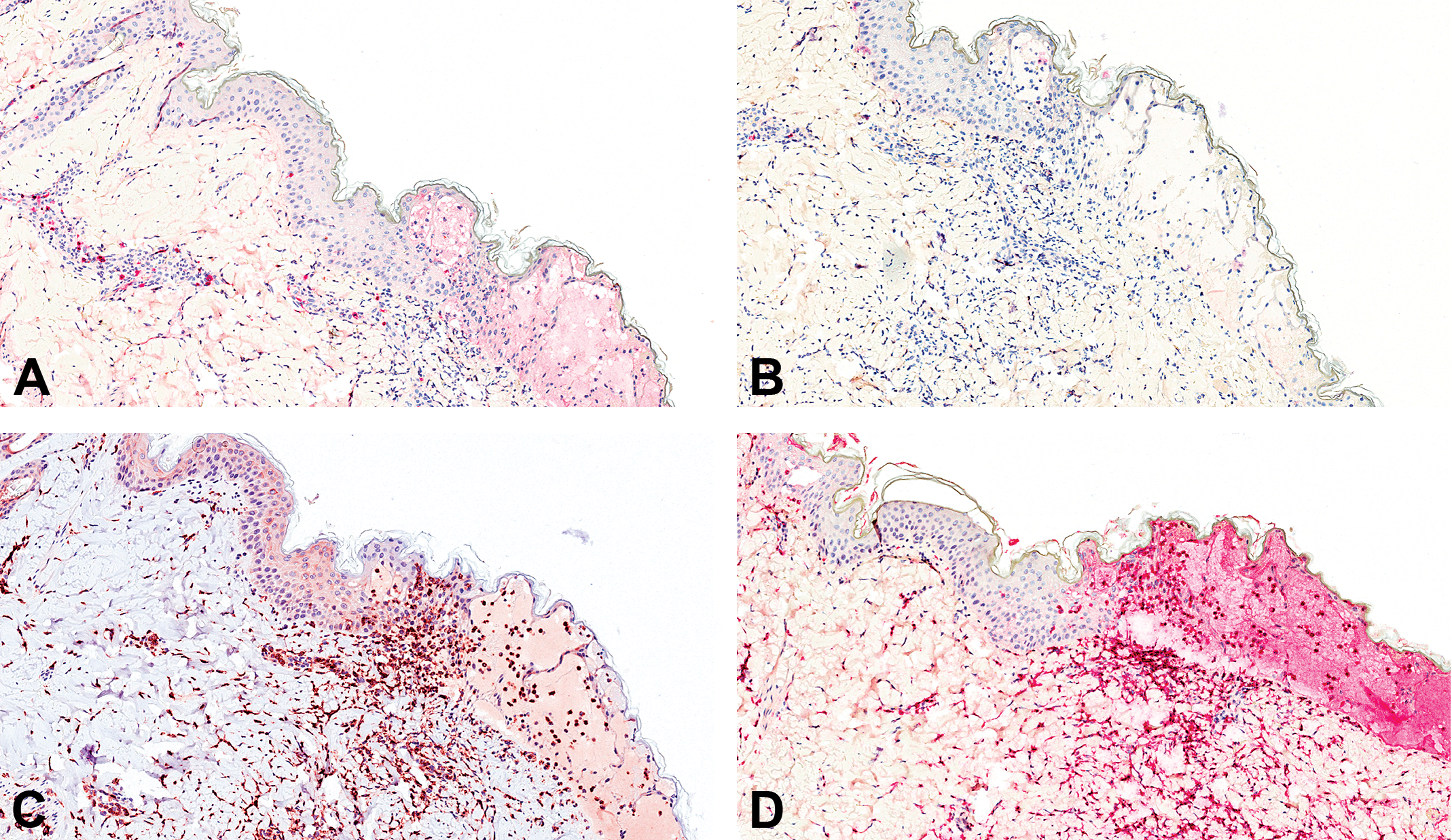
Immunohistochemistry staining with CD3 (A), CD20 (B), CD68 (C), and MPO (D). (A) There were few CD3-positive T lymphocytes mainly observed in the adnexal region. The expression level and distribution of CD3-positive cells were similar to what was observed in control animals and considered not to be associated with the skin lesion. (B) Inflammatory cells showed negative immunopositivity for CD20. (C and D) There were increased numbers of CD68-positive macrophages (C) and myeloperoxidase-positive neutrophils (D) in the skin lesion, and predominant inflammatory cells were confirmed as macrophage and neutrophils.
Differential Diagnosis, Hypotheses, and Investigation of the Cutaneous Toxicity
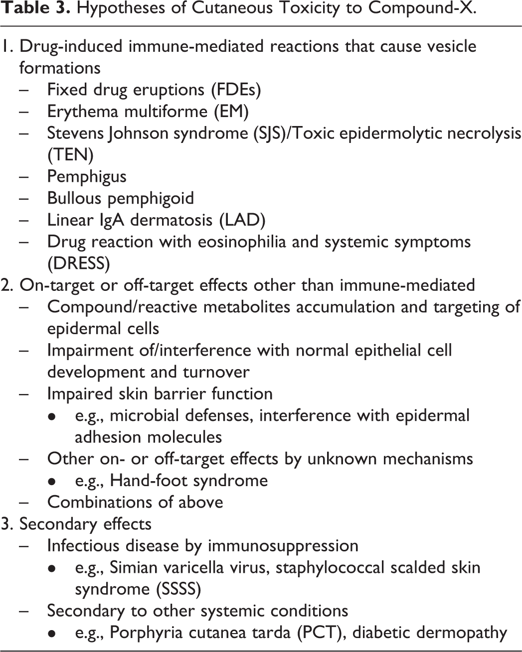
The hypotheses for the mechanisms of drug-induced skin lesions can be categorized into three broad types: (1) immune-mediated reactions, (2) other on-/off-target effects, and (3) secondary effects. Our preliminary work touching upon each of these categories (summarized in Table 3) is presented below.
Hypotheses of Cutaneous Toxicity to Compound-X.
Immune-mediated Reactions
The drug-induced immune-mediated skin lesions reported in humans that cause vesicle formation includes fixed drug eruptions, erythema multiforme, Stevens Johnson syndrome/toxic epidermolytic necrolysis, pemphigus, bullous pemphigoid, linear IgA dermatosis, and drug reaction with eosinophilia and systemic symptoms.
The key microscopic features that can be used for assisting diagnosis include precise characterization of the level of separation within the epidermis, inflammatory cell phenotype and distribution, and other findings including existence of keratinocyte apoptosis and/or the presence of vasculitis.
In our case, we used IHC for identifying the level of separation within the epidermal layers and confirming phenotypes of inflammatory cells. The IHC staining results confirmed that the levels within the epidermis where separation occurred were not consistent across sites or animals/groups and that predominant inflammatory cell infiltrate were comprised of neutrophils and macrophages without involvement of lymphocytes. These IHC staining results indicated that the conditions observed in our case probably did not fit classical elementary lesions associated with immune-mediated blistering skin lesions as reported in human or animal conditions (Lee and Thomson 2006; Kasperkiewicz and Zillikens 2007; Shiota 2009; Weyers and Metze 2011; Yager 2014; Roujeau 2015). However, because detailed planned collections and enhanced immune assessments (e.g., autoantibody detection or rechallenge test) were not conducted, possibility of immune-mediated reactions could not be completely excluded.
On- or Off-target Effects
The possible mechanisms of skin lesions underlying on- or off-target effects include (1) compound/reactive metabolites accumulation and targeting of epidermal cells, (2) impairment of/interference with normal epithelial cell development and turnover, (3) impaired skin barrier function (e.g., microbial defenses and/or interference with epidermal adhesion molecules), (4) other on- or off-target effects by unknown mechanisms, and (5) combinations of these mechanisms. Some examples of IHC markers for investigation of potential on- or off-target effects by compound-X are listed in Table 4.
IHC Markers for Investigation of On- or Off-target Effects by Compound-X.
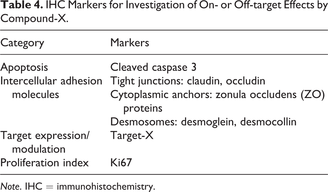
Note. IHC = immunohistochemistry.
In addition, we investigated the mechanisms of toxicity specific to our compound (i.e., compound-X) using Mechanism of Action (MoA) Central, a novel target/mechanism of action search engine developed at Novartis (Figure 4; Selinger et al. 2015). Briefly, MoA Central starts with a small molecule of interest and identifies compounds with related chemical structures or biological fingerprints. Biological fingerprints come from a number of sources, including transcriptional profiles, panels of high throughput screening assays, and cell line proliferation profiles (Barretina et al. 2012). To identify consensus targets, these chemically or phenotypically similar compounds are then searched against a number of sources of compound target information, including biochemical activity databases, chemical proteomics data, and chemical genetics analyses. An MoA Central search using compound-X identified structural analogs with cell line proliferation profiles similar to several phosphoinositide 3-kinase (PI3K) inhibitors. This suggested that direct or indirect modulation of PI3K could play a role in the observed skin toxicity.
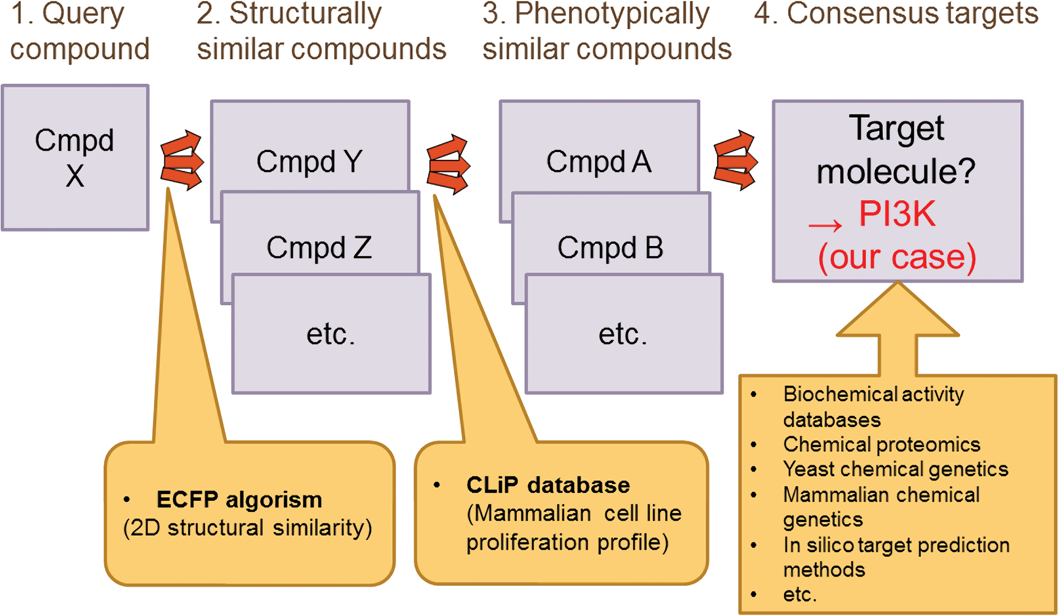
Mechanism of Action Central search strategy.
PI3Ks are a family of enzymes reported to be involved in cellular functions such as proliferation, differentiation, chemotaxis, survival, trafficking, and glucose homeostasis (Katso et al. 2001). PI3Ks are part of the PI3K/AKT/mammalian target of rapamycin (mTOR) pathway, a cancer signaling pathway (Pecorino 2008). Cutaneous toxicities are frequent findings with kinase inhibitors, including PI3K inhibitors (Curry et al. 2014). In addition, considering nature of the primary target of compound-X (i.e., target-X), it was thought to be possible that compound-X could modulate the PI3K/AKT/mTOR pathway. In this way, MoA Central raised another possible hypothesis—modulation of PI3K pathway. As a next step, further investigations of possible on- or off-target effects on our case, including PI3K pathway modulation, will be conducted.
Secondary Effects
Vesicular skin lesions have also been described in several viral or bacterial diseases in humans as well as nonhuman primates. Skin lesions in toxicology studies can be caused as secondary effects associated with immunosuppression or other systemic conditions caused by test articles. The examples of infectious diseases that are reported to cause skin lesions are simian varicella virus and staphylococcus (e.g., staphylococcal scalded skin syndrome). Examples of systemic conditions with effects on primary target organs that can cause secondary skin lesions include porphyria cutanea tarda secondary to liver disease and diabetic dermatopathy secondary to pancreatic disorders or other metabolic targets that cause hyperglycemia.
In our case, the hypothesis of secondary effects was ruled out based on the medical history, microscopic feature of the skin lesions, and other pathological evidences that no changes in the internal organs could be correlated with occurrence of skin lesions.
Conclusion
Drug-induced vesicular skin lesions can be categorized into three causes: (1) immune-mediated reactions, (2) on-/off-target effects, and (3) secondary effects. When assessing the etiopathogenesis of skin toxicity, investigations should be done holistically, as different mechanisms can cause similar morphological features but also the same mechanism can result in different morphological features. Although microscopic features are very important for diagnosis of skin lesions, microscopic features alone are not sufficient to determine the cause of skin toxicity. The clinical conditions such as dosing length until the occurrence of skin lesions, distributions and appearance of the skin lesions, existence of concurrent systemic conditions, and clinical pathology data are also very important factors to assist diagnosis of skin lesions.
Different approaches can be used to investigate mechanisms of cutaneous toxicities. First, depending on the project status, further investigative studies can be conducted to obtain supporting evidence to differentiate immune-mediated and nonimmune-mediated effects (e.g., rechallenge test with biopsy plan, etc.). Second, using existing formalin-fixed paraffin-embedded tissues, tissues-based investigative assessments (e.g., IHC and in situ hybridization) can be done. In our case, we found out that IHC staining with epidermal and leukocyte markers can help narrow down differential diagnosis listings. Also, the use of several IHC markers can be used to look for direct test article effects (e.g., target modulation). For the tissue collections, collection of skin lesions including margins is important, as it provided useful information while the nonlesion skins of affected animals did not provide any valuable information in our case. In addition, collecting concurrent control tissues from the identical location of skin lesions, and processing them in the same manner, can help in making decisive conclusions. Third, if available, nonfixed skin tissues (e.g., fresh/frozen samples) and serum samples can be utilized for some additional investigations (e.g., omics approaches, direct immunofluorescence methods, measuring compound concentrations within the tissues, or autoantibody detections). Lastly, computational approaches coupled with molecular biology follow-up can be used as a support. In our case, the in-house MoA Central search engine pointed to the PI3K pathway as a possible mechanistic component of compound-X-induced skin toxicity, a hypothesis that was not suggested by a pathological approach alone.
In conclusion, we are reporting here a unique skin toxicity case due to a small molecule given orally in cynomolgus monkeys. The mechanisms of cutaneous toxicity in our case could not be determined due to limitations in investigative possibilities. However, after running an MoA Central search, we are now investigating possible on- or off-target effects by compound-X in association with PI3K pathway modulation. Understanding the mechanism of the toxicity is very important when assessing human relevancy during developing drugs. Our investigative efforts can be utilized when unusual skin toxicity is observed in the future.
Footnotes
Acknowledgments
I would like to thank Dr. Daher Ibrahim Aibo and Ms. Sherry Nady for IHC discussions and staining; Drs. Douglas Selinger and Melissa L. Wilbert for computational support and collaboration; colleagues at Novartis for insightful discussions and supports for the investigation.
Author Contribution
The author (RK) contributed to conception or design; data acquisition, analysis, or interpretation; drafting the manuscript; and critically revising the manuscript. The author gave final approval and agreed to be accountable for all aspects of work in ensuring that questions relating to the accuracy or integrity of any part of the work are appropriately investigated and resolved.
Declaration of Conflicting Interests
The author(s) declared no potential conflicts of interest with respect to the research, authorship, and/or publication of this article.
Funding
The author(s) received no financial support for the research, authorship, and/or publication of this article.