
Abstract
The upper portion of the rat kidney pelvis has specialized anatomic structures referred to as fornices. Fornices have a role in urine concentration. Spontaneous lesions including mineralization, epithelial hyperplasia, and inflammatory cell infiltration may occur in the area of the fornices. However, little information regarding specific historical control data or the spontaneous development of these findings in male and female fornices is known. Understanding spontaneous age-related lesions in the area of the fornices versus other portions of the kidney pelvis may be relevant in the identification of test article–induced changes. A retrospective study was conducted of male and female Sprague-Dawley rat kidney fornices over several time points to determine the incidence and severity of mineralization, epithelial hyperplasia, and inflammatory cell infiltration. Based on this investigation, these lesions appeared to increase over time and, in general, occurred earlier and with a greater incidence in females. Regarding those chemicals that may result in lesions of the kidney pelvis, it may be important for pathologists to separately diagnose lesions of the fornices from other portions of the kidney pelvis to help differentiate between any spontaneous age-related and induced changes.
Introduction
Although the kidney pelvis of the rat is not an overly complex structure, it is noteworthy that the elaborate anatomic extensions of the upper portion of the kidney pelvis that extend into the renal medulla are termed the fornices (Pfeiffer 1968). Fornices begin at the junction of the renal papilla and the pelvis and are lined, in part, by the renal papillary epithelium, which continues into the pelvic urothelium (Verlander 1998; Cohen, Wanibuchi, and Fukushima 2002).The fornices are specialized vault-like folds that serve to increase the surface area of the pelvis, thereby enhancing exposure of the renal medulla to pelvic urine thus playing a role in renal concentration mechanisms (Pfeiffer 1968; Bulger 1986). The upper portions of the fornices might also allow for recycling of urea to short-looped nephrons in the outer medulla due to the increased number of microvillous lining epithelial cells reported in this region (Verani and Bulger 1982).
Spontaneous and induced lesions of the rat kidney pelvis may appear morphologically similar and, therefore, localization of the lesion site may become important in helping to determine the mechanism of an induced lesion. Most reported nonneoplastic lesions of the kidney pelvis represent hydronephrosis, inflammatory infiltrates or exudates, mineralization, urolithiasis, and urothelial hyperplasia (Hard et al. 1999; McInnes 2012; Frazier et al. 2012; Greaves 2012; Frazier and Seely 2013). The presence of renal and kidney pelvic mineralization occurs frequently in chronic rat studies and, at times, complicates study interpretation (Lord and Newberne 1990).
Understanding the development of spontaneous age-related lesions becomes critical in identifying biological or toxicological meaningful changes in a tissue. Furthermore, historical control background information is essential since it gives the pathologist additional reassurance when interpreting a lesion with a potential treatment-related incidence. Pathologists are aware, for instance, that spontaneous lesions of the fornices may be observed that are not present in other portions of the kidney pelvis. For instance, reflux from lower urinary tract obstructions may induce mechanical damage to the region of the fornices, which may lead to bacterial spread into the renal parenchyma and blood stream (Bitz et al. 2001). During the course of the study, if a pathologist observes a potential induced lesion limited to the fornices, it is likely that the laboratory historical control data may include only the general site location of “kidney pelvis,” leaving the concurrent controls as the single source for precise comparison. The purpose of this investigation is to report the incidence and severity of mineralization, epithelial hyperplasia, and inflammatory cell infiltration involving the fornices at different time points in control male and female Sprague-Dawley rats, which may be useful to toxicological pathologists.
Materials and Methods
Hematoxylin and eosin (H&E)-stained kidney sections of sacrificed control male and female Sprague-Dawley rats were reviewed from three 4-week studies, five 13-week studies, two 17-week studies, seven 26-week studies, one 30-week study, three 39-week studies, and two 104-week studies. Study specific information regarding the study length and number of studies investigated, strain, number of animals reviewed per study, age at dosing start (week), and the type of diet is presented in Table 1.
Studies Investigated.
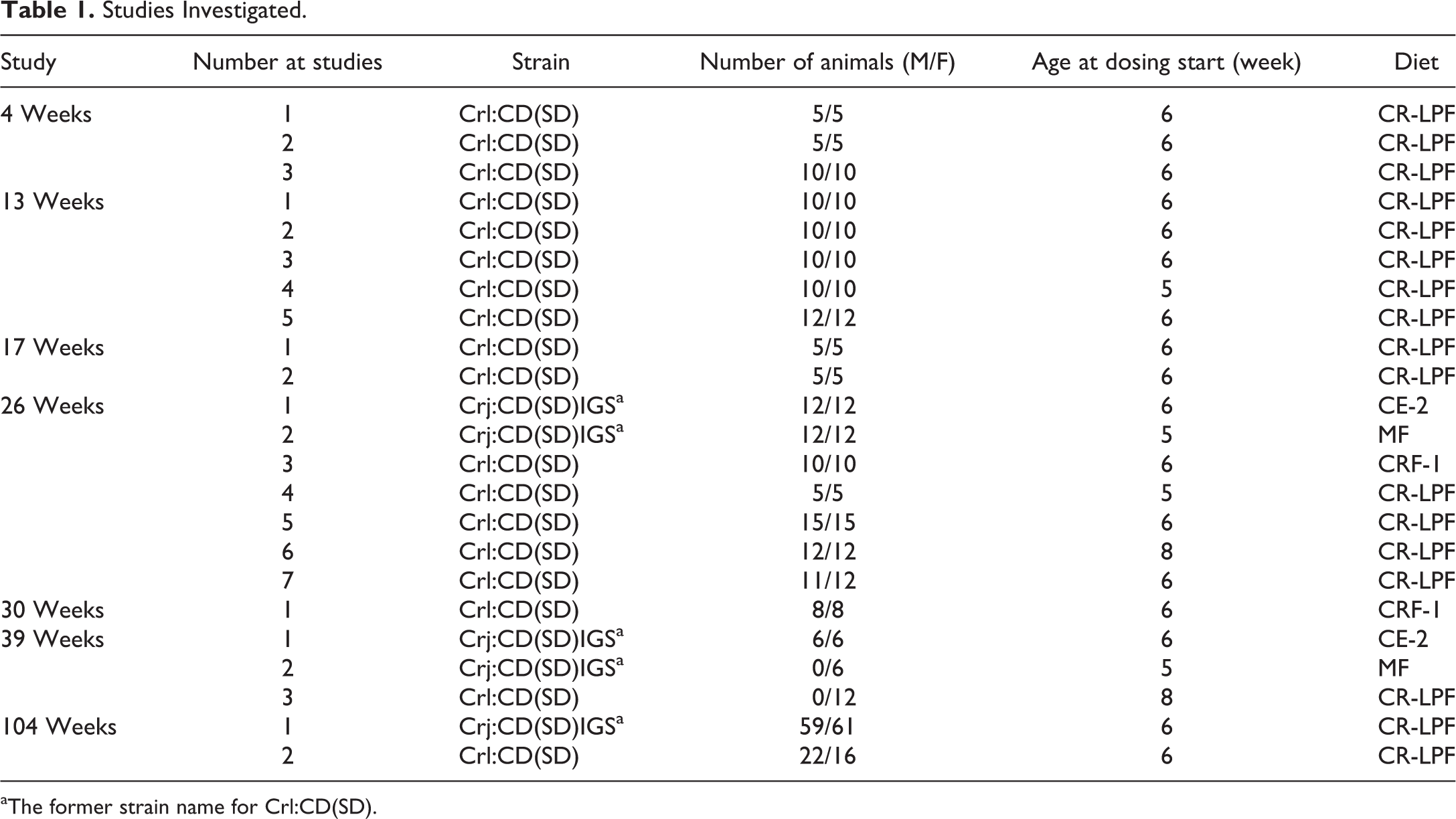
aThe former strain name for Crl:CD(SD).
The Sprague-Dawley rats [Crl:CD(SD) or Crj:CD(SD)IGS] were supplied by Charles River Laboratories Japan, Inc. (Kanagawa, Japan). They were given a commercial pelleted diet (Oriental Yeast Co., Ltd., Japan; CLEA Japan, Inc., Japan) and tap water ad libitum. The diet ingredients are shown in Table 2. Tap water was passed through a 5-μ filter and irradiated. The animals were singly housed in hanging-type stainless steel wire cages or polycarbonate cages. The animal room was maintained at a temperature of 20°C to 25°C, with a relative humidity of 55% ±15% with a 12-hr light/dark cycle. The animals were cared for and euthanized in accordance with the Guidelines for the Care and Use of Laboratory Animals prepared by the Japanese Association for Laboratory Animal Science and the guidelines of our institution.
Diet Ingredients (in 100 g).
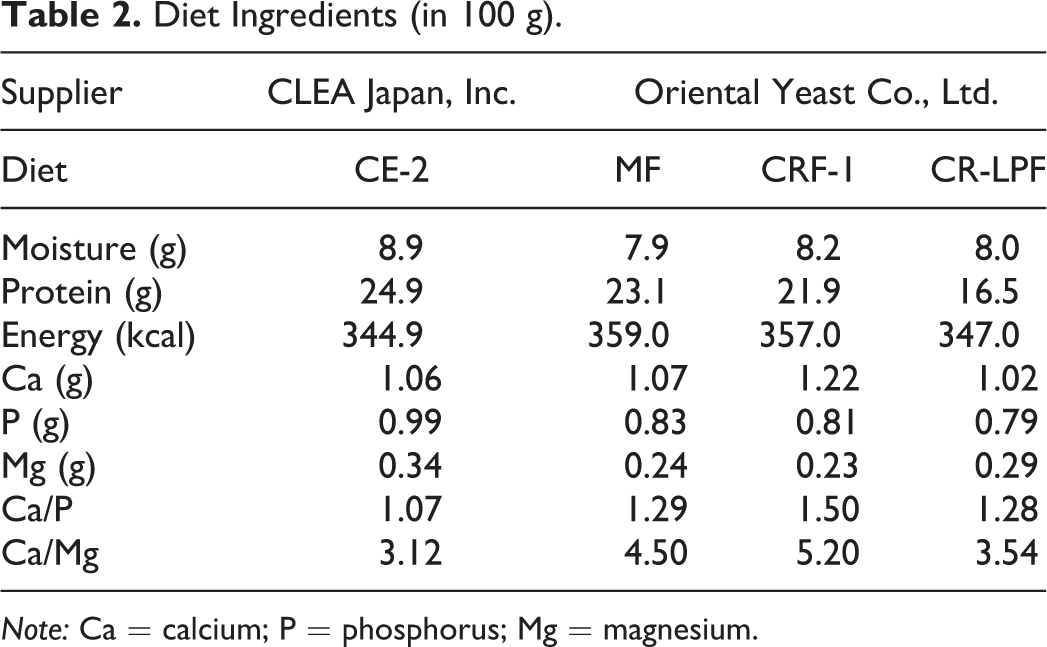
Note: Ca = calcium; P = phosphorus; Mg = magnesium.
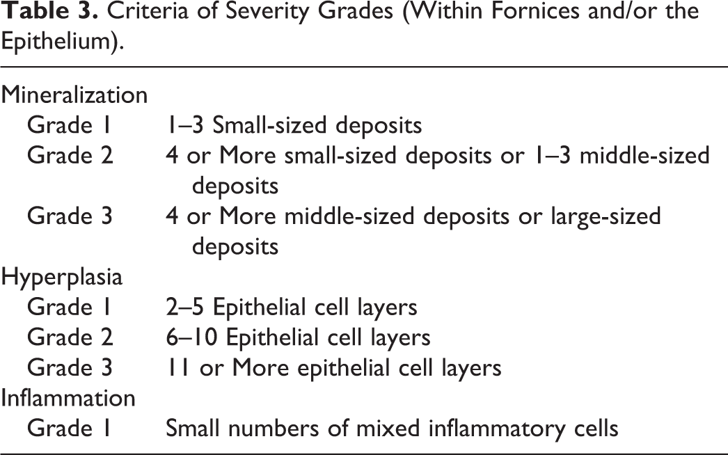
At necropsy, the kidneys were partially cut transversely and fixed whole in 10% neutral-buffered formalin. Tissues were embedded in paraffin and stained with H&E. Kidney cross sections having both fornices were evaluated for the presence and severity of mineralization, epithelial hyperplasia, and inflammatory cell infiltration. Only one diagnosis using the most severe lesion observed from both fornices was recorded for each animal. The severity grade scale used in this investigation is presented in Table 3.
Criteria of Severity Grades (Within Fornices and/or the Epithelium).
Results
Fornices normally appeared as specialized vault-like folds in the upper portion of the kidney pelvis lined, in part, by a single layer of epithelium continuing into the multilayered urothelium (Figure 1).
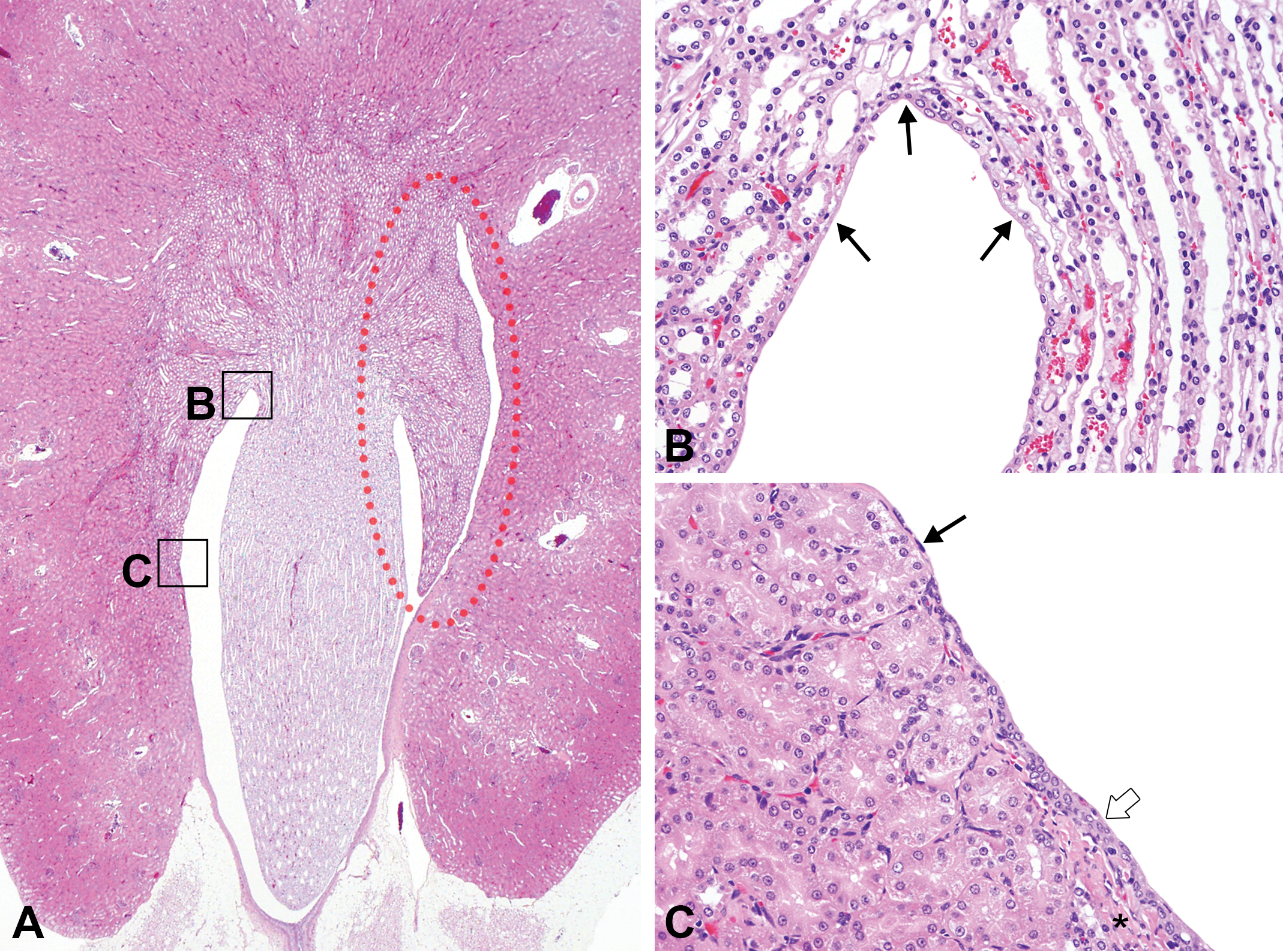
The fornices are specialized vault-like folds in the upper portion of the renal pelvis (A; red-dotted circle) lined by a single layer of epithelium (B and C, arrows). The remaining epithelium of the renal pelvis is lined by urothelium (C; open arrow) and consists of cells of 3–4 layers thick with smooth muscle (C; asterisk) beneath the epithelium. H&E = hematoxylin and eosin stain.
The incidence and severity of mineralization, epithelial hyperplasia, and inflammatory cell infiltration over several time points is presented in Table 4 and Figure 2.
Incidence of Each Pathological Finding.
aIncidence (%) = percentage given as number of animals with lesions per number of animals examined histopathologically.
Incidence of pathological findings. Severity represented by colored blocks. M = male; F = female.
Histologically, mineralization was characterized as small, irregularly shaped, basophilic deposits present within the fornices or closely associated with the epithelium (Figure 3A–C). Mineralization was first observed at 26 weeks in females but was not observed until 104 weeks in males. In females, the incidence of mineralization increased dramatically from 39 weeks to 104 weeks. At 104 weeks, the incidence of mineralization, in females, was nearly 3 times that of males at 104 weeks and, in general, more severe.

(A) Grade 1 mineralization. Small basophilic, multilamellar mineralized bodies (arrows) are deposited within the fornix and/or in the epithelium. (B) Grade 2 mineralization of fornix. (C) Grade 3 mineralization of fornix. H&E = hematoxylin and eosin stain.
Focal areas of epithelial and urothelial hyperplasia were observed in the vicinity of the fornices (Figure 4A–C). Grade 1 severity hyperplasia was first detected in the 13-week study in both males and females, with an incidence of nearly 2% and 4%, respectively. Over time, the incidence and severity of hyperplasia increased. By 104 weeks, the incidence of epithelial hyperplasia increased to 74% in males and 95% in females.
(A) Grade 1 hyperplasia (arrow) of fornix. (B) Grade 2 hyperplasia of fornix. (C) Grade 3 hyperplasia of fornix. H&E = hematoxylin and eosin stain.
Inflammatory cell infiltration consisted mainly of a mixed cellular population of mononuclear and polymorphonuclear inflammatory cells and was observed closely associated with the epithelium or in the fornices (Figure 5). In some cases, a lymphocytic to plasmacytic infiltrate predominated. Throughout this investigation the severity grade of inflammation was noted as grade 1. The inflammatory cell infiltration was first detected in the 13-week study in females but was not detected until week 26 in males. The incidence of inflammatory cell infiltration gradually increased over time. At 104 weeks, inflammatory cell infiltration was observed in 42% of males and nearly 56% in females.
Grade 1 inflammatory cell infiltration. Small numbers of inflammatory cells present in the fornix (arrows). The inflammatory cell infiltration generally consists of a mixed cellular population but lymphocytes and plasma cells predominate. H&E = hematoxylin and eosin stain.
Discussion
The fornices are routinely examined along with the other anatomic portions of the kidney pelvis. It is important, therefore, to have consistent sectioning of the kidneys to optimize examination of the fornices. In one of the author’s (J.C.S.) peer-review experiences, diagnoses involving the fornices tended to be variable from laboratory to laboratory and from pathologist to pathologist. Furthermore, diagnoses involving the fornices usually have the site localization recorded as the kidney pelvis. Therefore, historical control background lesions cited for the “kidney pelvis” may be too general and somewhat misleading for interpretative purposes involving the fornices.
This investigation was not conducted to determine the pathogenesis of mineralization of the fornices. However, mineralization within fornices probably represents a form of nephrocalcinosis. Nephrocalcinosis is commonly noted in female rats at the renal cortical–medullary junction and related to a dietary imbalance of calcium/phosphorus along with other factors (Ritskes-Hoitinga and Beynen 1992; Frazier et al. 2012). In this investigation, female rats also had an earlier and higher incidence of mineralization within the fornices than males. It appears that the age-related mineralization deposition within the fornices is a common histological finding, particularly in females, having an overall incidence that increases over time to nearly 27% in males and 82% in females at 104 weeks.
Because of the anatomic arrangement and histological narrow extensions of the fornices, urine flow in the vicinity of the fornices is undoubtedly decreased compared to other portions of the kidney pelvis. Therefore, urine stagnation may contribute to epithelial hyperplasia and inflammatory cell infiltration by longer exposure to urinary solids or mineralized particles representing an ongoing inflammatory–reparative cycle (Robertson 1980; Cohen, Wanibuchi, and Fukushima 2002). In addition, it is well known that urine composition varies among rat strains, sex, and diet and these factors may also contribute to lesion formation and result in differences in spontaneous historical incidences in rat studies (Cohen 1995; Tannehill-Gregg et al. 2009). Although hyperplasia of the fornices may be associated with mineralization, hyperplasia may also develop independently of mineralization. Hyperplasia was also a common histologic finding in both males and females at 104 weeks.
It is important to distinguish between the different forms of hyperplasia that may be observed in the pelvis. For instance, hyperplasia of the lining of the papilla is associated with an increased incidence and severity of chronic progressive nephropathy and is limited to the lining epithelium of the papilla. Hyperplasia within the fornices does not appear to be related to chronic progressive nephropathy since females had an earlier and higher incidence of these findings. Furthermore, it is significant that hyperplasia within the fornices can be noted in the absence of pelvic hyperplasia in other locations reinforcing the unique anatomical relationship of the fornices and the development of hyperplasia.
In summary, commonly observed spontaneous lesions of the rat kidney fornices develop in an age-related manner with a high incidence, particularly at 104 weeks. In general, females had a higher incidence of lesions within the fornices than males. Mineralization, epithelial hyperplasia, and inflammatory cell infiltration were often observed together in the area of the fornices, although each may be noted separately. Limited urine flow in the vicinity of the fornices and urine composition probably contributes to the pathogenesis of the lesions. As an outcome of this investigation, when test article–related findings are observed in the kidney pelvis or in other urothelial lined tissues, it is recommended that pathologists separately diagnose lesions within the fornices to assist in differentiating spontaneous lesions from induced changes.
Footnotes
Acknowledgments
We gratefully acknowledge the language editing by Mr. Steve Filiatrault and Ms. Kanae Tamatsukuri, image figure preparation by Ms. Emily Singletary and Mr. David Sabio, and critical review of the manuscript by Dr. Sam Cohen.
Declaration of Conflicting Interests
The author(s) declared no potential conflicts of interest with respect to the research, authorship, and/or publication of this article.
Funding
The author(s) received no financial support for the research, authorship, and/or publication of this article.