
Abstract
The spontaneous incidence of foci of oncocytic proliferation (oncocytic hyperplasia and oncocytoma) was assessed in a histopathological reevaluation of the kidneys of 2,391 male and female Fischer 344 (F344) groups of control rats from long-term carcinogenicity studies (involving 24 chemicals) that had been conducted by the National Toxicology Program. The overall incidence of oncocytic proliferation was 0.3%, with a male preponderance over females at 0.5% (6/1,236) versus 0.09% (1/1,155), respectively. In males, there appeared to be an association of oncocytic proliferation with advanced spontaneous chronic progressive nephropathy. Oncocytoma or oncocytic hyperplasia appear to be rare lesions in F344 rats, and observations from these carcinogenicity studies suggest that they are slow growing and tend to occur late in a rodent’s life span.
Introduction, Results, and Discussion
Renal oncocytomas and foci of oncocytic hyperplasia have been described in the commonly used strains of laboratory rat (Montgomery and Seely 1990; Bannasch et al. 1998), but to our knowledge there is no published survey data on the spontaneous incidence of these lesions in control rats. In a study we conducted recently, which correlated the grade of severity of the age-related renal disease, chronic progressive nephropathy (CPN), with occurrence of basophilic renal tubule tumors (RTT) and their precursor, atypical tubule hyperplasia, in control Fischer 344 (F344) rats (Hard, Betz, and Seely 2012), we also gathered information on the occurrence of oncocytic proliferative lesions.
Oncocytomas and foci of oncocytic hyperplasia are characterized as small tubule lesions with pale eosinophilic, granular cytoplasm, and centrally located round nuclei with an indistinct nucleolus (Montgomery and Seely 1990; Hard et al. 1995; Frazier et al. 2012). The granules have been identified by electron microscopy as abundant, morphologically aberrant mitochondria (Krech, Zerban, and Bannasch 1981). Serial sectioning has provided evidence that renal oncocytic lesions develop from the cortical and outer medullary collecting tubule system (Nogueira and Bannasch 1987). Because of their small size, the distinction between oncocytic tumors (oncocytomas) and foci of oncocytic hyperplasia is not always clear. While a diagnosis of either oncocytoma or oncocytic hyperplasia was made for each of the lesions noted in this review (see Table 1), there is no clear or established difference in the histogenesis of the two lesions, and both are considered a continuum (Frazier et al. 2012). Hence, for the purposes of this report and statistical analysis, we have opted to include both hyperplastic lesions and oncocytomas under the inclusive term of oncocytic proliferative lesions to cover both stages of development.
Occurrence of renal oncocytic proliferative lesions in control male and female F344 rats upon histopathological reevaluation of kidney from 24 carcinogenicity studies.
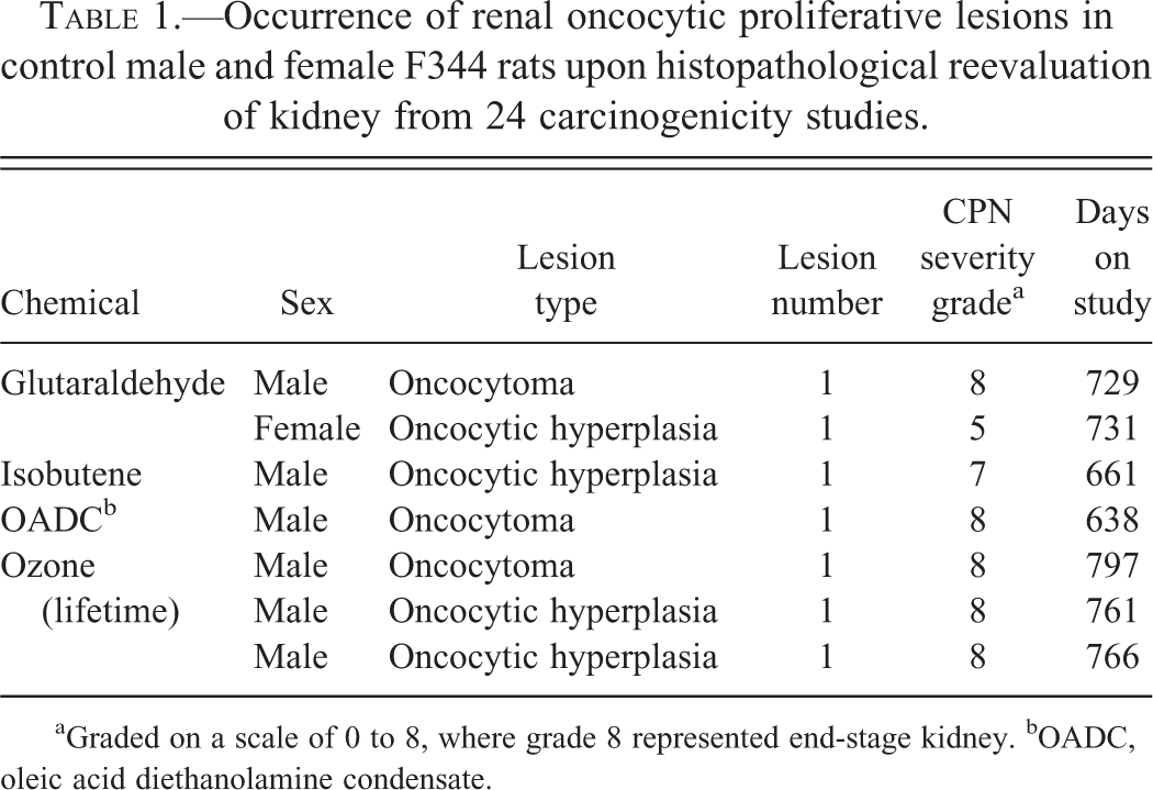
aGraded on a scale of 0 to 8, where grade 8 represented end-stage kidney. bOADC, oleic acid diethanolamine condensate.
In our CPN/RTT study, we reevaluated the renal histopathology of 2,391 untreated (control) F344 male and female rats from 24 carcinogenicity studies that had been conducted (and archived) by the National Toxicology Program, National Institute of Environmental Health Sciences, and National Institutes of Health. The chemicals comprising these 2-year studies (or in the case of ozone, lifetime as well as 2 years) are listed in Hard, Betz, and Seely (2012), along with the methodology for our approach, including severity grading of CPN, and statistical analysis of data generated. Briefly, for each of these 24 studies, the hematoxylin and eosin (H&E)-stained kidneys of 1,236 untreated male rats and 1,155 female rats comprising the control groups were microscopically examined by one of the authors (G.C.H.) for proliferative tubule lesions and severity grading of CPN using an extended scale of 0 to 8, where grade 8 represented an end-stage kidney. It should be noted that the 0 to 8 grading scheme for CPN was developed as a research tool by one of the authors (G.C.H.) and is not recommended for general use. The diagnoses for each proliferative lesion (and questionable lesions) were confirmed by one of the coauthors (J.C.S.). Statistical relationship between different severity grades of CPN and proliferative lesion occurrence was analyzed by logistic regression using SAS proc logistic (Kleinbaum and Klein 2002). A Bonferroni multiple comparisons correction was also carried out, resulting in a p value significance limit of .005.
Oncocytic hyperplasia or oncocytoma was very rare, with only 7 lesions occurring in the long-term study pool of 2,391 male and female rats. This resulted in an overall incidence rate of approximately 0.3%. The lesions were small, and located through various depths of the cortex (Figure 1) and outer stripe of outer medulla, in keeping with the origin indicated by the study of Nogueira and Bannasch (1987). Individually, the lesions comprised 4 foci of oncocytic hyperplasia and 3 oncocytomas (Table 1). Six of the 7 lesions occurred in 1,236 male rats, and 5 of these were in a background of end-stage (grade 8) CPN kidney. The other male rat lesion was in a kidney with grade 7 CPN (equivalent to conventional grade 4 CPN). There was only one oncocytic proliferative lesion in the 1,155 female rats, a focus of oncocytic hyperplasia in a kidney with grade 5 CPN (equivalent to conventional grade 2 or 3). These data suggest a male predisposition for oncocytic renal lesions in the F344 rat strain, with the incidence in males being 0.5%, and in females, 0.09%.
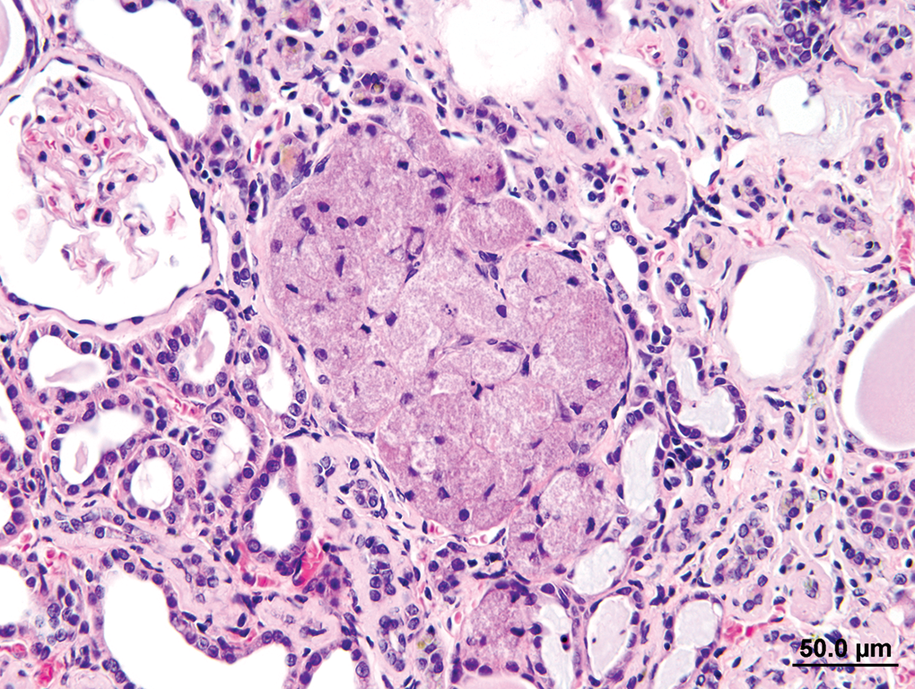
Oncocytic proliferative lesion, located in kidney cortex, that is borderline between oncocytic hyperplasia and oncocytoma. H&E = hematoxylin and eosin.
As shown in Table 1, oncocytic lesions usually involved single animals in individual studies except in one case (ozone) where 3 lesions were recorded in the males. It is probably significant that this was a lifetime study and the 3 affected animals survived 1 or more months beyond the usual 2-year study termination date. Coupled with their small size, this observation suggests that oncocytic proliferative lesions are not only slow growing, but late developing.
The lesion percentage per severity grade of CPN in male rats was 0% for grades 0 to 6 (651 animals), 0.24% for grade 7 (415 animals), and 2.94% for grade 8 (170 animals). As male rats with CPN severity grade 8 were statistically more likely to have an oncocytic proliferation than animals with CPN severity grades 6 or 7 (p = .04, .012, respectively), the evidence also suggests that at least in male rats, oncocytic proliferation may be associated with advanced CPN. However, the statistical associations were not significant using the multiple comparisons p value, and additional surveys would be needed to support this possible association.
The data indicate that oncocytomas and foci of oncocytic hyperplasia are very rare in control F344 rats. This rarity, and the lack of any published evidence for development of small oncocytic lesions into carcinoma, any reports of metastasis, and demonstration of different tubule origin from basophilic RTT (Nogueira and Bannasch 1987), supports the widely held viewpoint that these lesions should be counted separately from basophilic RTT and foci of atypical tubule hyperplasia in bioassays for carcinogenicity evaluation. However, the absence of malignant progression and different histomorphogenetic type remain the most important aspects of this perspective (Brix, Hardisty, and McConnell 2010).
Footnotes
Acknowledgments
The authors acknowledge the National Toxicology Program, National Institutes of Environmental Health Sciences, National Institutes of Health, and Experimental Pathology Laboratories Inc. (both Research Triangle Park, North Carolina, USA), for enabling access to bioassay specimens in the NTP Archives for the primary study.
The author(s) declared no potential conflicts of interest with respect to the research, authorship, and/or publication of this article.
The author(s) disclosed receipt of the following financial support for the research, authorship, and/or publication of this article: The information presented here was part of a study that had been funded by the European Industry Chemical Council and Plastics Europe, both from Brussels, Belgium, and the Tetrahydrofuran Task Force, Chemical Industries Council, Washington, DC, USA.