
Abstract
This study was designed to undertake a risk assessment to identify the health status of rats fed with somatic cell nuclear transfer (SCNT)-cloned Korean native beef cattle (Hanwoo) meat for 26 weeks. The rats were randomly divided into 5 groups, each consisting of 12 male (142.6 ± 5.23 g) and 12 female (113.7 ± 6.31 g) rats each. The animals were fed commercial pellets (control), pellets containing 5% (N-5) and 10% (N-10) of normal cattle meat, and diets containing 5% (C-5) and 10% (C-10) of cloned cattle meat. The mortality; clinical signs; body weight; food consumption; urinary, hematology, blood biochemistry, and histopathological analyses; and absolute and relative organ weights were analyzed and compared. During the 26-week test period, health status–related factors of the rats fed on cloned Hanwoo meat were found to have no test substance–related toxicities. The only difference was the increased uterus weight in female C-10 rats as compared to their counterparts counterparts (p < .05). On the basis of these health status results, it can be postulated that no food consumption risks might arise from the long-term feeding of cloned cattle meat in rats.
Introduction
Somatic cell nuclear transfer (SCNT) can potentially be used to produce various species such as sheep (Wilmut et al. 1997), cattle (Kato et al. 1998), pig (Polejaeva et al. 2000), and dog (Lee et al. 2005) or unlimited animal clones that naturally exhibit high-performance or other desirable traits. The U.S. Food and Drug Administration (2009a) drafted that clones of domestic livestock are perceived to be beneficial for breeders and consumers when used to expand elite breeding stock.
Many previous studies have reported that cloned animals that survive until sexual maturity exhibit normal fertility (Lanza et al. 2001; Enright et al. 2002; Shiga et al. 2005; Bae et al. 2007). In South Korea, cloned Korean native cattle (Hanwoo) have produced the fourth generation of progenies via artificial insemination or natural mating among clones or between clones and conventionally bred cattle (unpublished data). Their progenies exhibit normal growth characteristics and reproductive physiology as compared with their normal counterparts (Bae et al. 2009). However, normal fertility of the clone may not guarantee its safety for use in food. It has been forbidden to include clone- and clone progeny–derived milk or meat because of the associated potential hazards (Galli et al. 2004) and public controversy concerning the reliability of such products (Suk et al. 2007).
Although food safety experiments carried out a risk assessment of the potential hazards and characterized any risks that could arise from those potential hazards due to consumption (Rudenko et al. 2004; Rudenko and Matheson 2007), the composition and nutritional value of meat and milk derived from both normal and cloned animals did not differ from those of conventionally bred animals (Tian et al. 2005; Yamaguchi, Ito, and Takahashi 2007; Yang et al. 2007). And the European Food Safety Authority (2008) and the U.S. Food and Drug Administration (2009b) launched draft opinions and deemed the consumption of meat and milk derived from clones and their progeny as safe as the food products obtained from their conventionally bred counterparts.
The risk assessment and safety for assessing toxicological end points play an important role in many materials. Unfortunately, very little information was revealed regarding the long-term feeding (≥6 mo) effects of cloned progenies’ meat (Yamaguchi et al. 2008). Especially no results were reported related to cloned Hanwoo meat. This study was designed to undertake a risk assessment to identify the health status of rats fed with SCNT-cloned Hanwoo meat for 26 weeks.
Materials and Methods
Testing Facility
The examination of all the rats was conducted at the Laboratory Animal Research Center (Chungbuk National University) and Biotoxtech Co., Ltd. The study protocol and standard operating procedures were reviewed and approved by the Institutional Animal Care and Use Committee of the Laboratory Animal Research Center of Chungbuk National University. The experiments performed were conducted according to the “Guide Principles in the Use of Animals in Toxicology,” which was adopted by the Society of Toxicology.
Meat-based Diet
Two female cloned Hanwoo produced by SCNT at the National Institute of Animal Science (Suwon, Republic of Korea) were slaughtered and sirloin parts of the clones were used in the present study. SCNT procedure was carried out as described previously (Yang et al. 2008). The same parts of the normal Hanwoo meat were purchased from the local market. This normal sample and the meat obtained from the clones were freeze-dried and ground. The powders (5–10%) were then mixed with rat feed (38057, Agribrands Purina Korea) in the Korea Food Research Institute. The meat-based diets were sterilized by 17 kGy gamma irradiation for 18 hr. The nutritional values of the normal and cloned cattle-derived meat diets did not differ (Hwang et al. 2010).
Animals
We selected Sprague-Dawley (SD) rats for this study because extensive background information of this strain is available in literature, and these rats are commonly used in studies of reproductive and developmental toxicity. Animal pathogen- and virus-free (APF/AVF) SD rats were purchased from Samtako Bio Korea, Inc. (Osan, South Korea) certified by Institute for Laboratory Animal Research (ILAR). After acclimatization for 7 days, 5-week-old male and female rats were fed the meat-based diets. The animal housing chambers were artificially illuminated from 7:00 a.m. to 7:00 p.m. every day (150–300 lux), and the temperature (23°C–25°C) and humidity (50–60%) levels were controlled. All the rats were provided with pellets and filtered and sterilized water. The testing facility of the veterinary staff initiated an animal health monitoring program.
Experimental Design
According to the results of the dose range and maximum tolerated oral dose studies, 10% of the meat-based diet was selected as the highest dose. The animals were randomly divided into 5 groups of 12 male (142.6 ± 5.23 g) and 12 female (113.7 ± 6.31 g) rats per group. The animals were fed commercial pellets (control), pellets containing 5% (N-5) and 10% (N-10) of normal cattle meat, and diets containing 5% (C-5) and 10% (C-10) of cloned cattle meat. The mortality, clinical signs, and food consumption were monitored daily, and the body weights were measured every week. At the end of the experimental period, urinalysis was conducted, and both the absolute and relative organ weights of the rats were measured.
Urinalysis
During the last week of treatment, urinalysis of 6 males and females per group were conducted with fresh urine for item of specific gravity, pH, protein, glucose, ketone body, occult blood, bilirubin, urobilinogen, and nitrite by using a Miditron® Junior II (Hoffmann-La Roche Ltd., Mannheim, Germany). Urine sediment test was also carried out within 3 hr after taking samples during the last week of the administration period. The volume of urine collected for 24 hr was measured.
Hematology and Serum Biochemistry
Blood samples were analyzed using a Sysmex SE-9000 automatic hematology analyzer (Sysmex Corp., Kobe, Japan). Aliquots of whole blood were mixed with a fourfold volume of the manufacturer’s buffer containing 0.5% ethylenediaminetetraacetic acid (EDTA). The prothrombin time (PT) and activated partial thromboplastin time (APTT) were measured using 1/9 volume of sodium citrate solution mixed with aliquots of whole blood, and the mixture was analyzed on an ACL7000 (Instrumentation Laboratory, Bedford, MA). Serum biochemistry was carried out with Hitachi 747 (Hitachi Medical, Tokyo, Japan) using the sera obtained after centrifugation of whole blood (1,000g for 10 min).
Histopathological Assessment
For necropsy findings, the brain, heart, lungs, thymus, spleen, liver, kidneys, adrenal glands, uterus, epididymis, prostate, ovary, and testis were removed and weighed. In addition, the pituitary, eyes, Harderian glands, spinal cord, salivary glands, stomach, small intestine (duodenum, jejunum, and ileum), large intestine (cecum, colon, and rectum), pancreas, urinary bladder, skin, male and female mammary glands, trachea, esophagus, thyroid glands, tongue, aorta, thigh muscle, sternum, femur, sciatic nerve, seminal vesicles, and vagina were also removed. Thereafter, all the organs and tissues were fixed in 10% buffered formalin for 3 days for histopathological assessment. The tissues slices of all organs and tissues were routinely processed for paraffin embedding, and prepared sections were stained with hematoxylin and eosin.
Statistics
Results are represented as the mean ± standard deviation (SD). All the tests were applied at the .05 significance level. The data were tested for normality and homogeneity of variance using Levene’s test. Significance was determined using Dunnett’s test after analysis of variance (ANOVA). Statistical analyses were conducted using SAS Program (version 9.1.3, SAS Institute Inc.).
Results
There was no mortality or clinical signs in any of the rats fed the cloned cattle meat diet (Table 1). The only detected clinical sign was depletion of hair in the cervical multifocal areas in 1 out of 12 rats of both sexes in N-10 and C-10, respectively.
Mortality and clinical signs of the rats during 26 weeks of cloned-cattle meat feeding.
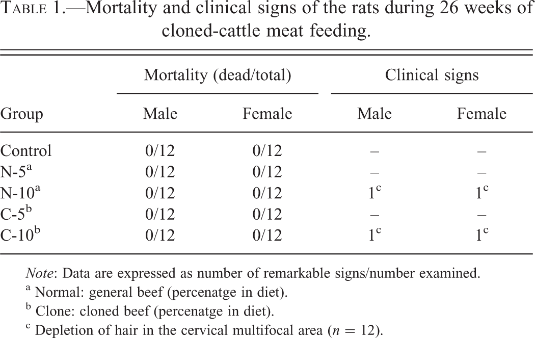
Note: Data are expressed as number of remarkable signs/number examined.
a Normal: general beef (percenatge in diet).
b Clone: cloned beef (percenatge in diet).
c Depletion of hair in the cervical multifocal area (n = 12).
The overview of food consumption in both male and female rats over 26 weeks is shown in Table 2. The difference between the cloned and normal meat diet groups was not statistically significant in both sexes.
Overview of food consumption in rats during 26 weeks of feeding.
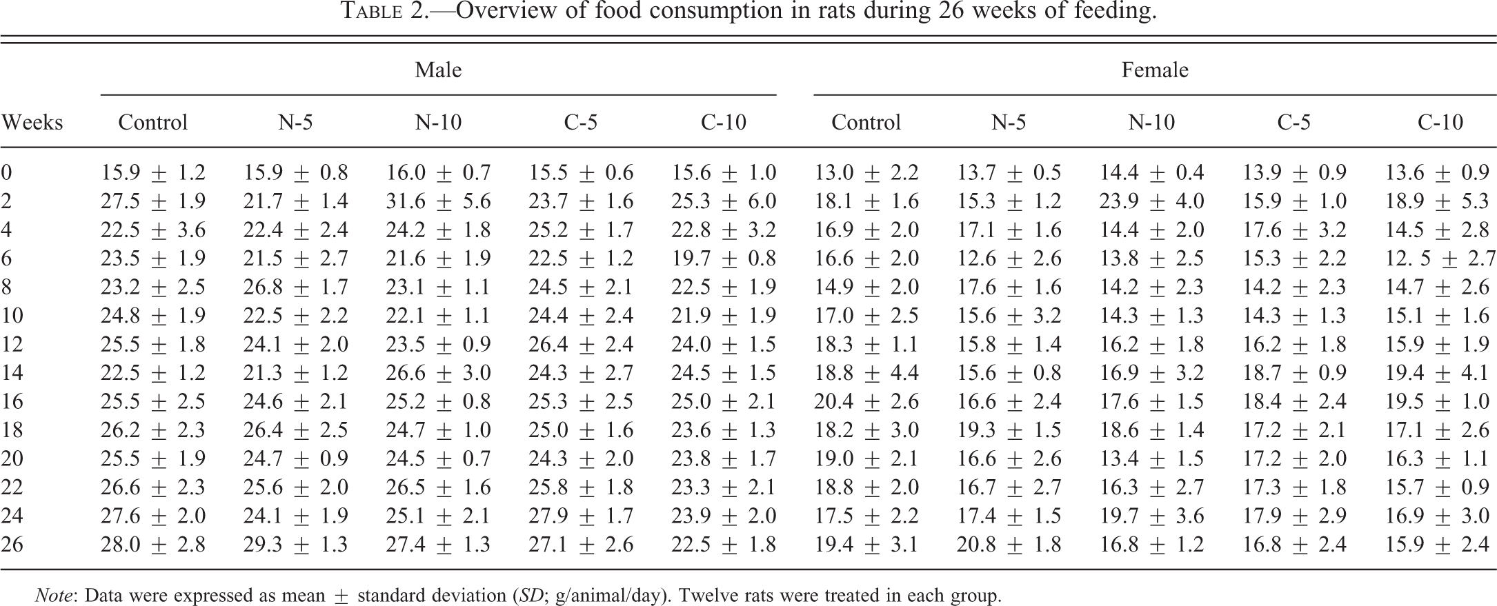
Note: Data were expressed as mean ± standard deviation (SD; g/animal/day). Twelve rats were treated in each group.
Significant changes in body weight were noted between 2 and 6 weeks in the N-10 and C-5 groups of male rats (p < .05), respectively (Table 3). However, no statistically significant differences were found for the female F1 rats across all the feeding periods.
Overview of body weight in rat during 26 weeks of feeding.
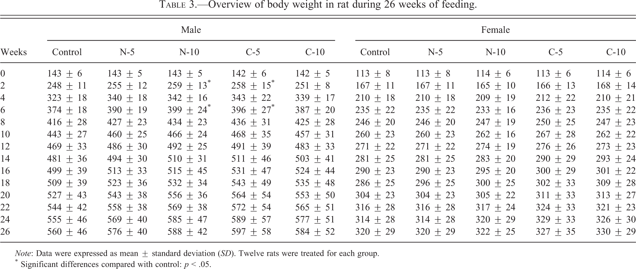
Note: Data were expressed as mean ± standard deviation (SD). Twelve rats were treated for each group.
* Significant differences compared with control: p < .05.
The results of the urinalysis of both male and female rats after 26 weeks are presented in Table 4. No significant differences were observed in the color, specific gravity, glucose, pH, ketones, leukocytes, hemoglobin, nitrite, urobilinogen, protein, bilirubin in all treatment groups.
Urinary analysis of the rats after 26 weeks of feeding.
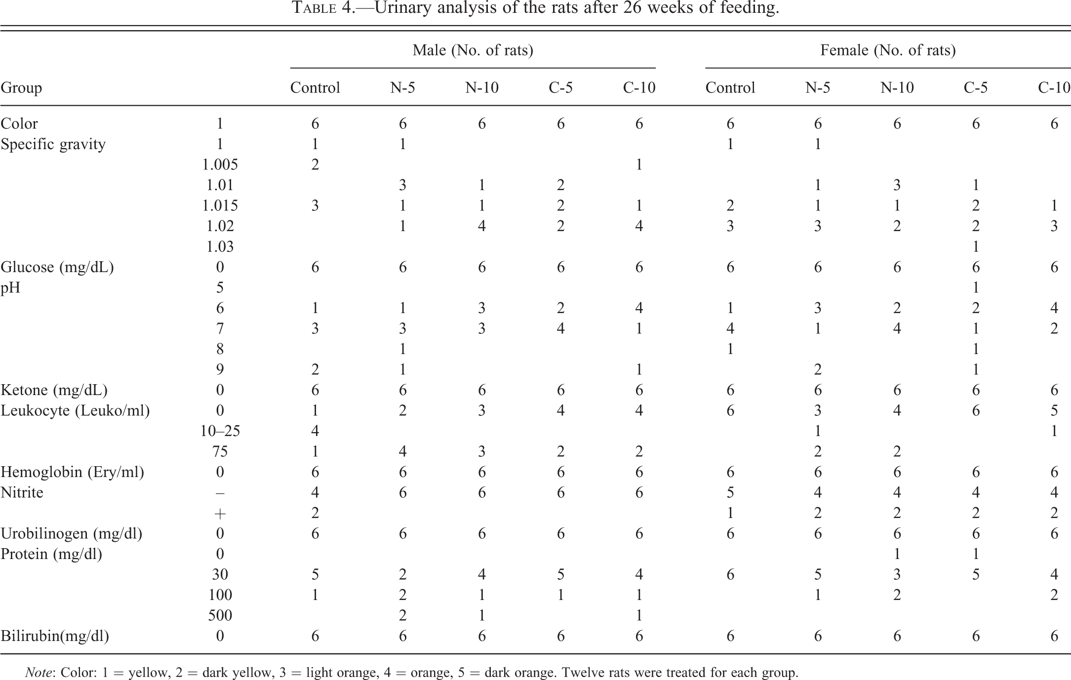
Note: Color: 1 = yellow, 2 = dark yellow, 3 = light orange, 4 = orange, 5 = dark orange. Twelve rats were treated for each group.
Results of the hematological examination (Table 5) and serum biochemistry analysis (Table 6) revealed no test substance–related differences in the rats as compared to the control and normal meat groups. Further, no remarkable signs of spontaneous lesions were detected in the abdominal cavity, brain, heart, kidneys, liver, lymph nodes (mediastinal or submandibular), mammary gland, pancreas, or urinary bladder in any of the examined animals (Table 7).
Hematological examination in rats after 26 weeks of feeding.
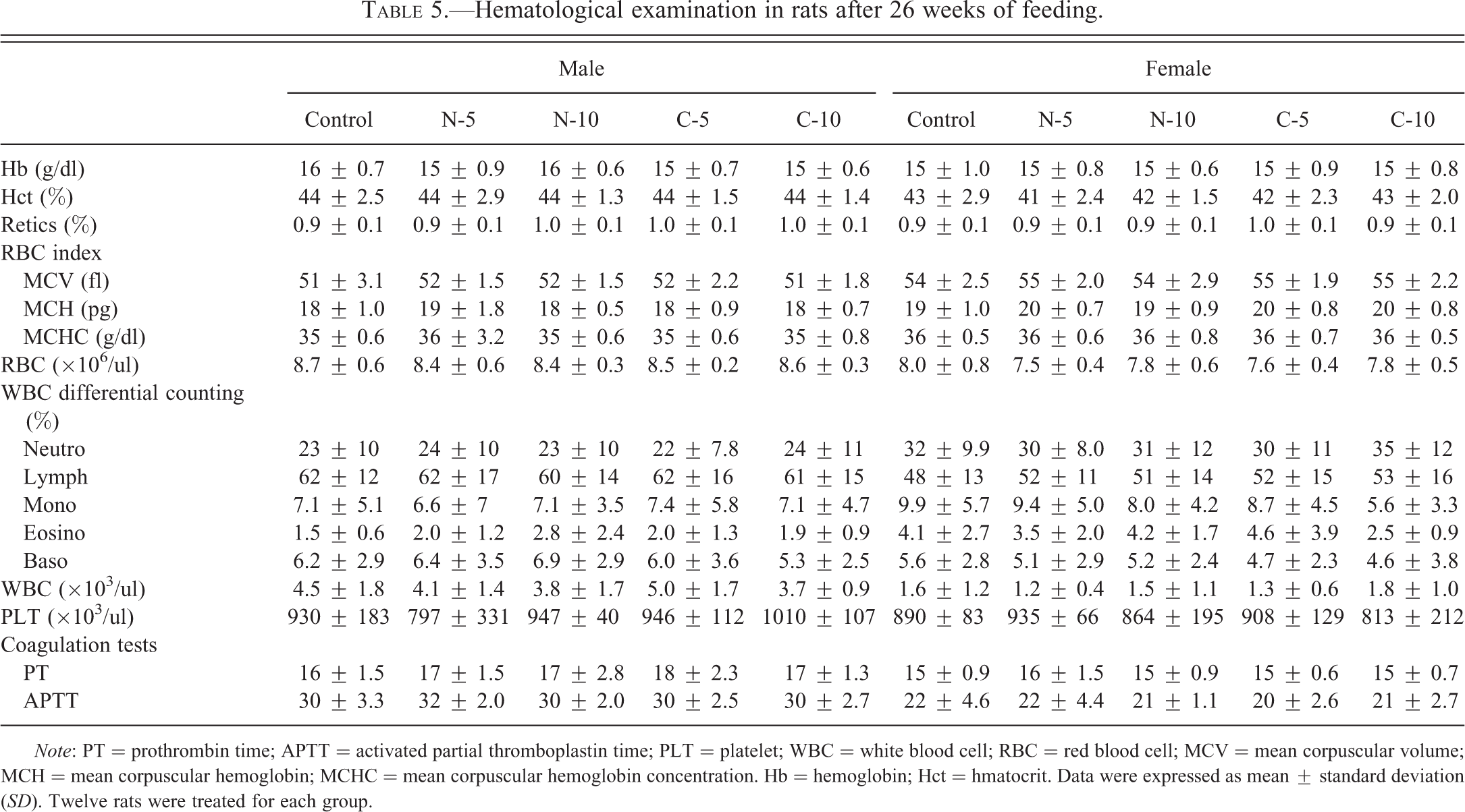
Note: PT = prothrombin time; APTT = activated partial thromboplastin time; PLT = platelet; WBC = white blood cell; RBC = red blood cell; MCV = mean corpuscular volume; MCH = mean corpuscular hemoglobin; MCHC = mean corpuscular hemoglobin concentration. Hb = hemoglobin; Hct = hmatocrit. Data were expressed as mean ± standard deviation (SD). Twelve rats were treated for each group.
Serum biochemical analysis in rats after 26 weeks of feeding.
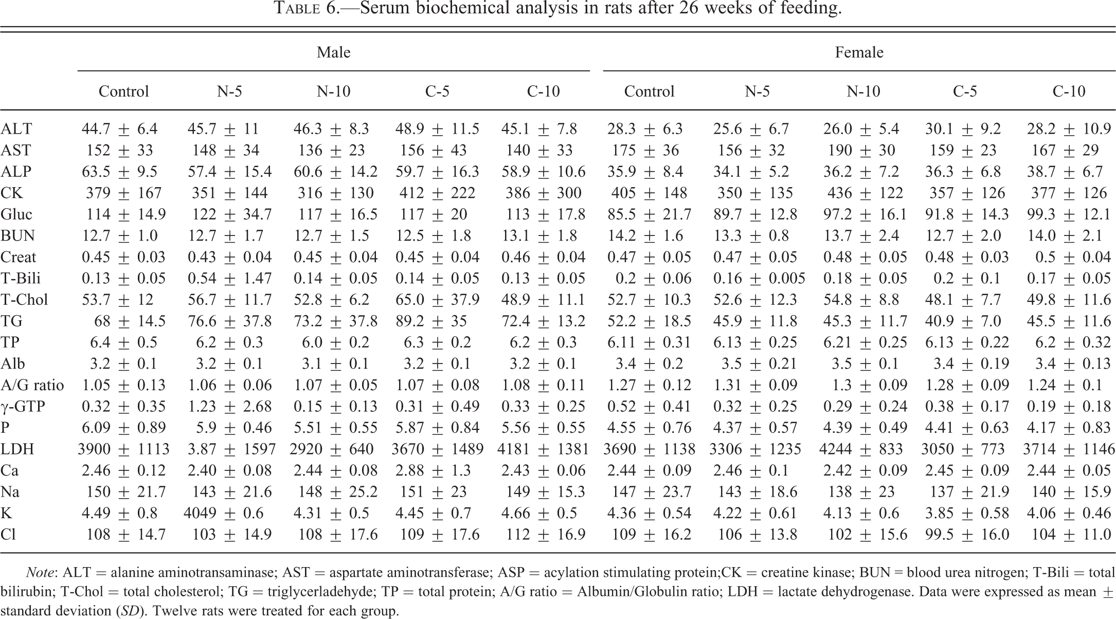
Note: ALT = alanine aminotransaminase; AST = aspartate aminotransferase; ASP = acylation stimulating protein;CK = creatine kinase; BUN = blood urea nitrogen; T-Bili = total bilirubin; T-Chol = total cholesterol; TG = triglycerladehyde; TP = total protein; A/G ratio = Albumin/Globulin ratio; LDH = lactate dehydrogenase. Data were expressed as mean ± standard deviation (SD). Twelve rats were treated for each group.
Histopathological assessment in rats after 26 weeks of feeding.
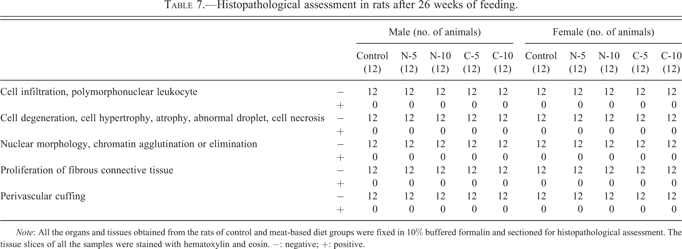
Note: All the organs and tissues obtained from the rats of control and meat-based diet groups were fixed in 10% buffered formalin and sectioned for histopathological assessment. The tissue slices of all the samples were stained with hematoxylin and eosin. −: negative; +: positive.
The absolute and relative organ weights of the male and female rats are presented in Tables 8 and 9, respectively. There were no significant differences in the absolute and relative organ weights in all treatment groups, except for the uterus weight in group C-10 (p < .05).
Absolute and relative organ weight of male rats after 26 weeks of feeding.
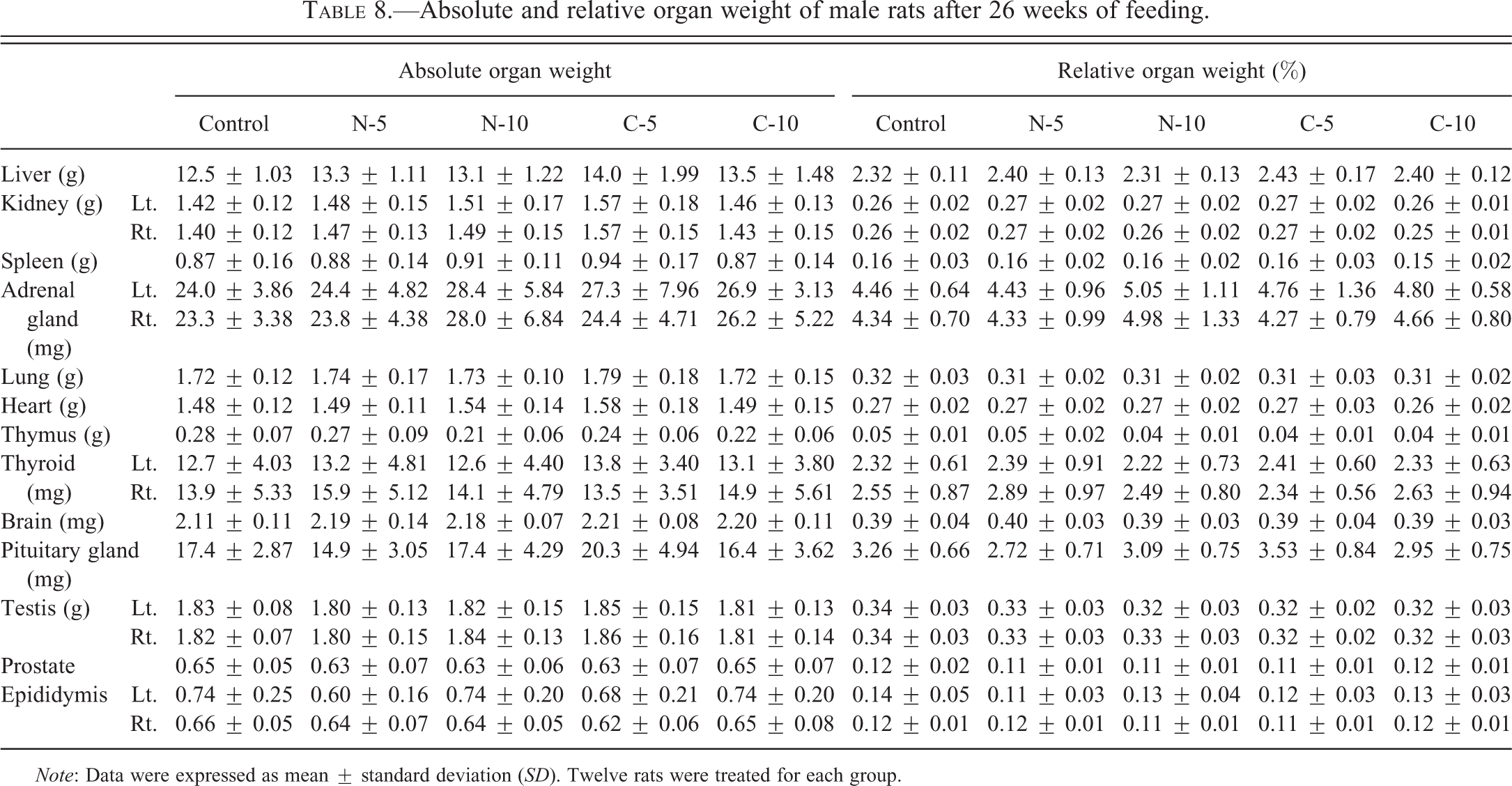
Note: Data were expressed as mean ± standard deviation (SD). Twelve rats were treated for each group.
Absolute and relative organ weight in female rats after 26 weeks of oral repeated administration toxicity study of clone-based meat diet.
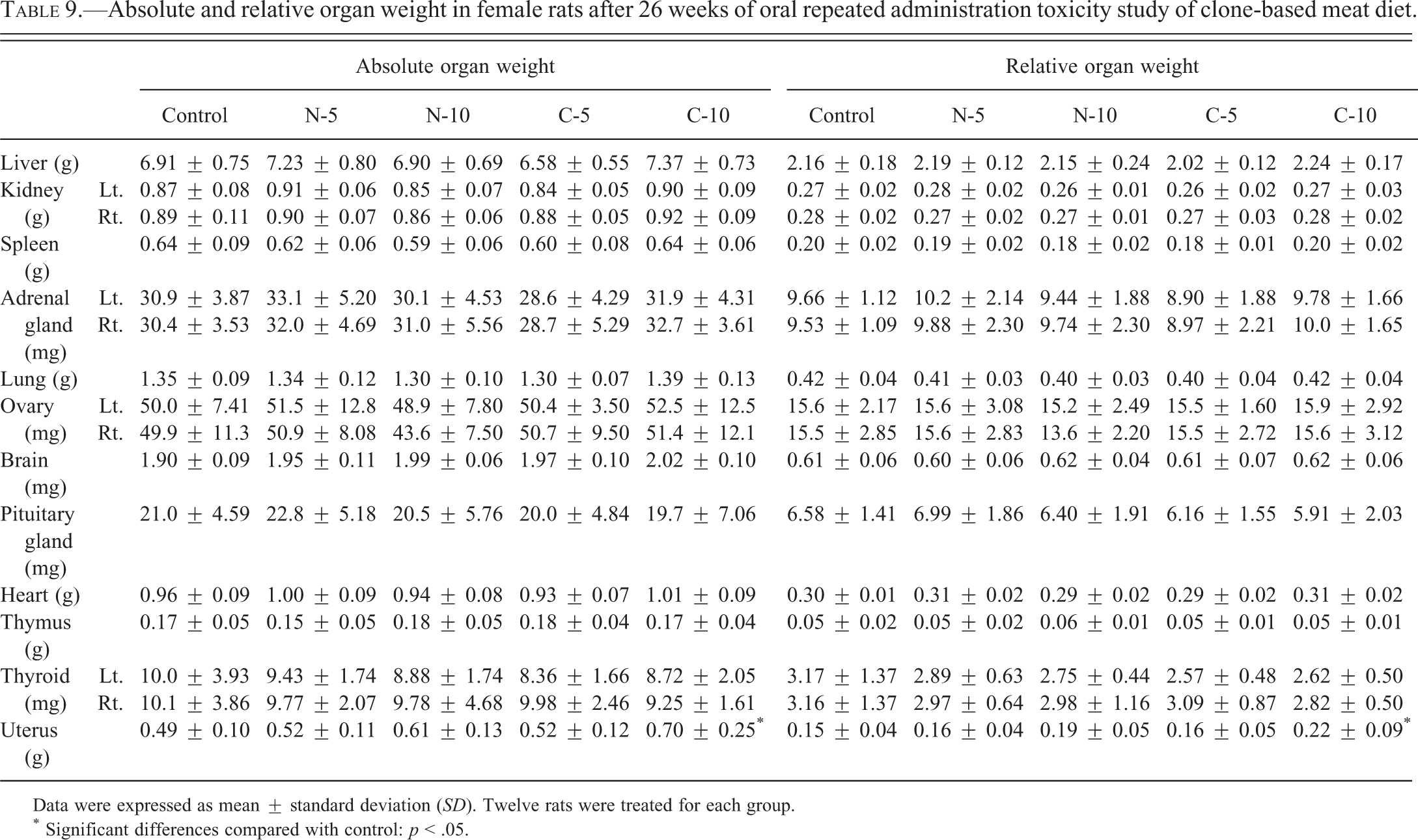
Data were expressed as mean ± standard deviation (SD). Twelve rats were treated for each group.
* Significant differences compared with control: p < .05.
Discussion
Despite the higher rates of embryonic, fetal, perinatal, and neonatal deaths, as well as offspring with various abnormalities (Edwards et al. 2003; Hwang et al. 2009), many of the somatic cell clones survived beyond puberty, and they were physiologically, immunologically, and behaviorally normal (Govoni et al. 2002; Savage et al. 2003). However, due to certain errors such as epigenetic reprogramming or physiological abnormality (Kang et al. 2001; Jaenisch and Bird 2003; Park et al. 2011), food products such as meat and milk derived from clones and their progenies are still being considered unsafe and unsuitable for consumption.
To adequately evaluate the safety of any food product in a rat model, it is important to not only monitor the physiological, developmental, or reproductive functions of dams and their offspring, but also determine the toxicity associated with long-term feeding. The authors of this study have previously evaluated cloned cattle meat-related toxicities in both rats and rabbits (Hwang et al. 2010; Lee et al. 2010). In the studies, no harmful effects of the diet were observed in female (estrus cycle, implantation, fertility, delivery, external findings, necropsy findings, and physiological development) or male (weight of epididymis and testis, epididymal sperm motility, morphology, and sperm counts) reproductive parameters in both laboratory animals and their offspring. Furthermore, no mutagenic toxicity (bacterial mutation, chromosome aberration, and micronucleus genotoxicity), F1 behavioral toxicity (sensory reflex, motor function, spatial learning, and memory tests), or reproductive toxicity (mating, fertility, and implantation) was detected in the animal fed diets containing cloned cattle-derived meat (Lee et al. 2011; Yang et al. 2011).
In the present study, all lesions observed here were diagnosed as spontaneous lesions. There were no significant differences in the occurrence rates of these lesions or the histological findings between the rat groups fed normal cattle-based meat diet and clone-based meat diet. Especially, histological observation of uterus in the rats fed the 10% of clone-based meat diet indicated significantly increased absolute and relative weights, and no distinctive lesions in the rats fed normal cattle-based meat diet and clone-based meat diet were observed. Based on these hematological and histopathological findings, the data for the different kinds of edible products demonstrated that there are no biologically significant differences in composition among the clones, their progenies, and conventionally bred cattle.
Only one previous study reported that feeding rats with meat or milk derived from the progeny of Japanese Black SCNT-cloned cattle for 12 mo produced no differences in physiological, developmental, or reproductive parameters (Yamaguchi et al. 2008). However, a risk assessment has been focused on clones rather than on their progeny, because most of the food products from SCNT technology will be derived from clone progeny, the sexually reproduced offspring of clones (Rudenko 2008).
Up to now, various types of cattle have been produced in many countries such as Japan, New Zealand, United States, Brazil, and Argentina (Kato et al. 1998; Heyman et al. 2004; Wells et al. 2004; Panarace et al. 2007), but food safety assessments have not been performed with each type of clones and their progenies. In conclusion, it can be postulated that there are no harmful effects associated with long-term consumption of cloned Hanwoo meat in rats.
Footnotes
The authors declared no potential conflicts of interest with respect to the research, authorship, and/or publication of this article.
The authors disclosed receipt of the following financial support for the research, authorship and/or publication of this article: This work was supported by the Ministry of Food, Agriculture, Forestry and Fisheries and the Agenda Program (No. PJ008587; PJ009095) by the Rural Development Administration, Republic of Korea.
Authors’ Note
Nam-Jin Lee and Byoung-Chul Yang have contributed equally to this article.