
Abstract
Cyclodextrins are oligosaccharides which are used in the pharmaceutical industry and research as vehicles for application of apolar substances such as steroids. The aim of this study was to examine the long-term effects of parenteral administration of 2-hydroxypropyl-β-cyclodextrin (HP-β-CD) on bone. Sham-operated (SHAM) or ovariectomized (OVX) adult rats were subcutaneously injected with physiological saline, 50, or 200 mg/kg HP-β-CD daily. After 4 months, body weight in OVX rats and uterine weight in SHAM rats were significantly lower after administration of 200 mg/kg HP-β-CD, relative to vehicle controls. At 200 mg/kg, HP-β-CD was hepatotoxic as measured by increased serum transaminases, and reduced serum albumin. Moreover, 200 mg/kg HP-β-CD led to decreased vertebral and tibial bone mineral density (BMD), and to cortical thinning at the tibial shaft. Bone loss in HP-β-CD-treated rats was associated with increased bone resorption as measured by increased renal deoxypyridinoline excretion. Although 50 mg/kg HP-β-CD was devoid of overt signs of organ toxicity and did not impair BMD, bone resorption was already increased. In summary, subcutaneous long-term administration of HP-β-CD at a daily dose of 200 mg/kg led to increased bone resorption and subsequent bone loss. Minor alterations in bone metabolism were also seen at 50 mg/kg.
Introduction
Cyclodextrins (CDs) are cyclic oligosaccharides which originate from starch degradation. The “parent CDs,” namely α-, β-, γ-, and δ-CD, are composed of six to nine glucose molecules forming a cone-like cavity. Hence, CDs have the ability to form inclusion complexes with apolar substances and, due to a polar outer surface, to enhance their water solubility. Moreover, other positive effects like improving physical and chemical stability have raised the interest of the pharmaceutical industry and of research to employ CDs as absorbance enhancers or as vehicles for the administration of lipophilic drugs (Stella and He 2008).
When applied directly to cells in culture, CDs can cause non-specific cell lysis and are potentially toxic. After oral administration, CDs are poorly absorbed in the gut, and, therefore the toxicity of orally administered CDs is low. However, systemic administration of CDs can cause toxic effects in various tissues (Irie and Uekama 1997; Olivier et al. 1991; Stella and He 2008). One of the hallmarks of the toxicity of CDs after parenteral administration is nephrotoxicity, manifesting in vacuolar alterations in the proximal convoluted tubules (Frank et al. 1976). As the kidney is the main organ for clearance of CDs from circulation, CDs were chemically modified to increase their solubility in an attempt to eliminate the renal toxicity. One such derivative is 2-hydroxypropyl-β-cyclodextrin (HP-β-CD). However, side effects were also reported after administration of HP-β-CD (Gould et al. 2005).
Based on its favorable chemical properties, HP-β-CD has been used as a vehicle for oral and parenteral administration of several drugs in animals and humans (Leeson et al. 2007; Tansho et al. 2006), and especially for administration of steroid hormones in different fields of research (Kan et al. 2008; Mello et al. 2008; Rabbani et al. 1997). Sex steroids play a major role in skeletal development and maintenance (Erben et al. 2004; Lapauw et al. 2009; Vandenput et al. 2001). However, the naturally occurring sex steroids such as 17β-estradiol or testosterone are not orally available, mainly due to a pronounced first pass effect in the liver. Therefore, they have to be administered parenterally. In addition, osteotropic conjugates of CDs with other drugs such as bisphosphonates have been tested as locally injected bone anabolic drugs (Liu et al. 2008). However, almost nothing is known about the skeletal toxicity of CDs. Therefore, we sought to examine the toxicity of parenterally administered HP-β-CD on bone in the current study. Because sex steroids are often given to estrogen deficient ovariectomized (OVX) rats in long-term studies, we examined the effects of a 4-month subcutaneous treatment of adult sham-operated (SHAM) and OVX rats with aqueous HP-β-CD solutions at dosages of 50 and 200 mg/kg body weight on bone turnover and bone mineral density (BMD) in the axial and appendicular skeleton. Surprisingly, we found major untoward side effects of long-term treatment with HP-β-CD at higher doses.
Material and methods
Animal Procedures
All animal procedures were approved by the Ethical Committee of the University of Veterinary Medicine Vienna and the local government authorities. At the beginning of the experiment, 6-month-old female Fischer 344 rats were weight-matched and either SHAM or OVX under anesthesia with isoflurane. SHAM and OVX groups (n = 5–8 each) received daily subcutaneous injections of either vehicle or of HP-β-CD solutions in physiological saline (pH 7.4) at dosages 50mg/kg and 200mg/kg body weight. These dosages were found to be commonly used in experiments or pharmaceutical formulations. The injection volume was 1 ml/kg for all rats. HP-β-CD was purchased from Sigma-Aldrich Inc. (St. Louis, MO, USA). The animals were housed at 24°C with a 12 hour/12 hour light-dark cycle, and were fed a standard laboratory diet (Ssniff R/M-H V1534, Ssniff Spezialdiäten GmbH, Soost, Germany) containing 1.0% calcium, 0.7% phosphorus, and 1,000 IU/kg vitamin D3. SHAM rats were fed ad libitum at all times. OVX rats were pair-fed according to the food intake of the SHAM rats. Calcein (Sigma-Aldrich Inc.) was subcutaneously injected at a dosage of 20 mg/kg on days 8 and 3 before sacrifice. All animals were sacrificed 4 months after surgery by exsanguination from the abdominal aorta under ketamine/xylazine anesthesia (50/10 mg/kg intraperitoneally; Ketasol® and Xylasol® was purchased from aniMedica GmbH, Senden-Boesensell, Germany).
For the collection of urine, all animals were kept in metabolic cages for 15 hours overnight before necropsy. Urine samples were stored at −20°C. Blood was kept at room temperature for one hour. Subsequently, the clotted blood samples were centrifuged at 1,800 × g for 5 min. Until processing, serum samples were stored at 80°C. At necropsy, the wet weight of the uterus was determined, and samples from the mid segment of the uterine horns were taken for measurement of the endometrial epithelial height. Left tibiae and 4th lumbar vertebrae (L4) were harvested for measurement of bone mineral density (BMD) using peripheral quantitative computed tomography (pQCT). The proximal right tibia was collected for fluorochrome-based histomorphometry.
Histology
Uterine and kidney samples were embedded in paraffin by routine procedures. Five-µm-thick cross-sections of the uterine horns or of kidneys were cut using a HM 355 S microtome (Microm, Walldorf, Germany). Sections were stained with hematoxylin and eosin. Bone specimens were fixed in 40% ethanol at 4°C for 48 hours, and embedded in methylmethacrylate as described previously (Schenk et al. 1984). Five-µm-thick undecalcified midsagittal sections from the proximal tibiae were prepared using a SM2500 S microtome (Leica Microsystems GmbH, Wetzlar, Germany), and were stained with toluidine blue (Erben et al. 2000) or mounted unstained with Fluoromount® (Serva electrophoresis GmbH, Heidelberg, Germany).
Measurement of Endometrial Epithelial Height and Myometrial Area
Measurements of endometrial epithelial height in SHAM and OVX rats and of the total cross-sectional uterine area as well as of the area covered by endo- and myometrium in SHAM rats were performed with a semiautomatic image analysis system (OsteoMeasure 3.0, Osteometrics, Decatur, GA, USA) and a microscope with a drawing attachment.
BMD Measurements
BMD was measured by pQCT using a XCT Research M+ pQCT machine (Stratec Medizintechnik, Pforzheim, Germany). Specimens stored in 70% ethanol were measured with a collimator opening of 0.2 mm, and a voxel size of 100 µm. A threshold of 710 mg/cm3 was used for calculation of cortical BMD. One slice in the mid-diaphysis of the tibiae located 2 mm proximal to the tibiofibular junction and one slice in the tibial proximal metaphysis located 2 mm distal from the growth plate were measured. In the L4 vertebra, three slices were measured, one in a mid-transversal plane, and two located 2 mm cranial and caudal of the mid-transversal plane. BMD values of the L4 vertebral body were calculated as the mean over three slices.
Bone histomorphometry
Fluorochrome-based histomorphometric measurements were made with a semiautomatic image analysis system (OsteoMeasure 3.0) and a microscope with a drawing attachment as described (Erben et al. 2000). The area within 0.5 mm from the growth plates was excluded from all measurements. The bone formation rate was calculated by multiplying the mineralizing perimeter (percentage of calcein double-labeled bone perimeter) with the mineral apposition rate.
Biological Chemistry
Serum aspartate aminotransferase (AST) activity, alanine aminotransferase (ALT), gamma-glutamyl transferase (γ-GT), alkaline phosphatase (ALP) activity, creatine kinase activity, total bilirubin, creatinine, urea, albumin, calcium, and phosphorus as well as urinary calcium, creatinine, and phosphorus were determined using a Hitachi 912–Automatic Analyzer (Roche, Mannheim, Germany). Total deoxypyridinoline (DPD) concentrations in urine were measured after acid hydrolysis by EIA (Metra® DPD EIA Kit, Quidel Corporation, San Diego, CA, USA), and were expressed as a ratio to renal creatinine excretion. Urine was assessed for proteinuria by using urine test strips (Roche Diagnostics, Mannheim, Germany).
Statistical Analyses
Statistics were computed using SPSS for Windows 17.0 (SPSS, Chicago, IL, USA). Data were evaluated by analysis of variance (ANOVA). When the ANOVA performed over all groups show a significant (p < 0.05) difference among the groups, statistical differences between individual groups were subsequently analyzed using Student-Newman-Keuls multiple comparison test. In addition, two-way ANOVA was performed to assess the effects of OVX and of HP-β-CD, together with their mutual 2-way interactions. P values of less than 0.05 were considered significant for all statistical analyses.
Results
Body Weight, Uterine Weight, Endometrial Epithelial Height, and Myometrial Area
Final body weight, relative uterine weight, endometrial epithelial height, myometrial area, and histological images of uteri and kidneys are shown in Figure 1. Two-way ANOVA revealed a significant detrimental effect of HP-β-CD on body weight. OVX rats treated with 200 mg/kg HP-β-CD showed 11% reduced body weight compared with OVX rats receiving vehicle or 50 mg/kg HP-β-CD. As expected, uterine weight (Fig. 1B) and endometrial epithelial height (Fig. 1C) were significantly decreased in OVX animals. Uterine weight was reduced in SHAM rats receiving 200 mg/kg HP-β-CD by 44% relative to vehicle-treated SHAM rats, and by 58% relative to SHAM rats treated with 50 mg/kg HP-β-CD. However, HP-β-CD had no effect on uterine weight in OVX animals (Fig. 1B), and did not influence endometrial epithelial height in SHAM or OVX rats (Fig. 1C). In agreement with the uterine weight data, administration of 200 mg/kg HP-β-CD to SHAM rats decreased total uterine cross-sectional area by 34% compared to vehicle-treated SHAM rats (Fig. 1D–E). In contrast, the relative area covered by endometrium and myometrium remained unchanged by HP-β-CD treatment (data not shown), suggesting that the adverse effect of HP-β-CD on the uterus was not specific for myo- or endometrium. At necropsy, all rats treated with 200 mg/kg HP-β-CD showed a macroscopically visible yellowish discoloration of the kidneys. Histologically, the 4-month treatment with 200 mg/kg HP-β-CD led to distinct vacuolation of proximal convoluted tubules (Fig. 1F).
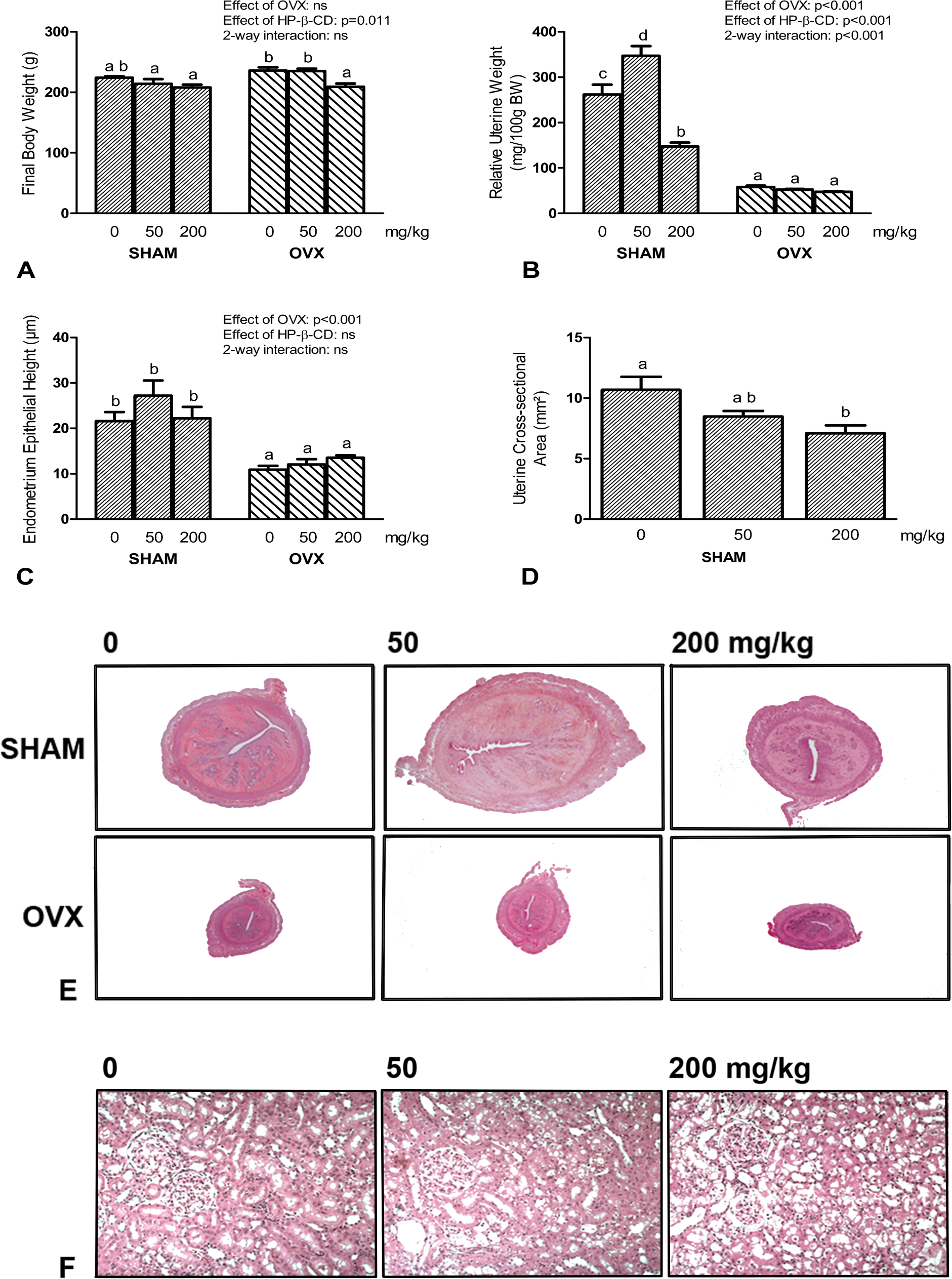
Effects of a daily subcutaneous 4-month treatment of sham-operated (SHAM) and ovariectomized (OVX) rats with vehicle, 50 mg/kg, and 200 mg/kg HP-β-CD on final body weight (A), relative uterine weight (B), endometrial epithelial height (C), and myometrial area (D, only SHAM rats). Microscopic images of H&E-stained paraffin sections from uterine horns and kidneys are shown in (E) and (F), respectively. BW, body weight; HP-β-CD, 2-hydroxypropyl-cyclodextrin; ns, not significant. Columns marked with the same letter are not significantly different from each other by one-way ANOVA followed by Student-Newman-Keuls test. Error bars represent SEM. Insets show results of 2-way ANOVA. Original magnification 25× in E, 200× in F.
Clinical Chemistry
Results of serum and urine analyses are presented in Figure 2 and Table 1. Two-ANOVA revealed a significant effect of HP-β-CD on serum ALP, AST, albumin, ALT, γ-GT, total bilirubin, phosphorus, and calcium as well as on urinary calcium, phosphorus, and creatinine clearance. OVX rats had generally higher serum ALP and lower serum albumin and renal creatinine clearance relative to SHAM rats by 2-way ANOVA. With the exception of serum phosphorus (Table 1), 50 mg/kg HP-β-CD did not change any serum or urinary parameter in SHAM or OVX rats. However, 200 mg/kg HP-β-CD profoundly increased serum ALP by 243% and by 93%, serum AST by 115% and by 260%, serum ALT by 157% and by 533%, serum γ-GT by 215% and 161%, and urinary phosphorus excretion by 92% and 72%, and decreased serum albumin by 32% and 18%, relative to vehicle-treated SHAM and OVX rats, respectively (Fig. 2). Serum creatinine and urea remained unchanged by HP-β-CD administration (Table 1). Although 2-way ANOVA picked up a significant effect of HP-β-CD on creatinine clearance (effect of HP-β-CD p = 0.009), the effects were not dose dependent. At the dose of 50 mg/kg HP-β-CD, creatinine clearance tended to be non-significantly lower compared with vehicle-treated SHAM and OVX rats, but returned to the values found in vehicle-treated rats at 200 mg/kg HP-β-CD (Table 1). To exclude proteinuria as a reason for reduced serum albumin, we used urine test strips. We found no evidence of proteinuria in HP-β-CD–treated SHAM and OVX rats (data not shown).
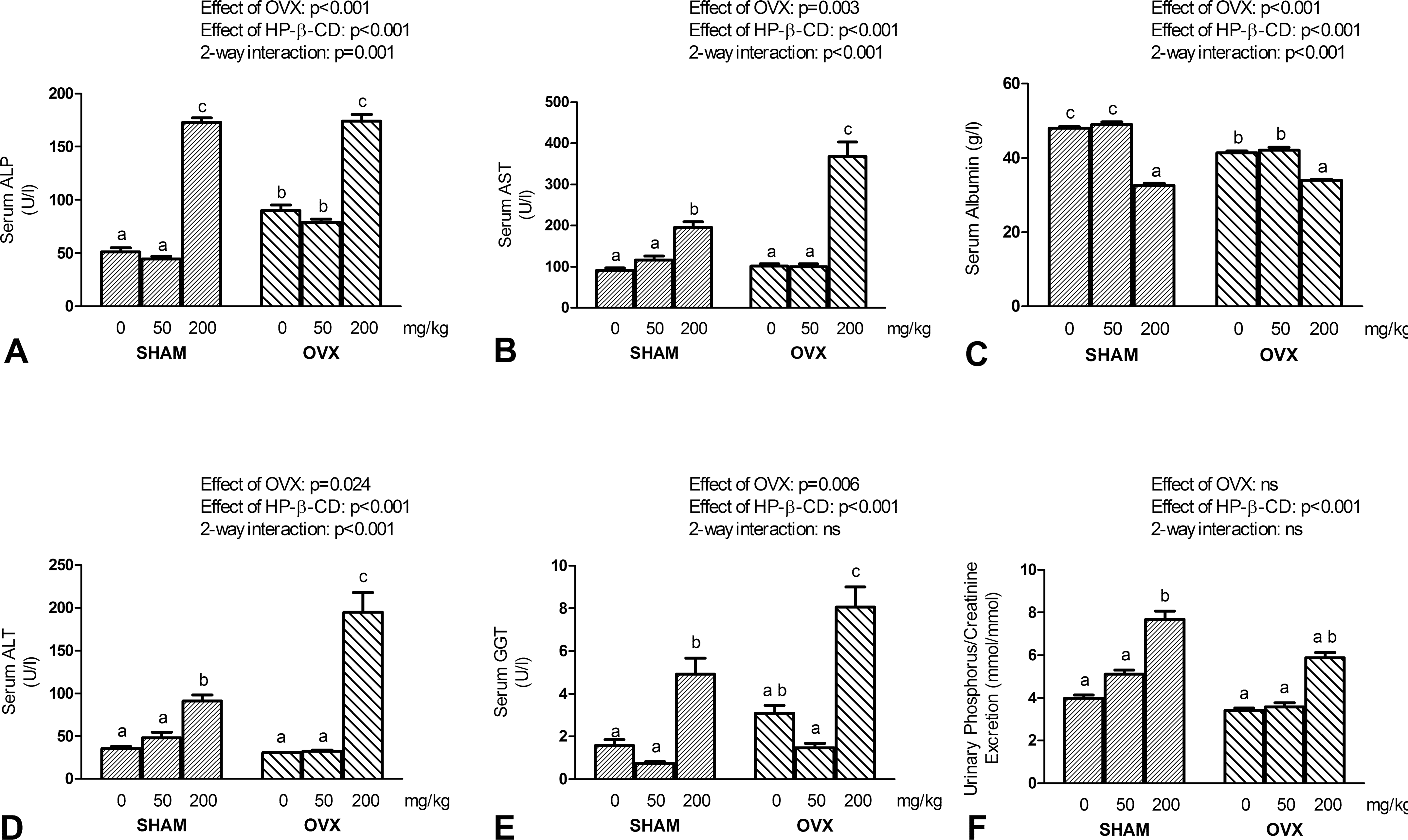
Effects of a 4-month administration of vehicle, 50 mg/kg, and 200 mg/kg HP-β-CD on serum ALP (A), serum AST (B), serum albumin (C), serum ALT (D), serum γ-GT (E), and urinary phosphorus excretion (F) in SHAM and OVX rats. ALP, alkaline phosphatase; AST, aspartate aminotransferase; ALT, alanine aminotransferase; γ-GT, gamma-glutamyl transferase. Columns marked with the same letter are not significantly different from each other by one-way ANOVA followed by Student-Newman-Keuls test. Error bars represent SEM. Insets show results of 2-way ANOVA.
Serum and urinary parameters after 4 months of administration of vehicle, 50 mg/kg, and 200 mg/kg HP-β-CD in SHAM and OVX rats.
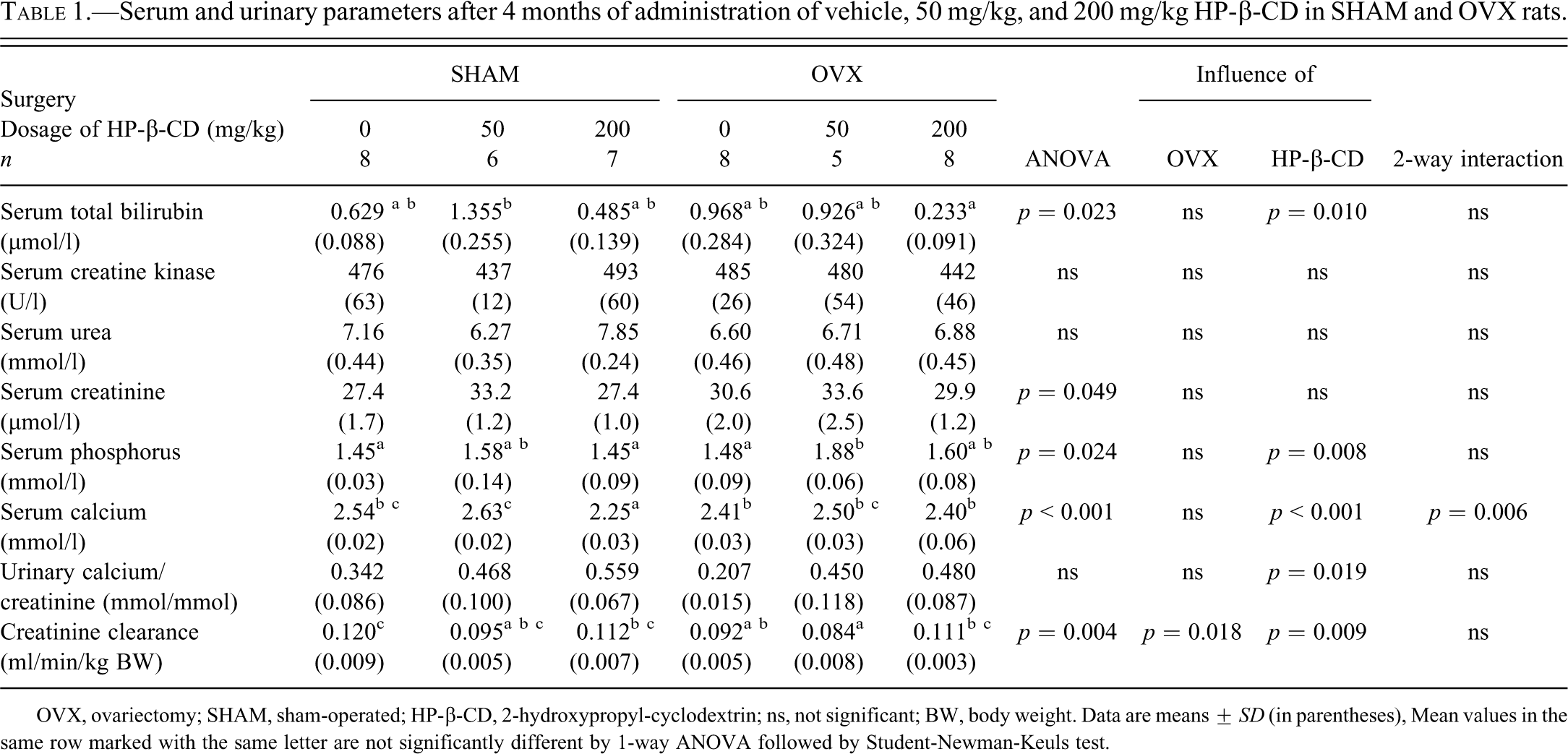
OVX, ovariectomy; SHAM, sham-operated; HP-β-CD, 2-hydroxypropyl-cyclodextrin; ns, not significant; BW, body weight. Data are means ± SD (in parentheses), Mean values in the same row marked with the same letter are not significantly different by 1-way ANOVA followed by Student-Newman-Keuls test.
BMD Measurements
As expected, OVX led to a decrease in volumetric BMD and cortical thinning at the proximal tibial metaphysis, at the tibial shaft, and at the L4 vertebra (Fig. 3). Cortical thinning at the tibial shaft was associated with an enlargement of the marrow cavity in OVX rats (increased endocortical perimeter, Fig. 3J). At the dose of 50 mg/kg, HP-β-CD did not cause bone loss at any of the sites examined in SHAM or OVX rats (Fig. 3A–J). However, 200 mg/kg HP-β-CD was associated with trabecular and cortical bone loss as evidenced by lower total and trabecular BMD (Fig. 3A–B) at the proximal tibial metaphysis (by 12% and 27% compared to SHAM rats and by 7% and 18% relative to OVX rats treated with vehicle, respectively). Proximal tibial cancellous bone osteopenia was clearly evident in bone sections from SHAM, and to a lesser extent also from OVX rats treated with 200 mg/kg HP-β-CD (Fig. 3D). At the L4 vertebra, total and trabecular BMD (Fig. 3E–F) were reduced by 14% and 20% in SHAM rats and by 6% and 9% in OVX rats receiving 200 mg/kg HP-β-CD in comparison with SHAM and OVX vehicle control rats. In addition, SHAM rats treated with 200 mg/kg HP-β-CD showed 24% lower cortical bone thickness at the tibial shaft (Fig. 3I), and 10% reduced cortical-subcortical BMD at the vertebral body (Fig. 3G), relative to vehicle-treated SHAM rats.
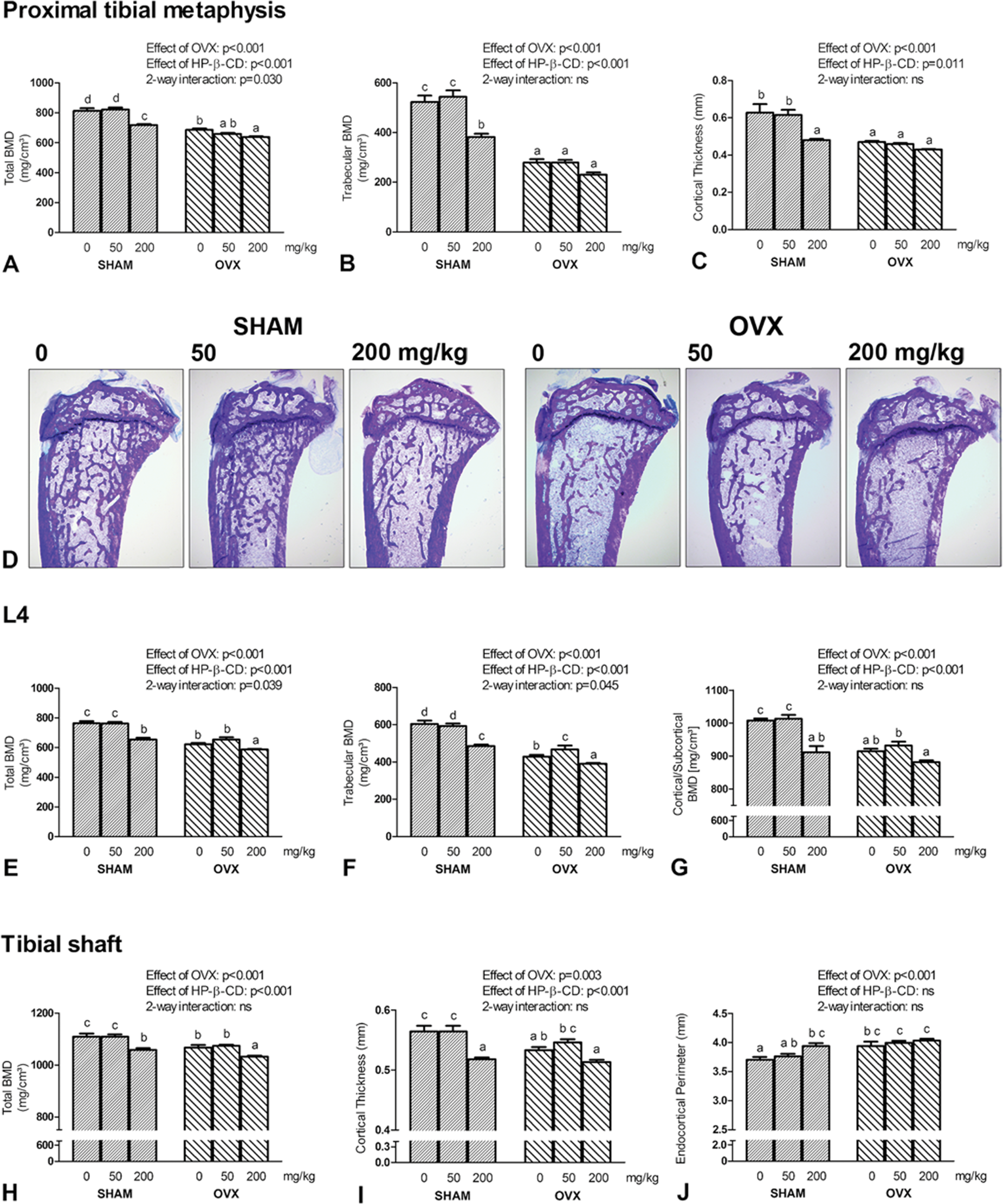
Influence of a 4-month administration of vehicle, 50 mg/kg, and 200 mg/kg HP-β-CD on tibial and vertebral trabecular and cortical bone in SHAM and OVX rats. Total bone mineral density (BMD, A), trabecular BMD (B), cortical thickness (C) at the proximal tibial metaphysis, proximal tibial bone histology (D), total BMD (E), trabecular BMD (F), and cortical/subcortical BMD (G) at the 4th lumbar vertebral body (L4), and total BMD (H), cortical thickness (I), and endocortical perimeter (J) at the tibial shaft are shown. Columns marked with the same letter are not significantly different from each other by one-way ANOVA followed by Student-Newman-Keuls test. Error bars represent SEM. Insets show results of 2-way ANOVA. Bone sections shown in (D) are 5-µm-thick undecalcified sections stained with toluidine blue, original magnification 25×.
Bone Turnover
To assess HP-β-CD-induced changes in bone turnover, we determined cancellous bone formation rate in tibial cancellous bone of SHAM and OVX rats by dynamic histomorphometry and measured urinary excretion of collagen crosslinks. As expected, tibial cancellous bone formation rate and urinary excretion of DPD was higher in OVX compared with SHAM rats (Fig. 4A–B), documenting the well-known high turnover osteopenia in OVX rats. We detected a weak stimulating effect of HP-β-CD on bone formation rate by 2-way ANOVA (Fig 4A). However, a significant difference between vehicle- and HP-β-CD-treated groups was seen only for OVX rats treated with 50 mg/kg HP-β-CD. In contrast, HP-β-CD treatment dose-dependently increased urinary DPD/creatinine excretion in SHAM and OVX rats (Fig. 4B). Urinary DPD/creatinine excretion was 204% and 55% higher in SHAM and OVX rats administered 200 mg/kg HP-β-CD, relative to vehicle SHAM and OVX control rats, respectively. Interestingly, already 50 mg/kg HP-β-CD significantly augmented urinary DPD excretion in SHAM and OVX rats by 51% and 48% compared to the respective vehicle controls.
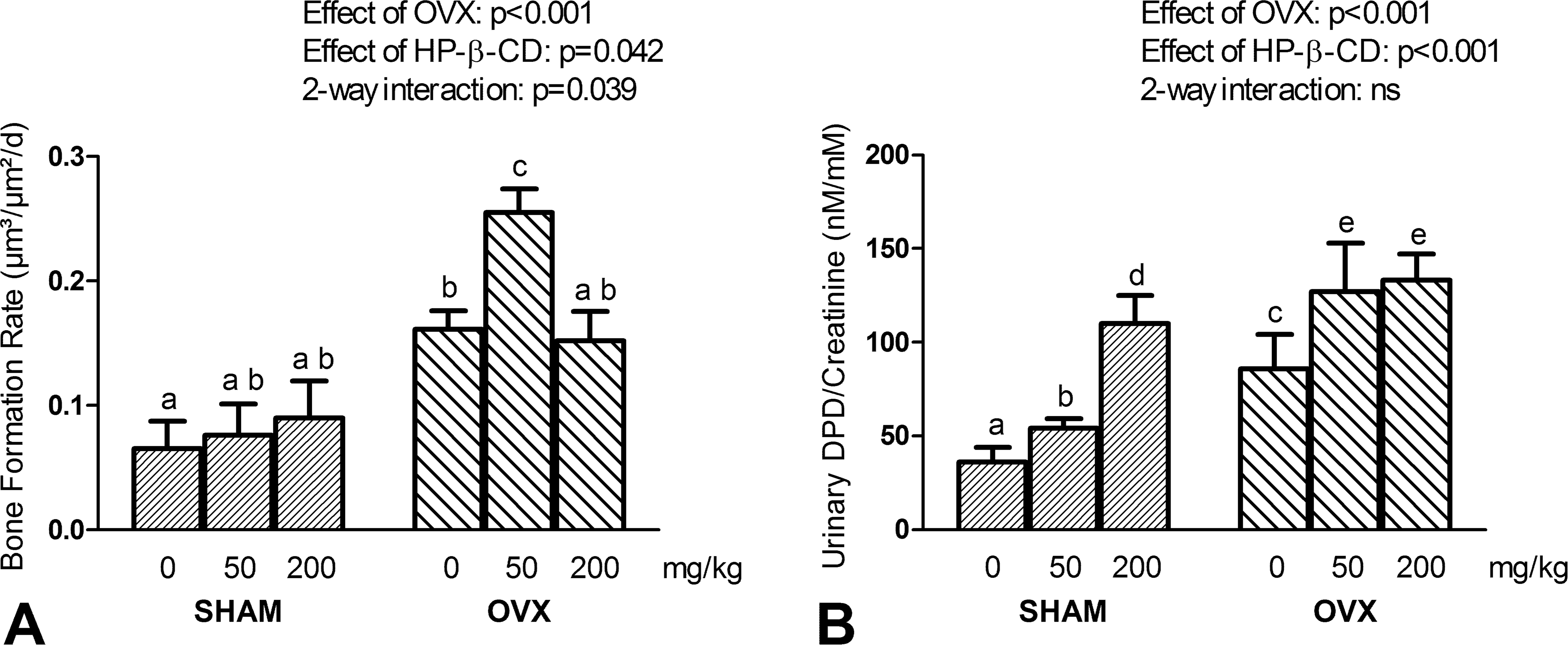
Bone formation rate measured by histomorphometry in cancellous bone of the proximal tibial metaphysis (A) and urinary deoxypyridinoline (DPD)/creatinine excretion (B) in SHAM and OVX rats treated for 4 months with vehicle, 50 mg/kg, and 200 mg/kg HP-β-CD. Columns marked with the same letter are not significantly different from each other by one-way ANOVA followed by Student-Newman-Keuls test. Error bars represent SEM. Insets show results of 2-way ANOVA.
Discussion
Parenteral administration of CDs may lead to toxic effects on different organs, particularly on the kidney. In addition, diverse behavioral and clinical symptoms have been linked to parenteral application of HP-β-CD (Gould et al. 2005). In our study, we examined for the first time the skeletal effects of long-term parenteral HP-β-CD at doses of 50 and 200 mg/kg in SHAM and OVX rats. At the daily subcutaneous dose of 200 mg/kg, HP-β-CD had pronounced negative effects on vertebral and tibial BMD, and profoundly increased bone resorption. In addition, 200 mg/kg HP-β-CD showed toxic effects on liver function and uterine weight in our 4-month study. Although 50 mg/kg HP-β-CD was devoid of any overt untoward effects on BMD or on organ function, this low dose already slightly increased bone resorption in SHAM and OVX rats. Therefore, toxic effects of parenteral HP-β-CD on bone metabolism need to be considered in long-term studies at daily doses of ≥50 mg/kg.
In the current study, OVX rats receiving daily injections of 200 mg/kg HP-β-CD showed reduced final body weights relative to vehicle-treated OVX animals. In a similar fashion, loss of body weight or failure to adequately gain body weight was described in other long-term rat experiments after intravenous and even oral application of CDs at higher concentrations (Gould et al., 2005; Perrin et al., 1978). It is not clear whether the reductions in body weight in HP-β-CD-treated rats can be explained by lower food and/or water consumption due to behavioral changes, or by liver or renal toxicity. Our findings of increased serum AST, ALT, and γ-GT, together with a decrease in serum albumin suggest that liver toxicity might be the reason for reduced body weight in OVX rats treated with 200 mg/kg HP-β-CD.
In addition to the HP-β-CD-induced changes in serum transaminases and albumin, we also found elevated serum ALP activity in SHAM and OVX rats treated with 200 mg/kg HP-β-CD. Because a robust discrimination between bone and liver isoforms of ALP is not possible in rats in our experience (unpublished data), we can only speculate about the tissue source of increased serum ALP. However, it is probable that the increased serum ALP was mainly caused by liver dysfunction, because bone formation rate measured by dynamic bone histomorphometry was only slightly influenced by administration of HP-β-CD, whereas transaminases and γ-GT were elevated. In a similar fashion, increased serum AST and ALP in HP-β-CD-treated rats were reported in earlier experiments (Gould et al. 2005). Furthermore, hepatotoxicity of long-term dietary administration of β-CD has also been suggested by a previous study (Bellringer et al. 1995). The authors observed increased centrilobular enlargement of hepatocytes which was interpreted as an adaptation response of the rodent liver to xenobiotics. In addition, a rise of single cell necrosis and portal inflammatory cell infiltration was noted, indicating hepatotoxicity.
The current study revealed a previously unknown adverse effect of HP-β-CD on the uterus. Treatment of SHAM rats with 200 mg/kg HP-β-CD induced a significant decrease in uterine wet weight. Histomorphometric analysis suggested that the HP-β-CD-induced reduction in uterine weight was not due to a specific effect on the myo- or endometrium, but rather affected the organ as a whole. The mechanism of the adverse effect of parenteral HP-β-CD on the uterus is unknown at present.
Interestingly, we found no evidence of a major nephrotoxic effect of HP-β-CD in our study. Although we also observed the vacuolation of the proximal convoluted tubules described in earlier studies (Gould and Scott 2005), serum creatinine and urea remained unchanged and creatinine clearance was not influenced in a dose dependent fashion by HP-β-CD. Thus, our data do not indicate major nephrotoxicity of HP-β-CD at the doses used. However, the increased urinary phosphorus excretion we observed in SHAM and OVX rats receiving 200 mg/kg HP-β-CD may indicate a beginning functional impairment at the proximal tubule. The major site of renal phosphate reabsorption is the proximal tubule, where the sodium-phosphate cotransporters NaPi-2a and NaPi-2c are expressed (Tenenhouse and Sabbagh 2002). Therefore, increased urinary phosphate excretion in the presence of unchanged serum phosphate levels may reflect a beginning HP-β-CD-induced reduction in proximal tubular transcellular phosphate transport.
The current study is the first to observe a negative influence of HP-β-CD on bone metabolism. At the daily dose of 200 mg/kg, HP-β-CD reduced tibial and vertebral BMD, and upregulated urinary DPD excretion, a biochemical marker of whole body bone resorption. Currently, we do not know the molecular mechanism behind this effect. Because the untoward skeletal side effect of HP-β-CD was almost identical in SHAM and OVX rats, it is very unlikely that this effect was primarily mediated by a suppressive effect on ovarian estrogen production. In addition, endometrial epithelial height, another indicator of estrogenic activity, was not reduced in HP-β-CD-treated SHAM rats. Collectively, the data in rats treated with 200 mg/kg HP-β-CD suggest that the bony effect may not be specific, but rather a part of general toxic effects of HP-β-CD on a variety of different organ systems. However, 50 mg/kg HP-β-CD already stimulated osteoclastic bone resorption in the absence of any other signs of general toxicity. Therefore, we cannot exclude that HP-β-CD might have direct bone resorption-stimulating effects in bone. Clearly, further experimentation is required to find out the mechanism which drives osteoclastic bone resorption in the presence of circulating HP-β-CD.
In summary, we demonstrated that daily parenteral administration of 200 mg/kg HP-β-CD over 4 months resulted in adverse effects on bone and liver in SHAM and OVX rats, and on the uterus in SHAM rats. Therefore, our study strongly suggests that untoward side effects have to be considered in long-term pharmacological studies when HP-β-CD is used as a vehicle for parenteral administration. Because the daily dose of 50 mg/kg already increased bone resorption, we cannot specify a “safe” dose of HP-β-CD in long-term studies. In addition, we do not know whether the negative influence of HP-β-CD on bone is transient or permanent. Several side effects are known to be reversible after discontinuation of administration of CDs (Stella and He 2008). Our study may also be relevant for humans. HP-β-CD is used as excipient for oral and parenteral administration of several pharmaceutical formulations approved for use in humans, including children (Groll et al. 2002; Willems et al. 2001). Some of the administered parenteral HP-β-CD dosages (e.g. 100 mg/kg, U.S. Food and Drug Administration 2009) are in the range of those used in the current experiment. Therefore, it would be advisable to assess potential untoward skeletal side effects of parenteral HP-β-CD also in humans. Our study suggests that the most sensitive way to do this would be the measurement of biochemical bone markers. In addition, future experiments are necessary to address the questions of the molecular mechanisms behind and of the reversibility of the untoward skeletal side effects of HP-β-CD.
Footnotes
Acknowledgments
The authors thank Claudia Bergow, Kerstin Klien, and Christiane Schüler for help with histology and histomorphometry, and Verena Spielberger for help with the surgery procedures.
The authors declared no potential conflicts of interest with respect to the research, authorship, and/or publication of this article.
The authors received no financial support for the research, authorship, and/or publication of this article.