
Abstract
A unilateral non-metastatic embryonal carcinoma and teratoma of the testis was observed in a 12-week-old Swiss Albino mouse at the end of a 28-day repeated dose toxicity study. The teratocarcinoma almost completely replaced the parenchyma of the left testis. The tumor was composed of sheets and rosettes of primitive embryonal cells, anaplastic cells, skeletal muscle tissue, sebaceous gland tissue, keratinized stratified squamous epithelium, and ciliated cuboidal epithelium. The histomorphological characteristics of the tumor were reviewed and presented in this report. To the best of the authors' knowledge, this is the first report of spontaneous teratocarcinoma of testis in the Swiss Albino strain of mice.
Introduction
The combination of embryonal carcinoma and teratoma is a tumor with more than one histologic pattern and has been termed as “teratocarcinoma.” In humans, this is the most common multiple histologic variant of testicular germ cell neoplasia and is capable of metastasis (Burns 2001). Teratocarcinoma and embryonal carcinoma of testis are rarely found in domestic animals, and teratoma is virtually unknown in domestic animals except for the horse (Foster and Ludds 2007). A single case of metastatic embryonal carcinoma in testis has been reported in the horse (Valentine and Weinstock 1986). As far as laboratory rats and mice are concerned, teratocarcinoma of the testis is commonly seen only in mouse strain 129 (Stevens and Little 1954). The few other reports of teratocarcinoma or teratoma in the testis of rats and mice include teratocarcinomas in two SD IGS rats (Sawaki et al. 2000), a teratoma in an ICR strain mouse (Tani et al. 1997), and teratocarcinomas in a DBA/2J and A/HeJ mouse (Meier et al. 1970). A case of spontaneous ovarian teratoma has been reported in a Swiss Albino mouse (Fawcett 1950).
However, the occurrence of spontaneous testicular teratocarcinoma seems to be extremely rare in strains of mice other than 129, and to the best of the authors' knowledge, there has been no previous reports of testicular teratocarcinoma in the Swiss Albino strain. The purpose of this communication is to describe the histomorphological characteristics of a spontaneous non-metastatic teratocarcinoma in the testis of a 12-week-old Swiss Albino mouse.
Materials and Methods
A Swiss Albino mouse (National Institute of Nutrition, Hyderabad, India) aged 12 weeks was found to have a testicular mass at the time of necropsy. The mouse was 1 of 48 animals sacrificed at the end of a 28-day repeated dose toxicity study. The male mouse was from the vehicle control group and had received honey mixed with water (1:3 parts at dose rate of 10 ml/kg body weight) for 28 days by the oral route via a stainless steel mice gavage needle. All mice in the study were housed individually in polycarbonate cages with rice husk on the bottom and a stainless steel grill lid. The cage environment was maintained at a temperature range of 20–26°C, relative humidity of 40–70%, and a 12-hour light-dark cycle. The animals were fed standard laboratory mice feed (National Institute of Nutrition) provided ad libitum and drinking water purified by the reverse osmosis process. All animals were maintained and cared for as per the guidelines of the Committee for the Purpose of Control and Supervision on Experiments in Animals (CPCSEA), India. At the end of the 28-day study period, all mice were euthanized by CO2 asphyxiation followed by exsanguination. Both the testes were preserved in Modified Davidson’s Fixative for the first 24 hours and later on in 10% neutral buffered formalin along with all other organs and tissues collected as per the study protocol.
Observations and Discussion
No mouse in the study showed any abnormal clinical signs or haematological and biochemical changes indicating disease condition. All mice exhibited normal body weight gain and food consumption. Since the mice had not had daily physical examinations during the experiment, it is not known when the scrotum of the mouse with the tumor started to expand or how rapidly the scrotum increased in size. Gross examination at sacrifice revealed an enlarged left testis that measured approximately 10 × 10 × 15 mm. The affected testis had a nearly normal shape, with a smooth surface and intact tunica albuginea. The cut surface of the testicular lesion consisted of solid whitish tissue with small red areas inside. The right testis was reduced in size, and the gross appearance of the right epididymis was normal. However, the length of the left epididymis was increased. No other gross lesions were found in any other organ or tissue.
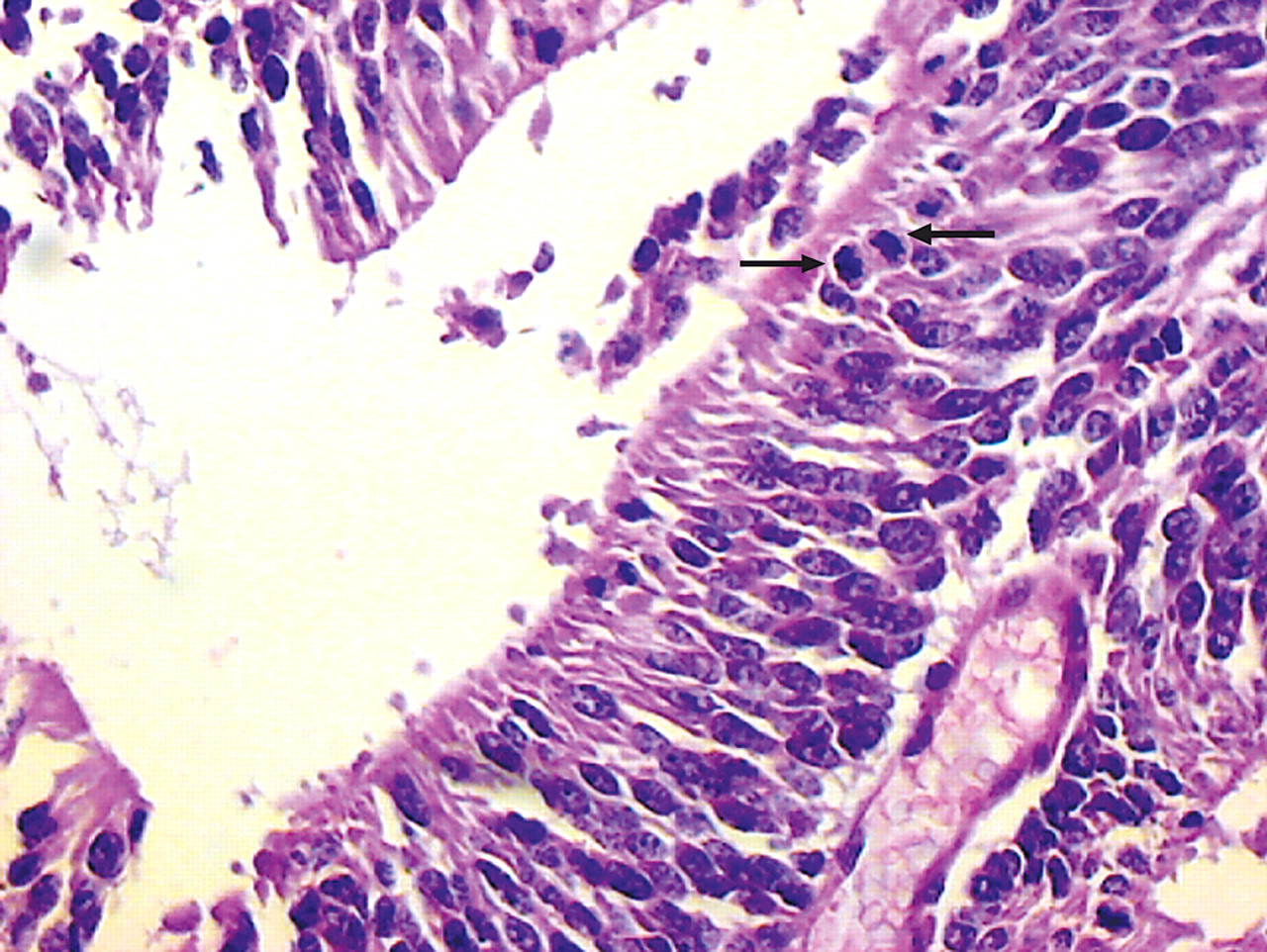
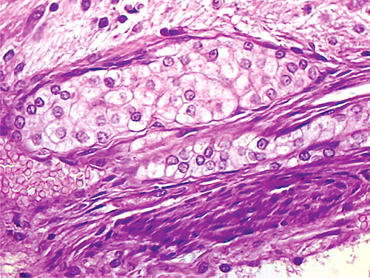
The testis was incised longitudinally at the midline and processed, embedded in paraffin, sectioned at 4 to 5 µm thickness, and stained with routinely used hematoxylin and eosin. Microscopic examination revealed that the testicular parenchyma was almost completely replaced by the cells of the embryonal carcinoma (Figure 1 ). The embryonal carcinoma was composed of solid sheets of primitive embryonic cells with crowded, large, oval, basophilic nuclei along with prominent nucleoli, scant cytoplasm, and indistinct cytoplasmic borders (Figure 2 ). The cells formed rosettes within the solid sheets at multiple sites (Figures 1 and 3 ). The sheets were surrounded by anaplastic cells with large, round vesicular nuclei and scant cytoplasm that were situated loosely within pink extracellular material (Figure 4 ). Mitotic figures were noticeably visible in the rosettes (Figure 5 ). Seminiferous tubules with one or two layers of germinal cells were observed at one end of the section.
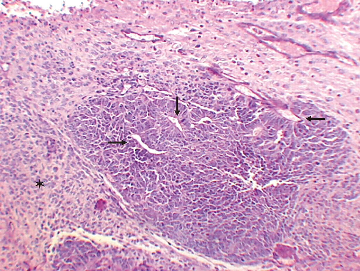
Testicular parenchyma replaced by embryonal carcinoma; rosettes formation in the sheet (arrows) and anaplastic cells (asterisk). H&E, 40×.
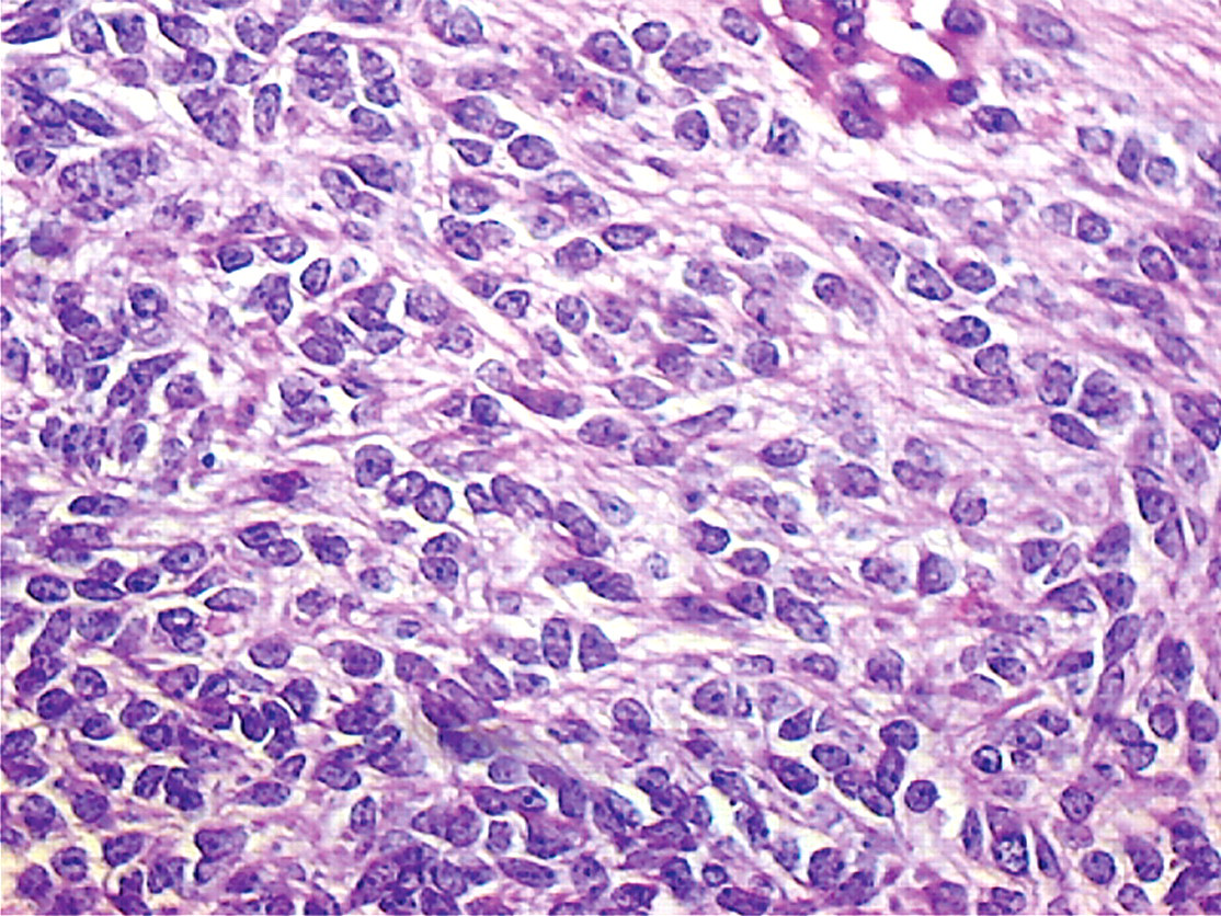
Sheet formation by primitive embryonal cells with large, oval, basophilic nuclei and prominent nucleoli, scant cytoplasm, and indistinct cytoplasmic borders. H&E, 400×.
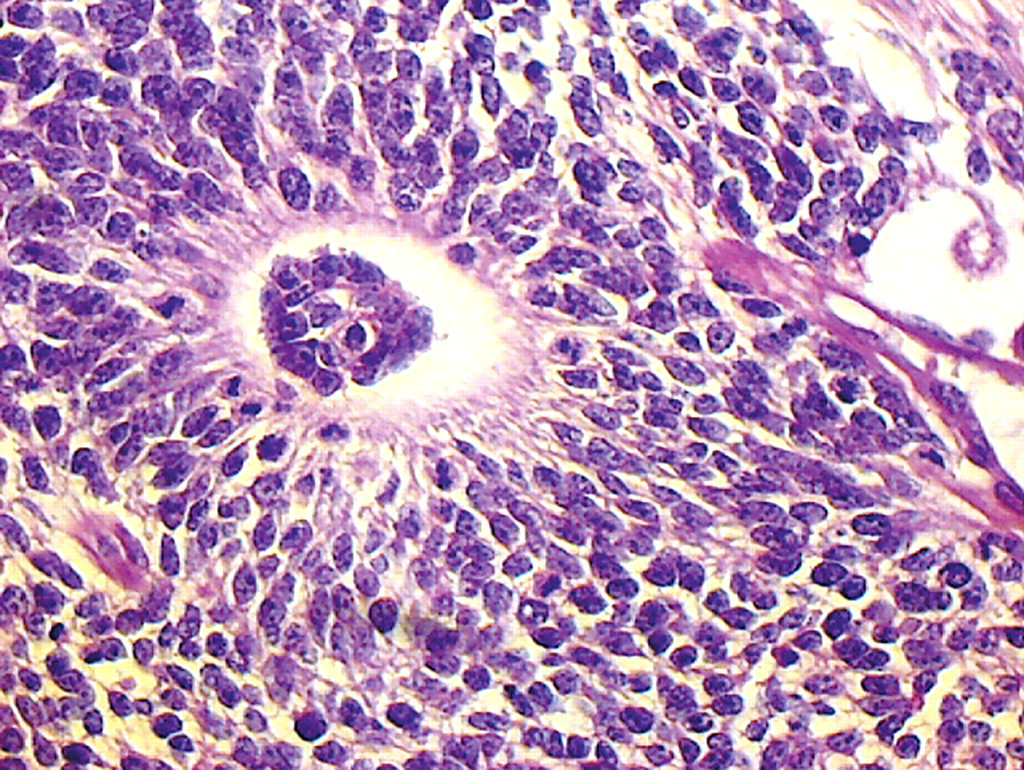
Primitive embryonic cells forming rosettes. H&E, 400×.
Anaplastic cells with large, round vesicular nuclei and scant cytoplasm within pink extracellular material. H&E, 400×.
Mitotic figures in the rosettes (arrows). H&E, 400×.
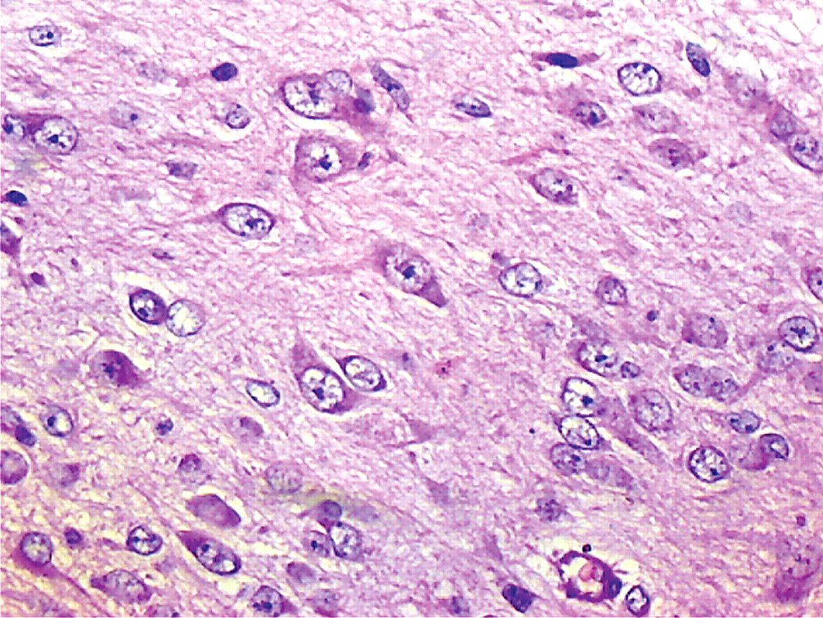
The tissue section also showed characteristic features of a teratoma with a focus of skeletal muscle (Figure 6 ), a cavity lined by ciliated cuboidal epithelium similar to respiratory epithelium (Figure 7 ), a duct lined by keratinized stratified squamous epithelium (Figure 8 ), and a sebaceous gland tissue (Figure 9 ). Subsequent serial sections of the testis revealed similar morphology. A focus of necrotic tissue was also identified.
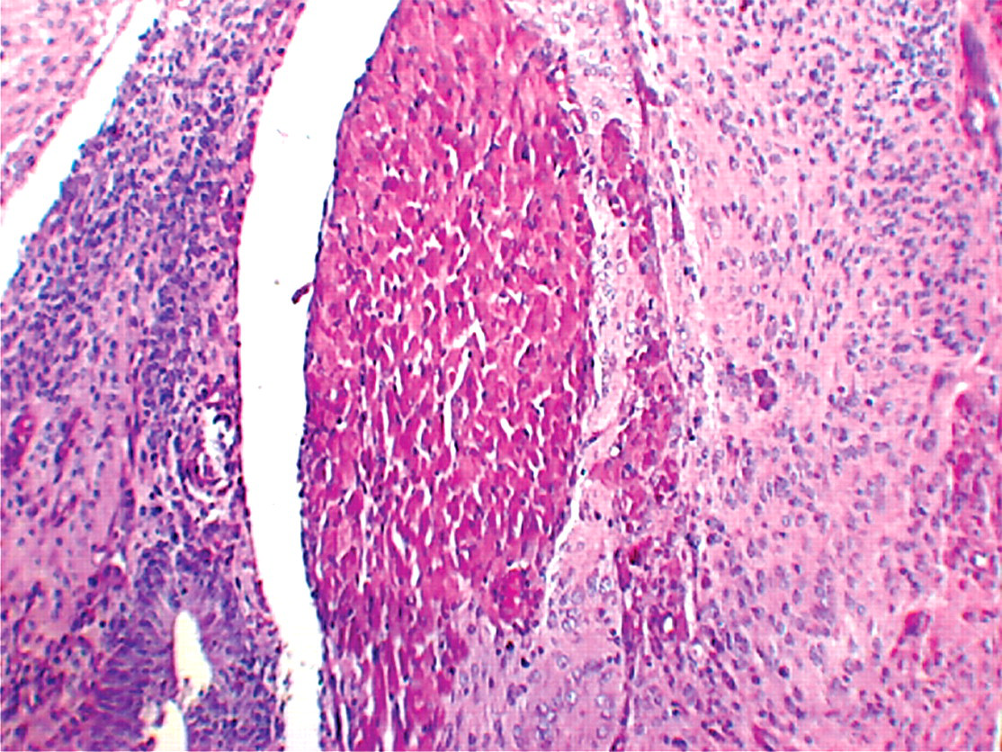
Skeletal muscle tissue surrounded by sheet of anaplastic cells. H&E, 100×.
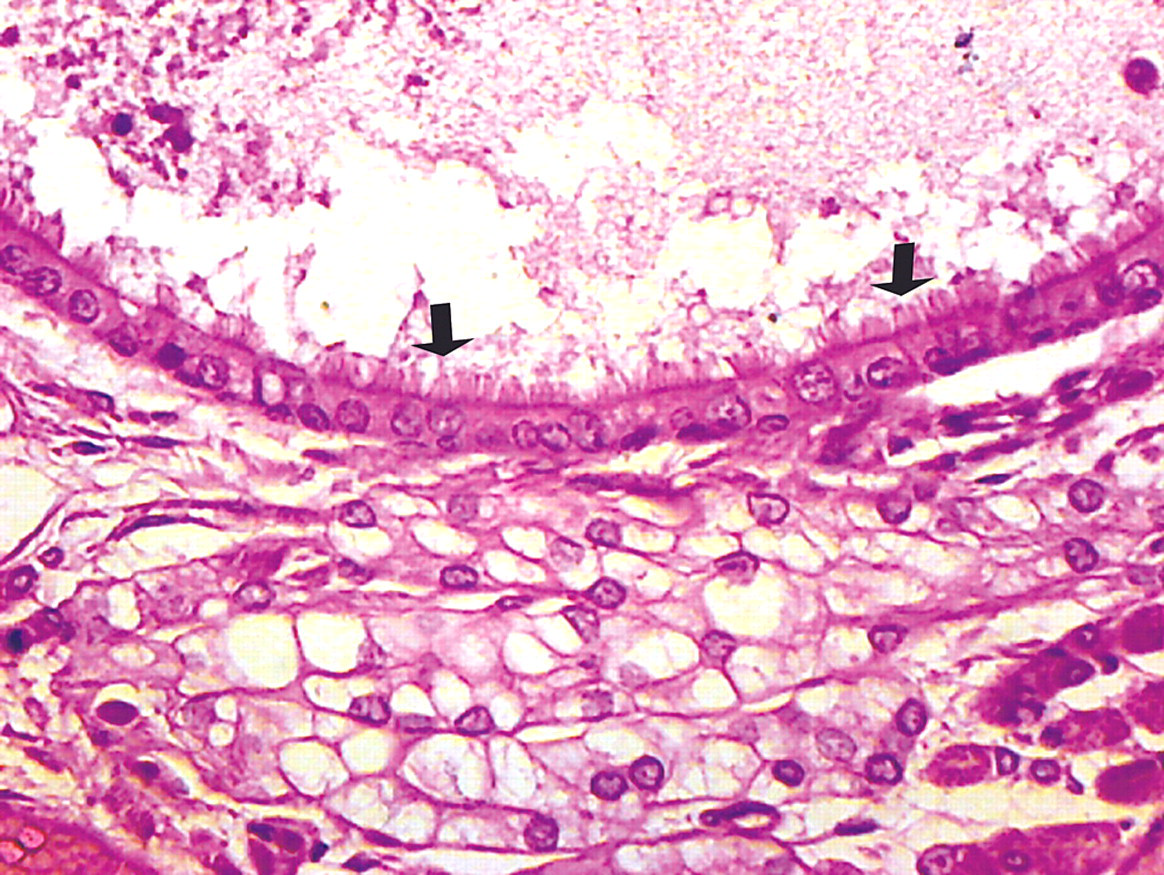
Cavity lined by ciliated cuboidal epithelium (arrows). H&E, 400×.
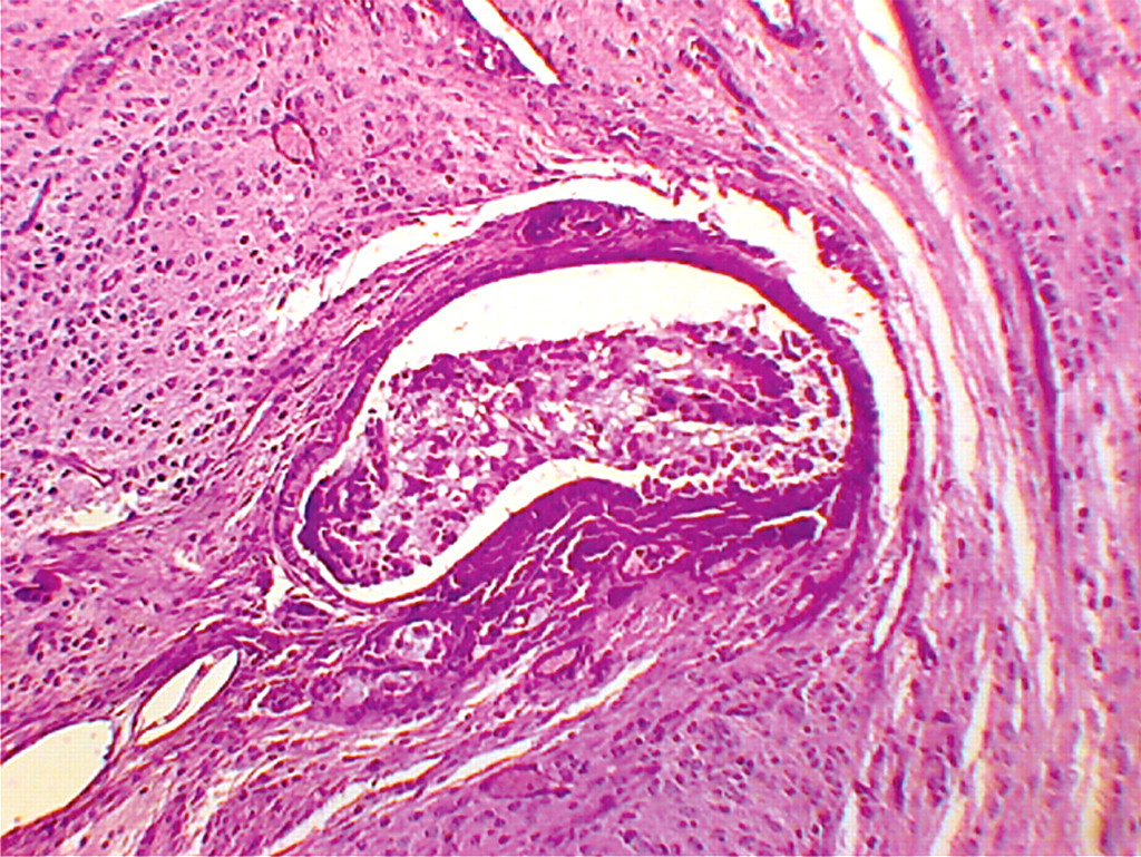
Duct lined by keratinized stratified squamous epithelium. H&E, 100×.
Sebaceous gland tissue within the teratoma. H&E, 400×.
Microscopic evaluation of the left epididymis revealed complete absence of spermatozoa in the tubules, which was attributed to replacement of spermatogenic seminiferous tubules by the tumor mass in the left testis. Other collected organs including heart, kidneys, brain, liver, and spleen did not show any lesions by microscopic evaluation.
The diagnosis of teratocarcinoma in this case is based on the histomorphological details of the lesion, which clearly matched with that of embryonal carcinoma and teratoma as reported in the literature (Sawaki et al. 2000; Burns 2001; Ulbright 2005). Immunostaining with alpha-fetoprotein (AFP) can be helpful for diagnosis of embryonal carcinoma (Valentine and Weinstock 1986; Foster and Ludds 2007), but there was no opportunity for an immunohistochemical workup in this case. Immunostaining with OCT 4, a nuclear transcription factor, is reportedly a more sensitive and specific tool for differentiating germinoma and embryonal carcinoma from other neoplasms in humans (Jones et al. 2004).
It is a well-accepted concept that the primordial totipotent germ cells of the embryo are the origin of embryonal carcinomas that eventually develop into the teratomas (Ulbright 2005; Göbel et al. 1998; Mostofi 1983; Nochomovitz and Rosai 1978). This concept is supported by the remarkable parallel patterns of allelic loss in teratomas of the postpubertal testis and other types of germ cell tumors that accompany these in humans (Kernek et al. 2003).
To the best of the authors' knowledge, the current report is first of its kind describing spontaneous teratocarcinoma of the testis in the Swiss Albino strain of mice, which implies that its occurrence is extremely rare in this strain of mice.
Footnotes
Acknowledgments
The authors gratefully acknowledge Dr. Bruce Williams from the C.L. Davis Foundation, USA, for his expert opinion on the case. The authors are also thankful to the Director General of the Central Council for Research in Ayurveda and Siddha for providing necessary facilities for conducting the research.