
Abstract
Phantoms simulating the human body play a central role in radiation dosimetry. The first computational body phantoms were based upon mathematical expressions describing idealised body organs. With the advent of more powerful computers in the 1980s, voxel phantoms have been developed. Being based on three-dimensional images of individuals, they offer a more realistic anatomy. Hence, the International Commission on Radiological Protection (ICRP) decided to construct voxel phantoms representative of the adult Reference Male and Reference Female for the update of organ dose coefficients. Further work on phantom development has focused on phantoms that combine the realism of patient-based voxel phantoms with the flexibility of mathematical phantoms, so-called ‘boundary representation’ (BREP) phantoms. This phantom type has been chosen for the ICRP family of paediatric reference phantoms. Due to the limited voxel resolution of the adult reference computational phantoms, smaller tissues, such as the lens of the eye, skin, and micron-thick target tissues in the respiratory and alimentary tract regions, could not be segmented properly. In this context, ICRP Committee 2 initiated a research project with the goal of producing replicas of the ICRP Publication 110 phantoms in polygon mesh format, including all source and target regions, even those with micron resolution. BREP phantoms of the fetus and the pregnant female at various stages of gestation complete the phantoms available for radiation protection computations.
1. Introduction
Phantoms simulating the human body or parts thereof play a central role in radiation dosimetry. The first computational body phantoms were based upon mathematical expressions representing planes, and cylindrical, conical, elliptical, and spherical surfaces describing the shape and position of idealised body organs (Fisher and Snyder, 1967, 1968; Snyder et al., 1969, 1978; Cristy, 1980; Cristy and Eckerman, 1987). For this first generation of computational body phantoms, the organ masses and volumes were in accordance with the International Commission on Radiological Protection's (ICRP) data of former Reference Man (ICRP, 1975).
With the advent of more powerful computers in the 1980s, various groups have developed voxel phantoms as an extension and improvement to these earlier models (Zankl et al., 1988; Dimbylow, 1996; Caon et al., 1999; Xu et al., 2000; Zankl and Wittmann, 2001; Petoussi-Henss et al., 2002; Kramer et al., 2003, 2004b, 2006; Caon, 2004; Fill et al., 2004; Dimbylow, 2005; Zaidi and Xu, 2007; Becker et al., 2008). Being based on three-dimensional images of single individuals, they offer a more realistic replication of human anatomy. Various authors have shown that the organ shapes of the earlier mathematical phantoms present an oversimplification that has an influence on the energy distribution which may deviate for some cases systematically from that calculated for voxel phantoms (Jones, 1997; Smith et al., 2000; Chao et al., 2001; Zankl et al., 2002, 2003; Kinase et al., 2003; Kramer et al., 2004a, 2005).
However, despite their obvious advantages compared with the previously used stylised phantom type, most of these phantoms do not represent the average Caucasian man or woman due to being derived from a specific individual. Hence, ICRP decided to construct voxel phantoms being representative of the adult Reference Male and Reference Female (ICRP, 2002) with respect to their external dimensions, their organ topology, and their organ masses (ICRP, 2009) for the update of organ dose conversion coefficients following the 2007 Recommendations (ICRP, 2007).
A further disadvantage of all voxel phantoms is their limited flexibility concerning changes in size, shape, and posture. Therefore, further work on phantom development has focused on the development of phantoms that seek to combine the realism of patient-based voxel phantoms with the flexibility of mathematical phantoms. So-called ‘hybrid’ phantoms may be based on voxel geometry, but their stepped surfaces are replaced by polygon meshes (PM) and/or non-uniform rational B-splines (NURBS), thus resulting in so-called ‘boundary representation’ (BREP) phantoms. Hence, this phantom type has been chosen for the family of paediatric reference phantoms being developed for ICRP.
Finally, as the voxel resolution of the adult reference computational phantoms is of the order of a few millimetres, smaller tissues, such as the lens of the eye, skin, and micron-thick target tissues in respiratory and alimentary tract regions, could not be segmented properly. Therefore, the calculated doses for these tissues have some limitations, particularly for weakly penetrating radiation. As a consequence, a series of stylised phantoms representing the skin, the lens of the eye, and high-resolution source and target regions in the human respiratory and alimentary tracts have been used recently for dose calculations of ICRP in addition to the reference voxel phantoms. In this context, ICRP Committee 2 recently initiated a research project with the goal of producing replicas of the Publication 110 (ICRP, 2009) phantoms in PM format, including all source and target regions, even those with micron resolution, so that only one set of phantoms can be used for all reference dose calculations.
2. Adult reference computational phantoms
2.1. Method of construction
Voxel phantoms were selected with external dimensions close to the reference data. The male phantom Golem (Zankl and Wittmann, 2001) is 176 cm tall, corresponding exactly to the reference height of the adult male, whereas his body mass of 69 kg is slightly below the adult male reference mass of 73 kg. The female phantom Laura (Zankl et al., 2005) is 167 cm tall, which is slightly taller than the ICRP female adult reference height of 163 cm, whereas her body mass of 59 kg is slightly lower than the corresponding ICRP reference mass of 60 kg. These two phantoms were adjusted to the reference values of Publication 89 (ICRP, 2002) in several steps: (1) adjustment of the body height and skeleton mass by voxel scaling; (2) adjustment of the individual organ and tissue masses to the reference values by adding or subtracting the required number of voxels; (3) additional organ and body region modifications, such as additional segmentation of the larger blood vessels and including a number of lymphatic nodes; and (4) adjustment of the whole-body mass to the reference values by adding or subtracting an appropriate number of adipose tissue voxels. The bones were subdivided into a cortical shell and a spongiosa region accommodating the bone trabeculae together with the marrow cavities – structures that are both much smaller than the voxel resolution of the phantoms. In the long bones, medullary cavities were segmented separately. The method of constructing the adult male and female reference computational phantoms is described in detail in Publication 110 (ICRP, 2009).
2.2. Applications and conceptual limitations
The adult reference computational phantoms of Publication 110 (ICRP, 2009) are the official computational models representing Reference Male and Reference Female (ICRP, 2002, 2007). Each of these reference computational phantoms has 140 different organs and tissues. The male phantom consists of approximately 1.9 million voxels with a resolution of 2.137 × 2.137 × 8.0 mm3, and the female phantom consists of approximately 3.9 million voxels at a slightly finer resolution of 1.775 × 1.775 × 4.84 mm3. Being based on computed tomographic (CT) data of real people, these phantoms represent digital three-dimensional representations of human anatomy. They are defined to enable calculations of the protection quantities – organ and tissue equivalent dose and effective dose – from exposure to ionising radiation. ICRP has recently published recommended values for dose coefficients for both external and internal exposures using these two phantoms: Publication 116 (ICRP, 2010), Publication 133 (ICRP, 2016a) Publication 134 (ICRP, 2016b) and Publication 137 (ICRP, 2017). Further dose coefficients issued by ICRP will follow (e.g. for environmental exposures, and further publications dealing with occupational and environmental intakes of radionuclides).
Although these phantoms have reference organ masses, they still have individual organ topology reflecting the CT data used for their construction. Obviously, both models cannot represent real individuals, and thus they should not be used to assess doses for specific individuals. While the reference computational phantoms were created for the purpose of deriving radiological protection quantities, they may have broader applications. However, the specific limitations related to their intended application have to be kept in mind.
2.3. Limitations due to voxel resolution
The reference computational phantoms’ voxel resolutions of the order of millimetres and, specifically, the slice thickness of several millimetres result in stepped organ surfaces, which is especially problematic for the skin and hollow organs as the contours are not closed. Also, the lens of the eye, a target that has recently become the focus of radiological protection, is represented too coarsely in the reference voxel phantoms. Furthermore, very fine structures, such as some specific source and target regions in the alimentary and respiratory tracts with micrometre dimensions, cannot be represented at all by the relatively coarse voxels. This resulted in the need to use several stylised phantoms in addition to the voxel phantoms for the calculation of reference dose coefficients for high-resolution source and target regions, and radiations of low penetration.
3. Adult reference phantom conversion project
This issue of limitations due to voxel resolution was discussed by ICRP Committee 2, and it was decided to propose a research project for creating BREP versions of the reference computational phantoms, with the aim of producing exact replicas of the Publication 110 (ICRP, 2009) reference phantoms in a high-quality PM format. These phantoms should include:
continuous and fully-enclosed walls for skin, stomach, gall bladder, and urinary bladder; thin target layers (10–300 µm) for the alimentary and respiratory tract organs; and detailed models for the skeletal system, eyes, lymphatic nodes, blood vessels, etc.
The research project is being undertaken mainly at Hanyang University in Seoul, South Korea, and is supported by a specific task group of Committee 2.
3.1. Method of construction
To construct PM models of simple organs, the voxel models of the Publication 110 (ICRP, 2009) reference phantoms were converted directly to the PM format. For this, various procedures were developed depending on the complexity of the individual organs. For very simple organs, the voxel model is first used to generate a PM model using a three-dimensional surface rendering method. Next, the number of polygons in the PM model is increased to facilitate the smoothing and refinement processes. After smoothing and refinement, the number of facets is reduced to a proper number; for high efficiency in computer simulations, the minimum necessary number of facets to keep the shape of the organ is used. The second procedure is applied to organs with a more complicated shape, such as the large intestine; again, the voxel model is first used to generate a PM model using a three-dimensional surface rendering method. Next, a contour line of the organ is created and subsequently converted to a NURBS surface model, which is then converted back into a polygon surface model (Yeom et al., 2013).
More specific procedures have been applied for individual organs and suborgan structures, such as the small intestine (Yeom et al., 2016a), the skeleton (Yeom et al., 2016b), the lens of the eye (Nguyen et al., 2015), and the thin target and source regions in the alimentary and respiratory tracts (Kim et al., 2017).
3.2. Results
The resulting PM phantoms are shown in Fig. 1, together with the Publication 110 reference voxel phantoms (ICRP, 2009). It can be seen that: (1) the PM phantoms closely resemble the Publication 110 phantoms in their main anatomical features and thus meet the requirement of being ‘exact replicas’; but (2) clearly demonstrate the high-quality smooth surfaces that are obviously lacking in the Publication 110 phantoms.
Details from the Publication 110 voxel phantoms (ICRP, 2009; left) and the high-quality polygon mesh phantoms (right).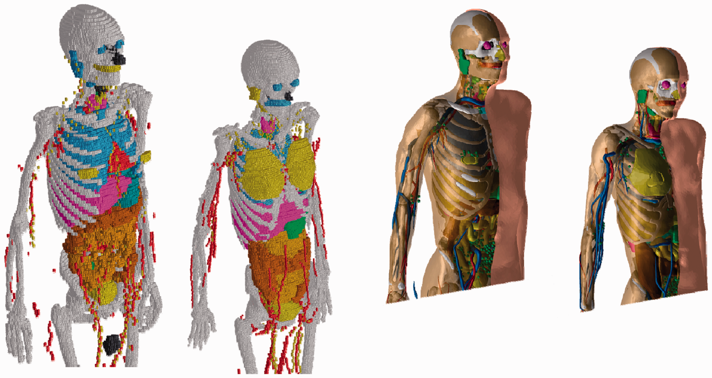
3.3. Computational results
It is expected that the converted phantoms will lead to the same, or very similar, dose coefficients as the Publication 110 reference phantoms (ICRP, 2009) for penetrating radiation, and provide more accurate dose coefficients for weakly penetrating radiation and small tissues. In addition, the reference phantoms in PM format would be easily deformable and could serve as a starting point to create phantoms of various postures to be used, for example, in accidental dose calculations.
4. Further developments
4.1. Paediatric reference computational phantoms
Researchers at the University of Florida (UF) have introduced a series of hybrid phantoms: newborn, and 1-, 5-, 10-, and 15-year paediatric phantoms (Lee et al., 2010). Major organs and tissues were segmented from CT data of patients at ages close to newborn, and 1, 5, 10, and 15 years. NURBS and PM surfaces were used to model individual organs and tissues, and subsequently match anthropometric data and reference organ masses (ICRP, 2002). These UF phantoms were modified to include the breast model with glandular and adipose tissue. Subsequently, the UF hybrid phantom series was used to produce the reference phantoms in voxel format, matching the reference data of Publication 89 (ICRP, 2002). In order to introduce these phantoms as ICRP reference paediatric phantoms, further modifications and refinements have been performed: blood vessels in the lungs have been incorporated; the anatomical realism of the muscles has been improved; and a sophisticated distribution of lymphatic nodes has been incorporated (Lee et al., 2013).
4.2. Phantoms of the developing human fetus
As concern grows over possible radiation-induced cancers from medical and non-medical exposures of the pregnant female, the need also increases to better quantify fetal radiation doses, particularly at the organ level. To address this need, two fetal hybrid computational phantoms were constructed at UF using high-quality magnetic resonance imaging and CT image sets obtained for two well-preserved fetal specimens aged 11.5 and 21 weeks post conception. Individual soft tissue organs, bone sites, and outer body contours were segmented from these images and converted to deformable NURBS surfaces. The two specimen-specific phantoms, along with a modified version of the 38-week UF hybrid newborn phantom, comprised a set of base phantoms from which a series of hybrid computational phantoms was derived for fetal ages 8, 10, 15, 20, 25, 30, 35, and 38 weeks post conception. The methodology used to construct the series of phantoms accounted for the following age-dependent parameters: (1) variations in skeletal size and proportion; (2) bone-dependent variations in relative levels of bone growth; (3) variations in individual organ masses and total fetal masses; and (4) statistical percentile variations in skeletal size, individual organ masses, and total fetal masses. The resulting series of fetal hybrid computational phantoms is applicable to organ-level and bone-level internal and external radiation dosimetry for human fetuses of various ages and weight percentiles (Maynard et al., 2011).
4.3. Phantoms of the pregnant female
Efforts to assess in-utero radiation doses and related quantities to the developing fetus should account for the presence of the surrounding maternal tissues. Maternal tissues can provide varying levels of protection to the fetus by shielding externally emitted radiation or, alternatively, can become sources of internally emitted radiation following the biokinetic uptake of medically administered radiopharmaceuticals or radionuclides located in the surrounding environment. Using CT image sets of pregnant patients, the eight UF fetal phantoms from the study of Maynard et al. (2011) were combined systematically with the UF adult non-pregnant female (Lee et al., 2010) to yield a series of reference pregnant female phantoms at fetal ages 8, 10, 15, 20, 25, 30, 35, and 38 weeks post conception. Deformable NURBS surfaces were utilised to alter contoured maternal anatomy in order to: (1) accurately position and orient each fetus and surrounding maternal tissues; and (2) match target masses of maternal soft tissue organs to reference data reported in the literature (Maynard et al., 2014).
5. Summary
The Publication 110 adult male and female voxel phantoms (ICRP, 2009) are the official computational phantoms representing ICRP Reference Male and Reference Female. They are defined to enable calculations of the protection quantities – organ and tissue equivalent dose and effective dose – from exposure to ionising radiation. Although these phantoms have reference organ masses, they still have individual organ topology reflecting the CT data used for their construction. Obviously, both models cannot represent real individuals, and thus they should not be used to assess doses for specific individuals. While the reference computational phantoms were created for the purpose of deriving radiological protection quantities, they may have broader applications. However, the specific limitations related to their intended application have to be kept in mind.
The reference voxel phantoms have limitations concerning the representation of small objects due to the voxel resolution of the underlying medical image data. These limitations are being addressed by the current phantom conversion project. The resulting PM-type reference computational phantoms closely resemble the Publication 110 phantoms (ICRP, 2009) in their main anatomical features, and thus meet the requirement of being ‘exact replicas’, but on the other hand they include micrometre-fine source and target regions that could not be represented in the Publication 110 phantoms. It is expected that the converted phantoms will lead to the same, or very similar, dose coefficients as the Publication 110 reference phantoms for penetrating radiation, and provide more accurate dose coefficients for weakly penetrating radiation and small tissues. In addition, the reference phantoms in PM format would be easily deformable and could serve as a starting point to create phantoms of various postures to be used, for example, in accidental dose calculations.
A series of hybrid phantoms representing the Publication 89 reference newborn, and 1-, 5-, 10-, and 15-year paediatric phantoms (ICRP, 2002) has been introduced. BREP phantoms of the fetus and the pregnant female at various stages of gestation complete the phantoms available for radiation protection computations.