
Abstract
The purpose of the International Commission on Radiological Protection (ICRP) Committee 3 Working Party was to update the 2001 web-based module ‘Radiation and your patient: a guide for medical practitioners’ from ICRP. The key elements of this task were: to clearly identify the target audience (such as healthcare providers with an emphasis on primary care); to review other reputable sources of information; and to succinctly publish the contribution made by ICRP to the various topics. A ‘question-and-answer’ format addressing practical topics was adopted. These topics included benefits and risks of imaging using ionising radiation in common medical situations, as well as pertaining to specific populations such as pregnant, breast-feeding, and paediatric patients. In general, the benefits of medical imaging and related procedures far outweigh the potential risks associated with ionising radiation exposure. However, it is still important to ensure that the examinations are clinically justified, that the procedure is optimised to deliver the lowest dose commensurate with the medical purpose, and that consideration is given to diagnostic reference levels for particular classes of examinations.
1. INTRODUCTION
We live in a time where tremendous amounts of internet-based, or web-based, information are accessible on almost every conceivable topic, with varying degrees of source credibility. It is encouraging to note that there are numerous credible sources of information on radiological protection in medical settings. The purpose of the International Commission on Radiological Protection (ICRP) Committee 3 Working Party was to update an internet-based ICRP module entitled ‘Radiation and your patient: a guide for medical practitioners’ (ICRP, 2001), first published in English in 2001 and subsequently translated into Spanish. In an environment with many sources of similar information, it is important to ensure that ICRP’s web-based resource is not superlative. To this end, the updated report will focus on ICRP’s contribution to radiological protection in medical settings, and directed interested readers to further resources from both ICRP and other organisations working on radiological protection. Primary healthcare providers represent the target audience. The language level for the report is therefore provided in a way that can be shared with patients and their caretakers if desired. A pragmatic ‘question-and-answer’ format was chosen to allow the document to be searched easily for specific areas of interest, and to facilitate rapid answers to questions raised by busy providers. One further goal is to provide the information as open source to a worldwide audience.
2. MethodS
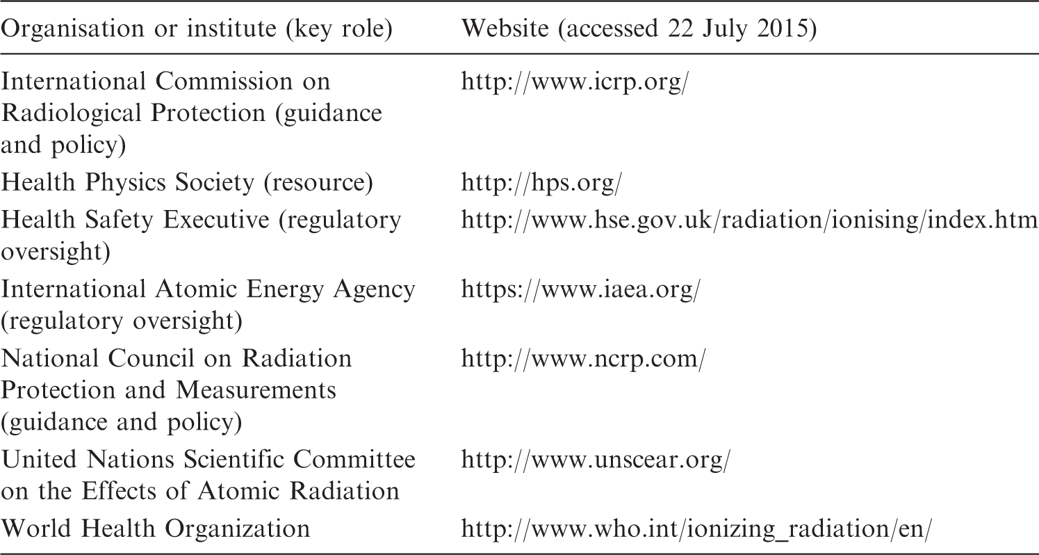
Relevant web-based sources of information on radiological protection in medical settings.
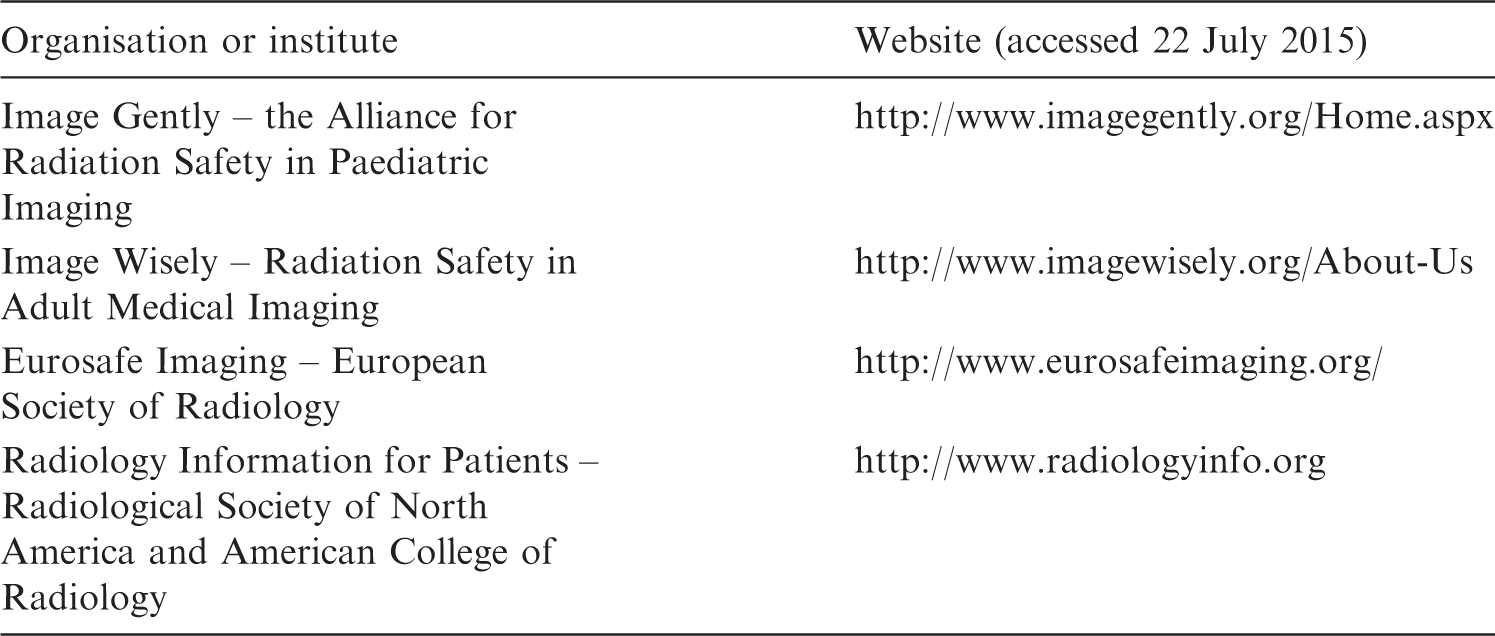
Relevant patient radiation dose reduction and imaging education websites.
3. Results
A list of commonly encountered radiological protection topics was compiled, and topics were described in a ‘question-and-answer’ format. These topics are not intended to be the final set, as the web-based resource can be modified and expanded at any time. The following is a brief description and summary of the key topics addressed in the report. Each of these topics is discussed in more detail in the web report. The main differences between this internet-based resource and the former are a shorter more focussed question-and-answer format, updated ICRP references, and an expanded audience of primary healthcare providers beyond physicians.
3.1. What is the International System of Radiological Protection?
‘Since 1928, ICRP has developed, maintained, and elaborated the international system of radiological protection used worldwide as the common basis for radiological protection standards, legislation, guidelines, programmes, and practice’ (www.icrp.org).
‘The International System of Radiological Protection has been developed by ICRP based on (i) the current understanding of the science of radiation exposures and effects and (ii) value judgements. These value judgements take into account societal expectations, ethics, and experience gained in application of the system’ (ICRP, 2007).
The international system of radiological protection is anchored by three key principles: justification, optimisation of protection, and the application of dose limits.
3.1.1. Justification
Justification means that any decision that alters the radiation exposure situation should do more good than harm (www.icrp.org).
In medicine, this means that a healthcare provider must only recommend an examination or procedure that uses ionising radiation when the potential benefit is greater than the radiation risk. ICRP, most recently in Publication 103 (ICRP, 2007), recommends justification of medical exposures at three levels: (1) use of radiation in medicine should do more good than harm; (2) a given type of procedure is justified for a particular clinical indication as it will improve the diagnosis or treatment of patients; and (3) a medical examination for an individual patient will do more good than harm by contributing to the management of the patient’s treatment.
3.1.2. Optimisation
Optimisation means that all exposures should be kept as low as reasonably achievable (i.e. the ALARA principle), while taking into account economic and societal factors, with restrictions on individual exposure to limit inequities in dose distribution (www.icrp.org).
In medicine, the principle of optimisation is best described as managing the radiation dose to the patient to be commensurate with the medical purpose. The goal is to use the appropriate dose to obtain the desired image or desired therapy. This process should ensure that procedure protocols are designed to use the best test and technique, based on the patient’s age, size, sex, and test availability, to address the clinical questions being posed.
Examples of optimisation include, but are not limited to:
reducing fluoroscopy time, such as by using pulsed fluoroscopy and limiting ‘beam-on’ time; proper shielding of staff (e.g. lead aprons, eye protection, and shielded control rooms) and patients (e.g. thyroid, breast, or gonadal shielding, when appropriate); optimisation of clinical protocols to achieve adequate image quality at the lowest exposure necessary. This involves ensuring that appropriate techniques are used, that the collimation/field of view is optimised, and that repeat imaging is minimized; and exploiting advances in technology such as automatic tube current modulation and iterative reconstruction algorithms for computed tomography (CT).
3.1.3. Dose limits
Except for existing (e.g. background) and emergency situations, dose limits apply to workers and the public, but not to patients undergoing radiological or nuclear medicine diagnostic or interventional procedures. However, diagnostic reference levels (DRLs) can be used in medical imaging to help achieve good image quality while keeping doses ALARA. DRLs are derived via national-, regional-, or local-modality- and procedure-specific radiation dose surveys. ICRP does not specify specific DRLs, leaving this decision to the involved ‘authorised bodies’.
‘Diagnostic reference levels are already being used in medical diagnosis (i.e., planned exposure situations) to indicate whether, in routine conditions, the levels of patient dose or administered activity from a specified imaging procedure are unusually high or low for that procedure.’ (ICRP, 2007)
3.2. What is ionising radiation?
Ionising radiation deposits energy in tissues and can damage DNA either directly or indirectly. Health effects of ionising radiation can be divided into threshold-related tissue reactions (e.g. hair loss, skin erythema, and cataracts) and stochastic effects (e.g. cancer or hereditary effects) with no threshold.
3.3. How is ionising radiation dose measured?
Radiation dose refers to the deposition of energy in tissues related to exposure to either external or internal ionising radiation sources. The Système International unit for radiation dose is the Gray (Gy; J kg−1). The sievert (Sv; J kg−1) is used for equivalent dose (when radiation weighting factor is applied) and for effective dose (when radiation weighting and tissue weighting factors are applied).
3.4. How much ‘natural’ radiation are we exposed to?
Natural, or background, radiation originates from terrestrial (i.e. the earth) and cosmic (i.e. outer space) sources (UNSCEAR, 2008). Natural radiation levels vary somewhat depending on geology and altitude above sea level. The average background radiation dose is approximately 3 mSv. Radon gas emitted from the ground is the largest contributor to natural background radiation dose, and radon is felt to be the most significant contributor to the incidence of lung cancer in non-smokers.
This compares with a per-capita medical radiation dose (i.e. x rays and nuclear medicine) in the USA of 0.2 mSv in the early 1980s, increasing by a factor of 7 to 3.0 mSv in 2006 (NCRP, 2009). A medical dose survey conducted between 2007 and 2010 in European Union countries reported a lower per-capita dose of approximately 1.1 mSv (EC, 2014).
3.5. What are the benefits and risks of diagnostic imaging and interventional procedures?
Modern diagnostic imaging and image-guided interventional procedures are integral for a good quality healthcare system. The relatively small risks of ionising radiation need to be put into the context of significant benefits to patient care related to justified diagnostic or interventional procedures with appropriate protocols. One of the most important concerns in long complex interventional procedures is to avoid the risk of radiation-induced skin burns. The theoretical risks of low-dose medical radiation, such as cancer or adverse hereditary effects, also need to be acknowledged and managed through the aforementioned principles of justification and optimisation. Table 2 provides credible sources of information on dose reduction strategies for medical imaging.
3.6. What are the risks of ionising radiation in pregnancy?
In general, the benefits of justified ionising radiation diagnostic procedures with appropriate protocols outweigh the risks to the mother or the fetus (Russel et al., 1997; McCollough, 2007; ICRP, 2015). Ionising radiation interventional procedures require expert consultation and consideration. There needs to be a standardised protocol to screen for pregnancy in female patients of reproductive capacity. In some instances (e.g. high-dose procedures or therapies), laboratory confirmation such as a negative serum or urine human chorionic gonadotropin β result may be required.
3.7. What are the risks of ionising radiation to babies during breast feeding?
In nuclear medicine, some radiopharmaceuticals are excreted into breast milk. As a result, in addition to the mother herself, her breast milk can be a source of radiation to the baby. Depending on the specific radiopharmaceutical administered, guidelines range from no interruption, to interruption for a prescribed period of time, to total cessation of breast feeding (Stabin and Breitz, 2000; ICRP, 2004). If a nuclear medicine examination is necessary while the patient is breast feeding, consultation with a nuclear medicine specialist is advised.
3.8. What are the risks of ionising radiation to children?
With radiation exposure from imaging increasing due to a rapid rise in use, it is important to consider the unique situation of children. Depending on their age, organ, and tumour type, children are reported to be, on average, two to three times more sensitive to radiation than adults, and the younger the infant or child, the more radiosensitive they are at high doses. A recent report by the United Nations Scientific Committee on the Effects of Atomic Radiation (UNSCEAR, 2013) found that 35% of cancers are associated with increased sensitivity in children compared with adults, with approximately one-third of other cancers not showing different sensitivity from adults, and the final third of cancers showing inconclusive information. In addition, children have a longer lifetime during which cancer may develop from exposure to radiation. For these reasons, a more cautious approach tends to be adopted towards the use of ionising radiation imaging in children than in adults.
The following are a few simple things that can be done to reduce childhood radiation exposure when using ionising radiation imaging tests (e.g. CT scans): (1) consider the patient’s age and size, and choose the level of radiation exposure accordingly; (2) most information can be gained from a single study, so do not repeat studies unnecessarily; and (3) only image the indicated area.
4. Discussion
In the field of radiation safety, there is very little published literature on the utility of web-based resources. Busby (2002) published a broad set of web-based resources on ‘radiation’ which included basic and applied science, health effects, regulators, and related organisations. The target audience was the general public, health professionals, policy makers, and scientists.
However, the internet is an ubiquitous source of information that individuals, professionals or otherwise, access to become better informed. The goal is to ensure that such individuals become ‘well’ informed rather than just ‘web’ informed. For example, internet-based information can be seen as a resource and tool to address radiation risk knowledge gaps between healthcare providers and their patients (Thornton et al., 2015).
This is reiterated in Jardine et al. (2003) on the essential role of communication in human health risk management.
5. ConclusionS
In an era of information overload, it is important for web-based resources to remain timely, relevant, and pitched at the appropriate level for the target audience. The updated ICRP web-based report on radiological protection in medical settings will achieve this for healthcare professionals.