
Abstract
Introduction
Of all the face surgeries, rhinoplasty is known to be the most difficult. The art and science of rhinoplasty outweigh the challenges of other surgeries, such as those involving limited anatomic access, the need for extreme physical strength, or long operating times that lead to surgeon weariness. 1 A precise preoperative assessment of the deformity, an in-depth familiarity with the possible correction options, a suggested course of action and sequence, and a thorough, unwavering execution of the surgical technique are necessary for a successful outcome in each instance. 2
Complications are inevitable in each surgical procedure; the only surgeon who never operates is never in a problem. Knowing pertinent complications and sequelae is vital to educating the patient and making an informed decision, decreasing the issues, minimizing the severity of an oncoming complication, and treating it once it has occurred. 3
Methods
PubMed, EBSCO, UpToDate, Proquest Central at Kırıkkale University, and Google and Google Scholar were used in the literature review. The search was performed with the keywords “open roof deformity,” “rhinoplasty,” “fillers” between 2024 and 1980.
Definition of Open-Roof Deformity
It is common for people undergoing primary rhinoplasty to have their hump removed, which can lead to open-roof deformity (Figures 1 and 2). It is common practice to perform lateral osteotomies to seal this space. However, lateral osteotomy becomes tricky when the patient’s bony vault is small. Another well-known option is to shape and replace the hump or to use a spreader graft, flap, sliced cartilage, or some combination of these. Consequently, though, steps or dorsal abnormalities may develop.4-10
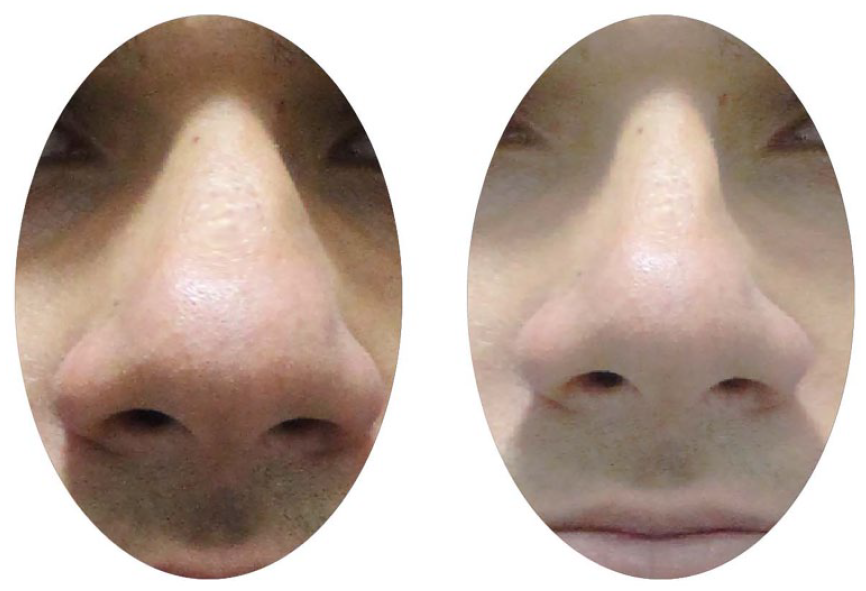
Case 1 open-roof deformity (preoperative and postoperative view).
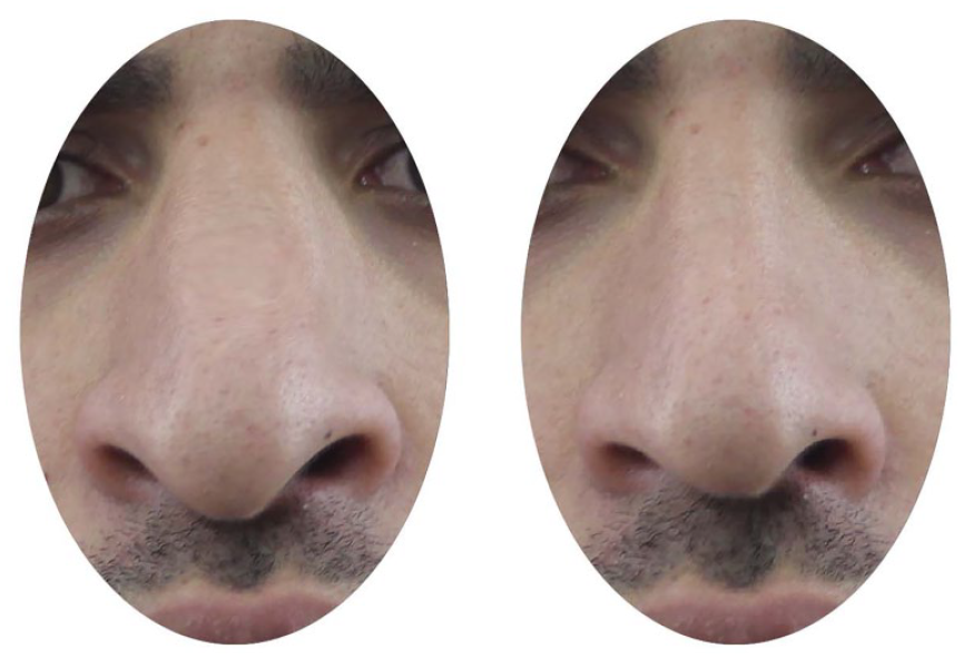
Case 2 open-roof deformity (preoperative and postoperative view).
A gap can be felt and seen when the septal dorsum and the lateral segments do not line up after osteotomies. If left untreated, a depression can form at the location where the intranasal mucous membrane attaches to the soft tissue above. When one side of the nose is out of alignment, it can make the nose look uneven. After an osteotomy, the septum must be brought into proper centering, and the lateral segments must be fully mobilized medially for correction. The following are common reasons for open-roof deformity:
The greenstick cephalic fracture will return to its original position after an osteotomy.
Inadequate medial mobilization of the fragmented segments.
Over-packing of the nose.
The medialization of the lateral segments may be hindered if the uncorrected deviating perpendicular plate of the ethmoid is not rectified.
Greenstick or partial lateral osteotomies can cause an open-roof deformity, manifesting as a visible or palpable separation of the nasal bones over the dorsum, preventing the open roof from completely closing. 11
The “open-roof” deformity is one of the most undesirable outcomes of removing a big bump. It is essential to thoroughly evaluate the dorsum following osteotomies when a big hump is excised. It is expected to need a spreader graft or a graft that only covers the dorsal surface to avoid open-roof deformities.12,13
Performing the Right Osteotomies to Stop Open-Roof Deformities
The osteotomies that finish reconstructive and cosmetic rhinoplasty can be done in various ways.14,15 One of the most common is the classic approach, which involves performing medial and transverse osteotomies after a lateral osteotomy and digitally creating greenstick fractures to separate the superior bony attachments. Although lateral nasal osteotomy is a fundamental approach to reshaping the nasal alignment, narrowing the nasal dorsum, and correcting the open-roof deformity that occurs after hump resection, 16 it can be associated with complications such as functional nasal obstruction, postoperative aesthetic deformities, asymmetric nasal wall deformities, long-lasting edema, and ecchymosis. 17 To avoid trauma to the soft tissues and periosteum, which can cause ecchymosis, some surgeons recommend using thin osteotomes. The increased risk of in-fractured bones relapsing to their preoperative position is the main disadvantage of this approach. Some orthopedic surgeons choose a more aggressive approach, using wider osteotomes. Although severe osteotomy reduces recurrence rates, it increases the unwelcome hazards of overcorrection and collapse and causes more periosteal tearing and soft tissue trauma. 18
Patients may be dissatisfied if splints do not correct their significant external deviations after an osteotomy. 19 Splints are always used to stabilize the nasal frame, reduce swelling, and maintain shape. Recently, “no-osteotomy rhinoplasties” have been preferred as a means to mitigate iatrogenic trauma. But splints have potential side effects, like a worse quality of life in hot weather, plaster burns, conjunctivitis, scleral issues, and in-fractured nasal bones relapsing because of improper splinting. 14
Four different kinds of osteotomies are performed 12 :
Lateral
Medial
Transverse
Double
In contrast to the endo-nasal method, the author favors the percutaneous method for osteotomy. 20 As a result, you have more command, and the nasal bones are less likely to be ground to powder. The severity of the issue dictates the osteotomy level. In cases involving a convex nasal bone, such as those resulting from trauma, a second-level lateral osteotomy might be necessary. In addition, a transverse osteotomy might be required after a trauma. 12
A web-like structure between the septum and nasal bone can form in cases of post-traumatic nasal deformities or people with broad noses. To move the bones of the nose inside, a medial osteotomy may be necessary to remove this. A vast, bony vault or open-roof deformity will remain. 12
A transverse osteotomy may be necessary to address the condition when a patient has a wide, post-traumatic nose or a crooked or twisted nose. 12
Airway Compromise in the Nose
Another unfavorable consequence of osteotomy is the medial displacement of the nasal bones, which narrows the nasal airways. A patient with a little to moderate amount of nasal deviation before surgery, who may not be experiencing any symptoms, may experience this. A patient who was asymptomatic before surgery may develop complaints of nasal airway blockage after a lateral osteotomy, which involves moving the nasal bones medially to narrow the airway. 12
Greenstick Fracture
The so-called “greenstick fracture” is an incomplete fracture that can go wrong after an osteotomy. Such a situation would lead to postoperative asymmetry as the nasal bones would return to pre-osteotomy positions. This ensures that the osteotomy is finished and that the nasal bones may move freely. 12
Step Deformity at the Osteotomy Site
A lateral osteotomy can result in a visible or palpable residual stump unless the incision is made low along the nasofrontal process of the maxilla. 12
Utilizing Grafts to Avoid Open-Roof Deformities
Grafts can be a lifesaver in rhinoplasty. Unfortunately, they also risk complications that could lead to unfavorable outcomes. Grafts can be grouped into 3 types. 12
Alloplasts
Homografts
Autografts
Alloplasts
A wide variety of alloplasts have found various applications over time. Jacques Joseph, regarded as the pioneer of rhinoplasty, utilized ivory. Silicone and pores are the most frequently used alloplasts. These grafts are effective in patients with thick nasal soft tissue envelopes but pose severe risks in secondary rhinoplasties and patients with fair or thin tissue canopies. 21
Because of their limitless volume, lack of donor site deformity, and accessible availability off the shelf, alloplasts are enticing to employ. Nevertheless, there are numerous drawbacks to alloplasts. 22 Some include aberrant mobility due to non-integration, the possibility of visible and palpable edges, and erythema caused by atrophy of the skin and subcutaneous tissues overlaying the implants over time. Infection and extrusion of these grafts are common complications. If this occurs, the following deformity will worsen the initial issue that necessitated the grafts. 12
Because of scarring and reduced blood flow, alloplasts are not an option for secondary rhinoplasties. 12
Homografts
Some preserved bone and cartilage are used, although this has been associated with a high resorption rate. An acellular tissue matrix like alloderm or allomax can provide soft tissue padding to cover grafts for patients with fragile and attenuated tissues. As a filler, they can help smooth out wrinkles and other imperfections in soft tissues. 12
Autografts
Because of their low complication rate and widespread use, autografts are the graft of choice for rhinoplasty. 23
Cartilage
Nasal septum Ear Rib
Bone
Perpendicular plate of ethmoid from the nasal septum Iliac crest Rib Cranial bones
Fascia
Dermis.
Transplant complications include graft displacement, bending, resorption, and apparent or palpable abnormalities. Dorsal enlargement via a conchal transplant carries the risk of irregularities. 12 As Daniel 13 suggests, diced cartilage wrapped in fascia is the way to go. Technical mistakes, rather than defects in the graft material, are the usual causes of these avoidable problems. 14
Treatment
Osteotomies and Grafts
Reducing the dorsal hump is one of the most typical rhinoplasty procedures. Inadequately conducted dorsal hump reduction, on the other hand, can lead to functional and aesthetic issues such as inverted-V deformity, mid-vault overresection or underestimation, and dorsal abnormalities.5-7 Upon removing the hump, open-roof deformity becomes apparent. Patients may experience hypersensitivity, irritation, and abnormalities with palpation due to this deformity, characterized by a wider dorsum and a discontinuity in the bony portion of the dorsum. It can also lead to unnatural dorsal aesthetic lines. To seal the open roof, lateral osteotomies are typically performed. It is challenging to attain a standard and optimal result during rhinoplasty with osteotomies since they are the most violent and least controlled treatment. Osteotomies, if not executed properly, can cause asymmetry, steps to form, lacerations of the mucosa, airway narrowing or collapse, open-roof syndrome, and visible scars in cases of external osteotomies.8-10 The patients included in this study have a limited bone vault, which means lateral osteotomies might not be an option for them. Among the well-known surgical options available to these patients, there are spreader flaps, grafts, diced cartilages wrapped in fascia or Surgicel, or the option to shape and replace the hump.7,24-26
Rohrich et al 6 have shown their method to preserve the dorsal aesthetic lines. Separating the upper lateral cartilage from the septum, reducing the septum and bone dorsum, palpation verification, and last modification (spreader graft, suturing procedures, and osteotomies) were the 5 phases they outlined for their technique. In 34 patients with a limited bony vault, Arslan 10 proposed a no-osteotomy idea. This approach involves closing the open roof with spreader flaps, using spreader grafts as necessary, and preventing depressions in the lower part of the upper lateral cartilage with onlay grafts.
In their study, Öreroğlu et al 27 described a method that utilizes diced cartilage, bone dust, and the patient’s blood to conceal dorsal abnormalities. After the operation, they took all the bone and cartilage parts and divided them up with a number 11 scalpel. They blended 3 to 5 ml of blood from a peripheral vein to make a clotting mixture.
Bone dust is preferable to fat or crushed cartilage, says Taş. 4 The hump is made up of cartilaginous and bony components. Most surgeons believe using autogenic tissues to repair a lesion is the best way to achieve a natural and optimal result. Also, think about a supply strategy for reconstruction to avoid resorption if it happens. For this reason, bone is recommended for a bony deficiency and cartilage for a cartilaginous one. The cartilaginous deficiency was filled using spreader flaps made from the cartilaginous hump, and the bony defect was filled with bone dust created from the bony hump, according to the method outlined in this article. After having a hump removed from a narrow nasal base, we feel this is the least invasive way to repair the problem. 4
One of the most annoying malformations that might occur after a large nasal hump excision is an open-roof deformity. Therefore, it is essential to examine every patient undergoing lateral osteotomy thoroughly. 28 Patients treated with thin osteotomes are more likely to experience open-roof deformity. 18 The nasal splint helps stabilize the nasal walls for approximately 1 week. Nevertheless, for the best outcomes, it is necessary to stabilize the broken bones for a minimum of 2 to 3 months. 21 Since the strength retention of Polydioxanone (PDS) is approximately 90 days, this method offers long-term support and prevents the deformity from returning. The nasal bones are still in the exact location as in before the fracture. 15
After osteotomy, the nasal bone collapses when the bones slide posteriorly toward the nasal vestibule. Correcting collapsed nasal bones is difficult but not impossible. The most effective methods for treating this condition are placing holes above the nasal bones, lifting the collapsed bone to the proper position with forceps, and stabilizing the bones with wire or surgical sutures.15,29,30
Fillers
To treat “narrow nose syndrome,” Sheen 31 first proposed using spreader grafts in 1984. These grafts are longitudinally orientated, typically paired, and fastened deep within the mucoperichondrium, between the nasal septum and the upper lateral cartilage. Restoring an open-roof deformity or preventing collapse of the middle section of the nose are 2 of the many uses for these grafts in surgical rhinoplasty. 31 HA filler can also be administered to achieve the same effect as a spreader graft. Along the length of the dorsum on both sides, HA is injected retrogradely. This procedure can enhance the look of the nasal bridge’s dorsal aspect or correct postoperative dorsal abnormalities. 32
Patients must be carefully chosen for nonsurgical rhinoplasty. Rhinoplasty with HA is typically indicated for patients with minor flaws in the dorsal contour or who exhibit nasal crookedness, depressions, notching, or asymmetry. Applying HA can improve the nasal radix or base, smooth out congenital furrows, even out small asymmetric dorsal lines, fix a bifid tip, make the tip more round or symmetrical, or fix a slight shortfall in tip projection or rotation. HA allows for reducing or eliminating skin depressions and other minor, aesthetic modifications in patients with nasal deformities caused by rhinoplasty or trauma. Another option for patients who want a slightly shorter nose is to rotate the tip of their nose via HA injection. 31
If a patient has a moderate deformity such as a drooping nasal tip, columellar show, depressed nasofrontal angle, hyperconvex nasal dorsum, or acute nasolabial angle, rhinofilling can be done instead of augmentation rhinoplasty. This operation is not recommended for patients with a prominent dorsal hump, severe tip projection, or major nasal deviation since they may require significant constriction or shortening of the nose. 31
HA injection is a popular substitute for surgical rhinoplasty since it is less invasive, takes less time to perform, costs little, doesn’t require general anesthesia, and doesn’t cause any downtime. 33 Applying HA can also help with small anomalies after rhinoplasty surgery and keep the nose’s natural height, which is not always possible with surgery.
In most cases, hyaluronidase can easily remove HA, so the substance is considered nonpermanent. 34 On the other hand, Mashiko et al 35 showed that HA can provide semipermanent volumizing effects on areas with a bony floor. The study’s authors theorized that HA activates periosteal stem cells, which enhance the volumizing effect. 35 Others have also verified the long-lasting impacts of HA rhinofilling.32,33,36
Conclusion
It is common for people undergoing primary rhinoplasty to have their hump removed, which can lead to open-roof deformity. Lateral osteotomies and the use of grafts are crucial in the prevention of open-roof abnormalities. It is common practice to perform lateral osteotomies to seal this space. However, lateral osteotomy becomes tricky when the patient’s bony vault is small. Another well-known option is to shape and replace the hump or to use a spreader graft, flap, sliced cartilage, or some combination of these. HA filler can also be administered to achieve the same effect as a spreader graft. Along the length of the dorsum on both sides, HA is injected retrogradely. This procedure can enhance the look of the nasal bridge’s dorsal region or correct postoperative dorsal abnormalities.
Footnotes
Acknowledgements
None.
Author Contributions
The authors contributed equally to the planning, literature survey, and manuscript writing.
Availability of Data and Materials
All data for this review is presented in this paper.
Declaration of Conflicting Interests
The author(s) declared no potential conflicts of interest with respect to the research, authorship, and/or publication of this article.
Funding
The author(s) received no financial support for the research, authorship, and/or publication of this article.
Ethics Committee Approval
Therere is no need for ethics committee approval because this is a review paper.
Informed Consent
Therere is no need to take informed consent because this is a review paper.