
Abstract
Osteoma is common in the temporal bone but extremely rare in the middle ear cavity and incus. Computed tomography plays an important role in the diagnosis of this slow growing benign osseous mass. The treatment of this lesion, which usually causes conductive type hearing loss, includes follow-up and surgery according to the patient's condition.
Significance Statement
Temporal bone osteomas are extremely rare in the middle ear cavity. The most common symptom is conductive hearing loss and the tympanic membrane appears normal on otoscopic examination. Computed tomography plays an important role in the diagnosis of these benign masses. Especially in young patients with hearing loss, osteoma is a pathology that should be kept in mind, albeit rarely.
A 47-year-old male patient was admitted to our clinic with left-sided hearing loss that had been present for several years and worsened in the last year. He had no history of surgery or trauma. On otoscopic examination, the tympanic membrane was normal and no mass lesion was observed. Hearing test revealed left-sided conductive type hearing loss. Non-contrast high-resolution temporal computed tomography (HRCT) images showed a hyperdense lesion with a bone density (1021 Hounsfield units) of approximately 4 × 6 mm in the medial part of the incus (Figure 1). The borders of the lesion were relatively regular, but there was no clear demarcation with the facial nerve trace medially. The stapezium and malleolus and inner ear structures appeared natural. The patient was diagnosed as middle ear osteoma. The operation was recommended and the risks in terms of facial nerve and inner ear structures were explained. The patient who refused the operation was given a hearing device and discharged with recommendations. Axial section non-contrast HRCT (A, B) and 3D CT scan (C) images show a pedunculated osteoma (red arrow) with bone density medial to the incus. (white arrow: incus).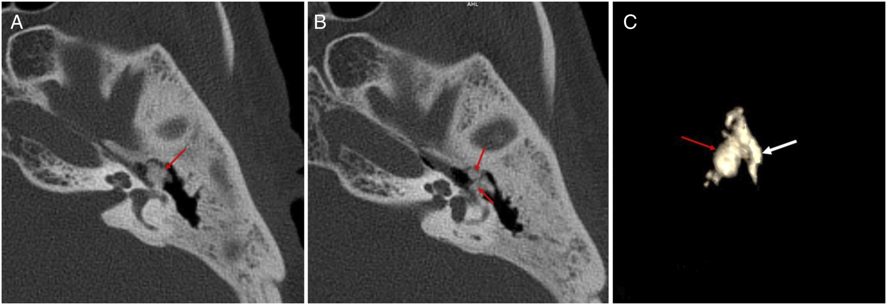
Temporal bone osteomas are benign solitary pedunculated osseous tumors usually seen in the external auditory canal and mastoid. Osteoma in the middle ear cavity is very rare and is usually presented as a case report in the literature. 1 It is 2 times more common in men and usually occurs at young age. 2 The etiology is not known exactly. Genetic factors and inflammatory processes are considered to be responsible. The most common symptom of middle ear osteomas is conductive hearing loss. This is due to mechanical obliteration of the ossicular chain and the oval window. Although some cases are asymptomatic, other symptoms include facial paralysis, mixed hearing loss, otorrhea, discharge, and tinnitus.2,3 As otoscopic examination of the patients is usually normal, HRCT is very important in the diagnosis. In the images, the osteoma seen in the bone density and its relationship with the bone chain and inner ear structures can be determined. Postoperative histopathologic evaluation can provide confirmation. The differential diagnosis mainly includes exostosis. Exostosis is multiple elevations in the external auditory canal after exposure to cold water and is bilateral. Osteomas are unilateral solitary and pedunculated mass lesions. Osteomas are benign masses that do not tend to grow much. Surgical removal may cause sensorineural hearing loss because of bone reconstruction. Therefore, hearing devices with follow-up are recommended in patients with asymptomatic or mild hearing loss. However, surgical resection can be performed in advanced cases.2,4
Footnotes
Declaration of Conflicting Interests
The author(s) declared no potential conflicts of interest with respect to the research, authorship, and/or publication of this article.
Funding
The author(s) received no financial support for the research, authorship, and/or publication of this article.