
Abstract
Hypopharyngeal liposarcoma is an extremely rare entity, and yet fewer than 40 cases have been reported in the literature. Lacking the possibility of distant metastases, local surgical resection with a clear margin can achieve the purpose of treatment. Endoscopic submucosal dissection has been widely used in the treatment of early upper gastrointestinal cancer and superficial hypopharyngeal cancer. Here, we present a case of a giant tumor originated from the hypopharynx that received primary resection by endoscopic submucosal dissection using a flexible upper gastroscope.
Case Report
A 70-year-old female patient had been diagnosed with a hypopharyngeal mass for almost 10 years. Fearful of surgical trauma and complications, the patient had chosen conservative treatments as her main symptom was just uncomfortable swallowing. However, the symptom of dysphagia had gradually worsened with pharyngeal soreness and low fever for half a year. Sometimes the mass would be vomited out of the mouth. Due to clinical experience with endoscopic resection for giant esophageal neoplasms, the patient was referred to our endoscopy center. Esophagogastroduodenoscopy (EGD) revealed a pedunculated tumor arising from the hypopharynx protruding into the esophageal lumen with mucosal erosion on the distal surface [Figure 1]. The computed tomography (CT) scan showed a lump-shaped soft tissue density lesion in the middle and upper esophagus. The lesion was unevenly enhanced, with a large cross-sectional range of about 33×27 mm. The physical examination was normal and no comorbidities were reported in her previous history. After a comprehensive evaluation and informed consent, peroral endoscopic submucosal dissection (ESD) was performed [Supplemental video]. EGD findings of the giant hypopharyngeal tumor. (A) The tumor root originated from the hypopharynx. (B) The tumor occupied most of the esophageal cavity. (C) Mucosa erosion on the distal surface of the tumor.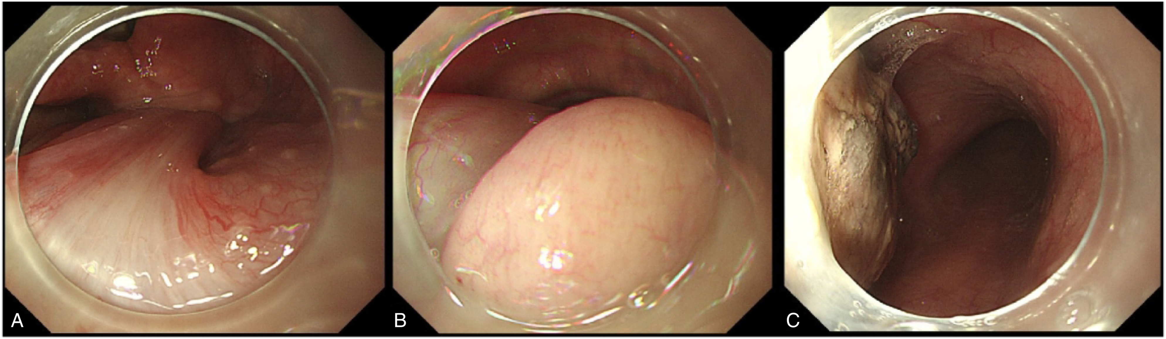
The patient was under general anesthesia with endotracheal intubation. At the base of the tumor, the mucosa was cut through after submucosal injection with normal saline and indigo carmine using a hybrid I-type knife (ERBE; Erbe Elektromedizin GmbH, Tubingen, Germany). The lesion was gradually separated along the submucosa layer from the oral side to the anal side combined with an IT knife (KD-611L; Olympus, Tokyo, Japan). After en bloc resection, the tumor was removed from the mouth with a snare. The specimen was 20 cm in height and weighed 48 g [Figure 2]. The procedure time was less than 20 min. The patient was discharged on postoperative day 2 with no adverse events or complications. Pathological results concluded in well-differentiated liposarcoma with mucinous degeneration. Photograph of the completed resected specimen.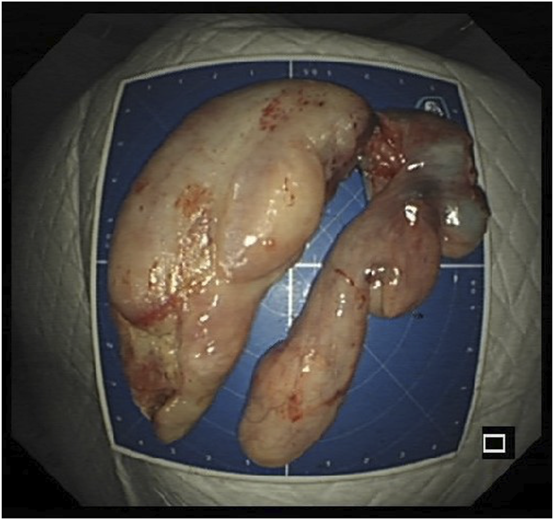
Discussion
Hypopharyngeal liposarcoma is an extremely rare entity. To our knowledge, less than 40 cases have been reported in the literature. 1 The most characteristic clinical manifestation described is progressive dysphagia as the tumor grows gradually. The mass might be spit out from the mouth. Some patients may have anemia-related symptoms such as hematemesis or black stool due to tumor rupture. The recommended treatment is surgical resection. Well-differentiated liposarcomas are low-grade malignant tumors that slowly grow with a 5-year survival rate of 100%. 2 The main concern is local recurrence rather than metastasis. 3 Thus, surgical resection without lymph node dissection can achieve a therapeutic effect. 3 With the development of medical techniques and instruments, surgical procedures have shifted from open surgery to minimally invasive surgery, such as transoral laser excision or endoscopic snare resection. 4 However, lateral pharyngotomy, the open neck incision, can be traumatic, while endoscopic laser or scalpel resection may not guarantee surgical margins, leading to a high risk of local recurrence. 5
Here, we present a case of a giant hypopharyngeal tumor that received primary resection by endoscopic submucosal dissection using a flexible upper gastroscope. ESD procedures have been widely used in the treatment of early upper gastrointestinal cancer and superficial hypopharyngeal cancer.6-8 Our case reported here demonstrated that ESD is also a minimally invasive, safe, and feasible approach for giant pedunculated hypopharyngeal tumors as it can easily achieve complete resection. Regular follow-up is still required to monitor local recurrence.
Supplemental Material
Footnotes
Declaration of Conflicting Interests
The author(s) declared no potential conflicts of interest with respect to the research, authorship, and/or publication of this article.
Funding
The author(s) disclosed receipt of the following financial support for the research, authorship, and/or publication of this article: This study was supported by grants from Major Project of Shanghai Municipal Science and Technology Committee (18ZR1406700).
Ethic Approval
This study was approved by the Ethics Committee of Zhongshan Hospital, in accordance with the Declaration of Helsinki.
Statement of Informed Consent
Informed consent was obtained from the patient for the publication of her information.
Statement of Data Availability
The data can be shared.
Supplemental Material
Supplemental material for this article is available online.
References
Supplementary Material
Please find the following supplemental material available below.
For Open Access articles published under a Creative Commons License, all supplemental material carries the same license as the article it is associated with.
For non-Open Access articles published, all supplemental material carries a non-exclusive license, and permission requests for re-use of supplemental material or any part of supplemental material shall be sent directly to the copyright owner as specified in the copyright notice associated with the article.