
Abstract
A pharyngeal recess cyst is a benign lesion, located at the nasopharyngeal recess with limited development. Pharyngeal recess cysts rarely occur. This case report describes a young male patient presenting with a foreign body sensation in the pharynx. Electronic nasopharyngoscope examination revealed a large nasopharyngeal cyst, whose root was located in the left pharyngeal recess. Complete surgical resection was performed, and the patient successfully recovered.
Pharyngeal recess cysts are rare lesions that can be diagnosed based on imaging and endoscopy findings. It is treated surgically and has a favorable prognosis.
Keywords
Introduction
The pharyngeal recess is located in the nasopharynx, between the superior posterior part of the Eustachian tube and the posterior pharyngeal wall. Most nasopharyngeal cysts are located in the middle region of the nasopharynx. They rarely develop in the pharyngeal recess region.
Case Report
The patient was a 37-year-old previously healthy male. One month ago, he noted a foreign body sensation in the pharynx with no apparent causes. This foreign body sensation gradually progressed, causing nasal congestion, eating obstruction, ear discomfort, and occasional breathing difficulties. On physical examination, the patient presented with dry retching. Large, smooth, round, erythematous organisms were detected in the oropharynx (Figure 1a). Electronic nasopharyngoscopy was performed to evaluate the presence of large obstructive masses in the nasopharynx (Figures 1b–1c). (a) The mass was observed from the oral cavity. (b) Complete obstruction of the nasopharynx by mass under electronic nasopharyngoscope. (c) The root pedicle of the mass is located in the left pharyngeal recess. (d) Pharyngeal recess cyst.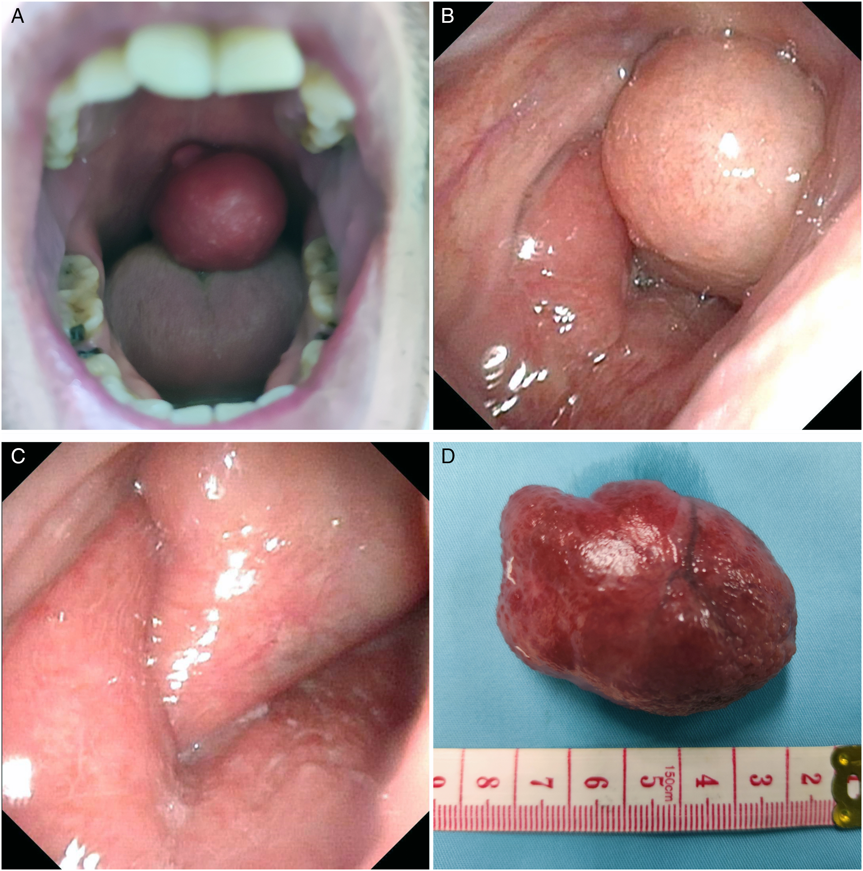
The enhanced computed tomography (CT) showed a non-enhancing soft tissue density shadow, measuring approximately 50 × 41 × 30 mm, in the nasopharynx. The nasopharynx was almost completely obstructed, while the pharynx and vertical inlet were partially blocked (Figure 2). Computed tomography (CT) images showing the soft tissue density of nasopharynx and oropharynx (a: sagittal CT and b: coronal CT).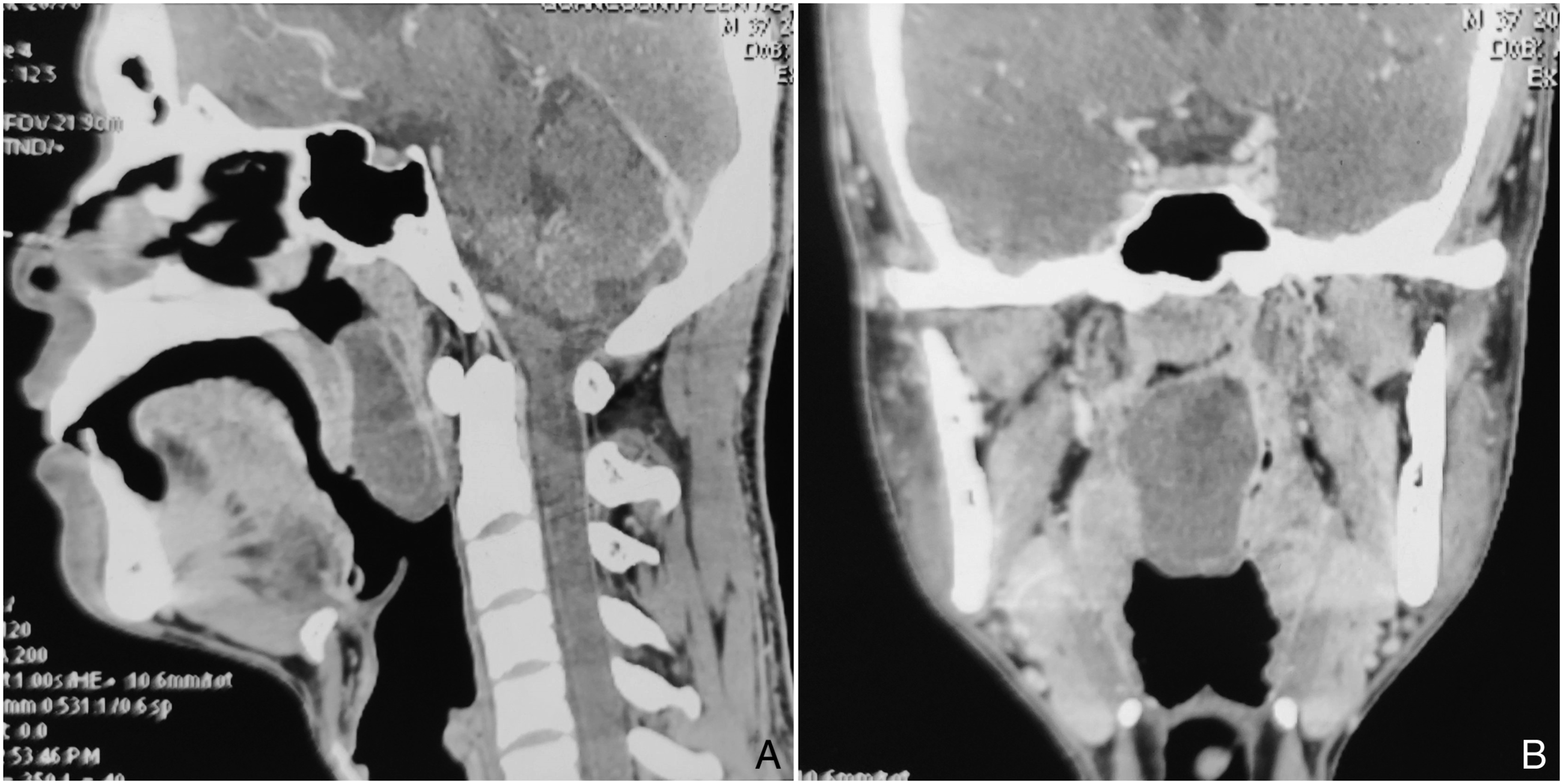
A transoral nasal endoscopic resection under general anesthesia was recommended.
During the operation, the nasopharynx was observed under a 70° nasal endoscope. The intraoperative surface of the tumor was smooth and erythematous. The root pedicle was located in the pharyngeal recess of the left nasopharynx. The tumor and root pedicle were completely removed using a low-temperature plasma knife. There was minimal intraoperative bleeding (Figure 1d).
The histological examination findings were consistent with a benign cyst.
Postoperatively, the patient’s symptoms of foreign body sensation, nasal obstruction, and ear tightness significantly improved. Six months later, the follow-up electronic nasopharyngoscopy revealed the scar tissue in the operative area. The patient successfully recovered.
Discussion
The pharyngeal recess is located between the superior posterior part of the round occipital of the Eustachian tube and the posterior nasopharyngeal wall. 1 Endopathies, which are difficult to detect, have been considered as malignancies. Nasopharyngeal carcinoma is the most common lesion in the pharyngeal recess. 2 Meanwhile, there have been some reports on pharyngeal recess polyps. 3 Pharyngeal recess cysts have not been reported in previous literature. The development of cysts induces common limitations. Individuals with a larger volume present with local compression symptoms. 4 In this case, the large cyst compressed the Eustachian tube, resulting in ear tightness.
Pharyngeal recess cysts are primarily diagnosed based on electronic nasopharyngoscopy. Alternatively, CT or magnetic resonance imaging examination can be performed, but malignancy needs to be excluded. Imaging examinations are critical because they facilitate a complete and safe surgical removal of the lesion.
Cysts are typically treated via surgery, which can be divided into the nasal and caliber approaches. The transnasal route involves a long path, where the nasal mucosa and posterior nostril are friable. Thus, the proximal oropharyngeal cyst is excised from the posterior lateral pharyngeal wall by means of caliber Louis. 5 The goals of the surgery were to completely remove the cyst and its mucosa, and to prevent a second operation. In this case, a better field of vision was obtained with the assistance of 70° nasal endoscopy. During the operation, the cyst was pulled to expose the root, and the tumor was completely removed with a low-temperature plasma knife. The root of the cyst was locally cauterized to prevent recurrence. Postoperatively, the patient’s symptoms of pharyngeal foreign body sensation, nasal obstruction, and ear tightness disappeared. Cyst recurrence was not noted during the follow-up 6 months later.
Conclusions
Large pharyngeal recess cysts are rare. Since it is a benign lesion, serious complications are unlikely. Imaging examination is critical because it characterizes the lesion and its relationship with the surrounding tissues. This case was treated via the transcaliber approach. Nasal endoscopy provided a more favorable visual field and adequate exposure, resulting in complete resection.
Footnotes
Acknowledgments
We would like to acknowledge Dr.Wensheng Ge for general guidance and providing clinical materials.
Authors’ Contributions
Dong Liu: data collection and manuscript preparation; Maocai Li: manuscript preparation; Lianqing Li and Lili Gong: collected pictures; Zuping Zhang: manuscript preparation and review. All authors read and approved the final manuscript.
Declaration of Conflicting Interests
The author(s) declared no potential conflicts of interest with respect to the research, authorship, and/or publication of this article.
Funding
The author(s) received no financial support for the research, authorship, and/or publication of this article.
Consent for Publication
The patient provided us informed consent for the publication of this case report.
Abbreviations
CT: computed tomography; MRI: magnetic resonance imaging