
Abstract

Schwannomas are a benign, slow-growing, usually solitary, and encapsulated tumor that is composed of Schwann cells. Schwannomas commonly arise in soft tissues of the head and neck as well as the upper and lower extremities. The most common site of intraoral schwannoma is the tongue, followed by the palate, floor of the mouth, buccal mucosa, gingiva, lips, and vestibule in decreasing order. 1 Intraosseous schwannomas of the jaws are even rarer.2-4 We report a rare case of intraosseous schwannoma of the mandible masquerading as radicular cyst of the molar.
A 45-year-old female with occlusal pain of right first molar of the mandible was referred to our department. She had no paresthesia in the mental region. Computed tomography showed a well-defined cystic lesion (10 × 12 × 24 mm) including the roots of the mandibular first molar (Figure 1). The lesion was diagnosed radiologically as radicular cyst. The patient underwent removal of the right first molar and cyst under general anesthesia. A buccal mucoperiosteal flap was elevated and buccal cortical bone over the lesion was removed. There was firm and rubbery mass below cystic lesion around the roots. Intraoperative rapid pathological diagnosis showed that the mass was schwannoma, and the lesion around roots was diagnosed with radicular cyst. After the extraction and cystectomy, the inferior alveolar nerve was identified and the tumor was directly attached to the neurovascular bundle. Careful dissection was performed to keep the neurovascular bundle integrated, and the tumor was removed completely. The right mental nerve paresthesia caused by inferior alveolar nerve damage was postoperatively identified by a Semmes Weinstein monofilament test. The final pathological examination of the tumor showed cellular areas (antoni A; Figure 2). Immunohistochemically, the tumor cells were positive for S-100 protein (Figure 2). Although there was no recurrence 7.5 years after surgery (Figure 3), the paresthesia in the mental region remained.
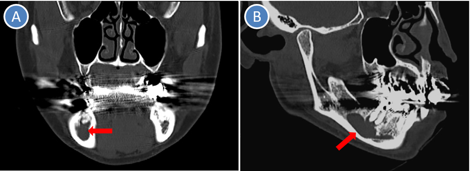
Computed tomography showed a well-defined cystic lesion (arrow) including roots of the mandibular first molar.
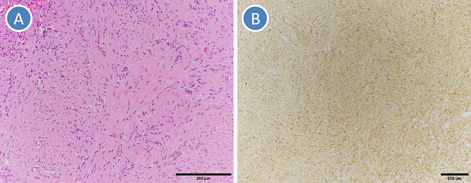
Microscopic pathology. A, Hematoxylin and eosin staining showed proliferated spindle-shaped tumor cells (antoni A area). B, Immunochemical staining showed tumor cells were positive for S-100 protein.
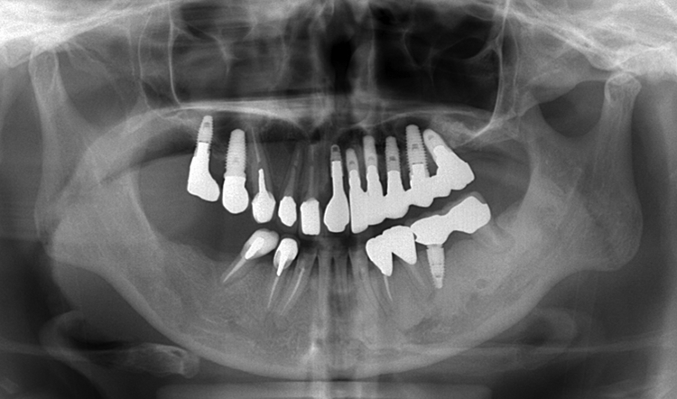
Panoramic radiograph showed no recurrence 7.5 years after surgery.
According to a recent review of intraosseous mandibular schwannoma, the average age was 36.9 years (range, 8-77 years) and the female to male ratio was 1.5:1. 2 Mandibular schwannoma most commonly involved posterior locations (78%). 2 Radiographically, intraosseous schwannomas typically present a well-defined unilocular radiolucency (76%), and multilocular and diffuse were 16% and 4%, respectively. 2 Accompanying features included external root resorption, cortical thinning or erosion, cortical expansion, tooth displacement or impaction, and spotty calcifications or focal radiopacities. 2 Direct association with the inferior alveolar neurovascular bundle was intraoperatively noted in 57% of cases. 2 The final diagnosis requires pathological examination, and conservative surgical enucleation of a schwannoma is generally curative. In the present case (45-year-old female), intraosseous schwannoma with a direct attachment to the neurovascular bundle was located at posterior mandible and presents a well-defined unilocular radiolucency. She had no characteristic accompanying features before surgery, and there was no recurrence 7.5 years after surgery.
The differential diagnosis for a radiolucent lesion of the jaws is wide, including odontogenic, fibro-osseous, vascular, and reactive lesions. The radiographic findings of intraosseous schwannoma may be similar to many other lesions such as odontogenic keratocyst and ameloblastoma. Radicular cyst is caused by pulpal necrosis secondary to dental caries or trauma, and its lining is derived from the epithelial cell rests of Malassez which proliferate to form the cyst. The cyst is radiographically a round or oval, well-circumscribed radiolucent image involving the tooth apex and can sometimes resorb the mandibular canal by the growth. Pimkhaokham et al 4 reported intraosseous schwannoma of the mandible in concurrence with radicular cyst. In this case, 4 the bilocularity of the lesion was due to such a coincidence and led to the radiographic appearance being atypical of both conditions. In the present case, intraosseous schwannoma of the mandible presented a well-defined unilocular radiolucency, and the lesion including roots of molar masqueraded as radicular cyst. Therefore, surgeons should consider schwannoma in the radiological differential diagnosis of mandibular cystic lesions including the inferior alveolar nerve.
Footnotes
Declaration of Conflicting Interests
The author(s) declared no potential conflicts of interest with respect to the research, authorship, and/or publication of this article.
Funding
The author(s) received no financial support for the research, authorship, and/or publication of this article.