
Abstract
This study describes a unique clinical presentation of trigeminal trophic syndrome (TTS), which is not well described within the otolaryngology literature. Trigeminal trophic syndrome classically presents with a triad of symptoms: trigeminal anesthesia, facial paresthesias, and crescent-shaped ulceration of the lateral nasal ala. The patient discussed in this report had a self-induced, waxing and waning ulceration of the frontal scalp for 7 years and was evaluated and treated ineffectively by multiple physicians, including otolaryngologists, before TTS was diagnosed and a targeted treatment was initiated. Although extranasal presentation is uncommon, this condition must be considered when ulcers are encountered in the trigeminal dermatome. This case highlights the variability in presentation and the importance of awareness of this rare syndrome. We aim to facilitate more prompt diagnosis and expedite the initiation of appropriate treatment for TTS in the field of otolaryngology.
Introduction
Trigeminal trophic syndrome (TTS) is an uncommon cause of cutaneous ulcers in the trigeminal dermatome, most often involving the nasal ala. 1 Although this disease has been described in dermatology and neurology literature, it is scarcely discussed in otolaryngology literature. Trigeminal trophic syndrome results from damage to the central or peripheral components of the trigeminal nerve pathway leading to facial dysesthesia and trigeminal hypoesthesia or anesthesia. 2 -4 Self-inflicted manipulation and trauma to the dysesthetic skin leads to cutaneous injury and ulcer development. 5 Diagnosis is often delayed because the clinical presentation is similar to more frequently encountered diagnoses in otolaryngology, including neoplasms (basal cell and squamous cell carcinoma), cutaneous vasculitis, infectious processes (herpetic ulcer), granulomatosis with polyangiitis, and pyoderma gangrenosum. 6,7 The management and treatment strategies used in TTS differ from those employed to address these more commonly encountered conditions. 4,6 Prompt recognition of TTS is important to prevent delays in diagnosis and initiation of targeted treatment.
Case Report
A 55-year-old female presented to a tertiary care otolaryngology practice with a 7-year history of nonhealing lesions of the frontal scalp region. The patient’s medical history was significant for herpes zoster virus and depression. The wound initially appeared after the patient burned her head with a curling iron. Over the years, the ulcer waxed and waned, with recurrent skull exposure and intermittent bleeding. The wound repetitively tested positive for staphylococcus, but no other infectious agents could be isolated. The patient described a deep itch sensation of the affected area and reported digging and picking the area for relief. She was evaluated by many physicians, including specialists in infectious disease, dermatology, and otolaryngology. Biopsies had eliminated oncologic etiologies. Multiple treatment attempts including topical cortisone cream, topical clindamycin, oral cephalexin, oral ciprofloxacin, wet-to-dry soaks, groin-to-forehead skin graft, and hyperbaric oxygen were unsuccessful.
Physical examination demonstrated a nonfriable 3-cm well-circumscribed ulceration located at the trichion of the scalp with 1 cm of bone exposure (Figure 1). There were also papular, crusting lesions in the skin adjacent to the affected area. Sensation was blunted in V1 distribution and normal in V2 and V3. Dermatopathology revealed epidermal excoriation covered by an inflammatory crust.
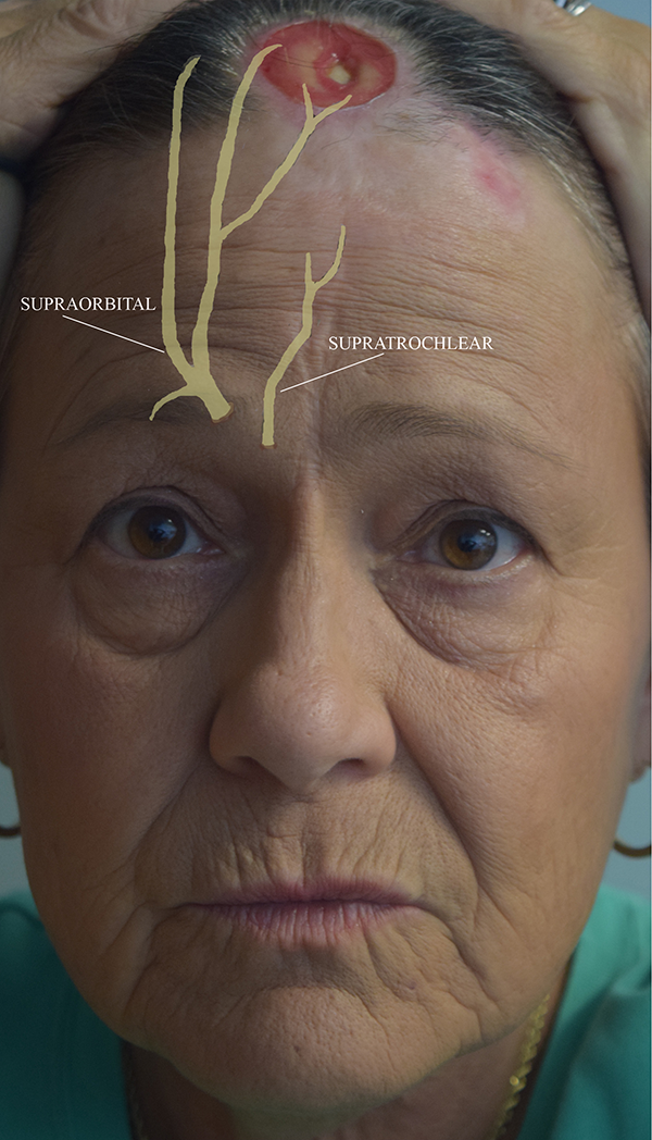
Frontal view of the trigeminal trophic syndrome (TTS) lesion located at the trichion of the scalp. The supratrochlear and supraorbital nerves are terminal branches of the ophthalmic division of the trigeminal nerve (V1) and have been superimposed on this image to depict the nerve distribution of this lesion.
There was an immediate concern that self-induced trauma to the wound may be exacerbating the disease, and she was advised to avoid manipulation of the skin. She was started on gabapentin 300 mg 3 times daily to address the sensation loss and paresthesias of the affected area. In addition, she was prescribed mupirocin 2% ointment to treat the skin infection. She was compliant with these recommendations and medications, and at her 1-year follow-up appointment, the wound was continuing to heal and had contracted to less than half of the original size (Figure 2).
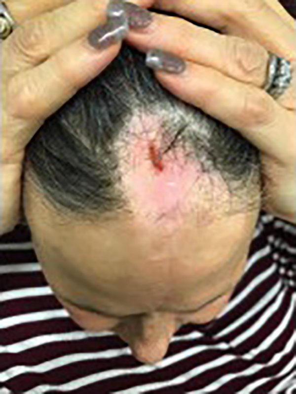
Trigeminal trophic syndrome (TTS) lesion at 1-year follow-up appointment. The patient was compliant with the treatment regimen (oral gabapentin, mupirocin ointment, and avoiding skin manipulation), and the ulcer contracted to less than half of the original size.
Discussion
Trigeminal trophic syndrome frequently presents with the following triad of symptoms: trigeminal anesthesia, facial paresthesias, and crescent-shaped ulceration of the lateral nasal ala. 2 -4,8 Unilateral facial ulcers in the setting of former trigeminal injury are pathognomonic for TTS; however, the primary causative condition may not be clear and many clinicians do not realize that the etiology of the lesions is a neurologic problem. 5,9 The majority of cases are caused by iatrogenic damage (eg, rhizotomy) to the trigeminal nerve. 1,8,10 Other causes include cerebrovascular disease, malignancy (eg, astrocytoma, acoustic neuroma, intracranial meningioma), herpes zoster virus, postencephalitic parkinsonism, syringobulbia, and trauma. 4,7,11 Cutaneous manifestations can appear 2 weeks to 30 years after nerve insult. 10,11
The 3 major branches of the trigeminal nerve are the ophthalmic (V1), maxillary (V2), and mandibular (V3) nerves. More than 80% of the reported cases develop ulceration in the distribution of a branch of the maxillary nerve, the infraorbital nerve. The area most frequently affected is the nasal ala, which is supplied by one of the terminal cutaneous branches of the infraorbital nerve, the external nasal branch. 5,10 Although the exact reason for this anatomic predilection is unclear, all cases, including our patient, appear to involve terminal sensory cutaneous branches of the trigeminal nerve (Figure 1). 10 The perceived sensation of cutaneous irritation prompts patients to rub, pick, or scratch the affected area. 2,12 Manipulation of the skin in TTS appears to be a reflexive action to alleviate dysesthesias and should be differentiated from dermatitis artefacta in which the self-inflicted trauma is a manifestation of psychiatric disturbance. 10
The patient described in this report did not present with the characteristic triad of symptoms. The lesion was located in the ophthalmic nerve distribution at the trichion of the scalp. Similar to previous reports, the affected area was innervated by specific terminal cutaneous branches, which in this case were the supratrochlear and supraorbital nerves (Figure 1). In addition, this case lacked a history of trigeminal neuralgia or an obvious causative condition. As a result of this patient’s unique presentation and the lack of awareness of this rare syndrome, she underwent an extensive workup and saw numerous physicians, including an otolaryngologist, before the condition was finally diagnosed and the correct treatment was initiated. Although the full symptom triad was not present, this case had many of the fundamental components of the syndrome including unilateral nonhealing ulcers, discomfort, some degree of neurological impairment (decreased sensation), and positive history of self-manipulation of the affected area. As such, TTS should have been included in the differential diagnosis, which highlights the importance of raising clinical awareness of this rare condition.
When TTS is suspected, workup should include thorough clinical history, neurological examination, serology, and biopsy of the lesion to rule out other causes of nonhealing ulcers. 8,12 Although histologic assessment is not specific, it rules out infection (herpes simplex/zoster, leprosy, syphilis, mucormycosis), granulomatosis, vasculitis, lymphomas, and skin cancer. 3 Once TTS is the most likely diagnosis, management should be multidisciplinary including behavioral modification, neurologic evaluation, pain management, medical treatment, surgical repair, and psychological management. 5,6,11,13 Patient insight into the self-induced mechanism of injury is a key component of treatment. Counseling and patient education play a pivotal role in reversing repetitive self-manipulation of injured tissue. 2,13,14 Soft gloves, short nails, and occlusive dressings can be used to prevent further injury. 14 Medical management includes carbamazepine, pregabalin, gabapentin, amitriptyline, and pimozide. 5,15 Transcutaneous electrical stimulation has also been studied in single case reports and aims to improve blood supply and healing of the area. 16 Although there are still mixed results, surgical management favors regional flaps to local flaps because if the adjacent tissue used in a local flap may be anesthetic as well. 11,17
Conclusion
Trigeminal trophic syndrome is a rarely encountered condition in the field of otolaryngology but, when properly diagnosed, can avoid unnecessary costly workups and delay in proper treatment. The evaluation of a patient with a nonhealing facial ulcer requires a comprehensive history and physical examination to exclude etiologies that can mimic TTS including malignancy, infection, autoimmune disease, and psychiatric disorder. Although TTS is most commonly encountered in the infraorbital nerve distribution, this case demonstrates that ulcerations caused by TTS can occur at any sensory branch of the trigeminal nerve. Raising clinical awareness of TTS is important because increasing numbers of surgeries that approach close proximity to the trigeminal nerve may increase the prevalence of TTS. We recommend keeping TTS on the differential and maintaining a reasonable level of suspicion for this diagnosis of exclusion.
Footnotes
Authors’ Note
This is the first submission on this topic and this research has not been presented at a scientific meeting.
Declaration of Conflicting Interests
The author(s) declared no potential conflicts of interest with respect to the research, authorship, and/or publication of this article.
Funding
The author(s) received no financial support for the research, authorship, and/or publication of this article.