
Abstract
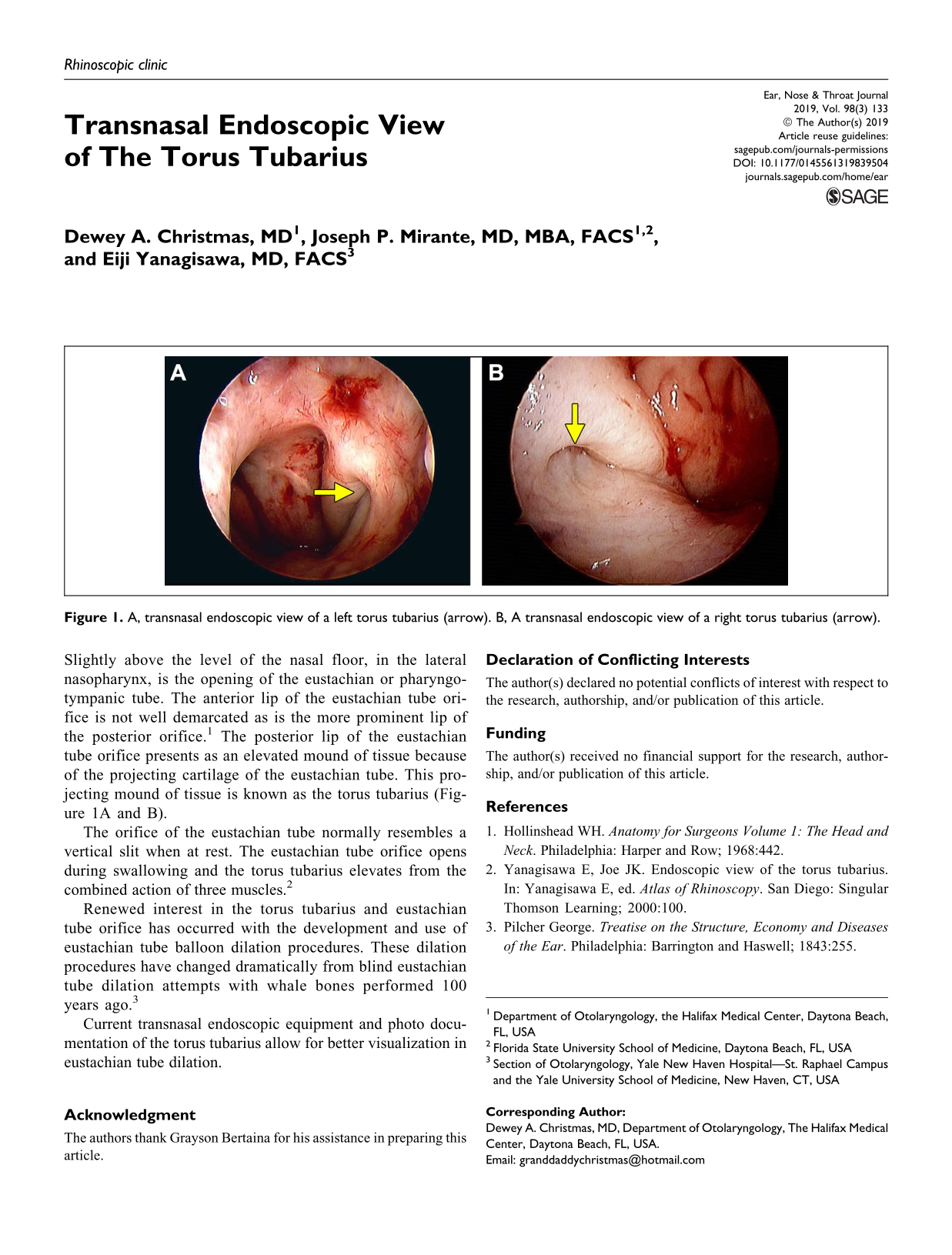
Slightly above the level of the nasal floor, in the lateral nasopharynx, is the opening of the eustachian or pharyngotympanic tube. The anterior lip of the eustachian tube orifice is not well demarcated as is the more prominent lip of the posterior orifice. 1 The posterior lip of the eustachian tube orifice presents as an elevated mound of tissue because of the projecting cartilage of the eustachian tube. This projecting mound of tissue is known as the torus tubarius (Figure 1A and B).
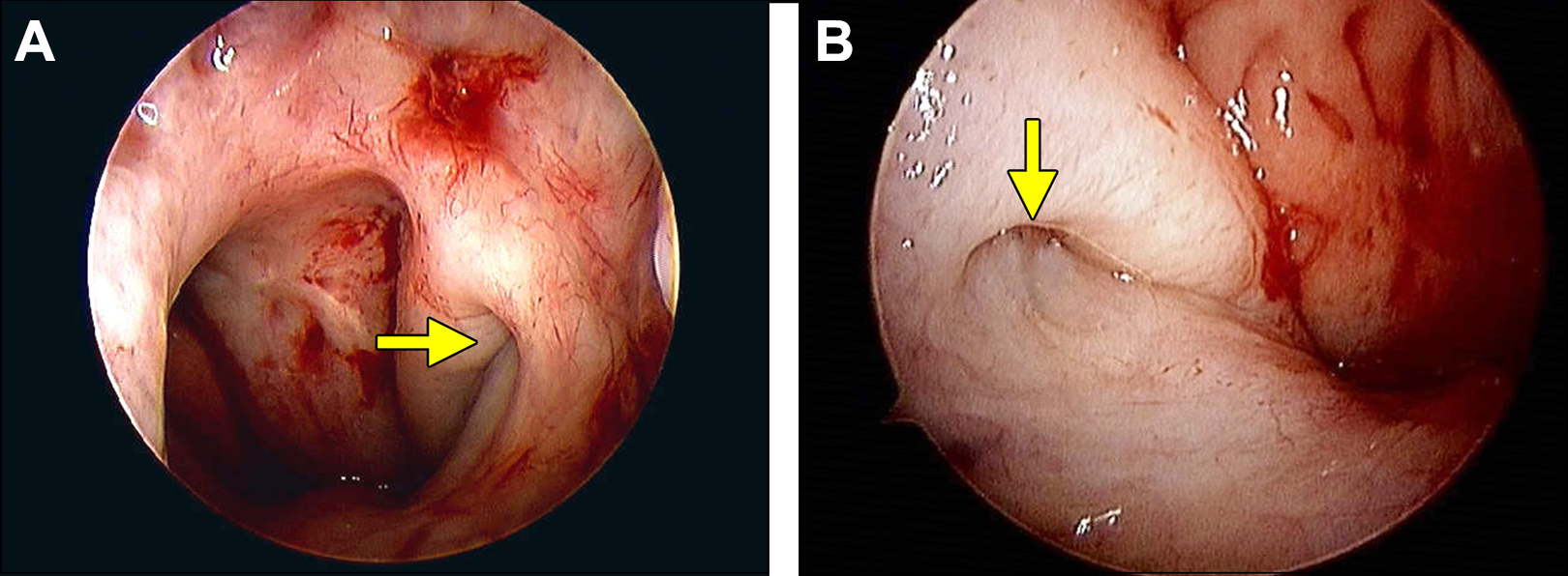
A, transnasal endoscopic view of a left torus tubarius (arrow). B, A transnasal endoscopic view of a right torus tubarius (arrow).
The orifice of the eustachian tube normally resembles a vertical slit when at rest. The eustachian tube orifice opens during swallowing and the torus tubarius elevates from the combined action of three muscles. 2
Renewed interest in the torus tubarius and eustachian tube orifice has occurred with the development and use of eustachian tube balloon dilation procedures. These dilation procedures have changed dramatically from blind eustachian tube dilation attempts with whale bones performed 100 years ago. 3
Current transnasal endoscopic equipment and photo documentation of the torus tubarius allow for better visualization in eustachian tube dilation.
Footnotes
Acknowledgment
The authors thank Grayson Bertaina for his assistance in preparing this article.
Declaration of Conflicting Interests
The author(s) declared no potential conflicts of interest with respect to the research, authorship, and/or publication of this article.
Funding
The author(s) received no financial support for the research, authorship, and/or publication of this article.