
Abstract
Osteomas of the turbinates are extremely rare. In this report, a case of inferior turbinate osteoma in a 24-year-old woman is described. The patient presented with a 6-month history of unilateral nasal obstruction. Computed tomography showed a bony dense mass in the anterior part of the left inferior turbinate. The lesion was removed endoscopically, and the patient recovered uneventfully. To the best of the author's knowledge, this is only the fifth case of a turbinate osteoma to be reported in the world literature, and only the second case that involved the inferior turbinate.
Introduction
Osteomas are commonly seen in the paranasal sinuses, but they are rare in the nasal cavity. 1 Only 4 cases of turbinate osteoma have been previously reported in the world literature,1–4 including only 1 case of an osteoma in the inferior turbinate. 1 In this report, a new case of inferior turbinate osteoma is described.
Case Report
A 24-year-old woman presented with a 6-month history of nasal obstruction on the left side. She had no history of surgery or facial trauma, and her medical history was not significant. Anterior rhinoscopy detected a hypertrophic left inferior turbinate, which was bony hard on palpation. Computed tomography (CT) of paranasal sinuses showed a bony dense mass in the anterior part of the left inferior turbinate (figure 1). With the patient under general anesthesia, the lesion was approached endoscopically and removed completely, and the patient recovered uneventfully. Histopathologic evaluation of the resected specimen identified it as an osteoma (figure 2). At 1 year of follow-up, no signs of recurrence were evident.
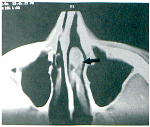
CT of the Paranasal Sinuses Shows the Bony Mass (Arrow) in the Anterior Part of the Left Inferior Turbinate.
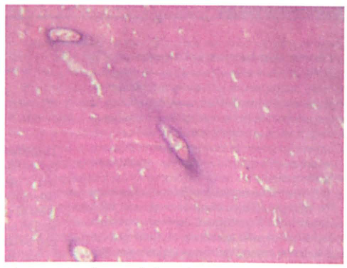
Microphotograph Shows Dense Compact Bone and Fibro-Vascular Channels (H&E, Original Magnification x40).
Discussion
Only 0.6% of all osteomas occur in the nasal cavity.3,5 These masses can occur at any age, but the highest incidence is seen during the second to fourth decades of life; there is a slight male preponderance.1,3 Most nasal osteomas are asymptomatic at an early stage, and they are usually found incidentally during routine radiographic examination. 3 The lack of signs and symptoms early and the nonspecific nature of signs and symptoms later frequently result in a delayed diagnosis. 1
When signs and symptoms do manifest, their presentation varies according to the location, size, and lesion's direction of growth. 3 Continued growth may completely obstruct the sinus ostia or nasal cavity and lead to sinusitis or the formation of a mucocele.1,3 CT is useful in determining the extent of the lesion. Magnetic resonance imaging is especially helpful in differentiating an inflammatory lesion from a neoplasm.1,3
Surgical excision is the treatment of choice for symptomatic osteomas. 1 Small osteomas can be removed via a transnasal approach. For lesions that involve the ethmoid sinuses, a lateral rhinotomy approach is suitable. 6