
Abstract
Kinesin family member 3A is an important motor protein that participates in various physiological and pathological processes, including normal tissue development, homeostasis maintenance, tumor infiltration, and migration. The wingless-related integration site/β-catenin signaling pathway is essential for critical molecular mechanisms such as embryonic development, organogenesis, tissue regeneration, and carcinogenesis. Recent studies have examined the molecular mechanisms of kinesin family member 3A, among which the wingless-related integration site/β-catenin signaling pathway has gained attention. The interaction between kinesin family member 3A and the wingless-related integration site/β-catenin signaling pathway is compact and complex but is fascinating and worthy of further study. The upregulation and downregulation of kinesin family member 3A influence many diseases and patient survival through the wingless-related integration site/β-catenin signaling pathway. Therefore, this review mainly focuses on describing the kinesin family member 3A and wingless-related integration site/β-catenin signaling pathways and their associated diseases.
Introduction
Kinesin is a linear molecular motor widely found in living eukaryotic cells. 1 It serves as a carrier by moving along the microtubules. It participates in many physiological activities, such as mRNA and protein transport, signal transduction, meiotic cilia, swinging of flagellum, and dynamic control of microtubules. 2 Kinesin proteins are divided into 14 families with hundreds of members, including the kinesin family member (KIF)1, KIF3, KIF4, KIF5, KIF13, and KIF17 family. 3 KIF3A plays an important role in kinesin protein family. 4 As a microtubule-orienting subunit, KIF3A is a member of the kinesin-2 family that regulates early development, ciliogenesis, and tumor formation. 5 Studies have shown that KIF3A is associated with the occurrence and development of bladder cancer, prostate cancer (PCa), thyroid cancer, lung cancer, and medulloblastoma (MB).6–8
The wingless-related integration site (Wnt)/β-catenin signaling pathway regulates cell proliferation during embryonic development and plays an important role in maintaining adult tissue homeostasis. 9 Abnormal Wnt/β-catenin signaling is associated with the occurrence and development of various human cancers. 10 In the absence of Wnt/β-catenin signaling, the β-catenin destruction complex composed of Axin, adenomatous polyposis coil (APC), and glycogen synthase kinase 3 (GSK3)β can inhibit β-catenin accumulation. 11 Upon stimulation by Wnt/β-catenin signaling, Wnt binds to its Frizzled family transmembrane receptor and coreceptor LDL receptor-related protein (LRP), leading to a phosphorylation cascade that stabilizes β-catenin by temporarily inhibiting its destruction complex via DVL. 11 When β-catenin enters the nucleus, it binds and activates transcription factors such as T-cell factor (TCF) and lymphoid enhancer factor (LEF), which activate the Wnt/β-catenin signaling pathway and mediate the expression of downstream target genes such as C-myc and Cyclin D1. 12
KIF3A is a critical element in many physiological and pathological mechanisms and is usually involved in the Wnt/β-catenin signaling pathway. The interaction between KIF3A and the Wnt/β-catenin signaling pathway and the outcomes are worth discussing and conducting in-depth studies. Therefore, in this review, we focus on how the motor protein KIF3A regulates development and maintains homeostasis in normal human tissues and the mechanisms by which it is involved in various diseases, especially cancer, via the Wnt–β-catenin signaling pathway.
KIF3A in kinesin family
A cell is a metropolitan city that organizes a community of biological macromolecules. Setting up meeting points and establishing timely schedules for molecular transactions is fundamental to cell behavior. Molecular motors, also known as nanomotors, consist of biological macromolecules that perform mechanical functions in muscle contraction, material transport, DNA replication, and cell division. 13 Molecular motors use chemical energy to directionally deliver cargo along cytoskeletal tracks. 13 Molecular motors include kinesin, myosin, dynein, DNA helicase, RNA polymerase, and rotary molecular motors. 14 Kinesin is the smallest identified molecular motor. Kinesin covers a large motor protein family called kinesin superfamily proteins. 6
Kinesin superfamily proteins (KIFs) are a group of molecular motors that move along microtubules and are involved in intracellular transportation, cell mitosis, and cell formation by the directed transport of various cargoes, such as membranous organelles, protein complexes, mRNA, and other materials. 6 Cumulative studies have shown that there are 14 subfamilies with 45 members in KIFs. 6 Among the KIFs family members, kinesin-2 and kinesin superfamily-associated protein 3 (KAP3) are the most highly expressed. Kinesin-2 exists mainly as a heterotrimer, including KIF3A, KIF3B, and KIF3C. KAP3 functions as an anterograde axonal motor for membranous organelle transport. 15 Kinesin family member 3A (KIF3A), a member of the kinesin family of motor proteins, serves as a microtubule subunit encoded by the human chromosomal region 5q31-33. 16 The KIF3A gene structure and location in human, Xenopus, and monkey retinas were first reported in 1999. A fragment of KIF3A from human retinal cDNA was amplified using the PCR products and highly homologous mouse KIF3A sequences. 17 KIF3A consists of three different structural domains: a spherical motor domain that contains a binding site with ATP and microtubules as the head of protein, a dimer slender coiled coil as the handle, and a combination site with KAP to transport cargo as the tail (Figure 1). 18 KIF3A serves as a microtubule-directed motor subunit to carry out many functions, such as regulating embryo development, ciliogenesis, and tumorigenesis. 18

Domain organization of components of the mammalian kinesin-2 machinery and schematic representation of kinesin-2.
Cilia are small organelles that are commonly found in mammalian cells. They are classified as primary and motile cilia. 19 Primary cilia are organelles protruding from the cell surface that act as chemical or mechanical sensors and provide the basis for signal transduction in various situations. 20 In single-celled organisms and sperm cells, motile cilia contribute to the movement of the entire cell. In multicellular organisms, 21 motile cilia gather at the cell surface and drive fluid flow in ductal cavities. 21 Motile cilia participate in cell movements, such as the clearance of lung mucous cilia and cerebrospinal fluid flow in the brain. 21 However, cilia cannot synthesize self-assembling proteins; they require intraflagellar transport (also known as IFT) to transfer them from the cytoplasm. 22 The IFT performs bidirectional movement along the axonemal microtubule for transport, and kinesin composed of KIF3A, KIF3B, KIF3C, and KAP3 are the main forces of forward transportation. 23 KIF3A can form a heterotrimer with KIF3B or KIF3C to bind with KAP3, but KIF3B cannot bind with KIF3C. 24 The combination of KIF3A and KIF3B is essential for forward transportation in the cytoplasm, especially during spemiogenesis. 25 The combination of KIF3A and KIF3C is responsible for particle transportation of axons and dendrites of nerve cells. 26 KIF3A knockout causes the loss of renal cilia, leading to polycystic kidney disease (PKD). Loss of cilia in pancreatic cells leads to metaplasia, fibrosis, and lipomatosis. 27 These examples indicate that KIF3A is necessary for ciliary structure and function.
In recent years, the relationship between KIF3A and the occurrence and development of various diseases has received increasing attention. KIF3A interacts with various signaling pathways, such as the Wnt/β-catenin and Hedgehog signaling pathways, which participate in many physiological and pathological activities and reveal essential biological functions.20,28
Overview of Wnt/β-catenin signaling pathway
The Wnt signaling pathway is one of the most evolutionarily conserved pathways. It plays an important role in embryonic development, adult tissue homeostasis, and various diseases, including cancer. 10 Wnt was initially discovered in mice as integration 1 (Int1). 29 Later, a homolog gene type was identified in Drosophila melanogaster with a wingless phenotype and was named Wnt. The pathway led by the Wnt gene is called the Wnt signaling pathway, and its mainly classified into the canonical Wnt signaling pathway, also known as Wnt/β-catenin signaling pathway. Wnt/β-catenin signaling activates the transcriptional activity of the target gene by β-catenin nuclear translocation and the noncanonical Wnt signaling pathway, which includes the Wnt Ca2+ signaling pathway, 30 Wnt PCP signaling pathway, and regulation of spindle orientation and intracellular signaling pathways in asymmetric cell division. 29 This review mostly focuses on the canonical Wnt signaling pathway because of its significant function in the human body. 16
The Wnt–β-catenin signaling pathway mainly consists of the Wnt family of secreted proteins, frizzled family transmembrane receptor protein, and LRP on the cell membrane. 16 In the cytoplasm, the Wnt/β-catenin signaling pathway positively regulates dishevelled (Dsh or Dvl), negatively regulates serine/threonine GSK-3, APC, axis inhibition protein (Axin), Casein kinase1 (CK1), and β-catenin. β-catenin is a cytoplasmic protein that acts as the central player and is the most important element of the Wnt/β-catenin signaling pathway. 16 Axin, APC, CK1, and GSK-3 form a protein complex called the cytoplasmic destruction complex. 11 In the nucleus, there are TCF/LEF and tumor-associated target genes (such as c-myc, cyclinD1, surviving, COX-2, CD44, PPAR, MMP7, and VEGF). 31
The Wnt/β-catenin signaling pathway has two conditional statuses: Wnt is activated when the Wnt/β-catenin signaling pathway is active. 31 In the absence of the Wnt/β-catenin signaling pathway, the cytoplasmic destruction complex phosphorylates β-catenin. Phosphorylated β-catenin is recognized by the F-box/WD repeat-containing protein β-TrCP, a component of a dedicated E3 ubiquitin ligase complex. 31 After ubiquitination, β-catenin is targeted for rapid destruction by the proteasome. 32 When Wnt binds to the frizzled/LRP co-receptor complex, the Wnt/β-catenin signaling pathway is active. 33 The Frizzled/LRP coreceptor complex transmits Wnt signals to Dsh and activates it. 34 Active Dsh suppresses the cytoplasmic destruction complex to maintain β-catenin under dephosphorylation conditions, while APC carries β-catenin to the nuclear membrane for nuclear translocation. 35 β-catenin can independently complete nuclear translocation independently. 35 In the nucleus, the cytoplasmic destruction complex displaces Groucho from TCF/LEF to promote the transcription of Wnt target genes (Figure 2). 10 Therefore, β-catenin is related to the Wnt/β-catenin signaling pathway. The Wnt/β-catenin signaling pathway is turned off when β-catenin levels decrease; otherwise, the pathway is active.
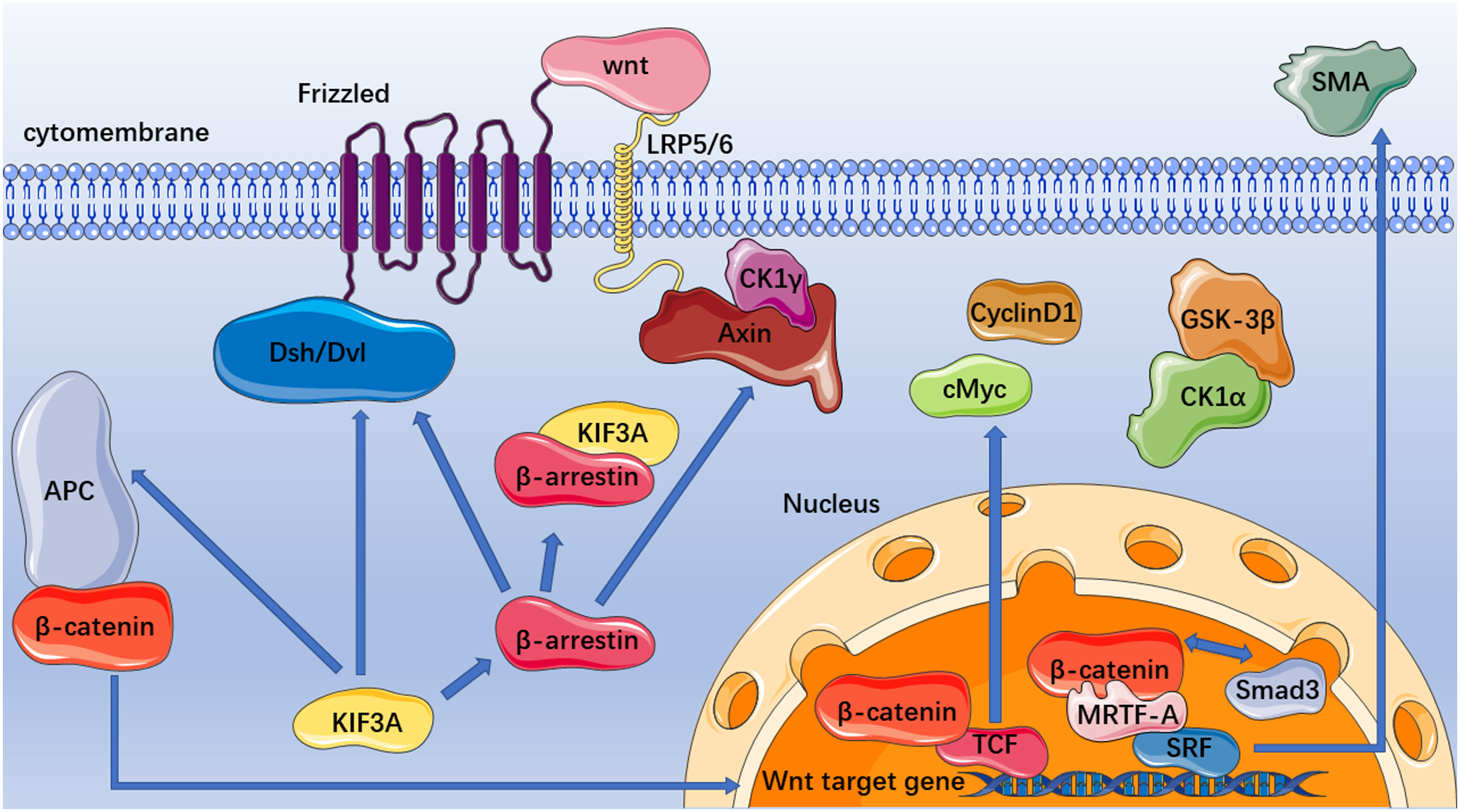
Interaction between KIF3A and Wnt/β-catenin signaling pathway. During the on-stage of the Wnt/β-catenin signaling pathway, KIF3A can bind with CID of APC to reduce β-catenin nuclear translocation. KIF3A also binds with Dsh/Dvl to increase β-catenin levels by stabilizing and phosphorylation of Dsh/Dvl. As KIF3A binds to β-arrestin, it promotes the β-catenin destruction complex formation which consists of Axin, APC, CK1, and GSK-3. Destruction complex phosphorylates β-catenin. Eventually, β-catenin is degraded. However, when KIF3A is decreased, β-arrestin is released from binding to KIF3A. Released β-arrestin binds to DVL2 and Axin, resulting in degradation of the destruction complex and increase of unphosphorylated β-catenin. In the nucleus, β-catenin displaces TCF to promote the transcription of Wnt target genes. The β-catenin competition combined with Smad3 to SRF to promote SMA transcription. APC: adenomatous polyposis coil; GSK-3: glycogen synthase kinase 3; CK1: Casein kinase1; TCF: T-cell factor; SMA: smooth muscle actin; SRF: serum response factor.
KIF3A regulation in spermatogenesis through Wnt/β-catenin signaling pathway
Spermatogenesis is a critical process in sexual reproduction. 36 The Wnt/β-catenin signaling pathway plays an important role in early and late sperm formation, and spermatocytes, spermatids, and slender mature sperm were severely missing in β-catenin knockout mice. 37 Recent studies have shown that KIF3A and KIF3B are involved in severe structural changes during spermatogenesis via intracellular transport. 38 Acrosome formation and nuclear remodeling are mediated by KIF3A/KIF3B/KAP3 heterotrimers in Palaemon carincauda. 39 In Larimichthys polyactis, KIF3A and KIF3B are highly expressed, which may indirectly facilitate the nuclear remodeling of sperm along the perinuclear microtubules and material transport during flagellum formation. 40 KIF3A regulates spermatogenesis through the Wnt/β-catenin signaling pathway in Eriocheir sinensis. 18 On the one hand, KIF3A can upregulate β-catenin via stabilization and phosphorylation of DVL, which can positively regulate the Wnt/β-catenin signaling pathway. 18 In contrast, β-catenin binding to KIF3A allows its nuclear translocation in the late stages of spermatogenesis in E sinensis. 18 Therefore, further investigation is required to explore the role of β-catenin in spermatogenesis.
KIF3A plays an important role in airway inflammation of asthma via Wnt/β-catenin signaling pathway
Asthma is a complex chronic respiratory disease that causes pathological changes such as excessive mucus production and reversible airway obstruction. 41 The detailed pathogenesis of asthma has not yet been elucidated; however, inflammation is the basis of disease pathogenesis. 42 Therefore, it is important to suppress airway inflammation and clarify the underlying molecular mechanisms. KIF3A's genetic locus, which is abnormally expressed in patients with asthma, has been repeatedly proven to be associated with susceptibility to asthma and eczema, and it has already been chosen as a new candidate gene marker of asthma.43,44
According to previous studies, KIF3A can combine with β-arrestin, a negative regulator of the G protein-coupled regulator signaling pathway, to interact with Dsh and axin to regulate the Wnt/β-catenin signaling pathway.32,45 The KIF3A expression downregulation is induced by house dust mite in a mice asthma model and in the 16 human bronchial epithelial (16HBE) cell line.46,47 Simultaneously, inflammatory factors such as COX-2 are secreted from bronchial epithelial cells. 48 KIF3A has a confirmed connection with asthma, and KIF3A goes through β-arrestin via the Wnt/β-catenin signaling pathway to regulate inflammation. It is believed that in asthma, KIF3A is downregulated by disruption of the β-arrestin/ KIF3A complex, allowing the upregulation of β-arrestin and Wnt/β-catenin signaling. Finally, β-catenin translocates to the nucleus to produce inflammatory factors that aggravate asthma. Therefore, KIF3A can be used as a diagnostic biological marker and potential therapeutic target for asthma. Finally, the regulation of KIF3A on airway inflammation in the in vivo asthma model needs to be gradually improved in future studies, and the specific role of KIF3A in the pathogenesis of asthma should be further explored.
Effect of KIF3A on pulmonary fibrosis process through Wnt/β-catenin signaling pathway
Exposure to silica is a common risk factor for pneumoconiosis, which occurs in many industries such as mining, quarrying, and ceramics. 49 During silicosis, epithelial cells undergo an epithelial-myofibroblast transition (EMyT), a source of myofibroblasts characterized by the positive expression of α-smooth muscle actin (α-SMA) and collagen deposition.50,51 KIF3A serves as a subunit of the microtubular motor and exerts a suppressive effect on EMyT. 52 Many transcription factors have been identified to play critical roles in EMyT, including serum response factor (SRF) and myocardin-related transcription factor (MRTF-A). MRTF-A is activated to promote α-SMA gene transcription. 53 β-Catenin is essential for EMyT. 45 When the Wnt/β-catenin signaling pathway is off-stage, Smad3 binds to MRTF-A and inhibits SRF activation. Simultaneously, β-catenin competes with Smad3 for MRTF-A binding. 54 When the Wnt/β-catenin signaling pathway is activated and nuclear translocation of β-catenin occurs, β-catenin allows MRTF-A to bind to SRF and promote α-SMA gene transcription. 55 However, the detailed mechanism by which KIF3A affects EMyt during silicosis via the Wnt/β-catenin signaling pathway requires further investigation.
Inactivation of KIF3A promotes PKD through the Wnt/β-catenin signaling pathway
PKD is a genetic disorder that leads to renal failure and abnormal proliferation of epithelial cells in the glomerular sac. 56 Recent research has found that KIF3A is essential for cilia formation in the renal epithelia. 57 In KIF3A knockout mice, the abnormal proliferation and apoptosis of cystic epithelial cells increased, β-catenin and c-Myc expression increased, and p21 expression decreased.57,58 However, the detailed mechanism of renal cyst formation by loss of KIF3A expression loss remains unknown. 57 However, the stable nuclear translocation of β-catenin activates TCF-responsive target genes.57,59 C-myc is a TCF-responsive target gene that represses the transcription of specific cyclin-dependent kinase inhibitors, leading to increased cell proliferation.57,60 It has been shown that the nuclear translocation of KIF3A downregulates p21 expression. 57 These results suggest that KIF3A plays a role in maintaining the differentiation of lumen-forming epithelia in PKD via the Wnt/β-catenin signaling pathway and that KIF3A has high research and clinical potential in the future.
Regulation of KIF3A in the pathologic process of nasopharyngeal carcinoma via Wnt/β-catenin signaling pathway
Nasopharyngeal carcinoma (NPC) is a malignant epithelial tumor that occurs in the nasopharyngeal mucosa. 61 Recent studies have revealed that KIF3A expression in NPC tissue is lower than that in para-NPC tissues, and patients with higher KIF3A expression have prolonged overall survival. 62 Therefore, KIF3A may play an important role as a potential tumor suppressor in NPC. Epithelial-mesenchymal transformation (EMT) plays an essential role in many biological processes. 63 Adherent epithelial cells can detach from neighboring cells, resulting in EMT-induced movement. 64 EMT occurs in areas with high research prospects, such as healing, fibrosis, and tumor development. 63 The effect of EMT on tumors mostly manifests in cell adhesion and cytoskeleton changes. 64 From a molecular perspective, cell adhesion strength changes mainly due to the regulation of cell adhesion protein expression, including reduced E-cadherin expression and increased N-cadherin expression. 63 Cytoskeletal changes are the transformation from a cytokeratin-based cytoskeleton to a vimentin-based cytoskeleton, and the molecular change is mainly the upregulation of the vimentin protein. 63
KIF3A overexpression inhibits N-cadherin and vimentin expression in NPC cells and upregulates E-cadherin expression. 65 Therefore, EMT is inhibited by reversing cell adhesion and inhibiting the transformation of the cytoskeletal structure from keratin to vimentin. 65 Conversely, the inhibition of KIF3A expression promotes EMT. 65 KIF3A has already been shown to be involved in the Wnt/β-catenin signaling pathway in various tumors, 45 and pathway activation promotes EMT change. 65 There are two GSK3transcripts, GSK3a and GSK3β . 66 GSK3β is phosphorylated at Ser9, Tyr216, and Thr390. 67 The phosphorylated serine tail causes the dimerization of GSK3β. Phosphorylation of Ser9 results in the competitive inhibition of GSK3β activity and leads to blocked phosphorylation-dependent ubiquitination degradation of β-catenin. KIF3A promotes GSK3β activity by inhibiting the phosphorylation of GSK3-Ser9. 68 Therefore, enhancing the phosphorylation-dependent ubiquitination degradation of β-catenin prevents its nuclear translocation.69,70 Therefore, KIF3A downregulates EMT via the Wnt/β-catenin signaling pathway by regulating phosphorylation of the GSK3-Ser9 site. Thus, it affected the invasion, migration, and proliferation of NPC. However, it remains unclear whether KIF3A affects downstream Wnt/β-catenin signaling or is involved in primary ciliary structures.
KIF3A participated in the triple-negative breast cancer pathological process through the Wnt/β-catenin signaling pathway
Previous studies have shown that KIF3A expression in breast cancer tissues is significantly higher than that in the adjacent normal tissues. 65 Analysis of the connection between KIF3A and clinicopathological features of breast cancer showed a strong relationship between KIF3A expression, lymph node metastasis, and the pathological grade of breast cancer patients. 71 Further studies have found that KIF3A expression and prognostic breast cancer parameters are relevant, such as ER, AR, EGFR, and Ki-67. 65 Triple-negative breast cancer (TNBC) is a subtype of breast cancer with high malignancy, strong invasion, and difficult treatment. 72 Triple-negative means that the diagnostic breast cancer indices, such as ER, PR, and HER-2, are all negative, but cancer still develops. 73
Like many other cancers, TNBC also undergoes EMT, and the Rb/E2F and Wnt/β-catenin signaling pathways have been associated with the EMT process. 74 Previous studies have shown that the expression of the epithelial marker E-cadherin can be suppressed during retinoblastoma (Rb) deficiency. E2F1 promotes EMT through the upregulation of fibronectin and vimentin, proving that the Rb/E2F signaling pathway intervenes in EMT. 75 When the Wnt/β-catenin signaling pathway is activated, a critical tumor-associated target gene, cyclinD1, is translated. 70 KIF3A also promotes the activation of cyclin-dependent kinases (CDKs) by inhibiting P21 expression. Subsequently, the cyclin/CDK complex induces Rb phosphorylation and leads to the dissociation of the Rb/E2F complex, increasing the expression of E2F and cycle-related proteins, and ultimately promoting the progression of the TNBC cell cycle and cell proliferation. 76 According to the above discussion, KIF3A is correlated with patient survival outcomes. It may be expected to become a new indicator for breast cancer diagnosis and a new parameter to evaluate the prognosis of patients with breast cancer. KIF3A affects the EMT process in TNBC by interacting with the Rb/E2F and Wnt/β-catenin signaling pathways. However, the detailed mechanism between KIF3A and b-E2F and Wnt/β-catenin signaling pathway interactions requires further examination.
KIF3A is a key part of non-small-cell lung cancer via the Wnt/β-catenin signaling pathway
In non-small-cell lung cancers (NSCLC), the most common malignant tumors, including squamous cell carcinoma, adenocarcinoma, and large cell carcinoma, grow and divide more slowly than in small-cell lung cancer. 77 Recent findings demonstrate that KIF3A is downregulated in NSCLC tissues compared to the adjacent normal tissue. Downregulation of KIF3A is closely associated with overall survival in patients with NSCLC. 8 Moreover, KIF3A and β-catenin are negatively correlated with each other in NSCLC tissue. 32 Recent findings demonstrate that the silencing of KIF3A in NSCLC cells using lentivirus upregulates β-catenin, phosphorylates Dsh expression, and promotes the proliferation and migration of NSCLC cells. 32 Phosphorylated Dsh can invalidate the cytoplasmic destruction complex of β-catenin to stabilize it for nuclear translocation.
Further studies show that KIF3A is binding β-arrestin to form a complex and inhibit β-catenin in the Wnt/β-catenin signaling pathway, and the silence of KIF3A can promote Dsh and Axin to form a complex to stabilize β-catenin. 78 Above all, KIF3A can suppress NSCLC genesis and development via the Wnt/β-catenin signaling pathway. More importantly, KIF3A is a valuable potential diagnostic or prognostic marker for the come. 78
KIF3A effect on PCa through Wnt/β-catenin signaling pathway
PCa is the second most common malignant tumor and cause of cancer-related death globally. 7 The Wnt/β-catenin signaling pathway is frequently activated during the progression of PCa. 34 Recently, researchers demonstrated that the motor protein KIF3A is an activator of the Wnt signaling pathway in human PCa. 79 The upregulation of KIF3A in PCa cells stabilizes β-catenin by increasing CK1-dependent DVL2 phosphorylation, thereby activating cyclinD1. CyclinD1 is a well-known PCa-associated gene that enhances the proliferative potential of human PCa cells and is a target gene of the Wnt/β-catenin signaling pathway. 79 Furthermore, MMP-9 and HEF1 regulate the migration and invasion of PCa cells, and their activation is KIF3A-dependent through the Wnt/β-catenin signaling pathway. 80 Based on the KIF3A's deep involvement in PCa development and migration via the Wnt/β-catenin signaling pathway, KIF3A may be a potential therapeutic target for advanced PCa in the future.
KIF3A inhibits hypoxia-induced EMT in thyroid cancer via the Wnt/β-catenin signaling pathway
Thyroid cancer is one of the most common endocrine malignancies worldwide, and its incidence has rapidly increased in recent decades. 81 Recent studies have shown that hypoxic conditioning promoted EMT. 82 It results in the downregulation of E-cadherin and upregulation of N-cadherin and vimentin while inducing migration and invasion of thyroid cancer cells. 83 Interestingly, silencing KIF3A significantly inhibited the EMT process and suppressed hypoxia-induced migration and invasion. 84 Furthermore, the knockdown of KIF3A in FTC133 cells inhibited the expression of β-catenin, c-Myc, and cyclin D1 under hypoxic conditions. 84 These findings suggest that the inhibitory effects of KIF3A gene silencing under hypoxic conditions promote EMT. This is most likely mediated by Wnt β-catenin signaling pathway suppression in thyroid cancer, suggesting that KIF3A may be a potential therapeutic target in the future.
The relationship of KIF3A to other signaling pathways and diseases
The other mechanism we are discussing here is mainly the Hh signaling pathway. The Hh gene was first discovered in Drosophila. 85 Subsequent studies have shown that there is only one Hedgehog gene in invertebrates, such as Drosophila while there are three hedgehog genes, including Sonic Hedgehog (Shh), Desert Hedgehog (Dhh), and Indian Hedgehog (Ihh) in mammals. 85 The Hh signaling pathway is mainly composed of the Hh ligand, transmembrane protein receptor Ptched (Ptch), transmembrane protein receptor Smoothened (Smo), downstream nuclear transcription factor Gli protein, and target genes. 86 The Hh signaling pathway is involved in embryonic development and homeostasis of the internal environment. The abnormal activation of this signaling pathway causes the generation and development of various tumors, such as basal cell carcinoma and MB. It affects the proliferation, differentiation, invasion, metastasis, and apoptosis of tumor cells. 85 Additional studies have shown that downstream of Ptch1 as a member of Ptch, KIF3A is required to activate normal Hedgehog target genes, and loss of KIF3A led to a decrease in the Gli subunit Gli3 repressor. 87 Furthermore, primary cilia also play an important role in the normal function of the Hedgehog signaling pathway. 88 Primary cilia protruding from basal cell carcinoma and MB cells are central to Hedgehog signal transduction. Any changes in the primary cilia can increase or decrease the incidence of tumors. 89 Meanwhile, it has been found that partial ciliary body disease is related to blocked Hedgehog signal transduction. 90
The bone is a highly dynamic organ that is constantly reshaping, sensing, and responding to signals to meet the needs of its surroundings. 91 Primary cilia that serve as chemical sensors are also mechanical sensors for bone formation and maintenance, first observed in chondrocytes and later in mesenchymal stem cells, osteoblasts, and osteocytes. 92 Primary cilia are assembled and maintained by the IFT system, which is involved in the differentiation of mesenchymal stem cells into osteoblasts, chondrocytes, and adipocytes. 93 Previous studies show that KIF3A deletion or destruction can lead to abnormal bone formation and morphology, abnormal growth plate development, and impaired adipocyte differentiation. Deleting KIF3A in mouse osteoblasts can lead to functional defects and reduced bone mass, 94 and inhibiting Hedgehog signal transduction pathways in osteoblasts can lead to abnormal cilia-related signaling pathways. 95 These results proved that KIF3A regulates osteogenesis and bone formation, and these changes appear to be caused by damage to the Hedgehog signaling pathway. 94 Additionally, KIF3A is involved in bone development and chondrogenesis. 96 Therefore, disruption of the IFT system can lead to subsequent cilia loss, postpartum dwarfism, and inhibition of chondrocytes. 97 Thus, KIF3A can maintain the growth plates by regulating chondrocyte proliferation, differentiation, and rotation.
MB is a tumor cell that loses growth control from granule neuron precursors (GNPs). 98 Under normal conditions, the GNPs were divided in response to SHH. 99 If one copy of the Ptch gene, as a receptor of Shh, is lost, MB may form. 100 Proper transduction of the SHH signal critically depends on primary cilia, and improper signal reception leads to failure in activation of SHH target genes resulting from primary cilia lost 88 KIF3a, part of the kinesin motor, is required for the formation of primary cilia. 16 Recent studies have concluded that instead of mitotic or intracellular transport defects, mutated Kif3a or an altered primary cilium affects the proper hedgehog signaling pathway leading to tumorigenicity, proving that KIF3a is necessary for MB formation. 101
Besides the Wnt/β-catenin and Hedgehog signaling pathways, there are discoveries in which detailed mechanisms have not been described. KIF3A knockout mouse embryos show neural tube degeneration, mesodermal and caudal hypoplasia, pericardial edema, developmental delay, and neural tube closure defects in morphology, ultimately resulting in embryo death. However, the most prominent manifestation was the randomization of lateral asymmetric development, indicating that KIF3A plays an important role in development. 102
Bladder cancer is the tenth most common cancer worldwide, and the ninth most fatal malignancy in men. 103 Patients with bladder cancer have a relatively low survival rate and a poor prognosis. 103 The expression level of KIF3A in bladder cancer tissues was found to be significantly higher than that in para-cancer tissues by qRT-PCR and immunohistochemistry detection. 6 Analysis of the relationship between KIF3A and clinicopathological features showed that KIF3A expression was related to clinical stage, pathological grade, and lymph node metastasis, and Km-plot analysis showed that increased KIF3A expression was associated with poor prognosis in patients with bladder cancer. 6 Meanwhile, interfering with KIF3A expression can inhibit the proliferation, migration, and invasion of bladder cancer cells. Further Western blot analysis showed that Ki-67, MMP2, and MMP9 expression in bladder cancer cells was decreased by interfering with KIF3A expression. 6 In conclusion, new targets, such as KIF3A, need to be discovered in disease studies to improve patient survival and prognosis.
Summary and prospect
KIF3A is involved in the occurrence and development of many diseases via the Wnt/β-catenin signaling pathway
KIF3A, like other members of the kinesin family, can use the energy generated by ATP hydrolysis to move along microtubules for cargo transport, which is necessary for the maintenance of intracellular balance and mitosis. 44 Many functions of cilia rely on the performance of KIF3A, 16 including cilia formation, movement of the sperm flagellum, and external perception of primary cilia. 104 In addition, as a critical factor influencing the Wnt/β-catenin signaling pathway, KIF3A plays a role in organohistogenesis, inflammation, and various cancers. When the Wnt/β-catenin signaling pathway is activated, inflammatory factors like COX-2, the main cause of asthma, are secreted. 44 Likewise, the nuclear translocation of β-catenin also promotes the transcription of tumor-associated target genes, such as CyclinD1 and c-myc, 12 which are the leading causes of thyroid cancer, PCa, and TNBC.65,83,105 Most importantly, the Wnt/β-catenin signaling pathway can give rise to EMT, a committed step in organ fibrosis and many cancers for tumor cell generation, infiltrating invasion, and migration, such as pulmonary fibrosis, thyroid cancer, TNBC, and NPC. 106
How KIF3A regulates Wnt/β-catenin signaling pathway
The Wnt/β-catenin signaling pathway is very important in various pathological and physiological mechanisms, and abnormal pathway activation leads to severe diseases such as cancer. 31 Recent studies have shown a clear and robust link between the KIF3A and Wnt/β-catenin signaling pathways. First, KIF3A downregulates the Wnt/β-catenin signaling pathway by binding to β-arrestin. β-arrestin acts as a positive regulator of the Wnt/β-catenin signaling pathway when it forms a complex with DVL2 and axin to stabilize β-catenin. However, when KIF3A binds to β-arrestin and forms a complex that interacts with the Wnt/β-catenin signaling pathway, β-arrestin changes its role as a negative regulator of the pathway. Second, KIF3A promotes β-catenin expression by stabilizing the quantity of phosphorylated Dsh. Third, KIF3A binds to the CID of the APC protein and downregulates β-catenin.
KIF3A might be a potential therapeutic target for cilia disease
In vertebrate cells, the centrosome is the center of the microtubule and its function changes with the cell cycle. 107 In some cells, during the G0 phase, the mother centrosome is transformed into a matrix, which initiates the assembly of microtubules and the formation of primary cilia. 108 When cells withdraw from the G0 phase, the primary cilia structure disintegrates. 108 The length of the primary cilia is prolonged during the G0 phase and shortened when cells withdraw from the G0 phase. 108 Recent studies have shown that primary cilia might have the ability to control or affect cell cycle process. 108 However, the mechanisms by which primary cilia control or affect primary cilia remain unknown. 108 According to our recent studies, silencing of KIF3A shortens primary cilia and suspends the cell cycle. 53 Therefore, KIF3A may be a key regulatory factor of primary cilia. Primary cilia are related to the Hedgehog, Wnt, TGFβ, Notch, and PDGFRα signaling pathways. 109 As primary cilia mediate many cell signals, their dysfunction can cause multiple tissue and organ diseases. 109 The disease caused by primary cilia dysfunction is called cilia disease and includes nephronophthisis, senior-loken syndrome, orofaciodigital syndrome, Jeune syndrome, autosomal dominant PKD, autosomal recessive PKD, Leber congenital amaurosis, and usher syndrome. 109 Based on its potential relationship with KIF3A and primary cilia, KIF3A may be a valuable therapeutic target for cilia disease.
The limitation of this study
The limitation of this review is that we discussed multiple diseases that are related to the KIF3A and Wnt/β-catenin signaling pathways, but we did not dig deeper into the molecular mechanisms of each disease, and we did not investigate further whether there are other potential links between each disease besides the KIF3A and Wnt/β-catenin signaling pathways.
Conclusion
In general, β-catenin is the key player in the Wnt/β-catenin signaling pathway, which leads to many critical activities in the human body, and KIF3A can affect it. Three interactions between them can occur simultaneously. KIF3A is also related to other signaling pathways, such as the Hedgehog signaling pathway. The appearance of both Wnt/β-catenin and Hh signaling pathways with abnormal regulation of KIF3A can be associated with the same disease. The molecular mechanism underlying the fluctuation of KIF3A expression in diseases such as bladder cancer is unknown. KIF3A is a valuable target with great potential for application in the treatment of maldevelopment, allergy and inflammation, organ fibrosis, and cancers. In addition, the molecular mechanism of KIF3A upregulation or downregulation in disease, the relationship between KIF3A and Wnt/β-catenin signaling pathway, and the interaction between Wnt/β-catenin and Hedgehog signaling pathways and KIF3A in disease need to be addressed. Questions such as molecular mechanism by up or downregulation of KIF3A in diseases, the relationship between the KIF3A connection with the Wnt-β-catenin signaling pathway, and the interaction between the Wnt-β-catenin signaling pathway and Hedgehog signaling pathway with KIF3A in disease need to be answered. The molecular mechanism of KIF3A and its clinical applications require further research and interpretation.
Footnotes
Acknowledgments
The authors would like to thank Professor Hong Xu for inspiring their interest in the development of this review
Authors’ contributions
All authors contributed to data analysis and drafting or revision of the article. All authors have agreed on the journal to which the article will be submitted, gave final approval of the version to be published, and agree to be accountable for all aspects of the work.
Declaration of conflicting interests
The author(s) declared no potential conflicts of interest with respect to the research, authorship, and/or publication of this article.
Funding
The author(s) disclosed receipt of the following financial support for the research, authorship, and/or publication of this article: This work was funded by the National Natural Science Foundation of China (No. 82003406), the National Natural Science Foundation of Hebei Province (H2020209287), the Science and Technology Project of Hebei Education Department (No. ZD2022127); the Science and Technology Project of Tangshan (No. 21130208c); Non-profit Central Research Institute Fund of Chinese Academy of Medical Science(2020-PT320-005); Project of Key Laboratory of Functional and Clinical Translational Medicine (XMMC-FCTM202203).
Author biographies
Shupeng Liu is a doctoral student in public health and preventive medicine. His area of research is research on prevention and treatment of organ fibrosis.
Yang He holds a master's degree in clinical medicine. Her area of research is dermatology and venereology.
Shifeng Li holds a PhD in pathology and pathophysiology. Her area of research is research on prevention and treatment of organ fibrosis.
Xuemin Gao holds a PhD in pathology and pathophysiology. Her area of research is research on prevention and treatment of organ fibrosis.
Fang Yang is a professor in pathology and pathophysiology. His area of research is research on prevention and treatment of organ fibrosis.