
Abstract
Human teeth have become a prominent source of DNA for human forensic identification as their biological structure is highly resistant to extreme conditions. Previous forensic identification was mainly dependent on the pulp and the other hard tissues of intact teeth. However, there is high likelihood that only carious teeth can be available for forensic analysis. This study aimed to validate the use of the carious part of the teeth for forensic identification and to compare two DNA extraction methods-the operative technique with the cervical cut technique for human identification using STR typing. The reliability of STR markers in carious part of the teeth was evaluated in 120 carious teeth (60 dental pulp and 60 dentinal carious tissues, respectively) with considerable coverage of gender type and age range to avoid false exclusions. The study was performed on genuine data set where samples have been extracted by proficient dentist during the treatment operation and collected for further analysis. Complete DNA was extracted and the corresponding human identification profile was obtained using the GoldenEye™DNA ID system 20A kit. The operative technique showed a conservative approach to the sampling of carious tissues and allowed safe access to collect carious tissues, whereas the cervical cut technique permitted access to the root canals and complete sampling of the pulp tissues. The findings indicated that there was no significant association between the cervical cut and operative cut techniques (p = 0.165). In addition, there was no statistically significant association between the various teeth types and the obtained profiles observed. The operative technique, by drilling holes on the defected surface of carious human teeth and gentle hand excavation of carious tissues, was indicated to be very efficient, preserving, time-saving, and cost-effective in the recovery of human DNA from carious teeth. The result gives new insights that the carious tissues of human carious teeth might be as valid as the healthy teeth for forensic human identification.
Introduction
The latest advantages in DNA technology have revolutionized the field of forensic sciences. Nowadays, the importance of forensic DNA profiling has increased tremendously due to the identification issues in instances of terrorist attacks and mass murders. Furthermore, it is critical to determine the exact identity results to establish paternity and maternity cases, property disputes and burial of victims of disasters. This becomes highly significant as the world faces many crises like acts of terrorism, earthquakes, tsunamis, and airplane crashes. 1 A comparison between fingerprint records and different medical ante and post-mortem records is often difficult and not practicable due to the lack of sufficient ante-mortem records. 2 DNA can be extracted from soft tissues as well as from hard tissues like bones and teeth. In accidents soft tissues may not remain in good conditions, therefore hard tissues are usually preferred. Among bones teeth are the hardest tissues in human body; it may survive in extreme temperatures and in adverse environmental conditions.
Teeth are considered as an important source of DNA3,4 due to their composition which provides protection from different microorganisms and environmental factors responsible for decay. While pulp is recognized as the main source of DNA in healthy fresh teeth, 5 many factors can reduce its value such as age, dental disease, and postmortem disintegration. 6
Dental caries is one of the most common oral diseases globally; approximately 2.43 billion people, 36% of the world’s population, have carious lesions in permanent teeth. 7 The prevalence is increasing day by day with changing dietary habits and lifestyles. It is widely accepted that the consumption of sugar-rich foods increases the incidence of caries. Caries affects tooth structure as it destroys enamel and dentin and also the pulp also undergoes necrosis depending upon the extent of the carious lesion. 8
Dental caries status in China shows typical characteristics found in developing countries. 9 According to the national third epidemiological report of 2005, the prevalence of dental caries is high in children of 5–6 years age (70.2%), adults of 35–44 (60.6%) years, and of 65–74 years age (75.5%). 9
In recent years different techniques for DNA extraction from teeth have been used, that are sectioning of teeth horizontally or vertically (cervical cut), crushing or cryogenic grinding, and conventional access cavity preparation. Crushing or grinding is not recommended as the tooth is ground and is not available for any further evaluations. 10 The access cavity preparation techniques or operation techniques: Occlusal perforation (Perforation of the occlusal surface), cervical perforation (through the tooth neck) are simple, relatively low cost and preserve the tooth. The cervical cut method, a longitudinal cut through the teeth using a very thin disc or saw, facilitates access to the cells of the dental pulp. The cervical cut is less conservative but allows direct access to the pulp cavity of the tooth. Cervical cut and use of endodontic files to access the pulp cavity were considered the most optimum conservative method for DNA recovery by Hervella et al. 11
Short tandem repeats (STRs) and Single nucleotide polymorphism (SNPs) are different types of DNA tmarkers which have revolutionized the field of genetic identification Among the 3 million or so DNA bases that do not code for proteins are regions with multiple copies of short repeating sequences of these bases, which make up the DNA backbone. These sequences repeat a variable number of times in different individuals. Such regions are called “short tandem repeats,” and they are the basis of STR analysis which are important in forensic analysis.
DNA may be extracted from various tissues present in the tooth, such as from pulp tissues, dentine and cementum. 12 Teeth are frequently subjected to decontamination processes preceding sampling to remove exogenous DNA, environmental contaminants, and micro-organisms. 13 Decontamination includes one or both of the following processes: removal of the outside covering layer of the tooth by sanding or grinding; 13 washing or soaking in bleach. 14
The caries are known to destroy enamel and dentin and also the pulp may also undergo necrosis due to caries. As the pulp tissues are major source of DNA in forensic evaluation, implications of caries need to be evaluated.
The aim of this study was to establish a DNA extraction technique from human teeth, which is valid for carious teeth, cost-effective, time-saving, and conserves the tooth integrity and to validate STR profiling from carious teeth.
Materials and methods
Sample demographics
The nature of study is based on clinical data set. The study was performed on genuine data set where samples have been extracted by proficient dentist during the treatment operation and collected for further analysis. It was not based on literature data set but from actual data set.
The rationale for sample size selection is highlighted as follows. The study was evaluated in 120 carious teeth with considerable coverage of gender type and age range to avoid false exclusions. The number of samples was chosen to adequately represent the case study as per the common researches published in this domain. Sample number for our study lies on the high band in comparison to other highly referenced/cited researches on the same topic indicating 20 to 120 samples used.11,15
All participants included in the current study were from the same geographical area and the same dentist performed dental extractions.
Total 37 females and 23 males participated in the operative cut technique group, whereas 42 females/18 males participated in the cervical cut technique group. The age range was of participants was 15–68 in the operative cut group, whereas the age range was 17–64 in the cervical cut group.
Teeth collection
In this study, 120 carious human teeth were collected during 2016–2018 in the First Affiliated Hospital of China Medical University (Shenyang, China). Each patient declared verbal consent that was approved by the ethical review committee of China Medical University, Shenyang, Liaoning Province, P.R. China. The inclusion criteria were the presence of carious pathology diagnosed either radiographically or visually, whereas the exclusion criteria were root canal treated teeth, internal root resorption, periapical pathology, and severe open root apex because they have minimal bacterial interaction. The carious teeth were extracted under strict sterilized conditions by professional dental surgeons and stored into sterilized gauze. Next, the teeth were decontaminated by gentle bleaching and complete soft tissue curettage and then preserved at −20°C until further process. The collection of carious lesion samples was performed by a skillful dentist, as per International caries detection and assessment system (ICDAS) classification, radiographic images and whether soft or hard carious lesion was present.
Whenever the sample showed presence of superficial debris/plaque, these contaminants were removed by reduced transport fluid (RTF; Syed &Loesche, 1972) twice.
Consequently, comparative study was performed between two different DNA extraction procedures on carious teeth which are listed below (a,b):
Operative technique: (40 molars, 11 premolars, and 9 incisors). We used sterile diamond bur cut around hard carious lesion and hand excavated soft carious dentine with sterile spoon excavators of different sizes. The collected hard and soft dentine caries fragments were preserved in micro-centrifuge tubes and analyzed separately (Figures 1 and 2). Crushing was needed for a few samples where hard carious enamel was collected.
Cervical cut: (45 molars, 7 premolars, 8 incisors) A cut at the neck level of the carious tooth using tungsten carbide surgical bur facilitates direct access to the pulp. Pulpal tissue samples were collected using endodontic K-files of different sizes from the pulp chamber, root canals, and the dental apex of the root (Figure 3). In both groups, 6–20 g of samples was collected in microcentrifuge tubes.

Operative technique: before and after collecting the carious lesion from the carious tooth, not invading any adjacent healthy parts.
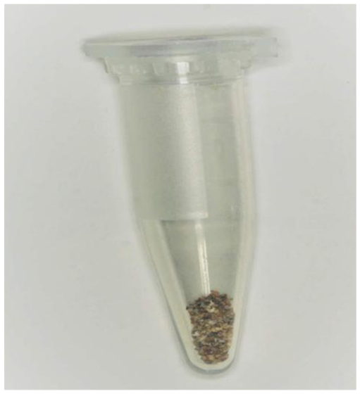
Carious lesion sample placed in micro-centrifuge tube.

Scheme of both sampling methods evaluated: cervical cut and operative cut techniques for DNA recovery from carious teeth.
Statistical analysis
The median and interquartile ranges (Percentile 25 and 75) of data chi-square test was applied to compare the obtained genetic profile proportion between the operative cut and cervical cut to access the pulp cavity and root canals. DNA purity and concentration between both groups was compared using the U of Mann-Whitney test.
Statistical analysis was performed using IBM SPSS Statistics V.20 (Cytel software Co., Cambridge, USA). The p-value of ≤0.05 was considered statistically significant.
DNA extraction from carious material
DNA from hard and soft carious materials and dental pulp tissues was extracted by the commercial QIAamp Mini Kit (Qiagen, Hilden, Germany) following the manufacturer instructions. The concentration of DNA was quantified by absorption at 260 nm using an ultraviolet spectrophotometer (UV-2800AH, UNICO).
PCR amplification and STR typing
PCR co-amplification of one sex locus and 19 autosomal STRs, including 13 combined DNA index system (CODIS) STRs and 6 other loci were performed in a fluorescence-based multiplex reaction using the Goldeneye 20A systems (D5S818, FGA, D3S1358, TH01, D13S317, D16S539, D8S1179, D21S11, D7S820, CSF1PO, vWA, TPOX, D18S51, Penta E, Penta D, D2S1338, D19S433, D12S391, and D6S1043). From 1 to 2 ng of the target DNA was amplified according to the manufacturer’s recommended protocol. Thermal cycling was conducted under the following conditions: 95°C for 5 min; 30 cycles of 94°C for 30 s, 60°C for 60 s, 70°C for 60 s; and a final extension of 60°C for 30 min. All loci were amplified in a GeneAmp PCR System 9700 thermal cycler (Applied Biosystems, Foster City, CA).
Analysis of the results was obtained in four different possibilities: (1) Full profile (where all 19 STR loci markers and amelogenin gene were present), (2) Partial profile (More than 10 STR loci could be identified and amelogenin gene), (3) Low profile (less than 10 STR loci could be identified and amelogenin gene), (4) No profile (No STR loci could be identified, whether amelogenin gene could be identified or not).
Results
The operative technique showed a conservative approach to the sampling of carious tissues and allowed safe access to collect carious tissues, whereas cervical cut technique permitted access to the root canals and complete sampling of pulp tissues. The process of DNA extraction was successful in both groups, purity (A260/280) values were symmetric, yet nucleic acid concentrations showed wide variations (Figure 4). Descriptive data for both groups are presented in Tables 1 and 2.

Correlation between nucleic acid concentration and purity from both groups: (a) operative technique group and (b) cervical cut group.
Descriptive characteristics of the 60 teeth underwent operative cut procedure and their corresponding human genetic profile proportions.

0: sound, 1: seen only after prolonged air drying or restricted to first visual change in enamel (confines of a pit or fissure) 2: distinct visual change in enamel, 3: localized enamel breakdown (without clinical visual signs of dentinal involvement), 4: underlying dark shadow from dentin, 5: distinct cavity with visible dentin, 6: extensive distinct cavity with visible dentin.
Interquartile range.
Descriptive characteristics of the 60 teeth underwent cervical cut procedure and their corresponding human genetic profile proportions.

0: sound, 1: seen only after prolonged air drying or restricted to first visual change in enamel (confines of a pit or fissure) 2: distinct visual change in enamel, 3: localized enamel breakdown (without clinical visual signs of dentinal involvement), 4: underlying dark shadow from dentin, 5: distinct cavity with visible dentin, 6: extensive distinct cavity with visible dentin.
Inter-quartile range.
Of the 120 samples, 118 (98%) samples obtained genetic profiles that could be used in forensic identification.
There was no significant association between the cervical cut and operative cut techniques (p = 0.165). However, the cervical cut method obtained higher complete STR genetic profiles with less variation in the distribution of complete (51), partial (6), and low (3) profiles. There was no “no profile” (No STR loci could be identified, whether the amelogenin gene could be identified or not) results.
The obtained profiles from operative cut were: 43 complete, 11 partial, 4 low, and 2 no profile results. There was no statistically significant association between various teeth types and the obtained profiles observed. Purity (A260/280) values and concentrations of DNA were obtained from both groups.
Discussion
According to the World Health Organization (WHO) Oral Health Data Bank, dental caries is the most prevalent bacterial pathology globally and its prevalence is increasing daily. 7 The aim of this study was to validate the use of carious enamel and dentine lesions to produce full short tandem repeats (STR) profiles in decayed dental tissues and establish the most suitable sampling method for carious teeth.
The carious lesions in both techniques comprised of both enamel and dentine caries (Tables 1 and 2). It is difficult to locate DNA from hard structure of enamel. Hence the samples in this study were carious enamel and some mild dentinal caries.
The limitation of this study was the use of freshly extracted teeth that are not exposed to postmortem, environmental and thermal changes. Despite this limitation, the study suggests that carious teeth may be used for human identification. However, further studies using forensic samples should be performed.
Higgins and Austin 16 proved that it is preferable to use the dental pulp of healthy teeth rather than carious ones to obtain human DNA for corpse identification in cases where classic identification methods are of no significance as in cases of mass disasters. With the evolution in the sensitivity and specificity of genomic DNA extraction and amplification procedures, we hypothesized that various tissues of carious teeth could be potential sources for obtaining human DNA. We also hope that it will benefit identification process in instances of mass disasters.
The major possible sources of dental human nuclear DNA are the odontoblasts and its odontoblastic processes which usually end at the Dentino-enamel junction or become embedded in the enamel as it forms or the reparative dentine in carious teeth. 17 Collection of the carious dental tissues using spoon excavators facilitated obtaining non-damaged genetic material from such places.
Several nondestructive methods for tooth sampling have been compared for DNA extraction, as the procedure used is a major factor that decisively affects the recovery of DNA profile.18,19 Most of the previous studies focused on nuclear DNA extraction from pulpal tissues. However, cells within pulpal tissues become nonviable within a short period of time. Contrary to expectations, even after pulpal tissues are completely destroyed, DNA is present in the hard tissues as the teeth of extended postmortem periods have demonstrated the presence of nuclear DNA. 20
The purity (A260/280) and concentrations of DNA obtained are limiting factors in determining the efficiency of obtained genetic profiles. For purity values in the range of 1.8–2.0 are considered to be adequate, while values less than 1.7 considered having protein or membrane contamination, whereas more than two are considered as RNA or minerals involvement.21,22 Most of our samples showed adequate purity with large variation in DNA concentration.
Full profile results were obtained from most of our samples. However, 10–15 samples were needed to re-amplification process two to three times when the first instance of amplification did not yield any genetic profiles.
The cervical cut technique and operative cut technique together yielded genetic profile in 98% of cases that could be used in forensic identification. Both techniques allowed successful extraction of DNA from the carious teeth. Thus our findings in the present study are consistent with the findings of Alia-García et al. 15 , which stated that carious teeth could be used for forensic identification purposes as in DNA extraction to the same extent as healthy ones.
Conclusion
It was concluded that the Operative technique by drilling holes on the defected surface of carious human teeth and gentle hand excavation of carious tissues would be an efficient, preservative, time-saving, and cost-effective in the recovery of human DNA from carious teeth.
In addition, soft and hard carious material could be used as a source for forensic identification purposes.
Footnotes
Author contributions
MF and YL contributed to the design of the study, data collection. PH, AA, did data analysis and interpretation. MF prepared the manuscript. AA modified the manuscript. All authors read and approved the final manuscript.
Declaration of conflicting interests
The author(s) declared no potential conflicts of interest with respect to the research, authorship, and/or publication of this article.
Funding
The author(s) disclosed receipt of the following financial support for the research, authorship, and/or publication of this article: The work was supported by Liaoning Province key research and development plan project (2020JH2/10300038) and Shenyang Science and Technology Project (20-205-4-099).
Ethical approval
The study has been approved by the Ethical Review Committee of China Medical University, Shenyang, Liaoning Province, P.R. China. Ethics and health compliance committee of China Medical University with Registration number (1006701211).
Informed consent
Informed consent was obtained from all individual participants included in the study.
Availability of data and material
The full dataset supporting the conclusions of this article can be obtained upon a reasonable request to the corresponding author