
Abstract
For four decades, genetically altered laboratory animals have provided invaluable information. Originally, genetic modifications were performed on only a few animal species, often chosen because of the ready accessibility of embryonic materials and short generation times. The methods were often slow, inefficient and expensive. In 2013, a new, extremely efficient technology, namely CRISPR/Cas9, not only made the production of genetically altered organisms faster and cheaper, but also opened it up to non-conventional laboratory animal species. CRISPR/Cas9 relies on a guide RNA as a ‘location finder’ to target DNA double strand breaks induced by the Cas9 enzyme. This is a prerequisite for non-homologous end joining repair to occur, an error prone mechanism often generating insertion or deletion of genetic material. If a DNA template is also provided, this can lead to homology directed repair, allowing precise insertions, deletions or substitutions. Due to its high efficiency in targeting DNA, CRISPR/Cas9-mediated genetic modification is now possible in virtually all animal species for which we have genome sequence data. Furthermore, modifications of Cas9 have led to more refined genetic alterations from targeted single base-pair mutations to epigenetic modifications. The latter offer altered gene expression without genome alteration. With this ever growing genetic toolbox, the number and range of genetically altered conventional and non-conventional laboratory animals with simple or complex genetic modifications is growing exponentially.
Human conditions are often complex and can have hereditary origins, be acquired or depend on both genetic and environmental components. To improve basic understanding of such disease mechanisms as well as to develop potential treatments, the generation of genetically altered animals is crucial. The extent of the alteration can vary greatly from single base pair substitutions to large deletions, insertions and chromosomal rearrangements; they can mimic a human mutation in the mouse gene sequence or swap in the human gene; or they make use of genes encoding reporters or other bioactive molecules derived from a wide range of organisms. Over the last 40 years, genetically altered animals have given valuable insights into biological phenomena and have often resulted in better understanding of human conditions. The most common laboratory animals used to alter genes or gene expression are the fruit fly (Drosophila melanogaster), clawed frog (Xenopus laevis and tropicalis), zebrafish (Danio rerio), chicken (Gallus gallus) and rodent, including mouse (Mus musculus) and rat (Rattus norvegicus).
Genome editing is a way to precisely modify the genome by insertion, deletion or replacement of genetic material. However, it was possible to alter DNA in lab animals prior to this, during what we can call the ‘pre-genome editing era’. The use of ionising radiation and chemicals to induce mutations began in earnest in mice after the Second World War. However, in general it is not possible to predict the specific alterations produced by these methods and certainly prior to whole genome sequencing it was difficult to identify affected genes. The first more designed genetic alterations accomplished in mice during the mid-1970s involved the use of specific viruses, such as SV401 and retroviruses such as Moloney murine leukaemia provirus that still integrated largely at random2,3 but provided a tag that could be used to identify mutated genes. In the early 1980s, direct injection of DNA into the pronuclei of the zygote could be used to insert any DNA sequence; although this was again at random into the host genome, this did allow study of the expression and/or effects of introduced gene. 4 However, advances in embryonic stem cell culture, homologous recombination and blastocyst injection, and improved targeting of DNA material to certain portions of the genome, finally made targeted genome alterations possible in the late 1980s. 5 In the subsequent decades, gene and genome alterations were performed resulting in the generation of more than 25,000 genetically modified mouse strains. 6 However, gene targeting in embryonic stem cells is often variable in efficiency, which can depend on the gene location, and it is lengthy. To help scientists with the process, numerous consortia generating null 7 and conditional mouse mutants 8 and phenotyping these mutants (International Mouse Phenotyping Consortium – www.mousephenotype.org) were put in place.
Different model systems can be modified using different approaches. For example, targeted mutations in flies were mainly generated by P transposable elements 9 or by homologous recombination, resulting in either insertion or replacement of sequences. 10 In zebrafish and clawed frogs, homologous recombination was not possible due to the lack of embryonic stem cells. However, due to high numbers of fertilised eggs, well-defined blastomere identity and fate, and easy zygote or early embryo injection, gene knock-down using morpholinos was mainly used in both. However, the short generation time in zebrafish, and to some extent in X. tropicalis, does make it possible to establish lines of transgenic and mutant animals. In chicken embryos, gene expression is altered either by electroporation or virus administration but, until very recently, few transgenic or mutant chickens have been generated for research purposes due to technical challenges. 11
Targeted modifications via homologous recombination rely on the generation of DNA double strand breaks (DSBs) and insertion of an exogenous DNA template with homology arms (homologous directed repair (HDR)), leading to insertion of the template at the desired location. When DSBs are not repaired by homologous recombination, an error-prone non-homologous end joining (NHEJ) mechanism takes place, often resulting in small deletions or insertions (indels). However, NHEJ activity is very low in embryonic stem cells, resulting in homologous recombination repair being predominant in these cells. 12 Generation of DSBs, while more prominent in embryonic stem cells than in one-cell embryos, 13 is still a rare event. In the mid 2000s/early 2010s, the start of the nuclease-based genome editing era, targeted DSB formation was achieved and led to the generation of genetically altered animals in different species using meganucleases, 14 Zinc Finger Nucleases15–17 (ZFNs) or Transcription Activator-Like Effector Nucleases18–21 (TALENs). More recently, CRISPR (Clustered Regularly Interspaced Short Palindromic Repeats), and its cleaving enzyme Cas9 (CRISPR-associated protein 9), has revolutionised genome editing by increasing the efficiency of DSBs at essentially any desired location.
The CRISPR/Cas9 system was discovered in bacteria (Streptococcus pyogenes – sp) where it is implicated in RNA-based adaptive immune response. 22 Modifications of the system by Doudna and Charpentier allowed it to be tailored for use in essentially any organism. 23 A guide RNA (sgRNA), composed of a scaffold sequence enabling Cas9 binding and a ∼20 nucleotide spacer sequence specific to the region to target, is used as a ‘location finder’. A small motif of 3–5 nucleotides (Protospacer Adjacent Motif (PAM)), which serves as a binding signal for Cas9, needs to be present between the scaffold and the spacer sequences. As it is the binding signal for spCas9, the 3′-NGG is the most commonly used PAM sequence. Modified Cas9 enzymes have also been engineered to recognise different PAM sequences. 24 Using this biological system, Wang et al. unleashed its potential in laboratory animals by generating, efficiently and simultaneously, mutations within five genes in the mouse. 25 Moreover, Cas9 and other Cas proteins have been employed and/or modified to offer a wide range of additional possibilities for genome editing with or without DSB and to change gene expression changes without genome alteration (Table 123,26–49). With an ever-growing toolbox, genome editing using the CRISPR/Cas system has taken the scientific community by storm due to its efficient ability to generate targeted mutations in a vast variety of species. In this review, we shall discuss the applications and implication of this system on laboratory animals.
Genetically altered rodents and modelling human conditions
In mice, very often, CRISPR components (sgRNA and Cas9 mRNA or Cas9 protein) are injected to the pronucleus of one-cell stage embryos; 50 likewise in zebrafish 51 and in clawed frog52,53 or by electroporation of mouse zygotes.54,55 The indels generated could provoke a frame shift resulting in premature stop codon and thus producing truncated proteins. However, CRISPR/Cas9 activities are not restricted to one-cell stage embryos and indel generation may happen in subsequent embryonic stages. In such cases, each cell may carry unique indels, leading to embryonic mosaicism. 56 In the context of indel generation, only the gRNA and Cas9 need to be injected. However, if homologous recombination is required, a DNA template (often single stranded oligonucleotide) is administered together with the CRISPR components. Using this approach, targeted mutants, either with gene replacement or conditional alleles, have been generated faster than when using previous methods. Furthermore, this advance in mutagenesis led to the generation of mutants which were difficult to generate using previous methods. For example, Y chromosome gene alterations, for still-unknown reasons, were proven difficult in embryonic stem cells. 57 The Y chromosome contains genes implicated in male sex determination as well as male fertility, 58 therefore understanding Y-gene function would have an important impact on men’s sexual health. To assess whether Y-genes could be targeted by CRISPR/Cas9, and as proof-of-principle, the sex determining gene on the Y chromosome (Sry) has been targeted. In many species using the XY sex chromosome system, lack of Sry leads to male-to-female sex reversal.59,60 CRISPR/Cas9-mediated deletion of Sry resulted in predicted male-to-female sex reversal 61 and thus proven that Y-linked genes can now be efficiently altered. The same is true for other loci, where the sequences may be heterochromatic in embryonic stem cells and difficult to target by the older homologous recombination methods.
While many human conditions have been modelled in mice and rats generated using previous methods or CRISPR/Cas9, other rodents have also been genome edited for biomedical research. Syrian hamsters (Mesocricetus auratus) are chosen instead of mice to study infection with hantavirus, ebola virus and Hendra virus as their symptoms are similar to those of humans.62–64 In that context, CRISPR/Cas9-mediated mutant hamsters for Stat2, a gene involved in viral infection, have been generated. 65 Nowadays, with the improved efficiency CRISPR/Cas9 offers, generation of such model mutants can be very much faster.
Investigating human conditions using non-rodent organisms
With CRISPR/Cas9 technology, the choice is now to select the best animal model to answer specific biological questions. Indeed, some human conditions are not fully recapitulated in rodents due to anatomical and physiological differences. Since 1933, ferrets (Mustela putorius furo) have been used as models for infections affecting the lungs, such as influenza virus infection or severe acute respiratory syndrome coronavirus.66–68 Previously, mutant ferrets were generated using somatic nuclear transfer, where somatic cell lines are first modified and targeted cell clones selected, before these are then used to generate embryos after nuclear transfer into enucleated oocytes. Notably, these methods were used to model cystic fibrosis.69,70 However, they are inefficient and with advances in genome alteration, mutant ferrets have been produced either using TALENs to study model microcephaly by mutating Aspm (abnormal spindle-like microencephaly-associated), the most common microcephaly gene mutated in humans, 71 or using CRISPR/Cas9 by mutating Doublecortin (Dcx), Aspm or Disc1, genes involved in brain structure and development. 72 More recently, ferrets containing a Cre-mediated conditional reporter allele inserted into the ferret Rosa26 locus have been generated, permitting lineage tracing. 73 With these new tools, ferrets will give a more in depth understanding of lung infections or brain development.
Very often larger animals are considered better models for human conditions as they present similarities both anatomically and/or physiologically. However, such animals that were until recently used only occasionally due to the lack to targeted mutations are emerging to become laboratory animals of choice and have not escaped from CRISPR/Cas9 genome editing. Indeed, targeted mutations have been performed in rabbits (Oryctolagus cuniculus),74–77 goats (Capra aegagrus hircus),78,79 sheep (Ovis aries),80–85 cattle (Bos Taurus)86,87 and pigs (Sus scrofus).88–99 While some CRISPR/Cas9-edited large animals have been generated particularly to improve productive traits,78–81 to generate disease-resistant animals88,93,94 or to improve their welfare,87,91 others have been generated as animal models for biomedical research. For example, in rabbits, congenital cataracts linked to GJA8 (encoding connexin50) 77 or X-linked hypophosphatemia (mutation in PHEX) 76 have been modelled as rabbits provide a good model for human eye- or immunological-related conditions. 100 Sheep have been used as biomedical models for a variety of conditions from vaccine development, neonatal development to respiratory diseases or biomaterial implant evaluation.101,102 In that regard, genome edited sheep to model cystic fibrosis, 83 hypophosphatasia (a rare bone disease in humans), 84 CLN1 disease, an infantile neurodegenerative disease, 85 or, more recently, human hearing impairment 82 have been generated.
In recent years, pigs have been brought forward as a good biomedical model due to their anatomical and physiological similarities with humans. 98 Together with refinements in somatic nuclear transfer methods, targeted mutations in pigs are performed readily. 89 For example, pigs have been used to model genetic skeletal disorders such as type II collagenopathy, which is due to loss-of-function mutations of COL2A1, 99 Huntington’s disease by mutation in Huntingtin 95 or Parkinson’s disease by mutation in both Parkin and Pink1. 97 Furthermore, pigs are currently ideal for organ donation in xenotransplantation. Generation of triple gene deletion (glycoprotein galactosyltransferase alpha 1, 3 (GGTA1); class II transactivator (CIITA); β2-microglobulin (β2M)) pigs permits longer organ survival in xenotransplant assay in mice hence providing therapeutic potential in pig–human xenotransplantation. 90 Moreover, CRISPR/Cas9 technologies have permitted the removal of porcine endogenous retroviruses, which prevents cross-species viral transmission and thus improves the safety of xenotransplantation. 103
Evolutionary-speaking, our closest relatives are monkeys (non-human primates) and therefore these are often considered ideal models for biomedical research, although it has to be remembered that they are not identical to humans, and there are both cost and ethical implications to their use. It is not surprising that mutant macaques (Macaca facscicularis, Macaca mulatta, Callithrix jacchus) have been generated using genome editing tools.104–110 For example, human conditions such as X-linked adrenal hypoplasia congenita and hypogonadotropic hypogonadism (DAX1 mutation 105 ), Duchenne Muscular Dystrophy (DMD), 106 severe combined immunodeficiency (Il2RG mutation using ZFNs and TALENs 108 ) have been modelled in monkeys. Recently, circadian rhythm has been studied in macaque monkeys by mutating BMAL1. Out of 88 edited embryos transferred to 31 surrogate females resulting in 10 pregnancies, eight live births were obtained and only five monkeys showed BMAL1 mutations, some of which displaying sleep reduction. 111 This outcome indicates that genome editing is still a somewhat inefficient way of generating mutant monkeys and that there is room for technical improvements.
Genetically altered non-mammalian vertebrates
Insights on human conditions can also arise from evolutionary studies. In addition to some model species of fish and frogs, mentioned above, new animal models are also emerging. Chicken embryos have been used by scientist for decades. While easily accessible and easy to manipulate via DNA electroporation of specific tissues in early chick embryos cultured in ovo, gene targeting and generation of genetically altered lines of birds has been very challenging. However, genome edited chickens have now been generated using TALENs 112 at first and now CRISPR/Cas9.113,114 To generate genetically altered chickens, primordial germ cells (PGCs) are isolated and cultured from early embryos, edited in vitro and then injected back into host embryos. Chimeric birds are then bred to give germline transmission of the modification. The first CRISPR/Cas9-edited chicken involved targeting the chicken immunoglobin heavy chain locus. 114 Another method in generating CRISPR/Cas9-edited chicken is by sperm transfection genome editing (STAGE). As proof of principle, Cooper et al. have targeted GFP in a line of GFP-expressing chickens, resulting in birds carrying the mutated gene, but not phenotypically expressing it. Similarly, the authors have also generated mutant chickens lacking the chicken sex-determining gene DMRT1; however, eggs were not analysed post sex-determination. 115 A recent study has used the PGC route to mutate DMRT1, where both embryonic and hatched chicks have been studied. 116 However, as STAGE uses sperm instead of PGCs, it could be used to generate mutant birds in species for which PGC culture methodology is not available.
Reptiles, a class which did not have targeted genome alteration due to technical difficulties, have also gained from CRISPR/Cas9 technology. As proof of principle, lizards (Anolis sagrei) mutant for the tyrosinase (Tyr) gene have been generated. Tyr was chosen as, in many species, Tyr mutants are viable and have clearly visible pigmentation defects. CRISPR/Cas9 components were injected into the ovarian follicles of female lizards, resulting in F0 animals, four of which were homozygous mutant for Tyr and phenotypically albino. 117 This finding opens the gate for the generation of mutant reptiles, a class which represents more than 10,000 species. 118 While for the ‘new’ laboratory animals (reptiles, pigs, birds, etc.) generation of mutants using CRISPR/Cas9 technology is emerging, for more established laboratory animals, it is proving possible to generate even more challenging genome alterations.
Using CRISPR for large genomic alterations
While using a single gRNA will generate DSBs that will be repaired by NHEJ, using several gRNAs targeting different loci in cis leads to the deletion of the DNA segment in-between. This method can generate deletions varying in size; from a few hundred base pairs (bps) to a few mega bps. To achieve such deletions, both DSBs need to happen simultaneously, because once DSB repair has generated an indel, the gRNA can no longer recognise the targeted sequence, and consequently subsequent DSBs cannot occur. While full length genes or specific exons could be excised using pairs of gRNAs, such methods have also proven essential for deletion in non-genic regions (e.g. regulatory elements containing enhancers). There is increasing evidence that many human conditions originate from mutations in such regions. For example, CRISPR/Cas9-mediated deletion of enhancers of the Sox9 gene have given insights in different human conditions; deletion of the rib-cage-specific enhancer to model human acampomelic campomelic dysplasia and campomelic dysplasia syndromes 119 or deletion of testis-specific enhancers to model disorders of sex development.120,121 Multiple enhancers have also been excised to assess the presence of redundant enhancers, otherwise known as shadow enhancers. For example, the regulatory landscape of Fgf8 was assessed in regard to its expression in the limb apical ectodermal ridge and in the midbrain–hindbrain boundary; regions thought to be controlled by a complex regulatory region with distinct sets of enhancers. CRISPR/Cas9-mediated removal of single enhancer or part of it has revealed an excessive inter- and intra-enhancer redundancy. 122
While a pair of gRNAs targeting a locus in cis will delete the DNA fragment between them, a pair of gRNAs targeting regions in trans can lead to chromosomal translocation. 123 Such rearrangements are often found in tumours and in some diseases (such as in a minority of individuals with Down’s syndrome or in infertility 124 ). Often chromosomal rearrangements are performed in embryonic stem cells and chimeric animals are generated. 125 However, such an approach is often not suitable for studying tumour formation, because the genetic alteration may be deleterious to embryonic development. To bypass this problem, methods to specifically deliver CRISPR/Cas9 to a given organ or cell type have been developed. Viruses can carry genetic material and thus they were employed to deliver genome editing tools. For example, adenovirus containing a pair of gRNAs targeting Myostatin has been injected intramuscularly in chicken, resulting in insights on postnatal muscle growth and development. 126 Similarly, viruses were used to deliver gRNAs and Cas9 to the lung to model ELM4-ALK chromosomal rearrangement often found in non-small cell lung cancers.127,128 To increase the efficiency of simultaneous DSB formation, Cas9 was fused to the nuclear localisation property of cdt1 (cdc10 dependent transcript 1) to enhance its presence into the nucleus (Cas9-mC). Cas9-mC was used to excise Tyr (∼72.2 Kb), resulting in albinos F0 or the largest coding protein gene (dmd – 2.2 Mb). 37 However, the latter showed a modest increase in efficiency when compared with standard Cas9. Conversely to chromosomal changes, small mutations such as base pair substitutions or those leading to mis-regulation also benefit from the CRISPR/Cas toolbox.
Point mutations, mis-regulation and the expanding toolbox
In the ever-growing CRISPR toolbox, additional components have been added to remove the need for a DNA repair template for generating point mutations. Point mutations can give rise to the same amino acid being produced due to redundancy in the genetic code (silent mutation) or result in a different amino acid, which may affect the protein produced profoundly. Such modifications were, and are still, made by HDR in embryonic stem cells and are often met with series of technical difficulties. However, catalytically inactive Cas9 (dead Cas9 – dCas9 38 ) fused to cytidine deaminase 39 (for C-to-T conversion) or adenine-deaminiase 40 (for A-to-G conversion) have been developed. Now, an increasing number of point mutant mice reflecting human conditions are being generated targeting single genes (Sf-1 or Sox9,129 Syce1,130 Wt1,131 etc.) or multiple genes simultaneously (vGlut3; otoferlin and prestin 132 ). Recently, Zhang et al. performed base editing in cynomologus monkey embryos, generating C-to-T conversion in FAH embryos or A-to-G conversion in APP (amyloid precursor protein, a gene mutated in familial Alzheimer’s disease) embryos.1333While no live monkeys were born in this study, targeted base editing in monkeys is feasible and will be used in future to generate disease models.
While base editing has been used to generate point mutants, efforts have been taken to assess the use of CRISPR/Cas-mediated base editing as a potential therapeutic tool. For example, in DMD, a progressive muscle wasting disorder, intramuscular injection of viruses containing gRNA and dCas9-adenine deaminase into a mouse model (dmdC>T) was able to correct the mutation, leading to the restoration of dystrophin in myofibres. 134 As an extension of base editing, prime editing has recently been developed. Cas9 nickase is fused to a reverse transcriptase and a template allowing writing of new genetic information at the targeted site. 135 Although the alterations made are limited in size, the flexibility and apparent efficiency of this system makes it potentially very valuable.
Alteration of the genome can potentially lead to undesired events (see Pitfalls and challenges below). To palliate such events CRISPR/Cas mediated gene mis-regulation could be employed. dCas9 is fused to a gene activator, repressor, or to a modifier of epigenetic marks (Table 1). Not only has this system has been employed in mice,1336 transient alteration of gene expression is also widely performed in chicken embryos.137Similarly, enhancer activity could be modulated by CRISPR/dCas9 mediated epigenetic modifications.1338As mentioned previously, for somatic genome alterations, CRISPR component delivery is often mediated by viruses. However, other delivery methods have been developed such as nanoparticles or cationic lipid.139,140
Cas9 toolbox.
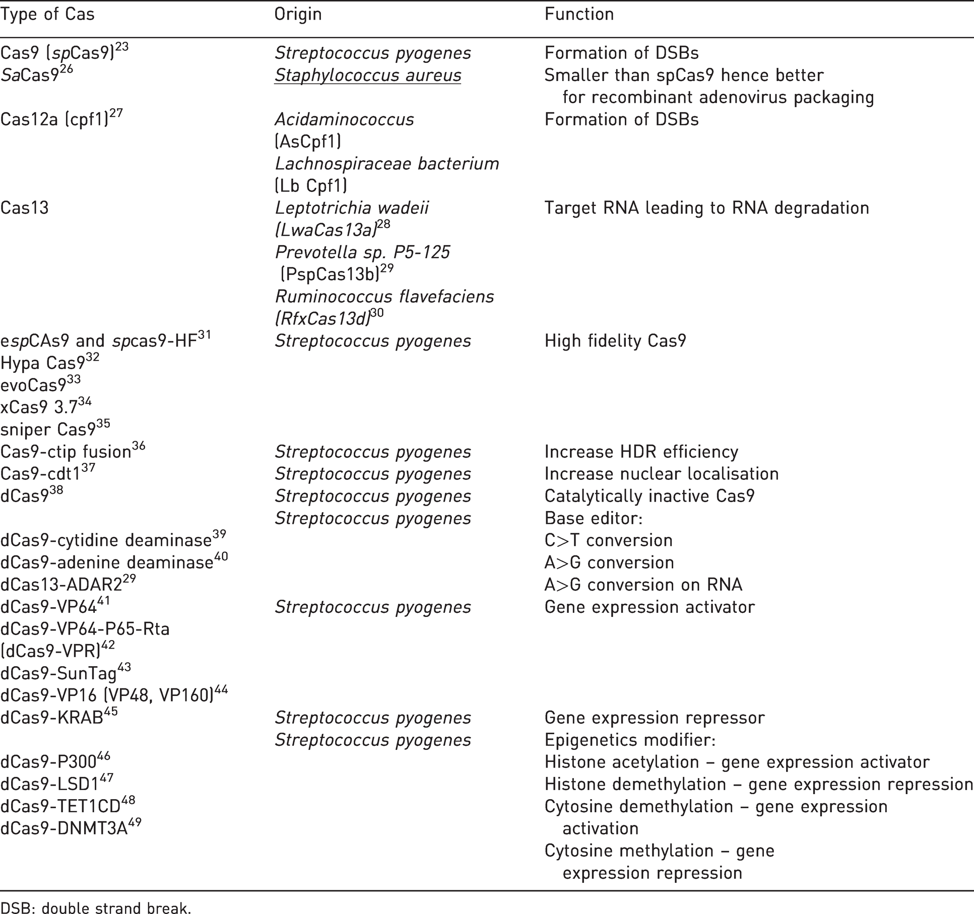
DSB: double strand break.
Pitfalls and challenges
gRNAs are designed to target a specific locus and several web-based software systems have been developed to assist with their design. However, it is sometimes not possible to exclude any off-target binding of the gRNA which could lead to undesired mutations. 141 Such potential off-target mutations could be analysed either focusing on the likely off-target sites or by whole genome sequencing, which is costly and laborious. However, off-target mutations seem to be very rare events in vivo. 142 It is worth noting that off-target events are not specific to CRISPR/Cas9 systems and can be found with genome editing in general. 143
However, the nature of the ‘on-target’ genetic alteration can still be a concern, especially if NHEJ DNA repair mechanisms are active. Once a gRNA has located its target and Cas9 has initiated DSBs, indels are unpredictable, therefore each allele might have a different indel. Any CRISPR/Cas component delivery methods (zygote injection, electroporation or virus infection) might suffer from this pitfall. For zygote administration, although injected at the one-cell stage, reports show that CRISPR/Cas activity may often occur at subsequent stages, leading to mosaic animals, 56 although these may be bred to segregate the different alleles. When using viruses, components are delivered to individual cells, each of which can carry different indels. Furthermore, virus administration may target unwanted cells. When DNA template is administered together with CRISPR/Cas components, random insertion of such templates into the genome may also occur as well as on-target replacement.
Recently, several groups have demonstrated that CRISPR/Cas9 systems can generate large DNA deletions or other unwanted changes, such as gene conversion, to the genome at the target locus in mouse embryonic stem cells 144 or in human embryos.145,146 Such phenomena have also been reported in vivo, in mouse embryos or founder mice. 147 Hence screening of CRISPR/Cas9-generated animals is of paramount importance.
Discussion
In the last seven years, the CRISPR/Cas9 toolbox has kept on growing from generation of indels to gene expression alteration without genome modifications (Table 1). Conventional laboratory animals are now harbouring more complex genetic make-ups to improve our understanding in both basic and biomedical research. An increasing number of CRISPR/Cas9-mediated genetic alterations are also being generated in non-conventional animal models, highlighting the feasibility of genome alterations in a wide variety of species. Indeed, due to its high efficiency, CRISPR/Cas9 permits the generation of genetically altered animals faster than ever before. This has increased exponentially our reservoir of mutants in both conventional and non-conventional animals, all of which are now on a path to become part of the scientists’ arsenal. As with every new system, a better understanding is necessary to fully harness its potential. In its current state, some pitfalls and challenges still exist. Indeed, greater screening and/or generating more than one founder for a given mutation might be needed. Furthermore, additional work needs to be done to better understand the unintended genetic modifications at on- and off-target sites, and for some types of genetic alterations, such as insertion of large DNA fragments, the methods still need improvement. Taken together, from the variety of methods now available and their relative ease of use, the number of genetically altered laboratory animals will only increase. However, while non-conventional animals may be more suited to reflect some human conditions, researchers are facing hurdles in using them. These include the time needed to start working with a new animal model, facility costs, logistics and ethical challenges, and the availability of relevant reagents and methods. Thus, to accommodate this new menagerie of conventional and non-conventional laboratory animals, animal facilities, species-specific reagents and regulatory systems will have to adjust.
Footnotes
Acknowledgement
We would like to thank Güneş Taylor for critical reading of the manuscript.
Declaration of Conflicting Interests
The author(s) declared no potential conflicts of interest with respect to the research, authorship, and/or publication of this article.
Funding
The author(s) disclosed receipt of the following financial support for the research, authorship, and/or publication of this article: This work was supported by the Francis Crick Institute, which receives its core funding from Cancer Research UK (grant number FC001107), the UK Medical Research Council (grant number FC001107) and the Wellcome Trust (grant number FC001107).