
Abstract
Animal models enable research on biological phenomena with controlled interventions not possible or ethical in patients. Among species used as experimental models, squirrel monkeys (Saimiri genus) are phylogenetically related to humans and are relatively easily managed in captivity. Quadrupedal locomotion of squirrel monkeys resembles most other quadrupedal primates in that they utilize a diagonal sequence/diagonal couplets gait when walking on small branches. However, to assume a bipedal locomotion, the human pelvis has undergone evolutionary changes. Therefore, the pelvic bone morphology is not that similar between the female squirrel monkey and woman, but pelvic floor support structures and impacts of fetal size and malpresentation are similar. Thus, this review explores the pelvic floor support structural characteristics of female squirrel monkeys, especially in relation to childbirth to demonstrate similarities to humans.
Introduction
Animal experimental models in biological research are necessary because they allow the study of biological phenomena. The choice of model is fundamental and must, therefore, be judicious. Animal models can improve understanding of human diseases and developmental injuries with controlled interventions, which is generally not possible with human subjects. 1 Although no animal model is perfect, some species are particularly suitable, such as rodents, pigs, and nonhuman primates (NHPs). 2 Animals have been used in research on infectious diseases, such as malaria,3,4 Zika virus, 5 and the pathogenesis of COVID-19, Middle East respiratory syndrome (MERS), and severe acute respiratory syndrome (SARS). 6 Recently, these authors concluded that SARS-CoV-2 causes COVID-19-like disease in macaques and provided a new model for testing preventive and therapeutic strategies. NHPs are good models for research on human diseases and have enabled remarkable scientific progress by elucidating cardiovascular, endocrinal, neurological, and reproductive diseases.7–9 However, the use of NHPs is costly and requires specialized resources. The ethics of using animals for research is another challenge.
In particular, animal models have improved understanding of some pelvic disorders in women.7,10 They are vital to basic and applied research on human female reproduction, prenatal development, and women’s health more generally. 11 Squirrel monkeys (Saimiri sp.) are New World primates that have been used in research on dystocia and changes in pelvic support.1,12–17 Knowledge of the pelvic floor support and delivery mechanism in these monkeys may help to improve understanding of women’s reproductive disorders with the potential to prevent debilitating injuries. Thus, this review aims explore the morphophysiological characteristics of the female squirrel monkey pelvic support structures. We also discuss the mechanics of childbirth to demonstrate similarities between this species and women.
Use of Saimiri sp. in human obstetrical research
Squirrel monkeys are neotropical primates of the Cebidae family. They have a short, thick coat, rounded head, short and black snout, rounded ears, and black-tufted tail. 18 These animals are insectivores and frugivores, and their food includes arthropods, mollusks, small vertebrates, fruits, and seeds. 19
Squirrel monkeys are among the most used neotropical primates in biomedical research, with the first studies reported by Klüver, 20 who provided information on their experimental use and management in captivity. Because of their small size (with a body mass of approximately 1 kg), squirrel monkeys can be kept in smaller spaces than necessary for other model NHPs. They acclimate well to captivity 21 and are easily handled because of their small size. Additionally, similarities in the development of pelvic changes in squirrel monkeys and humans make these monkeys good models for research on human obstetrics. 22 Another advantage of using squirrel monkeys as models is their reproductive longevity – the prime reproductive ages are between 3 and 13 years old. 23
Squirrel monkeys have a gestation period of about 150 days, with a range of 141–154 days,24,25 which enables one pregnancy per year to be monitored in captivity for experimental studies. 17 They are seasonal primates, and, in their native habitats, they reproduce from July to September, with births occurring from December to February; seasonality in this regard has also been observed in the northern hemisphere. 26 The average body mass of squirrel monkeys during the reproductive season is 914.58 ± 13.78 g for males and 752.5 ± 74.6 g for females. 27
Comparative morphology of female squirrel monkey and human reproductive organs and pelvis
Quadrupedal locomotion of squirrel monkeys resembles most other quadrupedal primates in that they utilize a diagonal sequence/diagonal couplets gait when walking on small branches. 28 However, to assume a bipedal locomotion, the human pelvis, forelimb, hindlimb, scull, and spine have undergone evolutionary changes. Therefore, the pelvic bone morphology is not that similar between the female squirrel monkey and woman, but pelvic floor support structures and impacts of fetal size and malpresentation are similar.
Squirrel monkey genital morphology has been described by Branco et al. 29 and Mayor and Plana. 30 Figure 1 shows the gross morphology of the genital organs of squirrel monkeys, and Figure 2 shows how these organs can be measured via ultrasound. Some similarities exist between the genital morphology of squirrel monkeys and women, such as the presence of a simple uterus with less prominent uterine horns. The ovaries are large and ellipsoid, have a smooth surface, and produce visible follicles, but the corpus luteum is not clearly distinguishable by ultrasound. The uterine tubes are long and straight muscular structures that are included in the mesosalpinx. These tubes merge caudally into a single small uterine body, where pregnancy occurs. The mesovarium and ovary participate in the formation of the ovarian bursa, which does not fully involve the ovary. The cervix is short and characterized by a thick and well-developed muscle wall. The vagina extends from the cervix (external uterine orifice) to the exit of the urethra (external urethral orifice). The vaginal vestibule is shared by the genital and urinary tracts. The vulva is formed by two lips that meet at the dorsal and ventral vulvar commissures and has a vertically elongated vulvar sulcus. Finally, the clitoris is located ventrally in the fossa, which is well-developed.
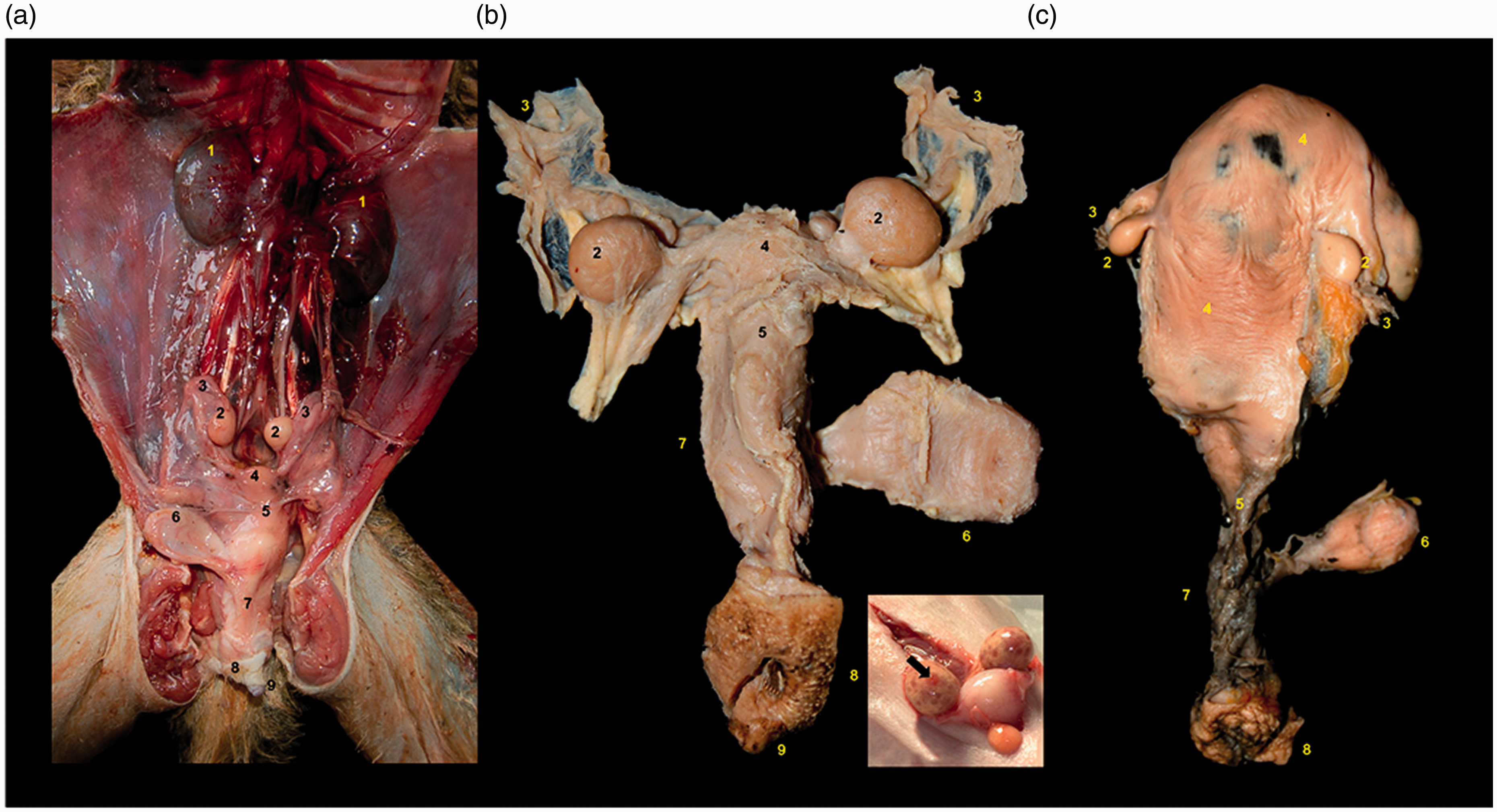
Gross anatomy of squirrel monkey (Saimiri sp.) genital organs. (a) In situ ventral view of nonpregnant; (b) dissected dorsal view of nonpregnant female and detail of ovary follicles (black arrow); (c) dissected dorsal view of pregnant female. 1. Kidneys, 2. Ovaries; 3. Uterine tubes; 4. Uterine body; 5. Cervix; 6. Urinary bladder; 7. Vagina; 8. Vulva; 9. Clitoris. Adapted from Mayor and Plana (2019). 30

Uterine and ovaries images in adult of Saimiri sciureus (10 years old and five deliveries). (a) Uterine variables at the sagittal scan (non pregnant female). (b) Transversal diameter (TD) at the transversal scan (non pregnant female). Hyperechogenic line in the central uterine region indicates the endometrium internal surfaces (arrow). In both ovaries the variable length was obtained at the sagittal scan (c) and (d), represented here by the variables right and left ovary length (RtOL and LtOL, respectively). The largest diameter of the follicles was measured at the sagittal or transversal scan.
The intrapelvic musculature of squirrel monkeys is similar to that of humans. For instance, the levator ani muscle consists of the pubocaudal and iliocaudal muscles, respectively, analogous to the pubococcygeus and iliococcygeus muscles in humans. In addition, the endopelvic fascia has connective tissue condensations that correspond to uterosacral and cardinal ligaments in women. 31 In women, the pelvic floor consists of muscles, ligaments, and fascia, whose contraction provides resistance to gravity and intra-abdominal pressure, and support the abdominal and pelvic organs, such as the uterus, vagina, urinary bladder, urethra, and rectum. Loss of muscle contraction and injury to ligaments, connective tissue, or nerves have been associated with the occurrence of pelvic organ prolapse (POP) 32 and similarly in squirrel monkeys. 14
Two main muscle groups line the pelvic floor in women: the pelvic and urogenital diaphragms. The pelvic diaphragm lines the lower and lateral region of the pelvis, extends from the pubis to the coccyx, and consists of the levator ani and coccygeus muscles. For most of the pelvic floor, the levator ani muscle is divided into the puborectalis, pubococcygeus, and iliococcygeus muscles, which connect and support the vagina, urethra, anus, and coccyx. The coccygeus muscle originates from the sciatic spine and inserts at the lower end of the sacrum and upper end of the coccyx. Therefore, coccyx flexion is important during defecation and fetal expulsion, in addition to assisting the levator ani muscle in supporting pelvic organs. 33
The urogenital diaphragm is in the superficial and distal layer of the pelvic floor and supports the distal portion of the vagina and urethra to the bony pelvis by fixing the ischiocavernosus, bulbocavernosus, and transverse muscles of the perineum. Muscles of urogenital diaphragm also stabilize the perineal body between the urogenital and the anal triangles. In the perineal body, the transverse muscle of the perineum supports the vagina and rectum and maintains urinary and fecal continence. 32
Fasciae and ligaments are formed by dense connective tissue and support the organs by connecting bones and muscles. The endopelvic fascia consists of connective tissue and is composed of two parts: a visceral layer, located below the peritoneum, which surrounds the pelvic diaphragm and supports the organs; and a parietal layer, which comprises ligaments and a septum, fixing the pelvic floor and enabling vascularization and local innervation. Ligaments originate from points of fiber condensation in the endopelvic fascia and actively participate in pelvic visceral support. The main ligaments are the uterosacral and cardinal ligaments, which support the vagina and uterus, and the pubovesical, pubourethral, and anococcygeus ligaments, which support the bladder, urethra, and anus. 34
Based on human anatomic dissections, the support of the vagina and uterus can be segmented into three levels. The upper level consists of the parametrium and paracolpium, which promote lateral support of the uterus and vaginal apex. In the second level, the cardinal and uterosacral ligaments act on the cervix, together with the pubocervical and rectovaginal fasciae, which laterally support the structures. In the lower level, the levator ani muscle and adjacent connective tissue support the vagina. 32
In humans, the pubovesical ligament supports the bladder by fixing the detrusor muscle of the bladder to the tendinous arch of pelvic fascia and pubis, while the pubourethral and puborectalis ligaments act on the bladder neck. The lateral ligaments of the bladder support the bladder trigone, while the pubocervical fascia maintains the base of the organ, suspended in the tendinous arch. 33 The bladder in squirrel monkeys is typically higher in the pelvis prior to pregnancy and lower post-partum with longer urethra relative to size than in humans. The urethra is about the same length as in humans. This is interesting considering the great difference in body size (Dr Kuehl, personal communication, 2020). In women, the urethra is supported by the surrounding tissues that are attached to the pelvic bones. The main tissues responsible for fixing periurethral tissues are the pubourethral ligaments, located in the proximal urethral region, and the levator ani muscle. 32
The anorectal fascia supports the rectum, laterally by the lateral ligaments of the rectum, anteriorly by the rectovaginal fascia, and posteriorly by fixing the presacral fascia to the sacrum. The anus is laterally supported by the pubovisceral and superficial transverse muscles of the perineum, while the perineal body and anococcygeus ligament support the anterior and posterior regions, respectively. 34
Regarding squirrel monkeys as models for research on the physiopathology of pelvic relaxation and prevention of loss of pelvic support in humans, the similarities and differences in vulvar and pelvic anatomy should be assessed, as this can inform obstetrics and the delivery mechanism in these species. 1 Comparative anatomy of the pelvic muscles, innervation, and connective tissue ligaments should also be considered. In this respect, some pioneering work has been reported.14,31,35,36 The main difference is the presence of the tail and muscle bundles in squirrel monkeys, which originate from the sacrum, located posteriorly in the pelvis and anterior to the sacral vertebrae. 12 The role of these muscles in the pathophysiology of POP remains unclear.
In women, the levator ani muscle plays an important role in supporting the pelvic organs. Comparative anatomy has indicated that this muscle in humans evolved from the tail muscles. Consequently, possible changes in pelvic muscles in squirrel monkeys have been evaluated, and the number of deliveries was found to promote changes in the structures of the levator ani muscle13,14 and coccygeus muscle. 15
Squirrel monkeys are dolichopelvic, and their pelvises have an oval cranial portion and are flattened laterally. The ischium is grossly excavated and arched ventrally at its caudal end, like that observed in ruminants and pigs.37,38 Aksel and Abee 39 measured squirrel monkey pelvises using lateral and anteroposterior radiographic projections. Significant differences were subsequently observed between the superior and inferior bi-iliac diameters of females with livebirth neonates (1.84 ± 0.09 and 1.9 ± 0.13 cm, respectively) and stillbirths (1.87 ± 0.08 and 1.92 ± 0.10 cm, respectively). However, a more recent study evaluated pelvimetric data in squirrel monkeys 40 and found superior and inferior bi-iliac diameters of 1.71 ± 0.08 and 1.67 ± 0.09 cm for adult females and 1.59 ± 0.07 and 1.63 ± 0.13 cm for subadult females, respectively.
Childbirth behavior and implications for pelvic biomechanics
Regarding behavior and posture at the time of delivery, female squirrel monkeys rest on their feet and base of their tail. The pelvis does not come into direct contact with the ground and remains suspended. This position can predispose the laboring female to prolapse due to gravity’s effect on the muscles that support the abdominal organs or during labor stress. 41 Furthermore, fetal rotation in female squirrel monkeys is similar to that in women, where the mean submentobregmatic diameter was significantly smaller than the mean occipitofrontal diameter. 22 Occipital–posterior presentation involves the fetal face facing in an opposite direction to that of the maternal face, which hinders the passage of the head and requires greater effort during delivery. 41
Dystocia in NHPs can be caused by cephalopelvic disproportion. Figure 3 shows the dimensions of the fetal cranium and those of the pelvic inlet in humans and NHPs, including squirrel monkeys. 42 Squirrel monkeys have a high incidence of dystocia because large fetal head sizes lead to losses due to stillbirths or neonatal mortality. 43 Approximately 12% of captive female squirrel monkeys, especially primiparous females, experience dystocia. 22 According to Hartwig, 44 the squirrel monkey cranium has a unique dolichocephalic shape, presumably to accommodate the relatively large infant brain while minimizing cranial breadth. While 90% of squirrel monkey deliveries are with the longitudinal presentation and mentum anterior position, other presentations lead to dystocia (Dr Ruiz, personal communication, 2020), which represents a difference compared to humans and may lead to an increase in the frequency of POP as first reported by Coates et al. 1 and Stoller. 22 In humans, discrepancies between the fetal size and maternal pelvis have also been reported and are associated with the evolutionary relationship between larger brain size and the bipedal position. 41
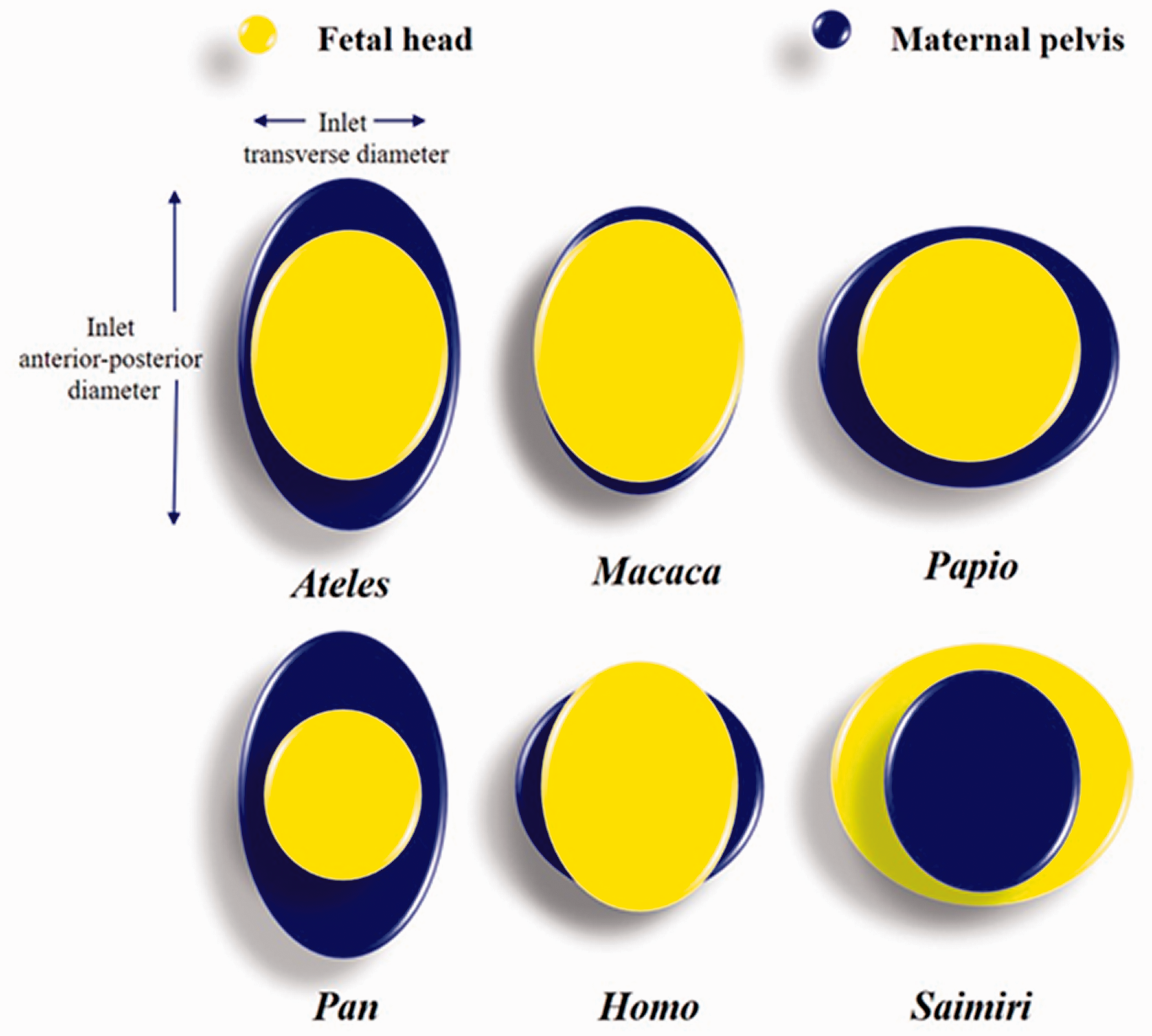
Schematic drawing illustrating the proportion of fetal head with maternal pelvis (transverse and antero-posterior diameters of pelvic inlet) in human (Homo) and non-human primates (Ateles, Macaca, Papio, Pan, and Saimiri). Adapted by Dr Ruiz from Rosenberg KR and Trevathan WR (1996).42
Despite the elongated cranium in squirrel monkeys, the presenting diameter of the infant cranium is still larger than the maternal pelvic in-/outlet (Figure 4). Furthermore, these monkeys have high perinatal mortality due to this disproportion, which impairs the reproductive performance of captive individuals. 39 Aksel and Abee 39 developed a pelvimetric method, radiographing pregnant females, as a predictor of pregnancy outcomes in squirrel monkeys, according to which 95.2% of live births and 92.1% of stillbirths could be predicted. This calculation can, therefore, be a useful tool in predicting pregnancy outcomes by observing narrow pelvic points in females who have stillbirths. Therefore, presentation is so critical in the squirrel monkey. Also, there are no squirrel monkey obstetricians to rotate/reposition the head during the delivery process. This leads to the need for a C-section/surgical intervention to salvage the mother as fetuses are usually lost by the time of discovery.
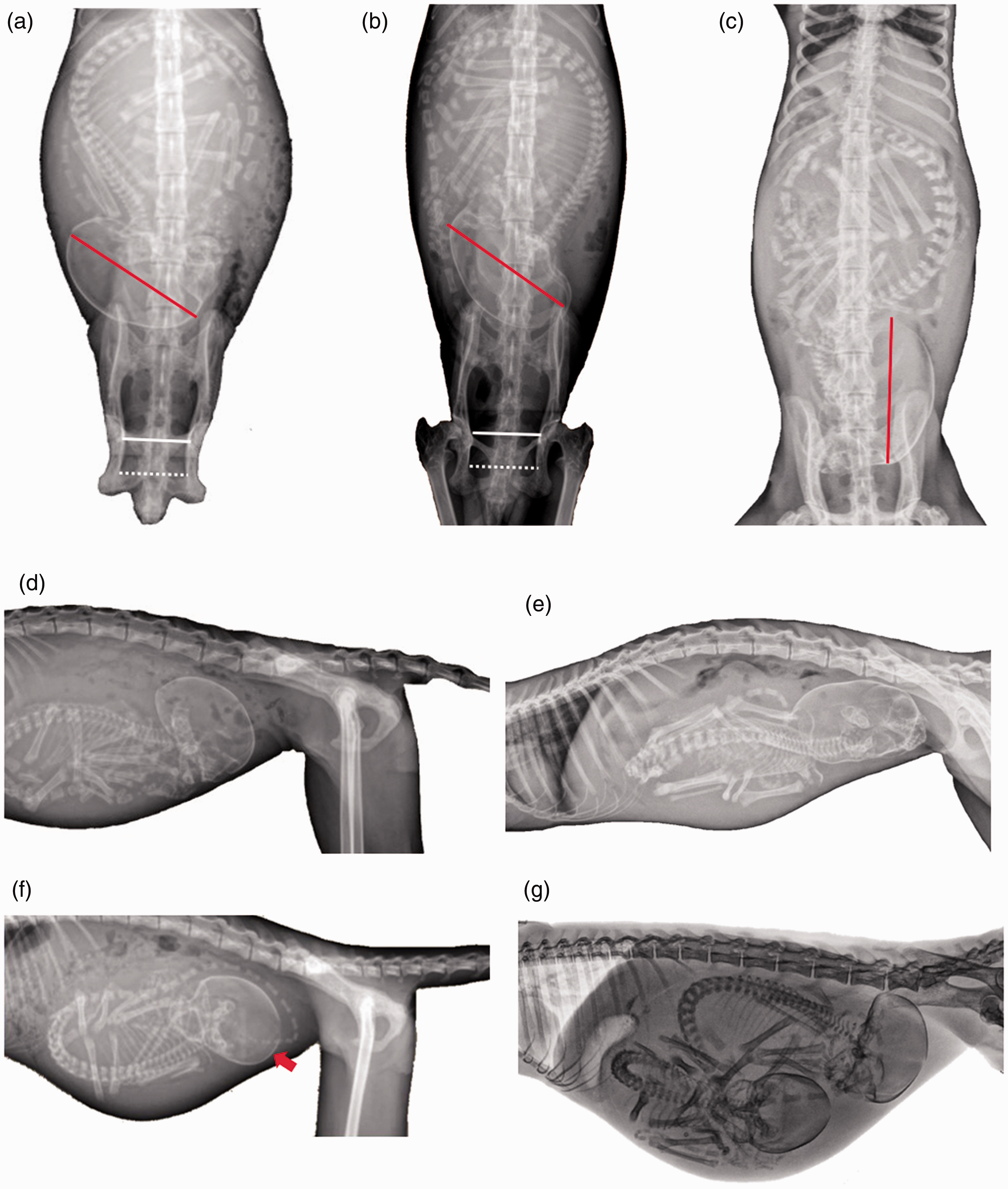
Ventrodorsal (a)–(c) and latero-lateral (d)–(g) radiographic views during delivery in Saimiri boliviensis. (a)–(e) Note that cranium diameter (red line) is still larger than the maternal inlet (white line) and outlet (dotted white line) pelvic; normal presentation (cephalic, vertex, or headfirst). (c)–(e) Fetal head is markedly extended with the face presenting. Fetal rotation as labor begins, the fetal neck is fully extended, and the sagittal axes of the mother and fetus are aligned ((d) and (e)). (f) Vertex presentation, longitudinal lie, occiput posterior position (red arrow). (g) Rare case of twin pregnancy in squirrel monkey (Saimiri sciureus). X-ray images by Dr Ruiz, JC.
The squirrel monkey is one of the neotropical primates with the largest brain size. 45 The development of the facial bones and neurocranium is mainly responsible for neonate size, which can be up to 18% of the non-pregnant female weight, larger than that for other neotropical primates. 46 Neonates are approximately one-sixth the size of the mother’s body weight and, thus, larger as a percentage than that reported in humans. The delivery of large neonates may increase the risk of perineal trauma, nerve damage, and prolapse. 12 Newborn squirrel monkeys have a large cephalopelvic disproportion at birth. In addition, the postnatal maturation of cranial bones is rapid, which is unusual for New World primates. 47
The length of the newborn squirrel monkey’s skull is substantially greater (136%) than the sagittal dimension corresponding to the maternal pelvic inlet. Furthermore, these monkeys have closely apposed orbits and a relatively large brain, reaching precocial prenatal development, a reflex in obstetric constraints imposed by the absolute limits of the pelvic inlet. 44 Favoretto et al. 40 described squirrel monkey pelvimetry using pelvic radiographs in ventrodorsal projections and compared the pelvis measurements of adult and sub-adult females to evaluate the occurrence of dystocia. It was observed that the latter had a smaller upper bi-iliac diameter and pelvic area than the latter did, suggesting a higher risk of dystocic birth.
In addition to trauma during labor, decreased estrogen levels with advancing age are associated with reduced strength and elasticity of the pelvic ligaments and muscles in women. 48 In squirrel monkeys, older females (>12 years old) have reduced hormone levels during the reproductive season of compared to those of younger ones. 49 Thus, the number of deliveries, newborn size, and aging may contribute to progressive denervation and subsequent pelvic prolapse in both squirrel monkeys and humans.
POP
A recent systematic review of the literature showed that several animal models have been used in the study of the pathophysiology of POP, such as lagomorphs, rodents, sheep, and NHPs. 50 The authors concluded that in several species there are measurable effects of pregnancy, delivery, and iatrogenic menopause, but there is not a single uniform pattern. However, only squirrel monkeys develop clinical POP spontaneously. Women and squirrel monkey females can develop disorders in their pelvic support muscles that may be related to age and number of previous deliveries. Coates et al. 12 found that almost 50% of older females in a squirrel monkey population showed pelvic changes compared to younger females. Multiparous females also had a higher occurrence of POP (4.0 versus 1.6).
Moreover, infant size and birth canal structure may render the pelvic floor more susceptible to injury and contribute to the occurrence of dystocic births. 1 Stratford et al. 51 first reported similarities in myogenic and neurogenic changes in the puborectalis muscle between women and squirrel monkeys via magnetic resonance evaluation. After delivery, no changes were observed in the volume of the levator ani and internal obturator muscles, but that of the coccygeus muscles increased, suggesting postpartum injuries. 15 Thus, the authors concluded that squirrel monkeys are suitable for comparative studies regarding women’s pelvic floor support.
In the histological evaluation of the levator ani muscle and paravaginal ligaments in squirrel monkeys with and without POP, no gross ruptures were observed in the aforementioned muscle and its innervation. Myogenic changes were more frequently observed in their pubocaudal muscles and were correlated with aging. In addition, in the paravaginal ligaments, increased apoptosis was associated with parity. 14 On the other hand, in the evaluation of the function of and defects in the levator ani muscle in women with and without POP, magnetic resonance images demonstrated that women affected by POP more frequently had defects in their levator ani muscle. 52
Joyce et al. 16 noted that the size of the pelvic outlet diameter was not related to POP occurrence in squirrel monkeys, but the number of deliveries was a risk factor. This finding was consistent with observations in women, with the number of births being the main predisposing factor for prolapse occurrence. 53 However, reduced pelvic support, related to pelvic musculature, is associated with age and number of births, which may be associated with obstetric complications. These observations inform ongoing research into the nature and cause of spontaneous pelvic relaxation in squirrel monkeys and support the potential use of these primates as animal models. 12
In women, prolonged births have been associated with pelvic denervation injuries, which can be a risk factor for future prolapse. 54 Therefore, childbirth is a risk factor associated with POP, since postpartum changes are detected, but long-term assessment and randomized research are difficult in humans. Consequently, the selection of an experimental animal model should be based on morphofunctional similarity and management practicality. 21 Based on this concept, the first randomized controlled trial of scheduled pre-labor C-section was performed in squirrel monkeys. 17 Animals in the control group demonstrated descent of pelvic structures and bladder similar to those subsequently reported in primiparous women undergoing their first vaginal delivery.55,56 However, animals undergoing scheduled C-section prior to the onset of labor did not show these changes. 17 Thus, there is the potential that such an intervention might reduce the impact of childbirth on human pelvic floor disorders. As this is a very hotly debated issue, follow-up and additional experiments are warranted.
Despite the progress made in recent decades in research conducted with the Saimiri genus, especially in captivity, substantial gaps in our knowledge remain. The relatively easy management of these monkeys in comparison with other primates, can improve the monitoring of birth-related disorders in women. Thus, squirrel monkeys can be used in studies that may inform the development of POP intervention in human obstetrics. Therefore, it is necessary that state-of-the-art imaging and assessments be carried out to understand the similarities between female squirrel monkeys and women. Such research is expected to advance scientific knowledge of reproductive research related to NHPs and human beings.
Footnotes
Declaration of Conflicting Interests
The author(s) have no conflicts of interest to declare.
Funding
The author(s) disclosed receipt of the following financial support for the research, authorship, and/or publication of this article: This was supported by the Coordination for the Improvement of Higher Education Personnel (CAPES, Coordination of Scholarships Abroad, grant number BEX 9255/13-3; National Academic Cooperation Program in the Amazon – PROCAD/Amazônia, grant number 1696/2018); and the National Council of Technological and Scientific Development (CNPq, grant number 305821/2017-2). We are also especially grateful to the Keeling Center for Comparative Medicine and Research at The University of Texas MD Anderson Cancer Center (SMBRR/NIH, grant number P40-OD010938-40).