
Abstract
Ultrasound is a powerful, low-cost, non-invasive medical tool used by laboratory animal veterinarians for diagnostic imaging. Sonohysterography and transvaginal ultrasound are frequently used to assess uterine anomalies in women presenting with abnormal uterine bleeding (AUB). In the present study, we have evaluated the abdominal ultrasound of bonnet monkeys (n = 8) showing spontaneous ovulatory (n = 5) and anovulatory (n = 3) AUB. The ovulatory (n = 5) macaques showed cyclic AUB for 7–8 days. The anovulatory (n = 3) macaques had irregular AUB with menstrual cycles of 40–45 days. The B-mode abdominal, colour Doppler and 3D ultrasound scans were performed during the proliferative phase of the menstrual cycle. Ultrasound examination revealed endometrial polyps in five macaques and endometrial hyperplasia in three animals. The width and length of endometrial polyps was around 0.5–1 cm (average 0.51 ± 0.23 cm × 0.96 ± 0.16 cm) with significant increase in endometrial thickness (P < 0.0002). 3D ultrasound also showed a homogeneous mass in the uterine cavity and colour Doppler ultrasound showed increased vascularity in the endometrial polyps. Endometrial hyperplasia characteristically appeared as a thickened echogenic endometrium (P < 0.0002). This study demonstrates the use of non-invasive ultrasound techniques in the diagnosis of AUB in macaques.
Abnormal uterine bleeding (AUB) is a major gynaecological disorder in women and is classified as either anovulatory or ovulatory. 1 Anovulatory AUB is associated with irregular or infrequent periods, occurring at intervals of more than 35 days in women. The ovulatory AUB with heavy menstrual bleeding for more than seven days during regular menses is also known as heavy menstrual bleeding or menorrhagia. 2 AUB is often associated with the development of anaemia. 3 AUB is also one of the most common indication for performing hysterectomies in women. 4 Structural abnormalities in the uterine cavity, 5 endometrial blood vessel dysfunctions 6 and blood coagulation disorders, 7 are aetiological factors of AUB in humans. Structural abnormalities in the uterine cavity include endometrial polyps, adenomyosis, submucosal fibriods, and endometrial or cervical malignancies. 5 Transabdominal or transvaginal ultrasound is the first-line imaging test for identifying structural abnormalities associated with AUB in women. 8
Non-human primates, especially macaques, are the animal model of choice for studying the physiological and pathological mechanisms involved in menstruation. Irregular patterns of endometrial bleeding are common in adolescent rhesus macaques 9 and heavy menstrual bleeding has been reported in rhesus macaques. 10 To our knowledge, this is the first ultrasound report investigating the causes of AUB in bonnet macaques.
Materials and methods
Animal husbandry practices
The bonnet macaques were housed individually at the animal house facility of the National Institute for Research in Reproductive Health (NIRRH). The sexually mature adult macaques (3–5 years) were captured from the wild forest of southern India in 2004. The age at of the macaques used in the present study is approximately 13–17 years (based on dental structure). 11 The macaques were fed daily with fresh seasonal vegetables, fruits, groundnuts, Bengal gram, and twice in week with eggs. Water was provided ad libitum. Housing conditions were a 12:12 h light:dark cycle, with a temperature of 24–28℃ and relative humidity of 30–70%. The ultrasound protocol was approved by the Institutional Animal Ethics Committee (IAEC) and the Committee for the Purpose of Control and Supervision of Experiments on Animals (CPCSEA). Humane care of animal was observed, and ultrasound examinations were performed during the routine half yearly health monitoring of bonnet macaques in the colony. The animal facility at the institute is registered with the CPCSEA, Ministry of Social Justice and Empowerment, Government of India (registration no. 78/GO/ReBi/SL/99/CPCSEA).
Monitoring of the menstrual cycle
Previous reports from our laboratory have shown that the length between each menstrual cycle in bonnet macaques is 26.0 ± 1.8 days with 3–4 days (group average 2.8 ± 1.4 days) of visible bleeding.12,13 Daily monitoring of the menstrual cycle by vaginal swab examination revealed an abnormal pattern of menstrual bleeding in eight macaques. Of these macaques five showed heavy AUB that continued for 6–8 days during their regular menstrual cycle. Three macaques had irregular AUB with their menstrual periods occurring at intervals of more than 40–45 days. An abdominal diagnostic ultrasound was performed on these eight macaques during the proliferative phase (days 11 to 14 after visible bleeding) of their menstrual cycle. The baseline ultrasound evaluation of the uterus for the normal menstrual cycle (n = 10) in the proliferative phase was generated.
Morphometric analysis
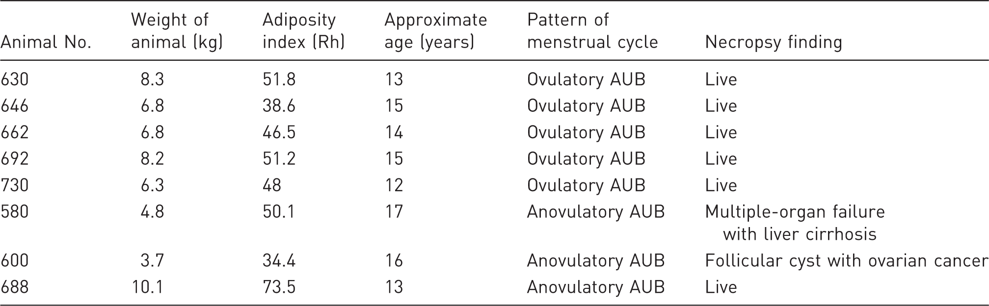
Species-specific adiposity index (Rh), menstrual cycle pattern and other characteristics of bonnet macaques with abnormal uterine bleeding (AUB) are shown.
Abdominal ultrasound examination
An abdominal ultrasound evaluation of the uterus was performed using the HDXE11 ultrasound system (Philips, Andover, MA, USA) in B-mode, colour Doppler (L12-5, C8-5, and C5-2, MHz transducer) and 3D (V6-2, MHz transducer) images. Macaques were sedated with ketamine (10 mg/kg body weight intramuscularly; Ketanir Nirma Ltd, Gujarat, India ) and placed in a supine position on a plain surgical table. The abdominal area was shaved with Savlon (Johnson and Johnson Ltd, Mumbai, India), and ultrasound gel (RTrace, RWave Cardio Products, Mumbai, India) was applied to the shaved area. The transducer was placed sagittally on the lower midline of the pelvic abdominal region to locate the uterus below the urinary bladder.
Histology of the uterus
Two of the macaques diagnosed previously with AUB on ultrasound evaluation died after a year. Their deaths may be due to liver cirrhosis and ovarian cancer (based on clinical signs and post-mortem reports). Post-mortem examination was carried out and the organs were collected in 10% buffered formalin. After formalin fixation the uterus was sectioned. Uterine sections were processed using haematoxylin and eosin staining for histological analysis.
Statistical analysis
The measurements of the length of the menstrual cycle, endometrial polyp sizes (width and length), endometrial thickness (echogenic endometrium in sagittal scan) and animal weights were expressed as mean ± SD using GraphPad Prism software (GraphPad Software Inc, San Diego, CA, USA). The mean significant difference (P < 0.05) was calculated by Student’s t-test using the GraphPad Prism software.
Results
Menstrual cycle pattern in the bonnet macaques with AUB
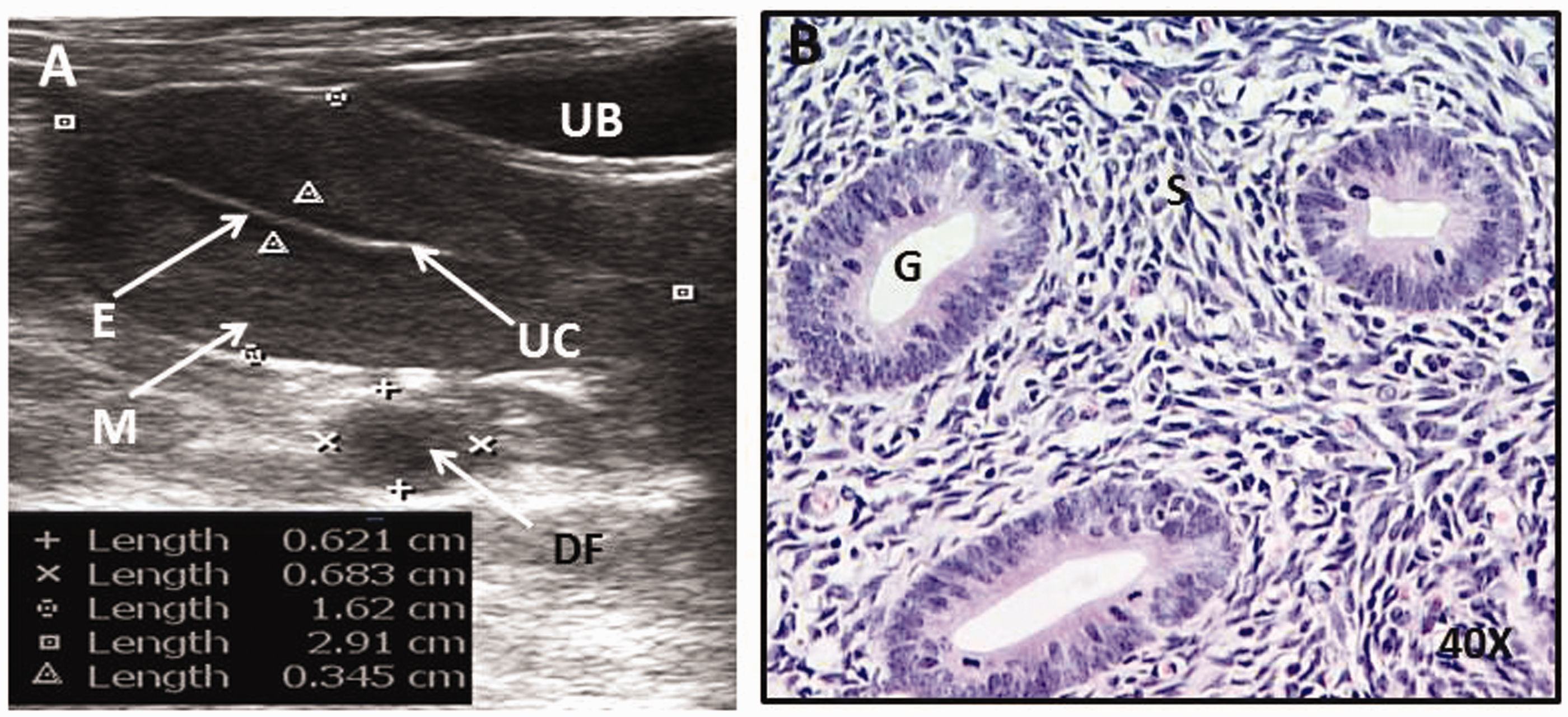
The menstrual cycle length of macaques (n = 5) showing ovulatory AUB was 31.2 ± 1.3 days with a visible bleeding period of 6.4 ± 1.4 days. The menstrual cycle length of macaques (n = 3) showing irregular AUB was 44.0 ± 1.0 days with a visible bleeding period of 2.6 ± 0.5 days (Figure 1). Ultrasound profile of the normal uterus in the proliferative phase showed a typical triple line pattern consisting of a middle white line surrounded by two black lines (Figure 2A). Histological analysis of the proliferative phase endometrium showed sparsely spaced and proliferating glands with abundant stroma (Figure 2B). Abdominal ultrasound examination in the macaques detected AUB with endometrial polyps (three with ovulatory and two with anovulatory AUB) and irregular thickening of the endometrium (two with ovulatory and one with anovulatory AUB).
A representative overlapping two-month menstrual cycle pattern of bonnet macaques showing two consecutive ovulatory abnormal uterine bleeding (AUB) for 6–8 days (animals 1 to 5) and anovulatory AUB (animals 6 to 8). Ultrasound examination was performed during the proliferative phase of the menstrual cycle. Sagittal ultrasound view of the uterus during the proliferative phase showing a typical triple line pattern of proliferative endometrium (between two triangle arrows) (A); and histology of normal proliferative phase endometrium of the bonnet macaques (B). E: endometrium, UC: uterine cavity, UB: urinary bladder, M: myometrium, DF: developing follicle of ovary, G: gland, S: stroma.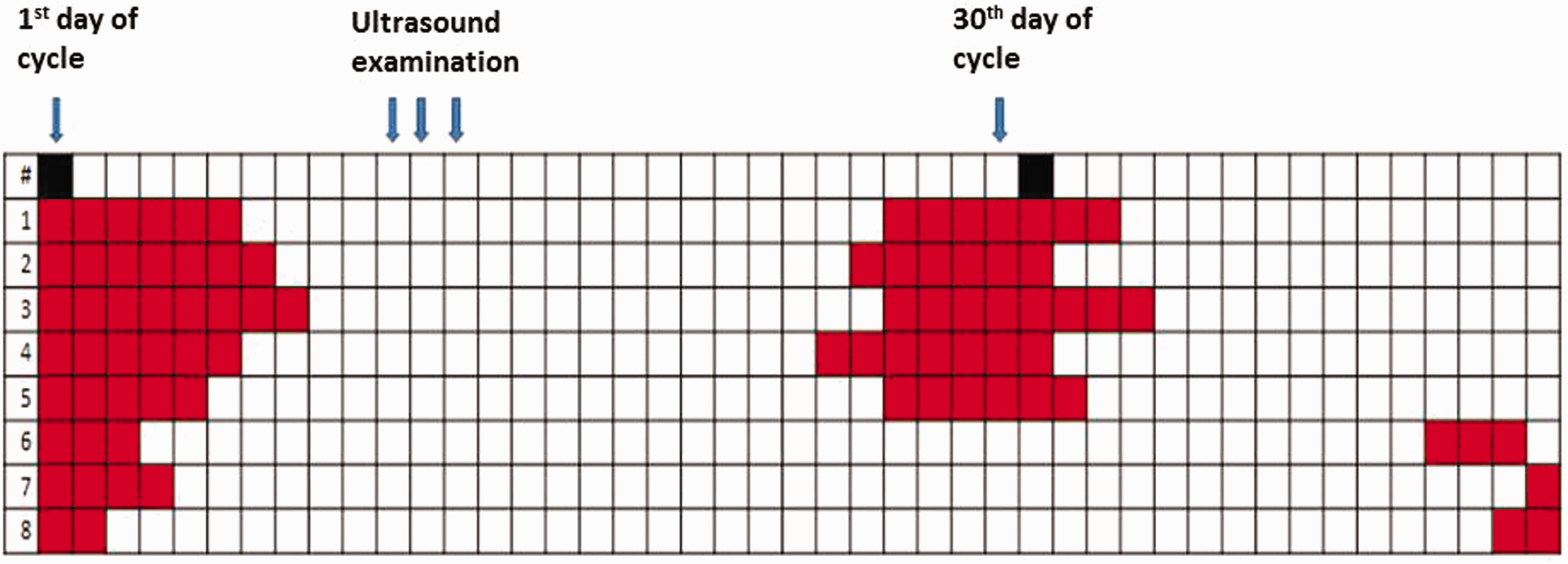
Endometrial polyps
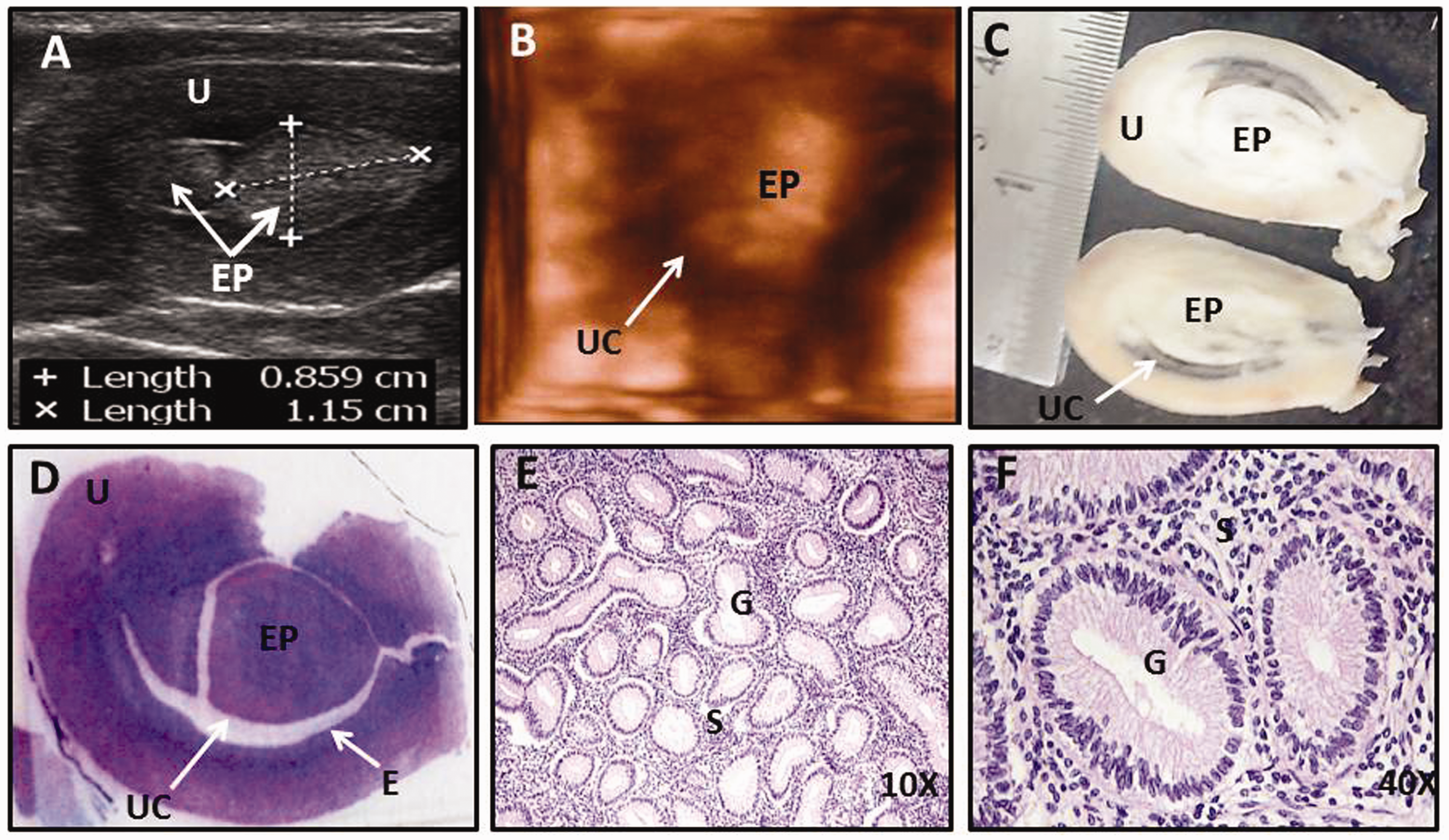
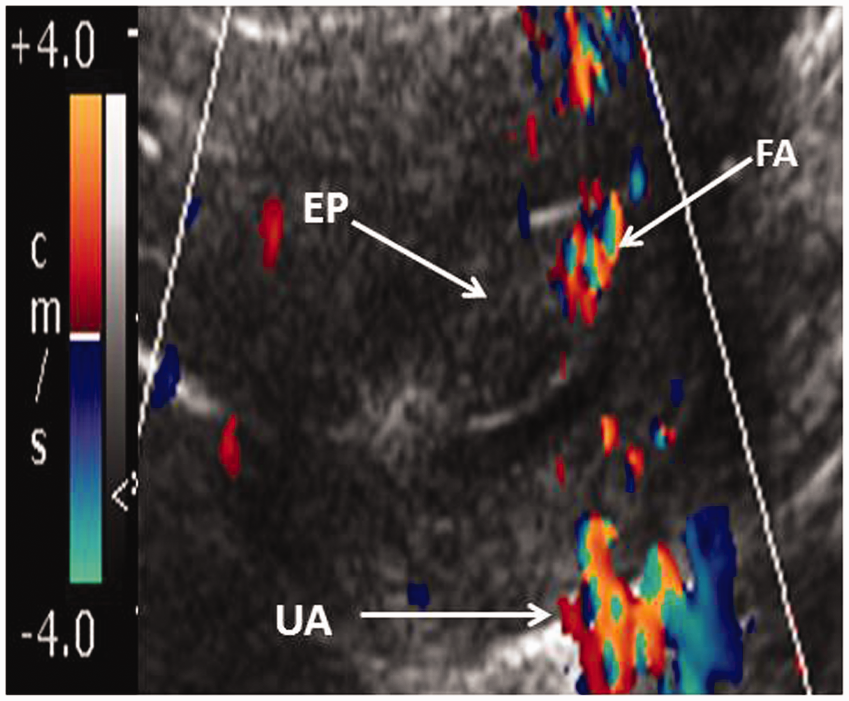
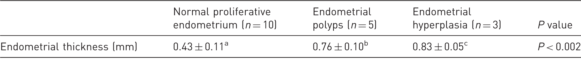
Endometrial polyps were detected as echogenic, well-defined, homogeneous, polypoid lesions in the fundus region of the uterus (Figures 3A–D). The polyps were isoechoic to the endometrium with intact endometrial and myometrial interfaces. One of the endometrial polyps detected in the three macaques showed cystic fluid accumulation (Figure 3B). Multiple polyps obstructing the entire uterine cavity were observed in two macaques (Figure 3C and Figures 4A–D). The endometrium of the macaques with polyps was found to have a significantly (P < 0.0002) greater thickness compared with the normal proliferative phase endometrium (Table 2). Histological analysis of one of the endometrial polyps showed complex endometrial hyperplasia with increased glands-to-stroma ratio (Figures 4E and F). The width and length of the endometrial polyps were around 0.5 × 1 cm (mean 0.51 ± 0.23 cm × 0.96 ± 0.16 cm). Colour Doppler ultrasound showed increased vascularity and a single feeding artery in the endometrial polyps (Figure 5).
Sagittal ultrasound view of the uterus showing smooth, well-defined, uniform echogenic endometrial polyps in the uterine cavity (A); endometrial polyps with accumulation of cystic fluid (B); multiple endometrial polyps at the fundus and meddle of the uterine cavity (C); small endometrial polyps at fundus (D). EP: endometrial polyps, E: endometrium, UC: uterine cavity, M: myometrium, C: cystic fluid. Sagittal ultrasound view of the uterus showing multiple endometrial polyps obstructing the entire uterine cavity (A); 3D reconstructed image of endometrial polyps in the uterine cavity (B); gross anatomy of the uterus with endometrial polyps (C), whole mounted uterus showing endometrial polyps stained with hematoxylin and eosin (D), histology of endometrial polyps showing complex endometrial hyperplasia with crowding of glands (E & F). U: uterus, EP: endometrial polyps, UC: uterine cavity, E: endometrium, G: gland, S: stroma. Colour Doppler ultrasound image of a single feeding artery in the endometrial polyps. EP: endometrial polyps, E: endometrium, V: vascularity, UA: uterine artery, FA: feeding artery of polyps. Endometrial thickness of bonnet macaques measured by ultrasound during the proliferative phase, with endometrial polyps and endometrial hyperplasia. versus b indicates P < 0.0002 by Student’s t-test. versus c indicates P < 0.0002 by Student’s t-test.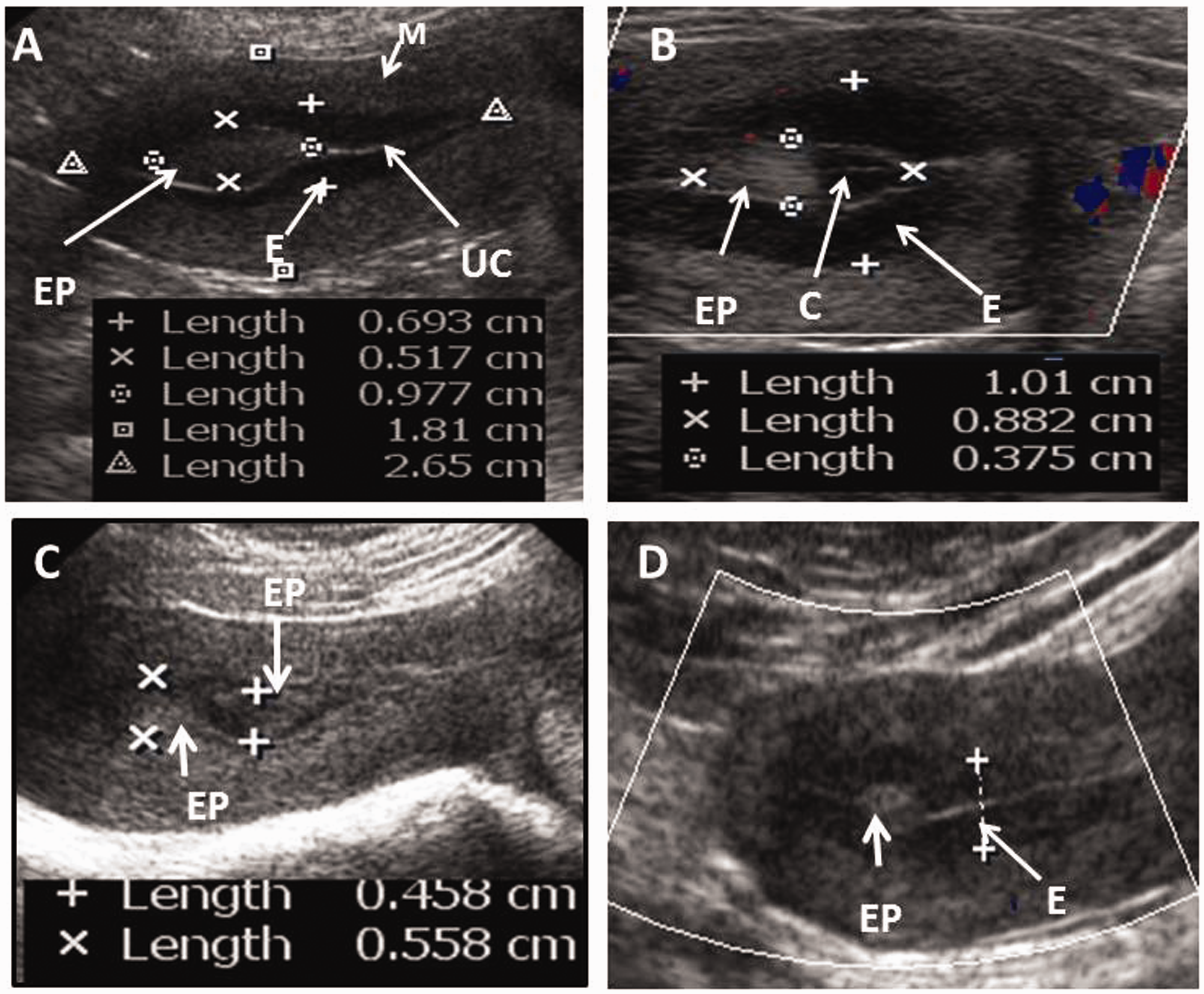
Endometrial hyperplasia associated with adenomyosis
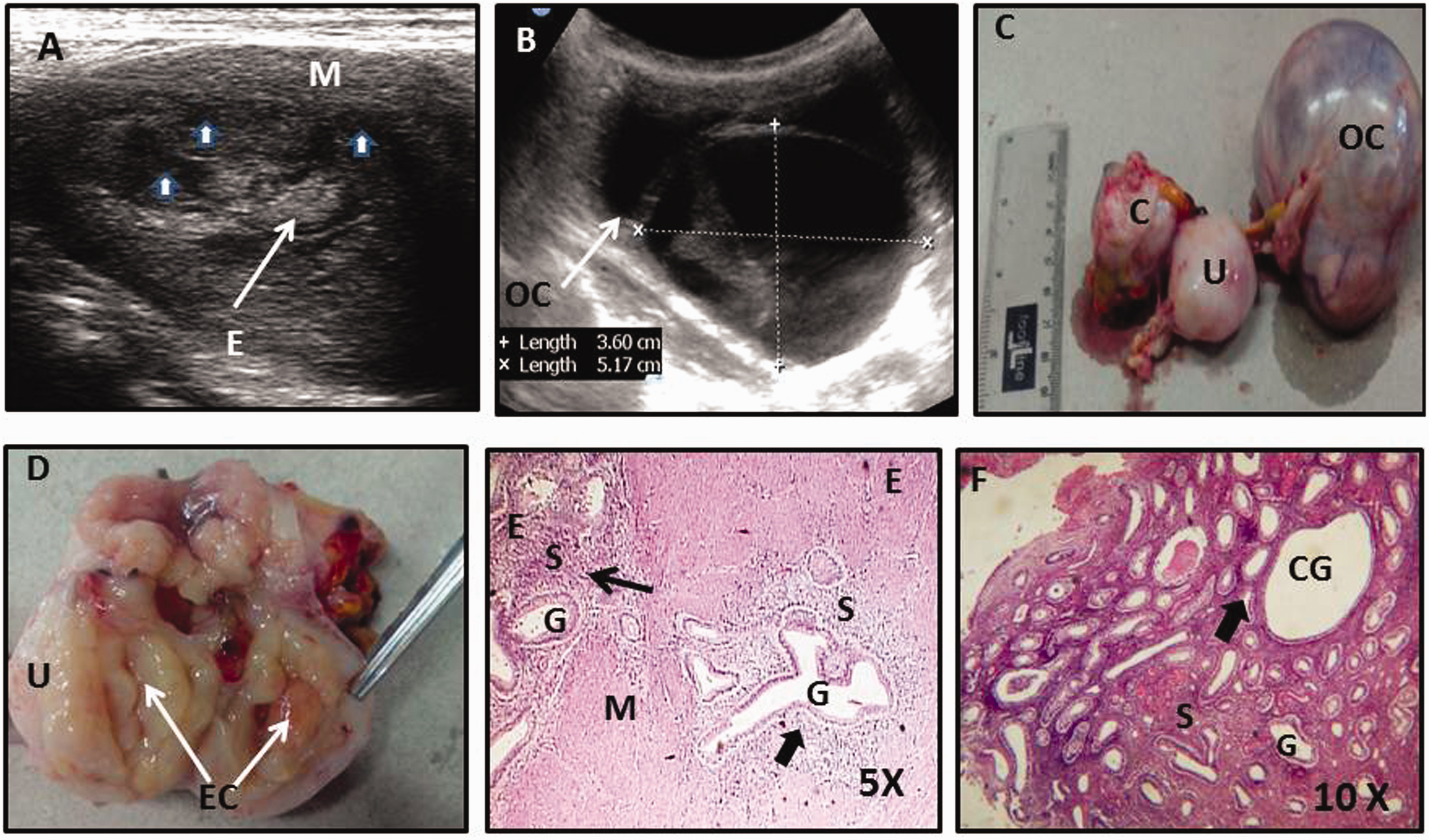
Endometrial hyperplasia was detected in three macaques (Figures 6A and B and Figure 7A). In these animals, the endometrium was thickened and the echogenic endometrium thickness was found to be significantly (P < 0.0002) greater with an absence of the typical triple line pattern of the endometrium observed during the proliferative phase. Endometrial hyperplasia in the animal with ovulatory AUB was non-homogenous with irregular endometrial margins (Figures 6A and B). In another macaque with anovulatory AUB areas of diffused echogenic endometrial thickening with multiple cysts were observed (Figure 7A). Interestingly this animal also demonstrated very large multiloculated ovarian cysts of around 3–5 cm (Figure 7B). During post-mortem examination large ovarian cyst was observed in the right ovary with cystic irregular endometrium boundaries (Figure 7D). Histological analysis of the uterine section showed cystic endometrial hyperplasia and adenomyosis (Figures 7E and F).
Sagittal ultrasound of complex endometrial hyperplasia showing diffuse, irregular thickening of the endometrium with focal hyperechoic polypoid mass (thick arrows) and irregular endometrial to myometrial border (triangle arrow) (A). Homogeneous diffuse simple endometrial hyperplasia with cysts (thick arrows) with preserved endometrial to myometrial border (B). E: endometrium, M: myometrium. Sagittal ultrasound of endometrial hyperplasia and adenomyosis (A); ultrasound of left ovarian loculated cyst showing sepated ovary filled with fluid (B); gross anatomy of cervix, uterus and ovarian loculated cyst (C); gross anatomy of uterine cavity with multiple cysts (D); haematoxylin and eosin (H&E) stained uterine tissue showed adenomyosis (thick arrow), normal endometrium distance on the left (thin arrow) separated by myometrium from adenomyosis foci (5×) (E); H&E stained histology of endometrium from the same animal showing cystic endometrial hyperplasia (thick arrow) (10×) (F). E: endometrium, M: myometrium, C: cervix, U: uterus, OC: ovarian cyst, EC: endometrial cyst, S: stroma, G: gland, CG: cystic gland.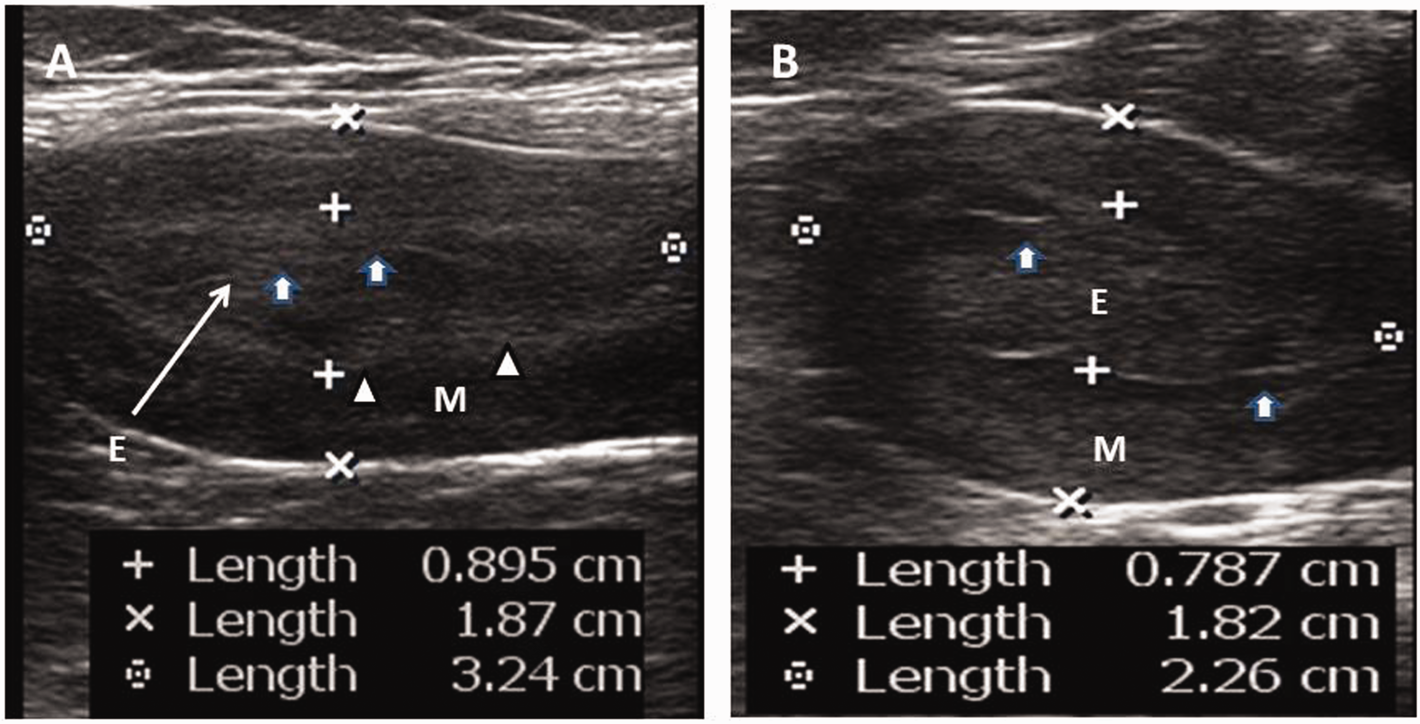
Discussion
The old world primates like rhesus, 15 bonnet 13 and cynomolgus 16 macaques exhibit menstruation parameters similar to those observed in humans and are the most ideal animal models to elucidate the molecular mechanism underlying normal and abnormal menstrual bleedings. In humans, transvaginal ultrasonography with 60–92% sensitivity and 62–93% specificity, is frequently used for the diagnosis of intracavitary lesions such as endometrial polyps and endometrial hyperplasia in premenopausal women showing AUB. 17
Endometrial polyps are localized intrauterine overgrowths and are one of the common gynaecological conditions associated with AUB, infertility and malignancy in women. 18 The typical appearance of an endometrial polyp on ultrasonography is a well-defined, homogeneous, polypoid lesion that may be isoechoic to the endometrium with preservation of the endometrial–myometrial interface. 19 Majority of macaques (n = 5) showing AUB in the present study were diagnosed with endometrial polyps. Atypical features like cysts were observed in one of the endometrial polyps. This has also been reported in human polyps. 20 Complex or atypical endometrial hyperplasia has rarely been reported in a human polyp biopsy specimen. 21 In one of the macaques complex endometrial hyperplasia within endometrial polyps collected during post-mortem examination were observed. Endometrial polyps have also been reported in oestrogen-treated, 22 and postmenopausal rhesus macaques. 23 Spontaneous endometrial polyps have been reported in young cynomolgus macaques 24 and baboons. 25
Endometrial hyperplasia is another major cause of AUB and is also considered to be a precursor for the development of endometrial cancer in women. 26 We observed typical diffuse, focal hyperechoic endometrial thickening in one animal and atypical focal hyperechoic thickening with irregular endometrial border in another. In humans irregular endometrial borders with thickening of the endometrium indicates complex endometrial hyperplasia. 27 Postmenopausal women with symptomatic endometrial thickness >5 mm is considered to be abnormal and need further confirmatory biopsy diagnosis. 28 We also observed a significant increase in endometrial thickness (P < 0.0002) compared with normal proliferative phase endometrium. We have previously reported ultrasound profiles of the proliferative and secretory phase endometrium from healthy bonnet macaques. 29 Oestrogen-induced endometrial hyperplasia has been reported in cynomolgus and rhesus macaques. 29 Spontaneous endometrial hyperplasia has been observed in rhesus macaques. 15
Adenomyosis is a non-neoplastic condition characterized by the presence of ectopic endometrium and stroma in the myometrium. 30 In women, it can cause pelvic pain (both cyclical and non-cyclical), AUB and infertility. 31 Spontaneous adenomyosis has been reported in many non-human primate species, including macaques and baboons. 32 A cystic space and heterogeneous zones of myometrium thickening by ultrasound indicate adenomyosis in humans. 33 Multiple cystic spaces with thickening of the myometrium along with thickening of the endometrium were observed in one animal. Histological analyses of the uterine sections showed cystic endometrial hyperplasia and the presence of endometrium within the myometrium. Endometrial hyperplasia and carcinoma have also been reported to co-exist with greater frequency in women with adenomyosis. 34
Obesity in women of reproductive age has been associated with the development of endometrial polyps35,36 and endometrial hyperplasia.37,38 An increase in obesity with aging has been previously characterized based on the adiposity index (Rh) in bonnet 39 and rhesus macaques. 14 . We also observed a progressive increase in obesity in our colony (unpublished data). This obesity may have contributed to the development of endometrial polyps and endometrial hyperplasia in the macaques included in the present study, which is in agreement with our previous report of endometrial polyps in obese bonnet macaques.40,41
In conclusion, AUB inbonnet macaques is associated with structural abnormalities such as endometrial polyps and endometrial hyperplasia, an observation similar to that reported in women with AUB. Ultrasound imaging may serve as a first-line diagnostic tool in determining potential causes of AUB, and this will further help in monitoring the prognosis of these lesions after therapeutic interventions.
Footnotes
Acknowledgments
We thank Mr Shinde, Mr Lokhande and Mr Salunkhe for their indispensable help during ultrasound examinations.
Declaration of Conflicting Interests
The author(s) declared no potential conflicts of interest with respect to the research, authorship, and/or publication of this article.
Funding
The author(s) disclosed receipt of the following financial support for the research, authorship, and/or publication of this article: The authors acknowledge the financial support received from the Science and Engineering Research Board (SERB), Government of India and the Indian Council of Medical Research (ICMR), Government of India.