
Abstract
A method is described for chronic cannulation of the cisterna magna to enable repeated sampling of cerebrospinal fluid from conscious, ambulatory sheep by means of a flexible vinyl tube. Ease of sampling and duration of cannula patency are similar to those obtained with rigid, metal cannulae, but this modified method minimizes the degree of surgical intervention, and possible trauma, occurring during placement of the cannula.
A variety of methods for cannulation of the cisterna magna and other cerebroventricular sites in sheep and goats have been described. However we consider the use of rigid, metal cannulae, with the attendant need to excavate a large section of the parietal bone and place securing attachments into the skull, to be unnecessarily invasive. To enable investigations of central roles of C-type natriuretic peptide, 1 we have sought to establish a method that enables repeated collection of cerebrospinal fluid (CSF) from the cisterna magna of sheep. CSF can be collected from sheep at cervical and lumbar sites, 2 although use of sedation is needed for this procedure and it is not amenable to repeated and/or chronic sampling. We describe a simple modification of a reported method, 3 which allows for repeated CSF collection with minimal surgical intervention.
Materials and methods
Six yearling Coopworth ewes (Lincoln University Research Farm, Canterbury, NZ, live weight between 30 and 51 kg) were fasted for 24 h prior to surgery, and water was withheld overnight. Each animal was given an intramuscular injection of analgesic (1 mL of 324 µg/mL buprenorphine hydrochloride, Temgesic injection; Reckitt Benckiser Healthcare (UK) Ltd, Slough, UK). Anaesthesia was induced with an intravenous injection of a combination of ketamine (10 mg/kg ketamine hydrochloride, Phoenix ketamine injection; Phoenix Pharm Distributors Ltd, Auckland, NZ) and diazepam (0.5 mg/kg, Pamlin injection; Parnell Laboratories New Zealand Ltd, Auckland, NZ). After endotracheal intubation, anaesthesia was maintained with 2% v/v isoflurane in oxygen. Post surgery, each sheep received analgesic (as above) every 8 h for two days, and rectal temperature was monitored daily. These procedures were approved by the Lincoln University Animal Ethics Committee.
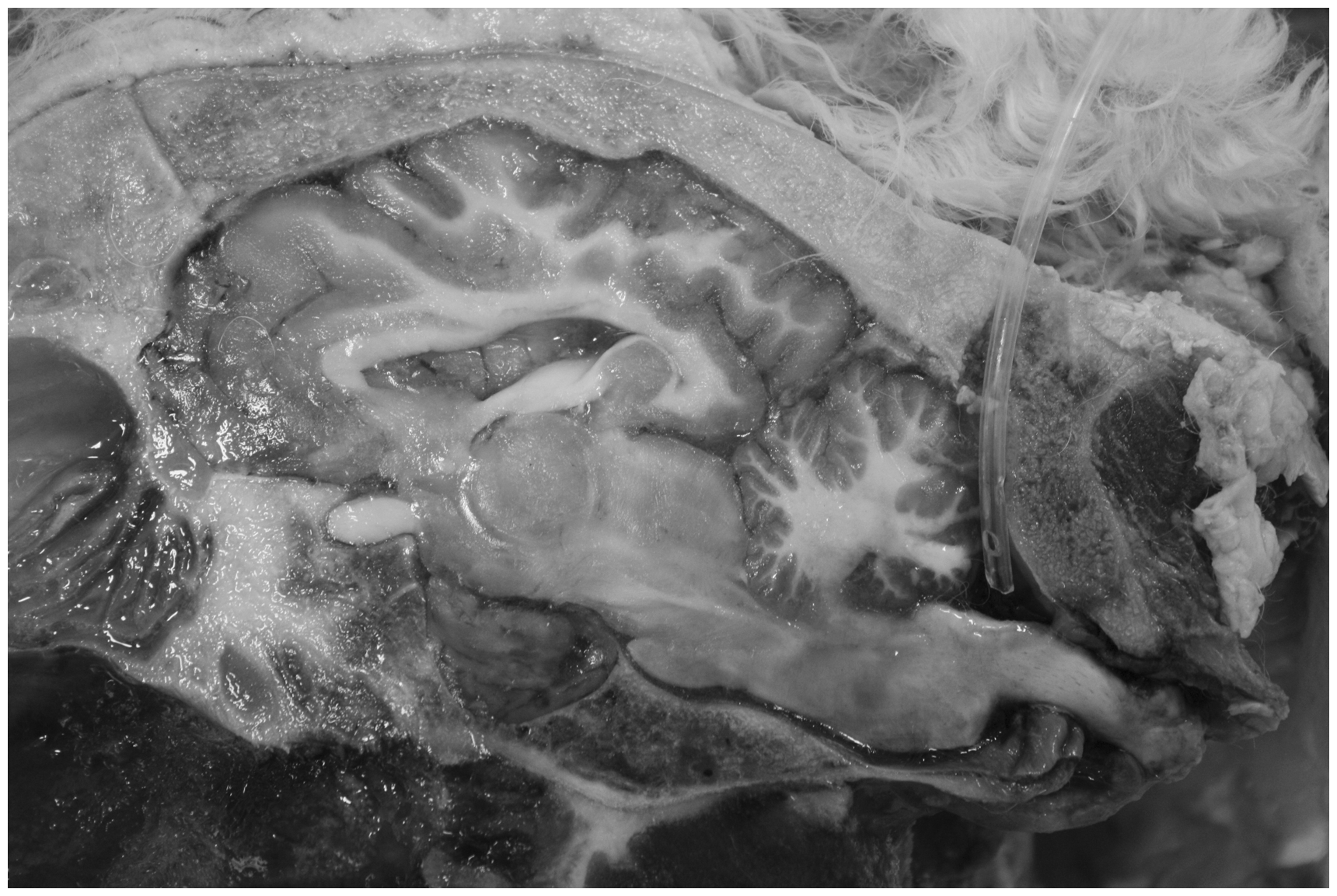
To maintain the sheep’s head in a stable position during surgery, its mandible was placed on a horizontal bar attached to an external steel frame on the operating table, and a tapered bar, clamped one on each side of the same frame, was secured into each auditory meatus (Figure 1). The surgical site was clipped free of wool, cleaned with povidone–iodine solution and disinfected with 70% v/v isopropanol. Using aseptic procedures a 6 cm sagittal skin incision was centred over the depression on the parietal bone and underlying tissues were revealed with the use of a retractor (e.g. Weitlaner) and further dissection. The periosteum was scraped from the underlying parietal bone to clear the entry site for drilling, with the midline about 3 mm rostral to the nuchal ligament aponeurosis. A 2.5 mm hole was drilled by hand through the bone at 15° rostral to the vertical (90°), until the bit passed through the bone. The hole was enlarged to a diameter of 5 mm and bone chips were removed with cotton swabs and by flushing with saline solution. Each cannula was made from a 30 cm length of clear polyvinyl chloride tubing (Dural Plastics & Engineering, Auburn, New South Wales, Australia) with an outside diameter (o.d.) of 2.5 mm (internal diameter 1.5 mm). At one end, the tip was sliced at an angle (45°) and two holes (2 mm diameter) were cut through the wall of the cannula on opposite sides, within 15 mm of the tip. Access to the distal end was provided by insertion of a sawn-off 1.65 mm o.d. (16 G) hypodermic needle with Luer hub. The dura mater and arachnoid membranes were carefully pierced in a caudal direction with a 10 G (3.4 mm outside diameter × 76.2 mm length) steel needle (Industrial Strength Llc, Wallingford, CT, USA), through which the tip of the cannula was introduced and advanced caudally towards the cisterna magna (about 45 mm) until CSF flowed freely in the tubing – see Figure 2. Alternatively, the cannula could sometimes be introduced directly into the cisterna magna after the meningeal membranes were pierced. The appearance of CSF around the needle during its insertion indicated successful penetration of the meninges. The cannula was secured by placing an absorbable suture on to subcutaneous tissue near the bone entry site and tying it to the cannula with Chinese finger trap knotting. The needle was placed subcutaneously within the incision site and directed outwards so that it exited through the skin 5 cm lateral to the incision and was used to exteriorize the distal end of the cannula. After removal of the needle and attachment of the Luer hub to the cannula, a sampling port (BD Q-Syte™ 0.10 mL; Becton Dickinson Infusion Therapy Systems Inc, Sandy, UT, USA) was fitted to the hub. Subcutaneous tissues were repaired above the bone entry site with continuous suturing (absorbable) and the skin was closed with interrupted sutures (non-absorbable). Plastic surgical adhesive tape (5 cm length) was placed on the cannula 10 cm from the skin entry site and secured to the skin with cyanoacrylate adhesive (‘instant glue’), and the skin entry site was sealed with a generous bleb of antiseptic ointment (Betadine povidine–iodine antiseptic–germicide ointment; The Purdue Frederick Company, Norwalk, CT, USA). The exterior section of the cannula and the surgical area were covered with sterile cotton swabs that were held in place with flexible synthetic netting. The cannula port was disinfected with 70% v/v isopropanol prior to sample collection, after which 0.5 mL of sterile saline solution (0.9% w/v) was injected into the cannula to occupy the dead space. Antiseptic ointment was reapplied and cotton swabs were replaced.
Position of the sheep for cannulation of the cisterna magna. The anaesthetized animal is placed in a prone position with its mandible resting on a steel rod and the head secured by tapered steel bars which extend into each auditory meatus. Mid-sagittal view of the head of a sheep showing the cerebrospinal fluid (CSF) cannula in situ. The cannula is introduced through a hole drilled in the parietal bone about 3 mm rostral to the nuchal ligament aponeurosis and inserted in a caudal direction so that the tip is located in the cisterna magna.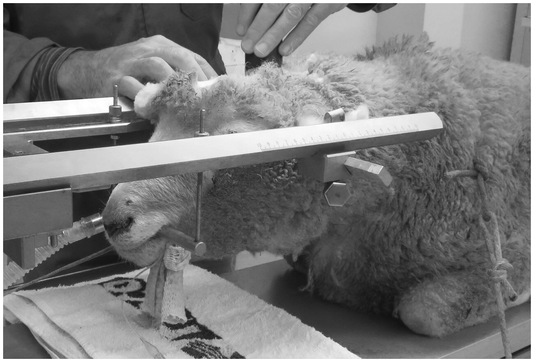
Results
All six sheep were successfully cannulated, and recovered completely to provide CSF samples. The cannulae remained patent for 9–19 days (mean 13.5 days) and 25–46 samples of CSF (1.0–1.2 mL), were collected from each sheep. In most cases at 1–2 days before cannula patency failed, CSF flow into the collection syringe became noticeably reduced, which could be alleviated sometimes by prior administration of a small volume (<0.5 mL) of saline solution through the cannula. After CSF ceased to flow, the cannulae were removed and all sheep were returned to the farm in good health.
Discussion
This method enables repeated sampling of CSF from sheep with minimal restraint, without need for sedation post surgery and without any ill effects. Sampling could be repeated on numerous occasions in a day for more than two weeks, which is a similar sampling performance to that described previously. 3 However, we consider that the present technique is a refinement because it is less invasive due to the use of flexible tubing, rather than the rigid metal needle used previously, 3 and requires less removal of the parietal bone. This contrasts with the relatively major excavation of bone that is required for placement of a rigid metal needle, and thus our method reduces the possibility of trauma to the dorsocaudal surfaces of the cerebellum. The utility of this technique for repeated collection of CSF from sheep will likely reduce the number of animals required to be subjected to the procedure.
In situ cannulae in the cerebral ventricles and cisternae of goats and sheep invariably become coated with a fibrous membrane, so that the functionality of such cannulae has a limited time-span. 4 Because the ability to collect CSF repeatedly from sheep does not extend beyond two to three weeks, it is desirable to have a cannulation technique that has minimal impact on welfare of the animals but provides ready access for sampling. The method described meets these criteria and makes some progress on two of the three Rs (replacement, reduction and refinement) of humane animal experimentation.
Footnotes
Acknowledgements
We are indebted to Dr Brent Higgins of Vetspecs, Veterinary Specialist Services, Christchurch, NZ, for advice on surgical procedures and to Martin Wellby for his technical assistance.
Declaration of conflicting interests
None declared.
Funding
This research received no specific grant from any funding agency in the public, commercial, or not-for-profit sectors.