
Abstract
The role of substantia nigra pathology in Alzheimer's disease (AD) is uncertain. Detection of pathology may be obscured by intraneuronal neuromelanin and influenced by stains. We determined methods for optimal visualization of nigral pathology in 45 cases of AD. For detection of Lewy bodies (LBs), we compared ubiquitin and α-synuclein immunostains to hematoxylin and eosin (H&E). For neurofibrillary tangles (NFTs) and neuropil threads (NTs), we compared Gallyas silver and paired helical filament (PHF) immunostains, after bleaching of melanin, to modified Bielschowsky, Gallyas, and PHF alone. The number of LB cases was not different using the three stains. However, more LBs per section were detected using α-synuclein (z=4.88, p<0.001). Twice the number of cases exhibited NFT (z=8.21; p<0.001) and the mean NFT number per section was 2.8–5.2-fold greater, using Gallyas and PHF after bleaching compared to without bleaching χ2=142.17; p<0.001). More NTs (z=6.54; p<0.001) were observed with PHF and Gallyas after bleaching. With optimal methods, we found LBs in 27%, NFTs in 89%, and NTs in all 45 AD cases. We show that detection of nigra pathology is influenced by histological method. Clinicopathological studies using these methods are needed to determine the role of nigral pathology in AD.
Keywords
C
Materials and Methods
Cases and Neuropathological Evaluation
Forty-five cases of AD were obtained from the neuropathology laboratory at the Rush Alzheimer's Disease Center. Subjects had all presented with dementia and had a clinical diagnosis of dementia that was confirmed as probable or definite AD by neuropathological evaluation (see below). Records from these cases did not contain uniform assessment for disease severity, extrapyramidal signs, or a diagnosis of dementia with LBs (McKeith et al. 1996,1999). No subject had a known clinical diagnosis of idiopathic Parkinson's disease. No exclusion criteria were applied. A total of 45 cases (23 female, 22 male) were evaluated with a mean age at death of 78.3 years (SD = 6.89; range 59–94).
The brains were removed from the calvaria 2–24 hr (mean postmortem interval 7.8; SD = 5.5) after death and processed using standard protocols. The brainstem was removed at the level of the diencephalic-mesencephalic junction and the hemispheres cut into 1-cm coronal slabs. The brainstem and slabs were then fixed in 4% paraformaldehyde for up to 7 days. Multiple cortical regions of brain (including midfrontal, superior temporal, inferior parietal cortices, and hippocampus) were embedded in paraffin, cut into 6-μm sections, mounted on slides, and stained with H&E and modified Bielschowsky silver stain or thioflavin S stain (Mufson et al. 1988). Neuropathological diagnoses of AD were rendered using the protocol developed by the Consortium to Establish a Registry of Alzheimer's disease (Mirra et al. 1991).
Evaluation of the Substantia Nigra
Consecutive 6-μm sections of midbrain, including the substantia nigra, were cut from the available paraffin-embedded block at approximately the level of exit of the third nerve. We applied eight methods of staining to adjacent sections of substantia nigra, including H&E stain, modified Bielschowsky silver stain (Mufson et al. 1988), Gallyas silver stain (Gall-yas 1971; Kobayashi et al. 1992), immunohistochemistry with antibodies to PHF [PHF-1, identifying a modification of the tau protein (Greenberg et al. 1992), 1:1000], ubiquitin (East Acres Biologicals, Southbridge, MA; 1:30,000) and α-synuclein (Chemicon International, Temecula, CA; 1:3750); and Gallyas silver stain and PHF-1 immunostain after applying bleach to tissue sections to remove neuromelanin (Uchihara et al. 1995). All sections were mounted on electrostatically charged plus slides (Daigger and Co.; Lincolnshire, IL), dehydrated in alcohols and xylene, and cover-slipped with Permount.
Gallyas silver staining was performed as originally described (Gallyas 1971) with the additional application of Schiff's reagent and counterstaining with hematoxylin after physical development, similar to the modified Gallyas technique or Gallyas-Schiff stain (Kobayashi et al. 1992) but without the pretreatment with periodic acid solution.
PHF, ubiquitin, and α-synuclein immunostains were performed using the avidin-biotin-peroxidase method (ABC Elite; Vector, Burlingame, CA). Briefly, paraffin-embedded tissues were mounted on electrostatically charged slides, de-paraffinized, washed, and blocked with normal serum. Sections were then incubated with the primary antibody overnight, washed, incubated with the biotinylated secondary antibody for 1 hr, and then with the avidin-biotin-peroxidase complex for 30 min. The chromogen for color development was 3,3′ diaminobenzidine. Immunohistochemical controls consisted of sections incubated without primary antibody. Positive controls included neocortical tissue from known cases of AD (for PHF immunostaining) and LB disease (ubiquitin and α-synuclein immunostaining).
Bleaching of the tissue sections for removal of neuromelanin was performed before immunohistochemistry with PHF and Gallyas silver staining (see above). The bleaching procedure was accomplished by 30-min incubation of mounted tissue sections in 0.25% potassium manganese peroxide followed by a 5-min incubation in 5% oxalic acid, as previously described (Uchihara et al. 1995). Immunohistochemistry was performed in groups of 10 and identically after bleaching as in cases where bleaching was not performed. For the Gallyas silver staining, length of time in the developer differed in those cases with and without bleach. After bleaching the length of time in the developer was generally longer, typically 10 min or until sections turned light brown, whereas without bleaching the length of time in the developer was typically 8 min for optimal staining of nigral NFTs and NTs. Preliminary experiments showed that bleaching before the modified Bielschowsky procedure did not enhance visualization of neuritic pathology compared to the Gallyas and PHF stains after bleaching (data not shown). Therefore, bleaching was not used before modified Bielschowsky. The bleaching technique was also attempted before ubiquitin and α-synuclein immunostaining. However, these antibodies stained the neuromelanin granules in addition to the inclusions (LBs), rendering no appreciable detection advantage in the use of bleach (personal observations).
Rating of Pathology in the Substantia Nigra
The same examiner (JAS) scored all 45 cases. Cases were separated into two batteries of stains and the examiner was blinded to the results of the initial battery when examining the second battery. The initial battery of stains included H&E for LBs, Bielschowsky silver staining, Gallyas silver staining, and PHF immunostaining for NFTs, and Gallyas silver staining and PHF immunostaining for NTs. Bielschowsky silver staining was not used to assess NTs because the dense staining of axonal elements and vessels interfered with the evaluation of NTs. The second battery of stains included ubiquitin and α-synuclein immunostaining for LBs and Gallyas silver staining and PHF immunostaining after applying bleach for removal of neuromelanin for NFTs and NTs.
The total numbers of NFTs and LBs in the 6-μm hemi-section were counted using a microscopic field at a magnification of ×100. We counted all positive structures within each frame from medial to lateral. We found that we were able to obtain reliable counts without the aid of a grid. The slide was manually moved across the stage in a stepwise manner, to avoid redundancy in counting, until all positive structures were counted. A magnification of ×200 was used when clarification was needed for structural identification (see below). The section was first visually scanned at low power to delineate the region of the substantia nigra and note was made if the section was incomplete or was significantly distant from the region of the third nerve exit. Quantitation of NFTs and LBs was completed on each slide from a medial to lateral direction. NFTs were identified when there was positive intraneuronal staining (brownish black on Bielschowsky, Gallyas, and PHF immunostaining; black with bleaching before Gallyas and PHF immunostaining) and a fibrillar morphology. Identification of LBs required a neuronal intracytoplasmic rounded structure (pinkish-red on H&E or brown on immunostaining), typically with a peripheral halo. Because LBs immunostained with ubiquitin and α-synuclein do not always show halos (McKeith et al. 1996, 1999), we also counted the stained structure as an LB if it was intraneuronal, round, and well circumscribed but showed no definite halo. Only structures that fulfilled the above criteria were included in the counts.
Neuropil threads (NTs) were scored in the region of substantia nigra exhibiting the greatest density within the hemisection. The slide was first scanned at low power to determine the location of greatest density of NTs within the substantia nigra and then scored on a semiquantitative scale, from 0 (none) to 5 (severe) at a total magnification of ×200. A score of 1 = rare NTs or <5 NTs/high-power field (hpf); a score of 2 = slight or about 6–15 NTs/hpf; a score of 3 = mild or approximately 16–35 NTs/hpf; a score of 4 = moderate or about 35–50 NTs/hpf; and a score of 5 = severe or about >50 NTs/hpf. NTs were defined as curvilinear structures of narrow width and of variable length that stained brown or black, depending on the staining method.
Statistical Analysis
Our data were quantitative (counts) for LBs and NFTs and semiquantitative (ratings) for NTs. We analyzed these variables as both dichotomous (present/absent) and counts and ratings. The primary outcomes of interest were the ability of different methods to detect (a) more cases with each pathological index, and (b) a greater number of each pathological index. Because this essentially amounts to measuring the same pathological index multiple times from the same subject under different experimental conditions, we employed repeated measures analyses, using the method of generalized estimating equations (Liang and Zeger 1986; Zeger and Liang 1986; Diggle et al. 1994). This method adjusts for the inherent correlation among measurements from the same subjects while also allowing one to model non-normal distributed data, such as dichotomous data, counts, and ratings. In addition, missing data are accommodated and need not be excluded. We fit these models using PROC GEN-MOD in SAS, V. 6.12 (SAS Institute 1997). We tested each given individual a priori hypothesis of interest using a Wald statistic. Significance was determined at a level of p<0.05.
Results
Lewy Bodies
We found that the number of cases detected with LB pathology did not significantly differ using α-synuclein immunostaining compared with H&E or ubiquitin (z=1.37; p = 0.085) (Table 1). In addition, ubiquitin immunostaining did not improve case detection rate compared to H&E (z=1.44; p=0.074). Conversely, the type of staining method significantly affected the number of LBs counted in hemisection. We counted a significantly greater number of LBs using α-synuclein compared to H&E and ubiquitin (z=4.88; p<0.001) (Table 1). In addition, ubiquitin immunostaining allowed detection of significantly greater numbers of LBs compared to H&E (z=2.88; p = 0.002). Of those with at least one LB, the median number of LBs was 5.5 using H&E, 10.5 using ubiquitin, and 19.4 using α-synuclein immunostaining.
Neurofibrillary Tangles and Neuropil Threads
The number of cases identified with one or more NFTs was highest using Gallyas silver staining and PHF immunohistochemistry in bleached sections (41 and 40 cases, respectively; z=4.06; p<0.001) compared to all of the other methods (Table 2). In addition, NFTs in the substantia nigra were observed in more cases using Gallyas silver staining (n=32) and PHF-1 immunostaining (n=37) compared to modified Bielschowsky silver staining (n = 18).
The overall numbers of NFTs counted in hemisection was significantly greater using Gallyas and PHF after the bleaching of tissue sections compared to all of the other methods without bleach (z=8.21; p < 0.001; Table 2). The absolute number of NFTs per hemisection observed with Gallyas or PHF after bleaching of melanin was more than 10-fold greater than the number observed by modified Bielschowsky and was more than 2.5 times greater than Gallyas or PHF staining alone (Figures 1A and 1B). Gallyas and PHF after bleaching allowed similar increases in detection of NFTs (33.9 ± 38.8 NFTs vs 30.6 ± 35.8 NFTs; χ2=1.86; p = 0.173). Gallyas silver staining and PHF immunostaining alone (without prebleaching) allowed detection of a greater number of NFTs on average, per case, compared to Bielschowsky silver staining (z=3.16; p<0.001).
The morphology of the NFTs using silver staining and PHF, with or without bleach, most often revealed the typical globose fibrillary structures (Figures 2A and 2B). Using Gallyas after the bleaching of tissue sections additionally revealed small thread-like and linear intraneuronal tangles (Figure 2C). PHF immunostaining, particularly after bleach, also showed small intraneuronal tangle-like inclusions, as well as diffuse or dot-like staining occasionally extending into neuronal processes (Figure 2D).
Detection and number of Lewy bodies (LBs) in the substantia nigra
∗ p<0.01.
∗∗ p<0.001 (compared to H&E).
Detection and number of neurofibrillary tangles (NFTs) in the substantia nigra in 45 patients with Alzheimer's disease
∗ p<0.01 (compared to Bielschowsky silver staining).
∗∗ p<0.001 (“after bleach” compared to all other methods).
We identified neuropil threads (Figure 3) in the majority of cases of AD. Depending on the method, 88–100% of the cases of AD showed at least a few NTs. Semiquantitative estimates of the number of NTs were increased by the use of bleach (z=6.54; p<0.001) as compared to no bleach. In addition, PHF after bleaching allowed visualization of more threads (mean score 3.2 ± 1.4) compared to Gallyas after bleaching (2.3 ± 1.4) χ2 = 50.75; p<0.001).
The distribution of neurofibrillary pathology within the substantia nigra was not specifically addressed in this study. However, neurofibrillary pathology was observed throughout the hemisection of nigra rather than confined to a particular anatomic locus within the nucleus.
Discussion
Specific stains and the bleaching of tissue sections to remove neuromelanin before staining dramatically improve detection of LBs and neurofibrillary pathology in the substantia nigra in AD. α-Synuclein and ubiquitin immunostaining did not increase the number of cases uncovered with LB pathology. However, α-synuclein and, to a lesser extent, ubiquitin allowed visualization of increased numbers of nigral LBs per case. Pretreatment with bleach significantly increased both the numbers of cases detected with neurofibrillary pathology and the amount of neurofibrillary pathology detected on average per case. Using the bleaching method, approximately 90% of AD cases showed NFTs, with NFT counts approximately 10-fold greater on average per case (compared to unbleached sections), and all cases displayed at least a few NTs.
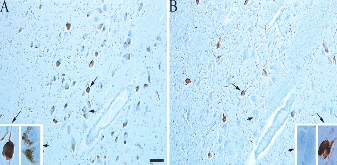
Improved detection of neurofibrillary pathology. (
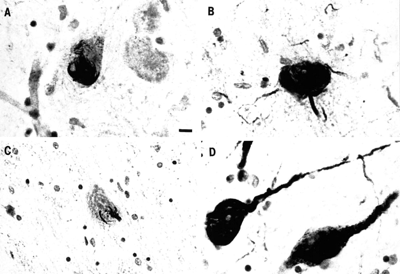
Neurofibrillary tangles: typical globose neurofibrillary tangle using (
Few previous studies have investigated detection of nigral pathology in AD comparing different histological stains or tissue bleaching before staining (Uchihara et al. 1995; Irizarry et al. 1998). In one study, the number of α-synuclein-immunopositive structures was found to exceed that of ubiquitin (Irizarry et al. 1998). LB case detection and the magnitude of the difference were not addressed. In a second study, Gallyas silver impregnation after bleaching revealed an unexpectedly large number of NFTs in five cases of AD (Uchihara et al. 1995). These investigators did not use immuno-stains after bleach pretreatment, nor did they apply these methods to cases with little or no pathology.
The protein α-synuclein is one of the integral components of LBs in Parkinson's disease, LB disease, and AD with LB (Irizarry et al. 1998; Spillantini et al. 1998). Double immunofluorescence studies have shown the presence of α-synuclein-immunopositive, ubiquitin-immunonegative LBs (Irizarry et al. 1998). Our study supports the notion that α-synuclein immunostaining has a higher sensitivity for LBs than ubiquitin. This may be explained partly by the fact that α-synuclein is present at an earlier stage of LB formation, e.g., pale bodies (intracytoplasmic pale eosinophilic inclusions lacking the peripheral halo) often considered a precursor to LBs. Unlike ubiquitin, which may immunostain other neuronal inclusions, e.g., neurofibrillary pathology, α-synuclein immunostaining is relatively specific for LBs (Irizarry et al. 1998; and personal observations). α-Synuclein positivity is, however, also found in oligodendroglial inclusions of multiple system atrophy (Wakabayashi et al. 1998).
Most previous literature of NFTs in AD suggest that neurofibrillary pathology in the substantia nigra is uncommon (Forno and Alvord 1971; Tomlinson and Corsellis 1984; Tabaton et al. 1985; Perry 1986; German et al. 1987; Parvizi et al. 2001), remains confined to the ventral tegmental area (Ishii 1966; Tomlinson and Corsellis 1984; Bondareff et al. 1994; Parvizi et al. 2001) and, when present, occurs late in the course of the disease (Braak and Braak 1991). Although one study reported widespread NFTs in up to 50% of the SN examined (Gibb et al. 1989), others describe nigral NFTs in less than 15% of their AD cases, and a more localized distribution (Forno and Alvord 1971; Parvizi et al. 2001). The magnitude of neurofibrillary pathology in the substantia nigra is also generally considered small, with as few as five NFTs per section (Tabaton et al. 1985) to three per microscopic field on 50-μm sections (Parvizi et al. 2001). None of these studies used bleach before silver or immunohistochemical staining.
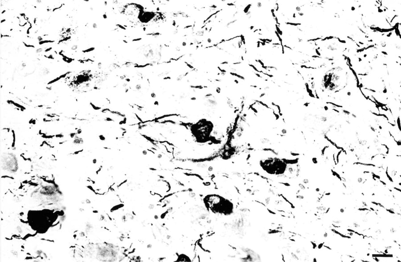
Neuropil threads. Many neuropil threads are seen with PHF immunostaining after bleach. Bar = 20 μm.
Improved visualization of nigral neurofibrillary pathology after bleaching could result from unveiling of perikaryal structures or from decreased neuropil staining. The occasional diffuse staining of cell bodies and extensions with PHF immunostaining after bleaching may suggest detection of an earlier stage of NFT development. During neurofibrillary degeneration, differential stages are recognized by ultrastructural and immunohistochemical changes (Bondareff et al. 1994). Oxidation-dependent epitopes, such as post-translational modifications, have been strongly implicated in the formation of NFTs (Smith et al. 1995). Epitopes may be made available through oxidation (bleaching). Artifactual staining is unlikely, given the variability of staining between cases, between NFTs and NTs, and the characteristic morphology of NFTs and NTs.
The impact of these findings on the pathologic diagnoses of diseases with LBs (e.g., dementia with Lewy bodies) remains uncertain. Although we did not increase case detection for LBs using α-synuclein, our study did not include evaluation of the cortex or assess severity of disease, and therefore it remains to be determined whether α-synuclein better detects cases with cortical LBs or nigral LB pathology in asymptomatic or early-stage disease. It is interesting to note that, compared to “pure” AD, there are fewer NFTs in the substantia nigra in cases of AD with nigral LB. Nevertheless, improved detection of neurofibrillary pathology is noted for both groups (Table 3). Fewer neocortical NFTs have been previously reported in AD with LBs (Hansen et al. 1993). Because LB cases typically have significant nigral neuronal loss, further studies evaluating nigral NFTs in AD with and without LBs will require a wide spectrum of change to account for neuron loss. Unlike the diseases with LBs for which the diagnosis relies on LBs at one or multiple sites, a pathological diagnosis of AD is not rendered on the basis of NFTs in the substantia nigra, and therefore the bleaching method is unlikely to affect diagnosis.
The implications of these findings for clinical pathologic and other research studies, where it is useful to use the most sensitive method to detect pathology, are apparent. Our results support the second Dementia with Lewy Body International Workshop (McKeith et al. 1999) suggestion of using α-synuclein for LB detection in clinicopathological correlative studies. Although the clinical correlate of the number of nigral LB has not been well studied in AD, absolute LB counts in the neocortex using ubiquitin immunohistochemistry may be related to cognitive status in LB disease (Samuel et al. 1996). In addition, our results suggest that Gallyas or PHF immunostaining with bleach to improve NFT detection may have an impact on clinicopathological correlative studies investigating NFTs in the substantia nigra. Only recently have NFTs been considered as an etiological factor for parkinsonism in cases of AD without LBs (Liu et al. 1997).
Alzheimer's disease without nigral Lewy bodies (AD/no LB) and Alzheimer's disease with nigral Lewy bodies (AD + LB): detection and number of neurofibrillary tangles (NFTs) in the substantia nigra a
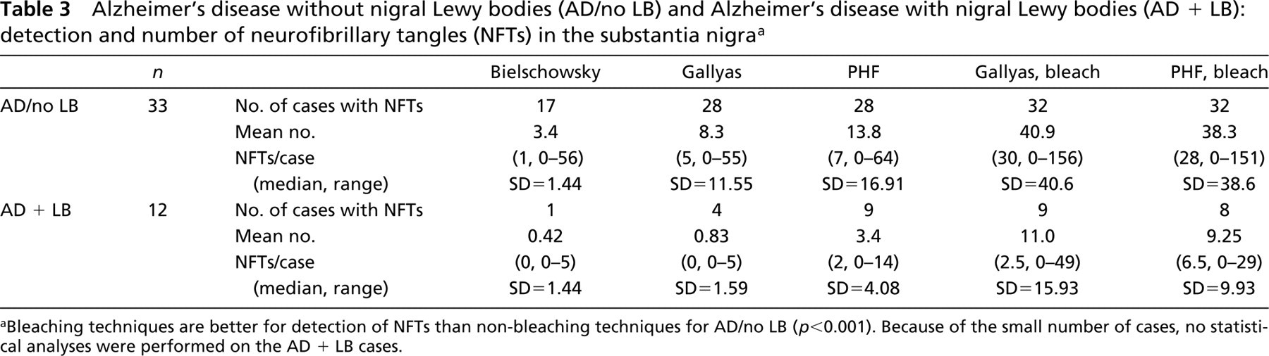
aBleaching techniques are better for detection of NFTs than non-bleaching techniques for AD/no LB (p<0.001). Because of the small number of cases, no statistical analyses were performed on the AD + LB cases.
In persons with AD and parkinsonism, the neuro-pathological substrate underlying the motor disorder often remains speculative (Morris et al. 1989). Indeed, the etiology of parkinsonism in AD may include more than one neuropathological substrate in the SN in addition to neuropathological changes outside the SN. One of the factors in determining the relationship of these pathologies to clinical signs is the method used to detect their occurrence in the postmortem brain. Because parkinsonism in persons with AD is common (Molsa et al. 1984; Chang Chui et al. 1985; Mayeux et al. 1985; Wilson et al. 2000a) and is associated with morbidity and mortality (Richards et al. 1993; Stern et al. 1994; Bennett et al. 1998; Wilson et al. 2000b), it is important to extend our findings by determining the overall contribution of these nigral cytoskeletal changes to clinical signs in AD. The present investigation provides striking evidence that use of specific stains and bleaching of neuromelanin from neurons of the SN greatly enhances the visualization of LBs and neurofibrillary pathology in these perikarya. These improved staining methods will provide basic researchers with a more sensitive tool for the evaluation of cytological degeneration in the SN and its relationship to cognitive and motor function.
Footnotes
Acknowledgements
Supported by grants from the National Institute on Aging (AG10161, AG14449, AG09966, AG09466, AG00849) and the Elsie Heller Fund.
We wish to thank Woojeong Bang for assistance with computer programming and Jia-liang Li for assistance in the laboratory.