
Abstract
Acid aspiration causes pulmonary vascular permeability and PMN sequestration. By increasing pulmonary mast cells through adoptive transfer of v-abl-transformed mast cells (V3MCs) into BALB/c mice, we now show that the greater mast cell number in the lung is associated with increased pulmonary injury.
Acid aspiration-induced acute alveolitis is mediated by several cellular and humoral factors (Kennedy et al. 1989; Knight et al. 1992; Weiser et al. 1997), and the pulmonary inflammation is characterized by an increase in vascular permeability and enhanced PMN sequestration. Mast cells are strategically positioned in the respiratory tract and are a source of several inflammatory mediators (Galli et al. 1993) that may play a proximal role in this inflammatory response. This response is significantly attenuated in the mast cell-deficient mouse (Kyriakides et al. unpublished data). We now demonstrate in a murine mastocytosis model that increased mast cells in the lung augment the acid aspiration-induced pulmonary injury.
One million v-abl-transformed mast cells (V3MCs) were injected IV into 6–8-week-old male BALB/c mice (Taconic Farms; Germantown, NY). Previous studies (Gurish et al. 1995) demonstrate that such adoptive transfer of the V3MCs resulted in mastocytosis of the spleen, liver, and intestine by Days 10–14 and the V3MCs localizing in these tissues matured into a phenotype indistinguishable from that of the indigenous mast cells. Two weeks later, anesthetized BALB/c and V3MC-injected BALB/c mice received an IV injection of bovine [125I]-albumin, followed by tracheal instillation of 0.1 N HCl at 2 ml/kg or saline through tracheostomy tubes placed in situ. Three hr later the lung vascular permeability was assessed. The ratio of the concentration of radiolabeled albumin retrieved in bronchioalveolar lavage (BAL) fluid to blood was used to calculate the lung permeability index (PI). Identical preparations save for the omission of [125I]-albumin were used for PMN quantification in the BAL for histopathology and immunohistochemistry. Neutrophils are expressed as 1 × 10⁴ cells/ml BAL. Sections of the lung were evaluated by chloroacetate esterase (CAE) reactivity to visualize mast cells and PMNs, as well as by standard histochemical stains. A grading of lung injury was performed histologically based on the alveolar neutrophil counts and interstitial edema per 10 randomly chosen HPF. Immunohistochemical staining for two of the mast cell secretory granule chymases, mMCP-1 and mMCP-2, also were performed. Animals used in this study were maintained in accordance with the guidelines of the Committee on Animals of Harvard Medical School and the Committee on the Care and Use of Laboratory Animals of the Institute of Laboratory Animal Resources, National Research Council [Department of Health, Education and Human Services, Publication no. 85–23 (National Institutes of Health) revised 1985].
Results (mean ± SEM) were analyzed after subtraction of sham value by one-way ANOVA and the Bonferroni procedure for multiple comparisons. Acid aspiration led to lung injury in the BALB/c mice, PI 0.023 ± 0.0025 compared to the saline sham control animals, PI of 0.008 ± 0.0015 (n = 3, p < 0.05). Acid-injured V3MC mice showed a marked increment in lung PI to 0.090 ± 0.0046 (n = 5, p < 0.05) in comparison with the saline sham V3MC mice, PI 0.037 ± 0.005 (n = 4, p < 0.05). A fourfold augmentation of the PI was observed in the V3MC acid-injured mice relative to the acid-injured BALB/c mice (n = 4, p < 0.05).
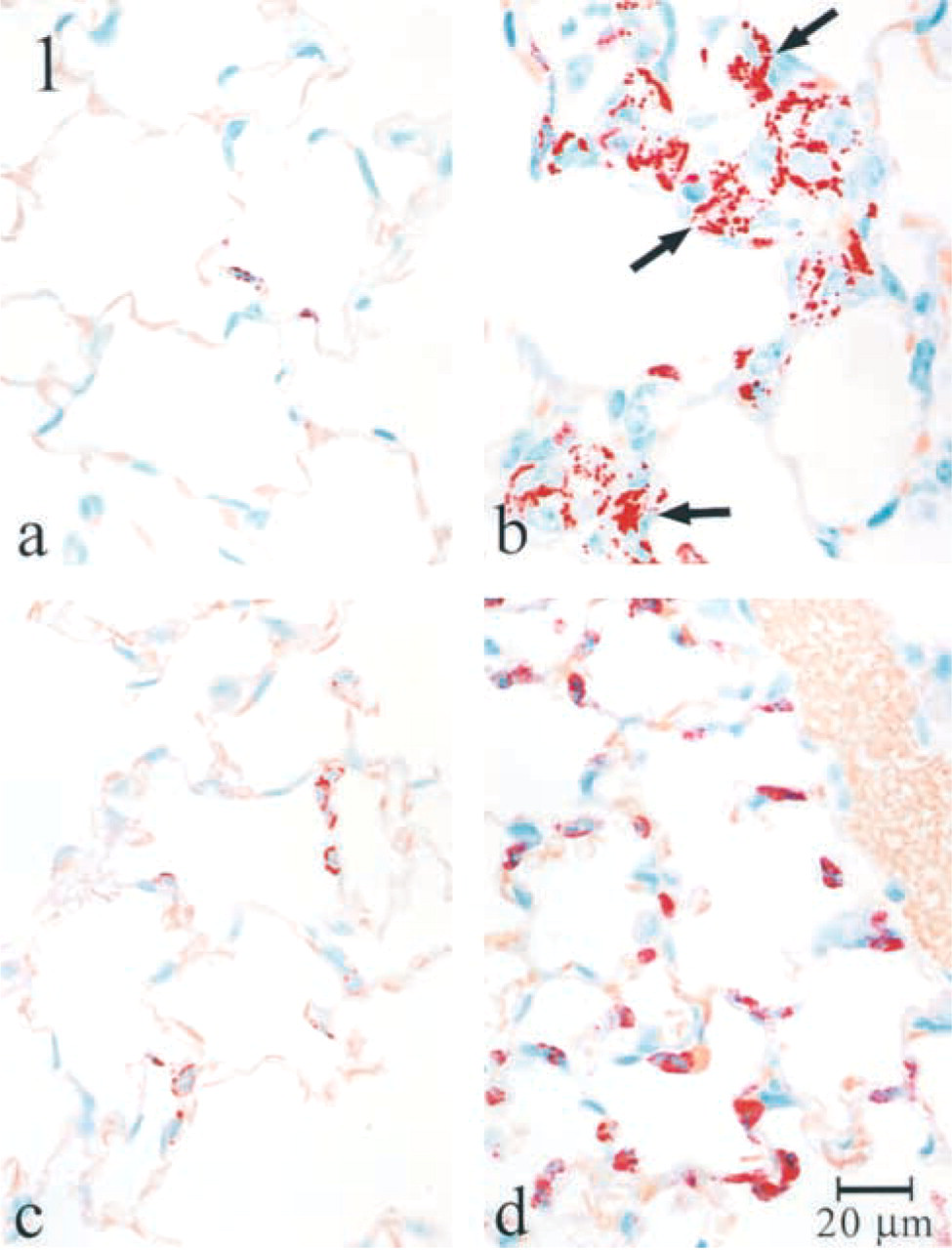
Chloroacetate esterase (CAE)-reacted section of a normal BALB/c lung (
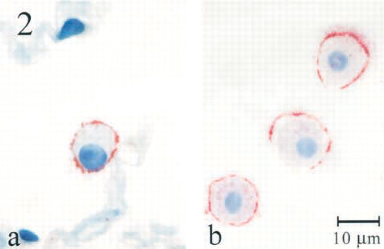
Mouse mast cell protease 1 (mMCP-1) localization by a rabbit anti mMCP-1 IgG peptide, in a section of lung from a V3MC-infused mouse 3 hr after acid instillation. Note that the surface of an alveolar macrophage is coated with this mast cell chymase (
The bronchoalveolar lavage PMN counts in the V3MC animals (1218 ± 165) that underwent acid injury was threefold higher relative to the acid-injured BALB/c animals (403 ± 85) and 30-fold compared to the saline-aspirated V3MC mice (40 ± 4) (n = 3, p < 0.05). Histological grading confirmed the lung injury in accordance with the PI and the BAL neutrophil quantification. Histologic analysis revealed that by Day 14 after injection, large numbers of the granulated mast cells populated the lung in the V3MC-injected mice (Figure 1b). They appeared principally in the alveolar septae. As assessed immunohistochemically, the V3MCs expressed the chymases mMCP-1 and mMCP-2 in their granules. After acid aspiration, some of the mast cells in both the V3MC and BALB/c mice were degranulated and mMCP-1, but not mMCP-2, was found to coat the surface of the surrounding alveolar macrophages in the acid- aspirated lungs (Figure 2a). Macrophages in the bronchoalveolar lavage of acid-injured mice also were coated with the chymase mMCP-1 (Figure 2b) but not mMCP-2.
We utilized a novel model of mastocytosis to define a direct role for mast cells in the pulmonary inflammatory response in acid aspiration injury and also to implicate mast cell protease mMCP-1 as functioning at the alveolar macrophage plasma membrane.