
Abstract
Counterflow centrifugation produces populations of gonadotropes or growth hormone (GH) cells enriched to 90% in a Beckman elutriator. The pituitary populations are first separated by size into three fractions applying different flow rates, stimulated with either gonadotropin-releasing hormone (GnRH) to enlarge the gonadotropes or growth hormone-releasing hormone (GHRH) to enlarge the somatotropes for 3 hr. The fractions are re-eluted, first at the original flow rates and then at higher flow rates to separate enlarged gonadotropes or somatotropes. Most other cell types are reduced to less than 5%. However, co-storage of GH and gonadotropin antigens is seen in either population. Enriched gonadotropes or somatotropes can be used in studies of proliferation, autocrine or paracrine regulation, or ion channel functions.
(
T
Our laboratory developed a method that produced a ninefold enrichment of pituitary corticotropes (Childs et al. 1988). More recently, we have worked on techniques that enrich gonadotropes or somatotropes. Male or female Sprague–Dawley rats are acclimated 7–10 days before use, as approved by committees at both University of Texas Medical Branch and University of Arkansas for Medical Science. During the dispersion, single-cell preparations are obtained (Childs et al. 1988, 1994, 2000). Pituitaries from four to six rats produce 10 million pituitary cells and 1 million somatotropes or gonadotropes.
The Beckman elutriator rotor is assembled with the Sanderson chamber, which allows work with small numbers (10,000–10,000,000) of cells. In the centrifuge, the rotor is attached to tubing and a peristaltic pump that delivers fluid into the chamber at regulated flow rates. Initially, 500 ml of 70% ethanol is run through the rotor at about 10 ml/min for sterilization while the centrifuge is off. Cold sterile water (on ice) is then run through the chamber, removing bubbles, followed by 200 ml Dulbecco's PBS with gentamyacin and bovine serum albumin (BSA). The centrifuge is then closed and run at 1960 rpm. This tests for leaks and bubbles in the system that must be corrected.
Single-cell dispersions are then loaded at 8–10 ml/min as 50 ml is collected (on ice) and the centrifuge is running at 1960 rpm. They are loaded through a syringe connected to the tubing, just before the pump. This allows the cells to settle into the Sanderson chamber, with the largest cells at the bottom and layers of smaller cells towards the top of the pellet. Then the flow rate is increased to collect larger cells sequentially in each of three fractions (Fr) Fr 1, 2, and 3 (50 ml each) are collected at 15, 25, and 35 ml/min, respectively. As Fr 3 is collected, the centrifuge is stopped, excess fluid in the tubing collected, the rotor removed, and chamber contents added. After reassembly, the rotor is prepared for the second elutriation by pumping more PBS through and eliminating bubbles and leaks. It is maintained at 0–4C until re-elutriation.
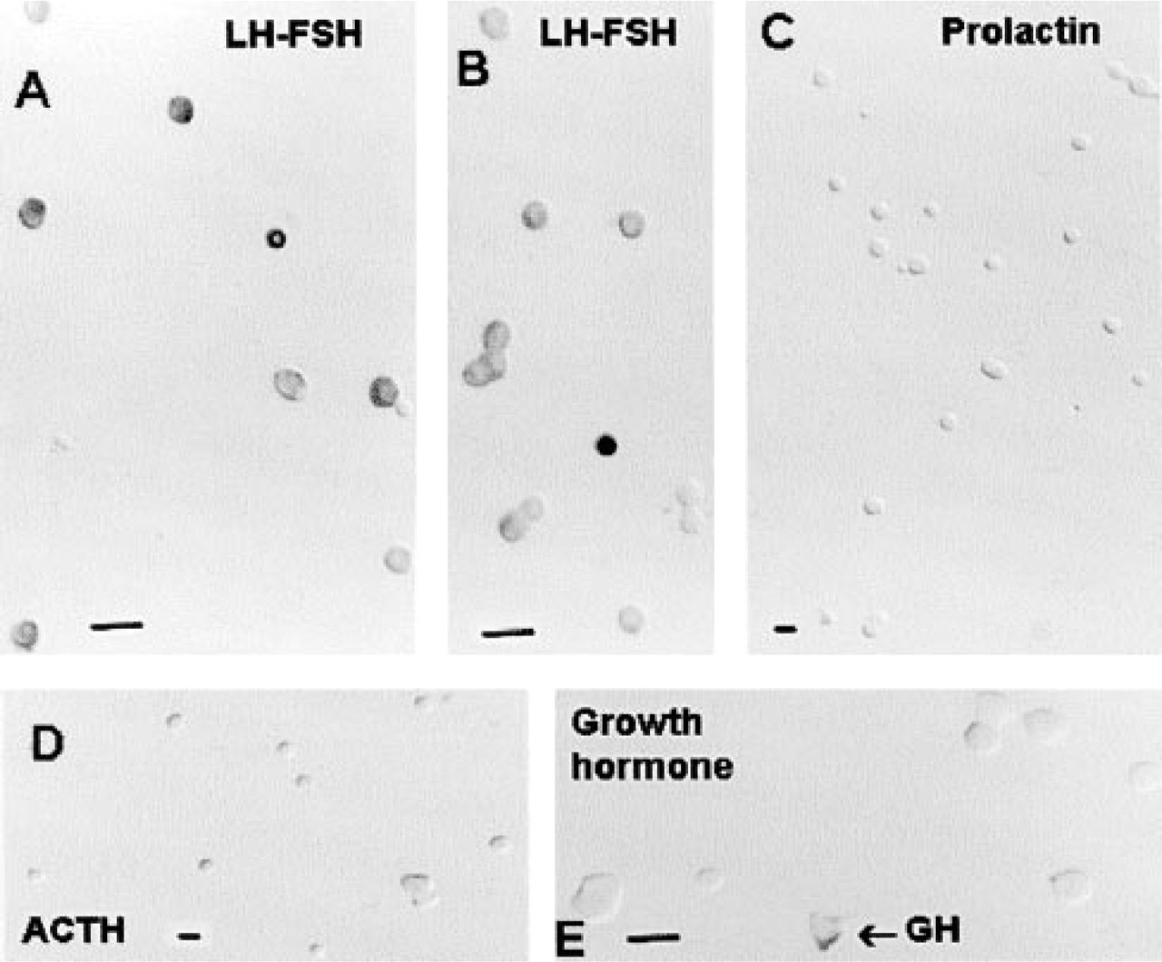
Immunolabeling for LH and FSH in enriched gonadotropes (
The three fractions are spun down at 900 rpm and pellets are resuspended in Dulbecco's modified Eagle's medium (high-glucose DMEM) including 2.5 μg/500 ml HEPES buffer, 0.3% BSA, 5 μg/ml insulin, 30 nM sodium selenite, 50 μg/ml transferrin, and 4.2 μg/ml fibronectin. The load fraction is added to Fr 1. After stimulation with GnRH (for gonadotropes) or GHRH (for somatotropes) for 3 hr at 37C, Fr 1, 2, and 3 are re-eluted separately. Each is loaded at 10 ml/min (50 ml is collected) and re-eluted first at the original flow rate (Fr 1, 15 ml/min; Fr 2, 25 ml/min; Fr 3, 35 ml/min) and then at a new flow rate to collect larger cells (5 ml/min higher for somatotropes or 10 ml/min higher for gonadotropes.)
Samples taken from the original and enlarged fractions were immunolabeled. For example, before re-elutriation, Fr 1, 2, and 3 contained 40 ± 8%, 45 ± 0.2%, and 36 ± 10% GH cells, respectively. After stimulation and re-elutriation, enlarged cells from these same fractions contained 84 ± 4%, 95 ± 0.3%, and 91 ± 2% GH cells, respectively. Pooled fractions from more than five or six somatotrope enrichments contain an average of 90 ± 2% GH cells. Similarly, pooled fractions of enriched gonadotropes contain 92 ± 3% cells with gonadotropins.
Most populations contain 5% other cell types. The storage overlap between GH and gonadotropins persists (Childs et al. 1994, 2000). At least 15% of GH cells contain gonadotropins and 15–30% of the enriched gonadotropes contain GH. Figure 1 shows immunolabeling for LH and FSH in enriched gonadotropes, with the label in various shades of gray. Enriched gonadotropes immunolabeled for prolactin (Figure 1C), ACTH (Figure 1D), or GH (Figure 1E) have few cells.
We run a two-step elutriation process because the smallest gonadotropes or somatotropes do not enlarge sufficiently to be separated from the largest cells in the pituitary. This protocol can separate cells that differ in area by 5–10 μm2. Small cells can easily enlarge by that amount. However they can be separated only if they are first eluted with their small cell counterparts. Although this technique does not give yields suitable for purifications or biochemical assays, it produces a >90% enrichment, uses few animals, and can be completed in one full day. It is useful in studies of proliferation, ion channel activity, RT-PCR, or cell communication.