
Abstract
The research article addresses, a novel natural cellulosic fiber namely Doum Palm Leaf Stalk Fibers (DPLSF) were extracted from Doum palm tree (Chamaerops humilis L.) are rich in cellulose, relatively inexpensive, and readily available in Algeria. The characteristic analysis on morphological, physiochemical, thermal and mechanical proprieties of the extracted raw and treated DPLSF with 20% sodium bicarbonate at various times of treatment were exanimated by optical microscope and scanning electron microscopy (SEM), X-ray diffraction method (XRD), Fourier transform infrared (FTIR) spectroscopy, thermos gravimetric analysis (TGA/DTG), differential scanning calorimetry (DSC). The SEM micrographs of the longitudinal topographic surface of the DPLSF after chemical treatment with sodium bicarbonate at various times treatment indicated that the removed the waxy layer and impurities from surface and formed a roughened surface. XRD analysis further confirmed the treatment’s positive effect on the fibers, with the crystallinity index increasing from 71.43% to 81.03%. However, FTIR analysis showed minimal changes in the peak position and intensity of transmittance. The TGA/DTG results revealed a significant mass loss in treated DPLSF, while thermal stability improved from 290°C to 330°C. Additionally, we analyzed the mechanical tensile properties of the treated fibers and observed that fibers treated at 20% NaHCO3 for 24 h exhibited the highest Young's modulus 8.63 GPa. To validate the experimental findings, we compared individual DPLSF results to a numerical simulation using ABAQUS code. This study underscores the potential of DPLSF as a sustainable and eco-friendly alternative to synthetic fibers, offering immense promise for diverse applications.
Keywords
Introduction
Natural fibers reinforced composites have gained significant attention in recent years as a viable alternative to synthetic fibers, driven by various factors. Natural fibers are renewable, recyclable, widely available, and have low energy consumption. Natural fibers' low densities and high performance have contributed to the development of environmentally-friendly materials, which could be a crucial factor in addressing some of the current ecological and environmental challenges. Natural fibers are mainly composed of protein, hemicellulose, and cellulose and are extracted mainly from plants, vegetables, and animals. Plant fibers are the most commonly used natural fibers as reinforcement in fiber composites.1,2 A variety of plant fibers are extracted from different parts of the plant, such as stems, leaves, seeds, bark, and fruits. For example, fibers such as bananas, jute, alfa, kenaf, bamboo, ramie, broom, flax, and hemp are extracted from stems. Moreover, fibers such as sisal and abaca are extracted from leaves, and fibers such as coir, cotton, and coconut are extracted from the fruit of the plant.3,4 In addition, there are fibers extracted from trunks, such as date palm and Doum palm. Agopyan 5 listed 18 types of natural fibers potentially useful for civil construction. These fibers are commonly used in textile fabrics for clothing and furnishings, but their potential applications go far beyond that. Lignocellulosic fibers, such as agave Americana, flax, hemp, sisal, and jute, are becoming increasingly competitive with synthetic glass fibers, which are commonly used in industry, due to their attractive features. These features include good thermomechanical characteristics, high electrical resistance, good acoustic insulating behavior, higher fracture resistance, relatively high strength and stiffness, and non-irritating properties, making them suitable for the production of various industrial products.6–8 The usage of natural fibers in composite manufacturing has increased recently, particularly in areas like construction, sports equipment, automobiles, aircrafts, naval, household appliances, textile and many more. 9 In recent days, there is an increasing use of green composites in composite manufacturing, researchers have focused on investigating newly discovered natural fibers from the stem of Himalayacalamus falconeri culms, 10 Strelitzia reginae, 11 Zmioculus Zamiifolia stem, 12 manau rattan, 13 Cyrtostachys renda, 14 Derris scandens stem. 15
Although natural fibers have these several advantages, they also have some limitations, such as moisture sensitivity, poor bonding with polymeric matrices, low resistance to high temperatures, and variable fiber properties. To overcome these limitations, suitable fiber surface treatment techniques have been developed, including alkalization (NaOH), silane treatment SiH4, sodium bicarbonate NaHCO3, benzoylation C6H5COCl, peroxide(C6H5CO)2O2, potassium permanganate KMnO4, and stearic acid CH3(CH2)16COOH. 16 In particular, alkaline treatment or mercerization is the least expensive, most efficient, and practical technique for enhancing the moisture resistance of lignocellulosic fibers compared to other chemical modification, 17 by encouraging fibrillation, limiting moisture absorption, and reducing amorphous hemicelluloses and lignin. Surface treatments improved the adhesion between natural fiber and polymer matrix by eliminating pectin and wax from natural fiber.
This research focuses on investigating the fiber extracted from Doum Palm Leaf Stalks (Chamaerops humilis L.). No research work has been carried out on extraction and characterization of the morphological, physiochemical, thermal and mechanical proprieties of DPLSF thus far, to the best of the author’s knowledge. The DPLSFs were analyzed using optical microscope and scanning electron microscopy (SEM), X-ray diffraction method (XRD), Fourier transform infrared (FTIR) spectroscopy, thermos gravimetric analysis (TG/DTG), differential scanning calorimetry (DSC). Additionally, a fiber tensile test was conducted on both untreated and treated with 20% sodium bicarbonate NaHCO3 at various times of treatment. In addition, the experimental results of individual DPLSF were compared and validated through numerical simulation results using ABAQUS/Explicit code.
Material and experimental procedure
Material source
The Doum palm tree, also known as the dwarf palm, is a medicinal plant from the areca family Chamaerops humilis L typically grows to a height of 1.0–1.5 m, and in protected areas it can reach up to 9–10 m.18,19 The Doum plants used in this study were sourced from Cap-Djinet, Boumerdes, Algeria, presented in Figure 1(a). This palm tree is the only one that naturally occurs in Europe and North Africa, and it can be found in the western Mediterranean basin, particularly in Algeria, Tunisia, Morocco, Libya, Italy, Spain, and Sardinia. This plant is highly adaptable and can thrive in a variety of conditions, including cold, drought, and salty environments. It is able to withstand temperatures as low as −12°C and is well-suited for Mediterranean ecosystems.20,21 The process of DPLSF extraction: (a) Doum palm tree; (b) Doum palm rachis and leaflets; (c) cut rachis; (d) section transversal; (e) peeled rachis; (f) rolling process and (e) extracted fibers.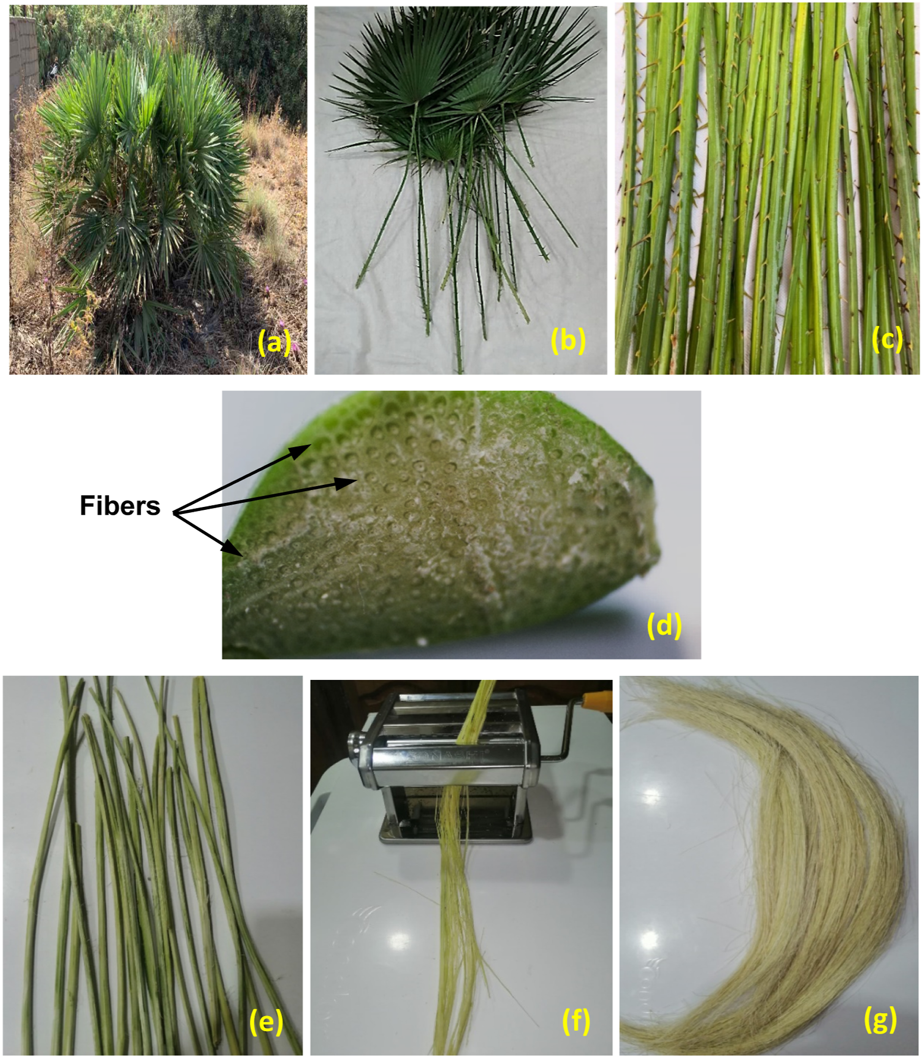
The fibers of the Doum palm tree have traditionally been used to make rope, canvas, pulp, paper and more recently for insulation and mats.19,22 Due to their high mechanical strength, Doum fibers are now being used to reinforce synthetic polymer matrices such as polypropylene, 21 low-density polyethylene, 22 and epoxy resins 23 in industrial applications.
Extraction of fibers
Several studies have been conducted on the extraction of fibers. 24 Three primary methods are used for fiber extraction: mechanical extraction, chemical extraction, and biological processes. 25 The choice of extraction method depends on the intended use of the fiber and the desired morphological, physical, and mechanical properties. Researchers are concerned with finding an economical extraction method that preserves the physicochemical properties of the fibers. The methods used for extracting fibers from Doum palm tree fronds are generally similar to those used for date palm fronds.
In this work, we applied a mechanical extraction method to extract fibers from Doum Palm Leaf Stalk Fibers. The method involves several steps: First, the leaf stalks are cut to a length of 60 cm (Figure 1(b)), tied, and immersed in water for 2 days to allow for microbial degradation after the leaves have been removed (Figure 1(c)). The fibers are then separated by a cold rolling process and grating the leaf stalk pulp to release the fibers (Figure 1(e)). After mechanically rolling the leafstalks (Figure 1(f)), the glued fibers are manually separated by brushing, and the raw fibers are separated into individual fibers with varying diameters and lengths. Finally, after being extracted and washed, the fibers are naturally dried by exposure to solar rays. The number of DPLSF obtained from each palm leaf stalk varies between 50 and 75 (Figure 1(d)), and their length can reach up to 0.50 m (Figure 1(g)). The separated Chamaerops humilis L. components are shown in Figure 1(f). The resultant DPLSFs extracted from the leaf stalks of Doum palm tree is shown in Figure 1(g).
Fiber diameter
It is widely recognized that accurately measuring the diameter of natural fibers can be challenging due to their irregular shape and variable thickness. These characteristics pose issues during tensile testing, particularly when the cross-sectional shape changes along the length of the fiber and when fibers split due to their microfibril structure.
17
In this study, the DPLSFs were found to have a polygonal shape with varying thickness along the length of the fiber. To simplify the analysis, each fiber was considered to be perfectly round, despite this variation. The OXION OX3245 binocular optical microscope, equipped with a computer-assisted camera, was used to measure the different fiber diameters of DPLSF. To account for the variability in fiber dimensions, three measurements were taken, and the average diameter was calculated assuming a quasi-cylindrical shape, as shown in Figure 2(a). The average fiber diameter for 50 samples of DPLSF was found to be 379 ± 48 μm. (a) Typical optical microscopy image of DPLSF for determining fiber diameters; (b) SEM image of the cross section of DPLSF.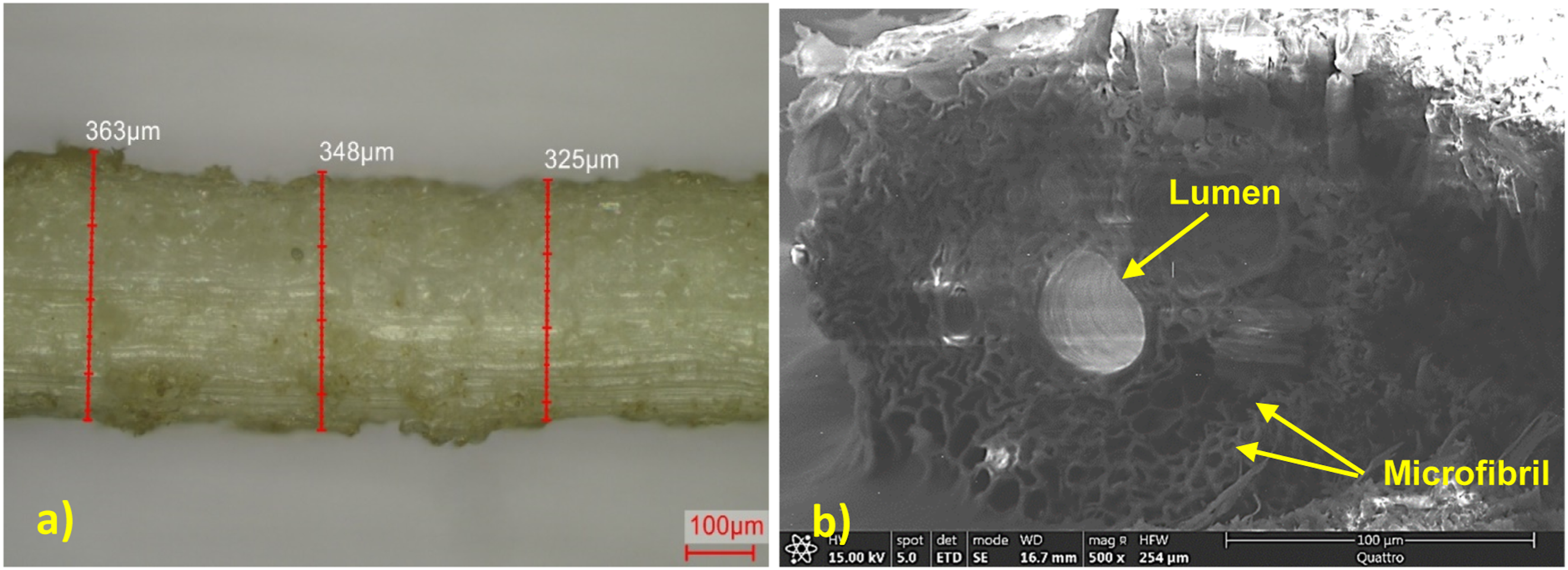
The typical cross-section of DPLSF was examined using a Scanning Electron Microscope (SEM), and is presented in Figure 2(b). The fibers were coated with a fine layer of gold to make them conductive. The cross-sectional shape of DPLSF varies notably from an irregular polygonal shape to a relatively circular one. These fibers are composed of numerous tubulars and hollow microfibrils, featuring a central hole called the lumen, as depicted in Figure 2(b). Remarkably, the cross-section shape of DPLSF closely resembles that of Sponge Gourd fibers.26,27
Treatment of the DPLSF with sodium bicarbonate NaHCO3
To enhance the adhesion between the DPLSF and the matrix, several surface treatments were employed. In this study, sodium bicarbonate was utilized to remove a portion of the lignin, waxes, and fats present on the outer surface of the fiber. Initially, the fibers were cut into 300 mm long pieces, washed with distilled water, and dried in an oven at 45°C until a constant weight was achieved. The DPLSF were then treated with 20% sodium bicarbonate NaHCO3 for varying durations of time (1, 3, 6, 12, and 24 h) at room temperature. Subsequently, the fibers were washed multiple times with fresh water to remove any residual NaHCO3 from the surface, neutralized with dilute acetic acid, and rinsed again with distilled water. Finally, the fibers were air-dried until they reached a constant weight.
Experimental procedure
Fourier transform infrared (FTIR) spectroscopy
The FTIR spectra analysis of the untreated and NaHCO3-treated DPLSF was carried out using an IRAffinity-1S infrared spectrophotometer (Shimadzu Corp., Kyoto, Japan). The scan was performed in the frequency range of 4000 to 500 cm−1 with a resolution of 2 cm−1 and an accumulation of 32 scans per analysis.
Thermogravimetric analysis (TGA/DTG)
The thermal decomposition of the untreated and treated DPLSFs with 20% NaHCO3 at various times of treatment was determined by TGA measurements performed using a thermogravimetric analyzer (Mettler Toledo TGA 2, Greifensee, Switzerland). Samples weighing approximately 8–15 mg were filled into a ceramic alumina crucible capsule and subjected to a constant nitrogen flow rate of 20 mL/min, heating from 30°C to 600°C at a constant rate of 5°C/min to prevent oxidation degradation. The derivative thermogravimetry (DTG) data was obtained from the analysis using STARe-Evaluation Software.
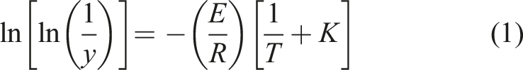
In addition to DTG, another essential parameter for evaluating the thermal stability of DPLSF is kinetic activation energy E, which can be determined using Broido’s equation (1)
28
:
Differential scanning calorimetry (DSC) analysis
Differential scanning calorimetry (DSC) analysis was performed to evaluate the thermal behavior of untreated and NaHCO3-treated DPLSF using a Mettler Toledo DSC instrument (Greifensee, Switzerland). Approximately 8–15 mg of fiber samples from each treatment group were placed in aluminum pans, and the samples were heated at a constant rate of 5°C/min over a temperature range of 25 °C–600°C. The data collected from the DSC analysis provides information about the thermal properties of the fibers, including their melting point and heat capacity
X-ray diffraction (XRD)
XRD analysis was employed to investigate the crystalline structure of untreated and sodium bicarbonate-treated DPLSFs with 20% NaHCO3 for various treatment durations (1, 3, 6, 12, and 24 h). The XRD patterns were collected at room temperature using a D8-Advance Davinci XRD diffractometer from Bruker AXS GmbH (Karlsruhe, Germany), operated at 40 kV and 20 mA, with a scanning rate of 0.3°C/min in the 2θ range of 5–40°.
The percentage of crystallinity (%Cr) was calculated using equation (3),
29
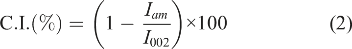
with the Crystallinity Index (C.I.) determined using Segal's empirical method equation (2)
29
:
The calculation was based on the maximum intensity of the (002) lattice diffraction peak I002 at a 2θ angle of approximately 22.80° and the minimum intensity of an amorphous area Iam at a 2θ angle of around 16–18°, following the method described by Liu et al.
30
and Mudoi et al.
31
The crystallite size (Crsize) of the DPLSF samples was also calculated using Scherrer's equation (4)
32
:

Results and discussions
Scanning electron microscope (SEM) analysis
The scanning electron microscopy (SEM) technique is an excellent tool for examining the longitudinal surface morphology of the untreated and treated DPLSFs with 20% of sodium bicarbonate at various times of treatment. Also, the examining the surface morphology of fibers is essential to evaluate their potential as effective reinforcement and their resistance to fiber pullout.
The longitudinal topographic surface of the fibers before and after chemical treatment with sodium bicarbonate at various times is presented in Figure 3 with some NaHCO3 residue present. Figure 3(a) presents the SEM micrograph of the longitudinal untreated fibre surface. The wax, lignin and other considerable impurities were present on the untreated fibre surface, as can clearly be observed. These impurities are known to cause poor fiber-matrix interface adhesion.
33
SEM images: (a) untreated DPLSF; NaHCO3-treated DPLSF (b) 1 h; (c) 3 h; (d) 6 h and (e) 12 h.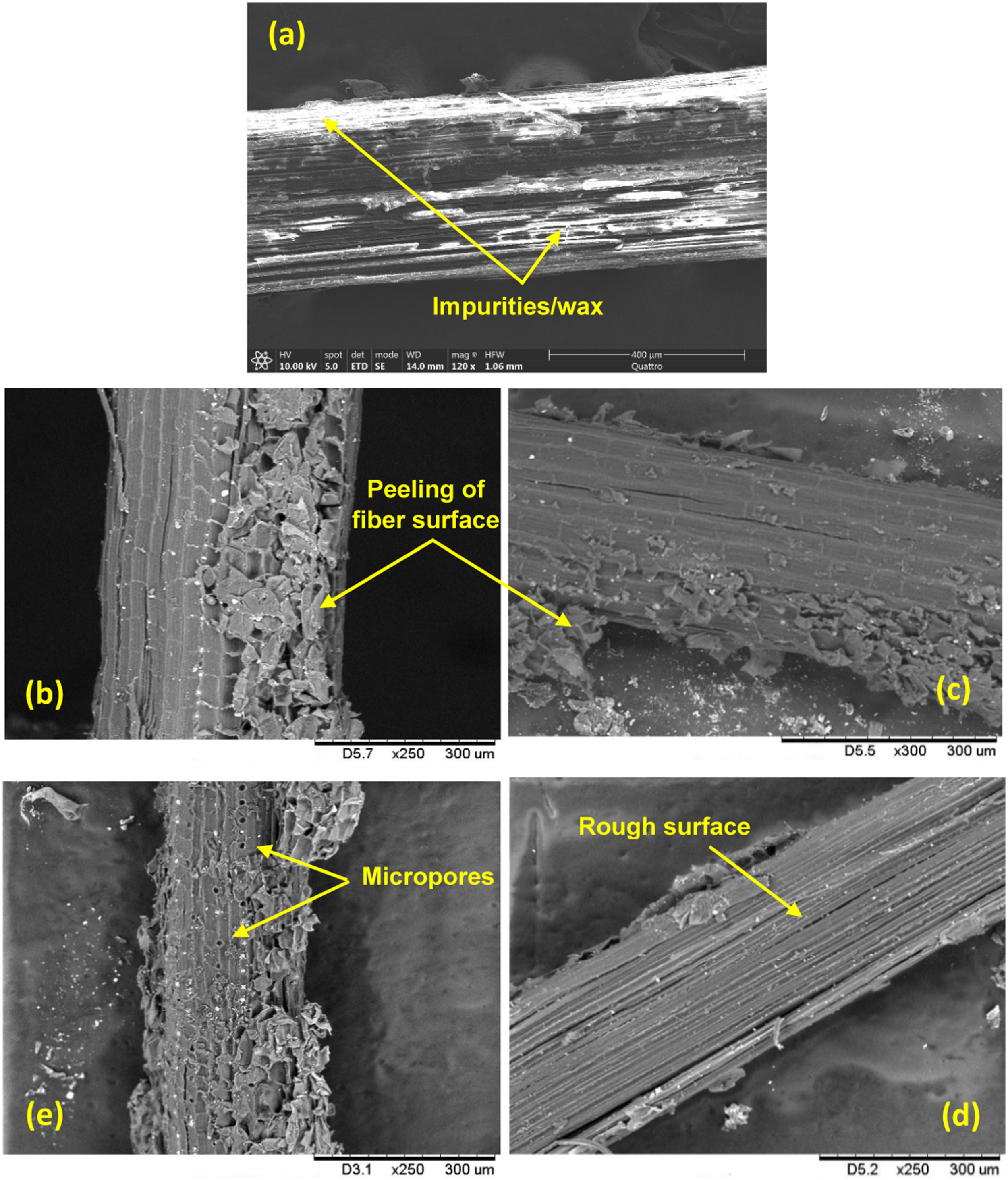
Figure 3(b)–(d) shows the longitudinal surface micrographs of fibers treated at various times of treatment (1, 3, 6, 12, and 24 h), respectively. It can be apparently seen that the surfaces of the treated fibers are much cleaner, rougher and porous with peeling of the fiber surface, due to the increase in the times of treatment. Notably, the samples treated for 6 h with NaHCO3 exhibit an abundance of micropores on their surfaces. These micropores may lead to a reduction in the tensile strength of the fibers. Whereas, the fibers treated for 12 h of display a few micropores on their surfaces, which could explain the observed improvement in tensile strength. Also, the analysis showed that the sodium bicarbonate treatment of DPLSFs at different treatment times improved the fibers surface roughness, enhancing their wettability and promoting good adhesion between the fiber and matrix in polymer composites. 34 Therefore, the SEM analysis suggests that the chemical treatment of DPLSFs with sodium bicarbonate has a positive impact on the fibers surface morphology, making them suitable for reinforcing polymer composites.
Chemical analysis
X-ray diffraction analysis
Figure 4(a) shows the XRD spectra for untreated and NaHCO3-treated DPLSFs at various times of treatment, with diffraction angles (2θ) ranging from 5 to 40°. The X-ray diffraction patterns of both fibers exhibited three peaks, which were assigned to the cellulose I plane (200), lattice plane (110), and cellulose I
β
plane (004), with the highest peak at 2θ = 22.29°, an average intensity peak at 2θ = 16.23°, and a low-intensity peak at 2θ = 34.68°. This is a common finding in natural lignocellulosic sources and their derivatives.16,30 (a) X-ray diffraction analyzes and (b) FTIR spectra of untreated and NaHCO3-treated DPLSF.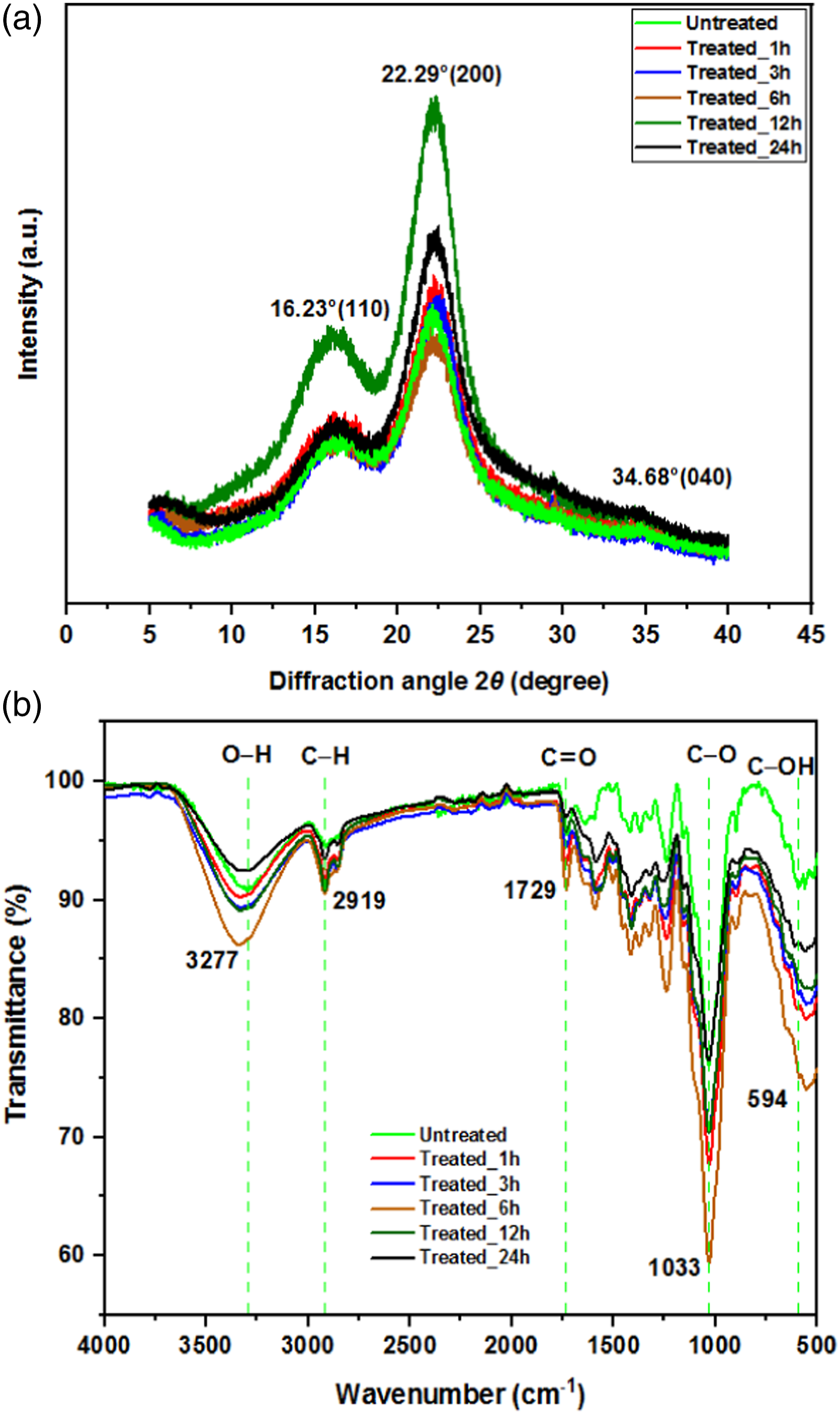
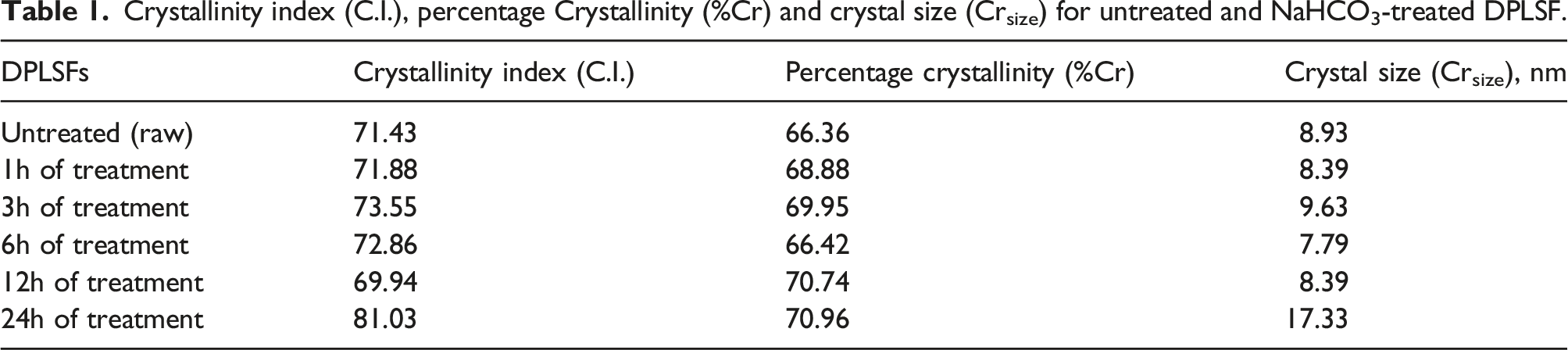
Crystallinity index (C.I.), percentage Crystallinity (%Cr) and crystal size (Crsize) for untreated and NaHCO3-treated DPLSF.
Infrared analysis of DPLSF (FTIR)
Peak positions and the corresponding function group with the chemical composition of untreated and NaHCO3-treated DPLSF.
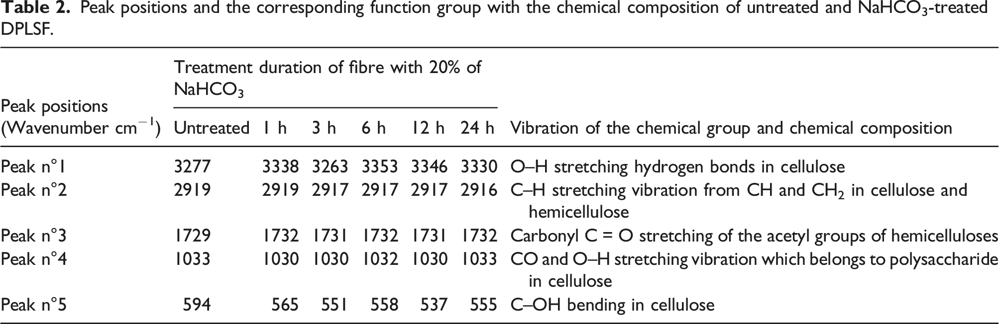
In the fingerprint region (500–1800 cm−1), the majority of the peaks were sharp, while in the functional group region (1800–4000 cm−1), common peaks were broader than others. Upon analysis of Figure 4(b), it can be observed that the broad and strong band around 3353–3277 cm−1 was from the hydroxyl O–H groups due to moisture in the fibers, as reported by Javier-Asteter et al. 38 The two small sharp peaks that appeared at 2919 cm−1 (symmetric) and 2855 cm−1 (antisymmetric) in Figure 4(b) are attributed to the C–H stretching vibration of CH and CH2 in cellulose and hemicellulose, according to Guo et al., 39 and Liu et al. 40 The absorption band in the region about 1729 cm−1 is attributed to the carbonyl group (C = O) stretching. The reduction in the absorption of the carbonyl region could be due to the removal of hemicelluloses during alkaline treatment, as reported by Liu et al., 40 and Chen et al. 41
Furthermore, the strong peak at 1033 cm−1 is ascribed to the C–O and O–H stretching vibration pertaining to polysaccharide in cellulose, as noted by Makhlouf et al. 42 The small peak at 594 cm−1 is associated with C–OH out of the plane bending in cellulose, according to Fiore et al. 34
In conclusion, the chemical treatment of DPLSFs with 20% of sodium bicarbonate NaHCO3 at various times of treatment had a minimal effect on peak position and intensity, with only slight variations in intensity, consistent with similar observations reported by Amroune et al. 43
Thermal analysis
Thermal degradation by TGA/DTG
The use of natural fibers as reinforcement in biocomposites is limited by their low thermal stability, leading to thermal decomposition or combustion under high-intensity heat sources.
34
This issue significantly hampers their applicability in high-temperature enviromrnts. To tachle this challenge, our study aimed to enhance the thermal stability of DPLSF by conducting thermogravimetric analysis. Through thermogravimetric analysis, we examined both untreated and NaHCO3-treated DPLSF, plotting the thermogravimetric analysis (TGA) and derivative thermogravimetry (DTG) as a function of increasing temperature while constant heating rate of 5 C/min, as shown in Figure 5. Comparison of the TG/DTG curves at a heating rate of 5°C/min with: (a) untreated DPLSF and treated DPLSFs with 20% NaHCO3 for: (b) 1 h; (c) 3 h; (d) 6 h; (e) 12 h and (f) 24 h.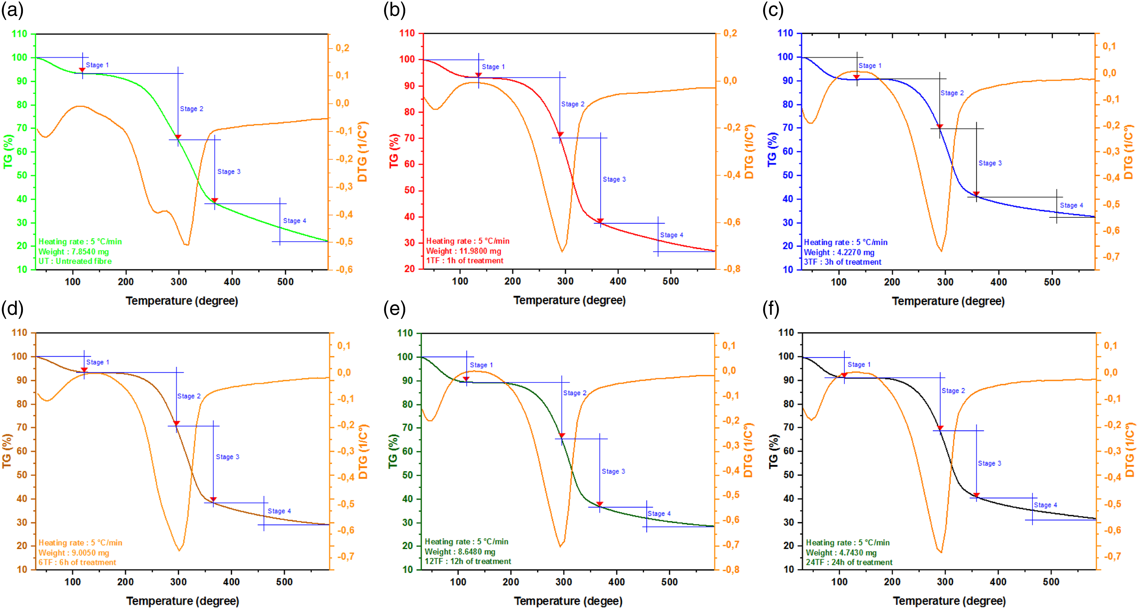
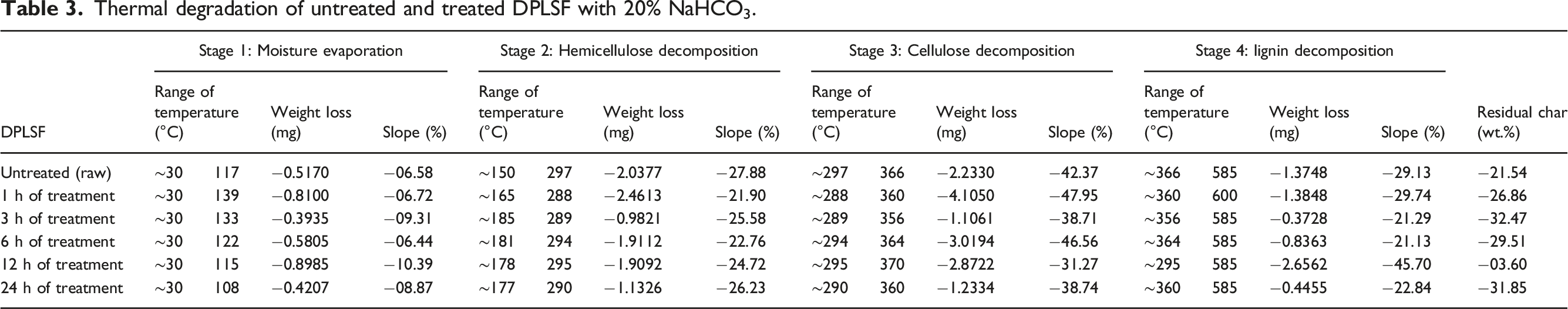
The TGA results revealed four stages of weight loss for both untreated and NaHCO3-treated DPLSFs: moisture evaporation, hemicellulose decomposition, cellulose degradation, and lignin decomposition. The first stage, which resulted in a small weight loss, was due to the elimination of absorbed moisture from the fiber and occurred within the temperature range of approximately 30°C–120°C.34,44 The thermal stability of DPLSF was noted up to about 180°C, where no significant peak was observed in the DTG curve. The second mass loss process, which represented the degradation initiation of the DPLSF, occurred in the temperature range of 180 °C–290°C and was attributed to the thermal decomposition of hemicelluloses and glycosidic linkages of cellulose. The third region, which appeared between 290 and 360°C, was related to the degradation of cellulose. Lignin degradation was expected to take place at temperatures above 360°C, but its degradation could take place over a wide temperature range due to the nature of lignin. Broido’s plot for untreated and NaHCO3-treated DPLSFs.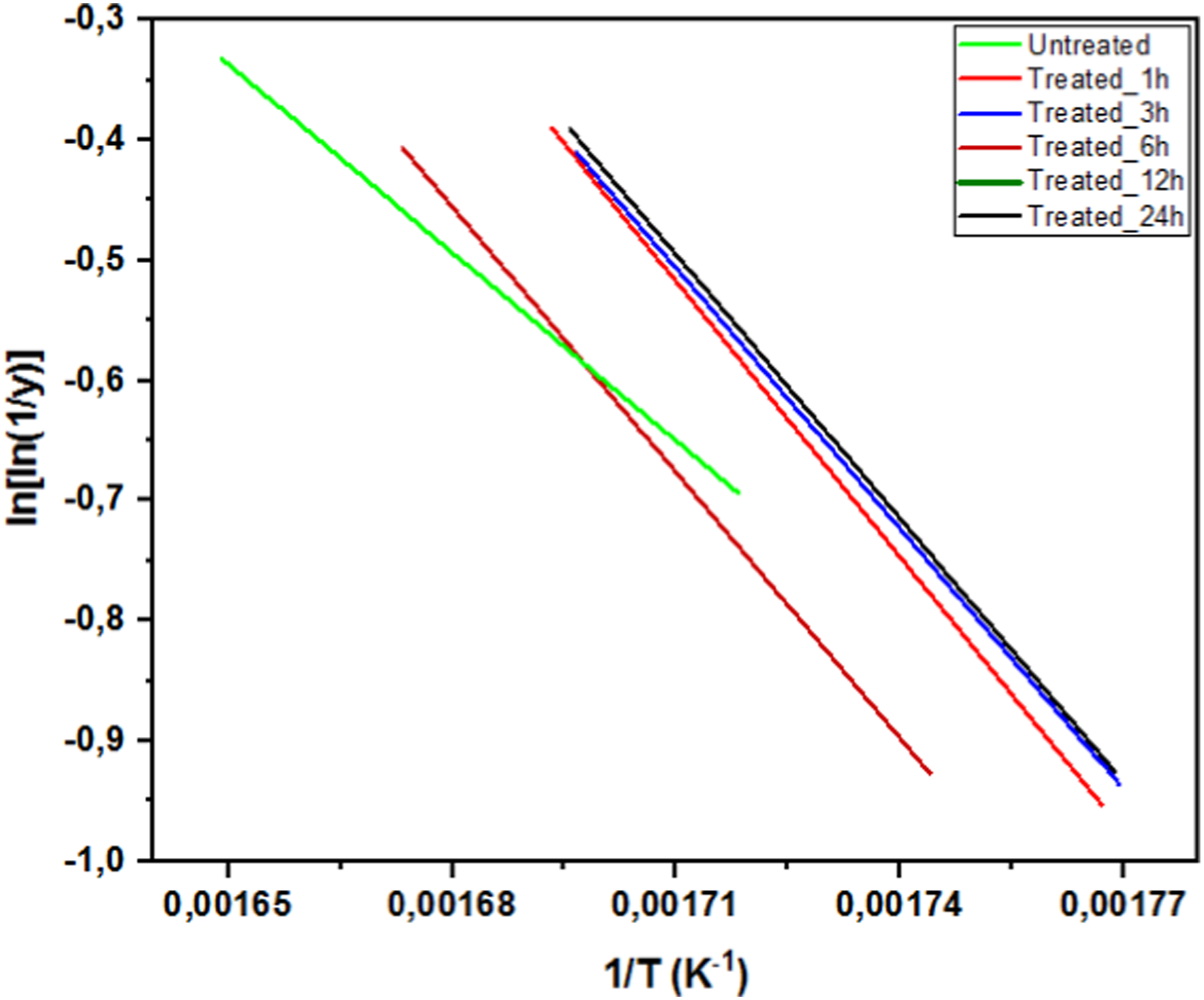
Thermal degradation of untreated and treated DPLSF with 20% NaHCO3.
The activation energy (E) holds pivotal significance in evaluating of reinforcement for high-temperature applications. The calculation of activation energy was derived from the slope of the ln [ln (1/y)] vs (1/T) plot, illustrated in Figure 6. For untreated fibers, the activation energy is 44.93 kJ/mol. Meanwhile, for the fibers subjected to treatment duration of 1, 3, 6, 12 and 24 h, the corresponding activation energies are 63.77, 60.53, 61.91, 61.36, and 61.22 kJ/mol, respectively. Evidently, these values exhibit an enhancement in activation energies following treatement, aligning well with findings reportes for other cellulose fibers described in the literature. 45 Notably, these values fall within the specified range (60–170 kJ/mol) characteristic of natural fibers. This alignment signifies the improves suitability of NaHCO3-treated DPLSFs for applications demanding high-temperature, such as in polymer composite manufacturing.
DSC analysis of DPLSF
The thermal behavior of DPLSF was further studied to investigate the effect of water content on their overall properties, given that the presence of water can adversely affect the performance of the polymer composites in which these fibers are used as reinforcement. Differential scanning calorimetry (DSC) measurements were carried out to analyze the thermal behavior of the untreated and NaHCO3-treated DPLSF under different treatment times. Figure 7 displays the typical DSC curves obtained for these fibers. The spectra for all fibers exhibit a broad exothermic peak between room temperature and 130°C, which shifts to 70°C for both untreated and NaHCO3-treated fibers. This peak corresponds to water loss or evaporation, consistent with previous studies.
46
The temperature range between 120 and 230°C shows negligible exothermic or endothermic changes, indicating that the fibers remain thermally stable within this range. The first endothermic peak observed at about 280°C for untreated fibers and at 320, 270, 292, 308, and 270°C for NaHCO3-treated fibers at different treatment times is attributed to hemicellulose decomposition. The second endothermic peak in the range of 330°C–470°C appears as a large exothermic peak and represents cellulose degradation, which is in agreement with previous studies.
47
These results provide valuable insights into the thermal behavior of DPLSF and their suitability for use as reinforcement in polymer composites. DSC analysis of the: (a) untreated DPLSF and treated DPLSFs with 20% NaHCO3 for: (b) 1 h; (c) 3 h; (d) 6 h; (e) 12 h and (f) 24 h.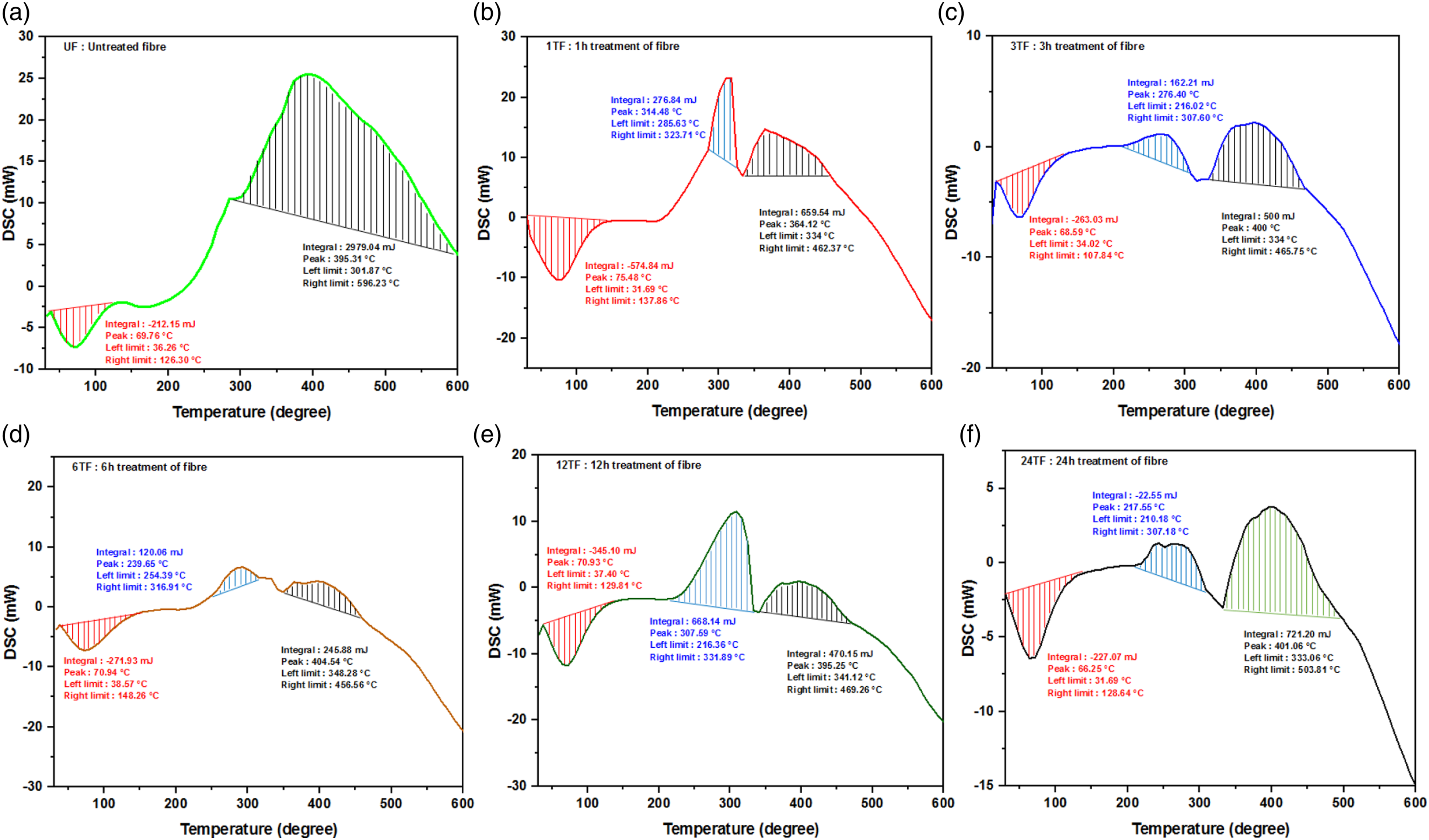
Mechanical tensile properties
The tensile properties of elementary DPLSFs were determined in accordance with ASTM D3379-75 standard using a Zwick/Roell Z2.5 (Zwick Roell, Ulm, Germany) tensile testing machine equipped with a 20 N load cell. The fibers underwent testing under standard conditions of 20°C and 60% relative humidity. Each fiber was mounted into the machine grips individually, using sandpaper. The tests were conducted using a crosshead speed of 1 mm/min until rupture occurred. The diameters and cross-sectional areas of the fibers were measured and recorded prior to testing, and their lengths were measured at a gauge length (GL) of 50 mm. Figure 8(a) presents the tensile behavior of untreated DPLSF, which show a quasi-linear variation of stress with increasing strains until a maximum value corresponding to a limit failure. Notably, some DPLSF exhibit a staircase form of stress-strain curves (Figure 8(b)), which is characterized by a sharp drop in stress without complete failure of the sample, and can occur once or several times during the load history before failure. This behavior has been described in the literature (e.g. Silva et al.
48
) as resulting from the collapse of the weak walls of primary cells in the natural fiber structure, as well as from delamination between fibrils. Stress-strain behavior of DPLSF tested.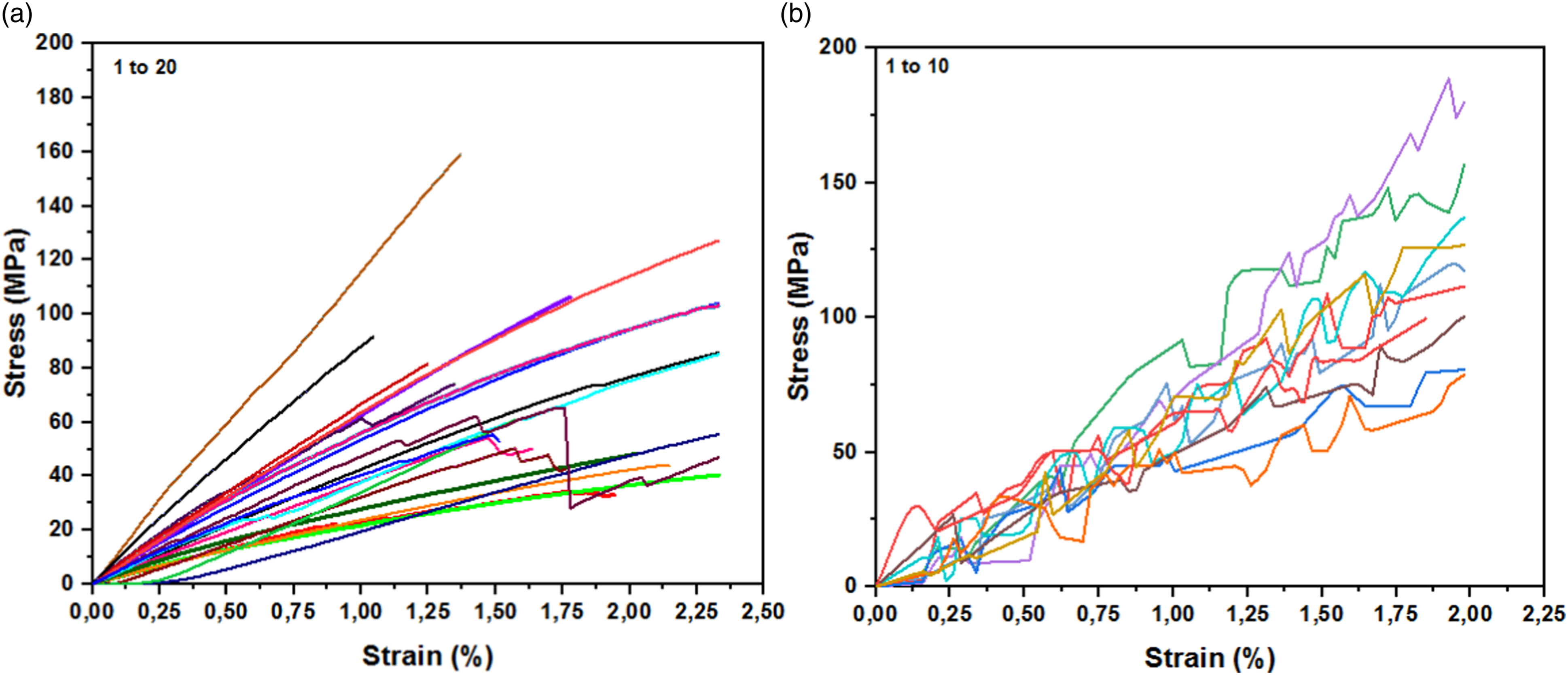
The comparison values physical, mechanical, crystallographic and thermal properties obtained in this work with recently explored natural fibers.
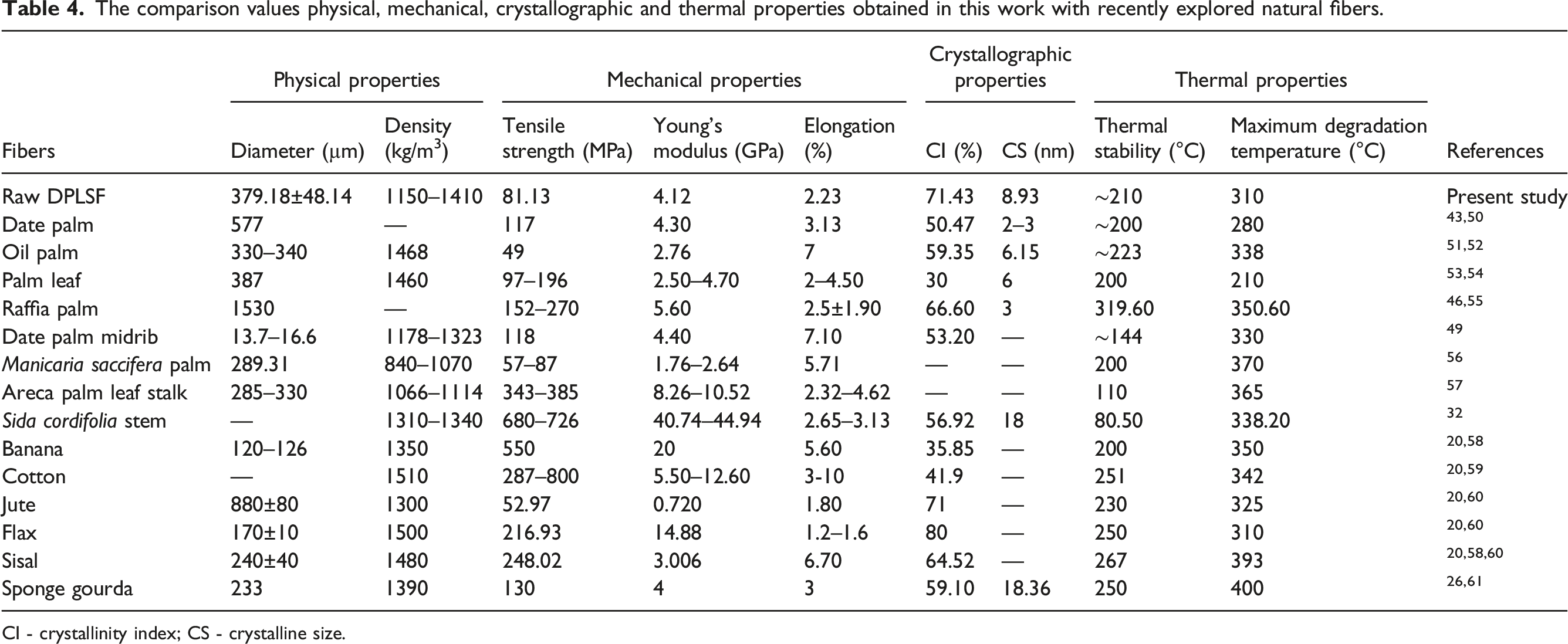
CI - crystallinity index; CS - crystalline size.
Figure 9(a)–(b) display Young’s modulus and ultimate tensile strength as a function of diameter for DPLSF extracted in this study. As expected for natural fibers, there is a significant dispersion of results, which can be attributed to various factors, including test parameters and conditions, area measurements, and plant characteristics (e.g., source and age of the plant, fiber extraction processes, location of the fiber in the plant, and the presence of defects).
30
Therefore, a statistical approach and numerical simulation of fibers are required to accurately evaluate their mechanical properties. The effect of fiber diameter on the tensile properties of DPLSF.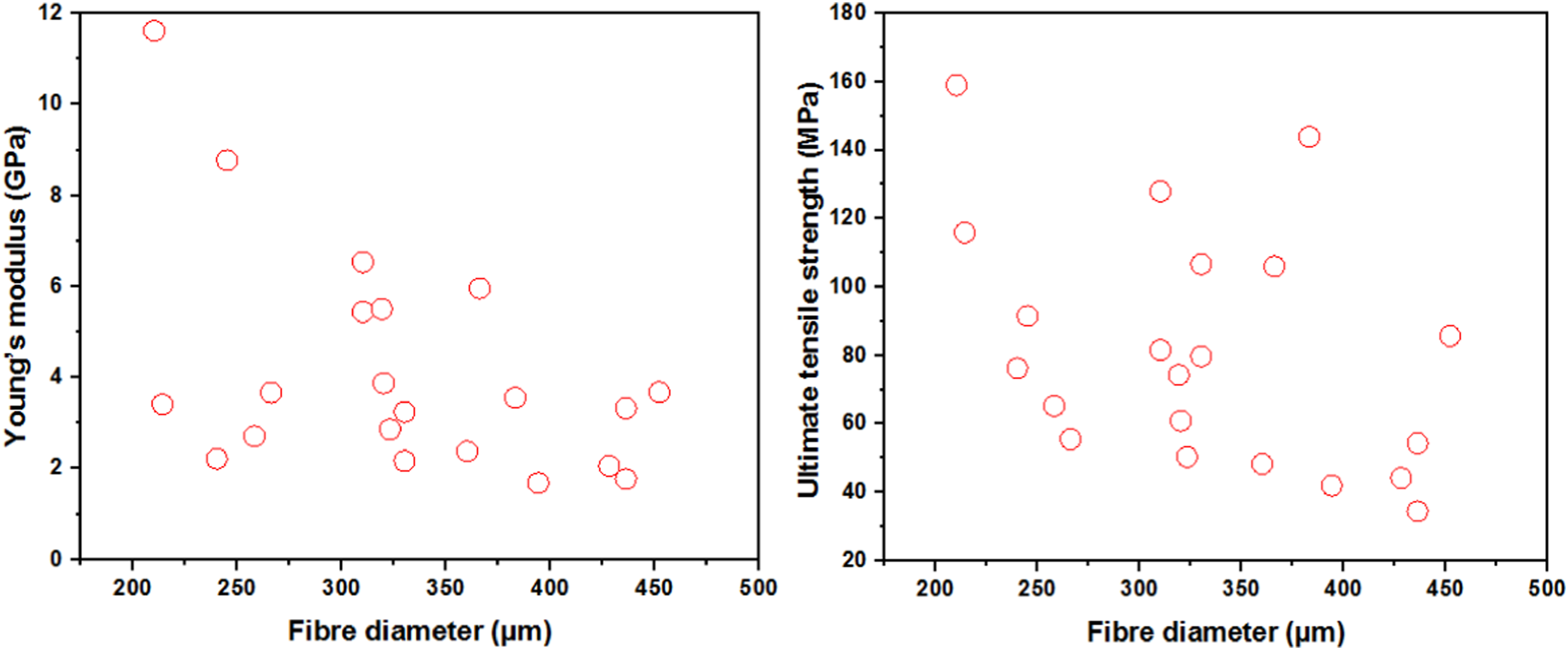
The mechanical properties of DPLSF before and after treatment.
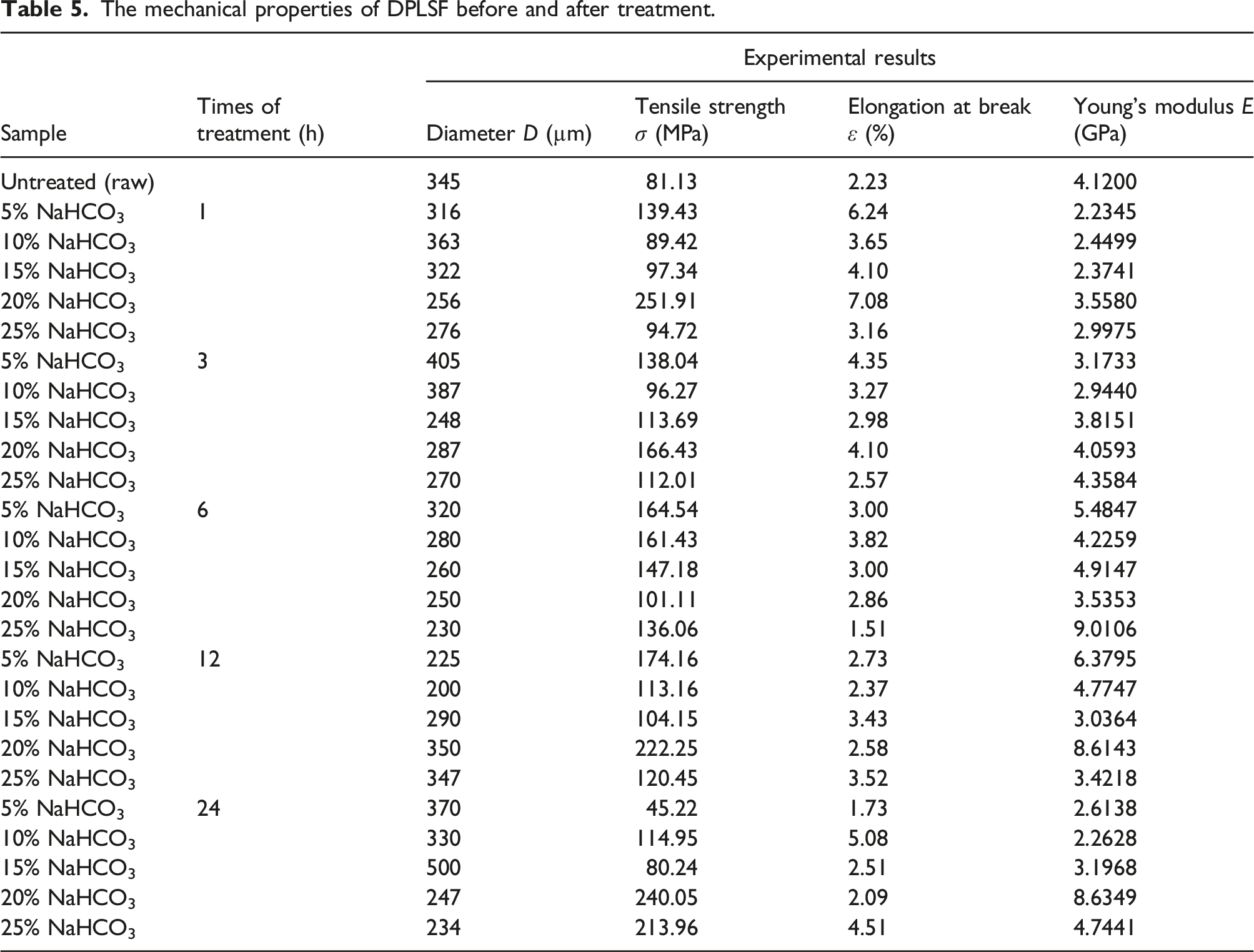
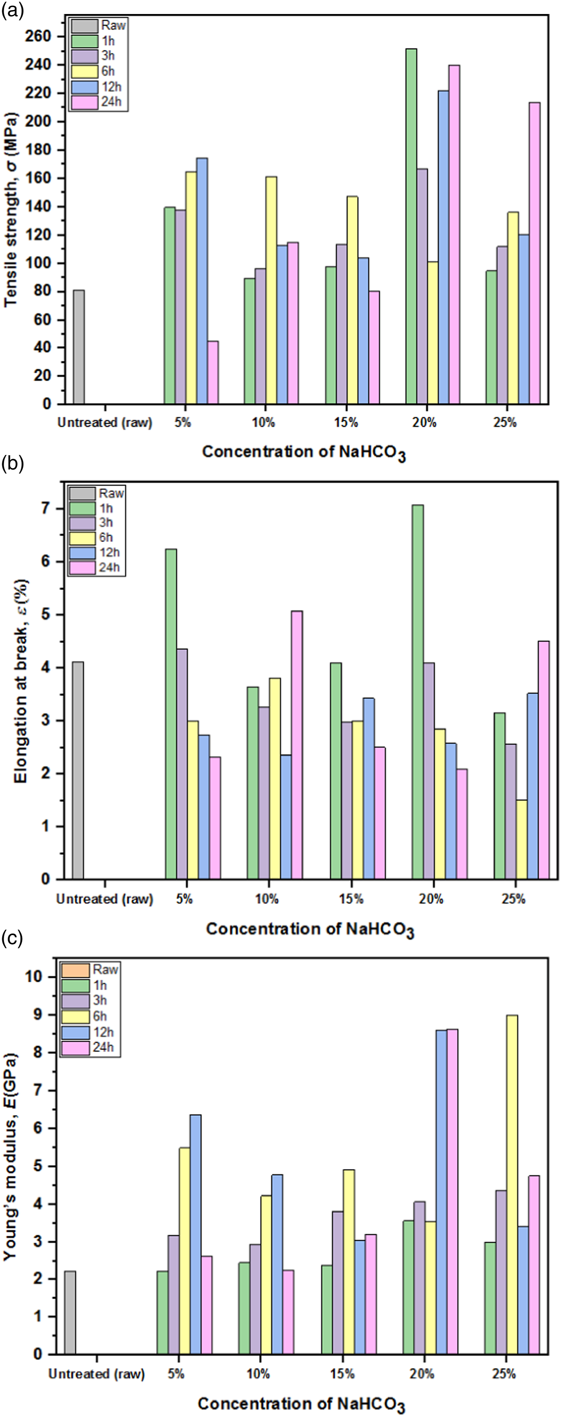
Effect of concentration of NaHCO3 and variation of treatment times on (a) tensile strength, (b) elongation at break and (c) Young’s modulus of DPLSF.
Numerical analysis
Numerical investigations in the present study are performed in order to validate the results found by experimental tensile test, numerical models based on finite elements method were developed by using commercial finite element code ABAQUS/Explicit. This simulation was applied to two models of raw DPLSF, the first is real was obtained from a picture taken by the SEM (Figure 11(a)) and is characterized by an irregular structure. The second model of signal DPLSF was proposed by a regular structure of classical honeycomb models (Figure 11(b)). A schematic RVE (Representative Volume Element) of the unit cell for the fibril lattice is shown in Figure 12(a), and a single fibril with 10 mm of gauge length is represented in Figure 12(b). The models were developed using 3D structural elements (C3D10 quadratic tetrahedron) defined by 10 nodes and three translational degrees at each node. The complete meshing of the model and 3D tetrahedron element is shown in Figure 13. The FE simulation is carried out with an implicit scheme and modeled as a load/displacement problem where the elastic tensile load or the observed total displacement until fracture is applied along the longitudinal axis (length of the specimen). In this study, to obtain the tensile Young’s modulus, we made a simulation with the following boundary conditions: surface A is loaded with a uniform elastic displacement condition: u1 = imposed, u2 = u3 uR1 = uR2 = uR3 = 0 and the opposite surface is set as fixed boundary condition by constraining all six degree of freedoms; u1 = u2 = u3 = uR1 = uR2 = uR3 = 0. The mechanical properties of lignin (Young’s modulus of 4.8 GPa and Poisson’s ratio of 0.23) have been chosen as the core material for the lattice ribs. Sections cross of the DPLSF (a) real section; (b) homogenized classical honeycomb. (a) Unit cell of the lattice hexagonal configuration for the Doum fibrils; (b) a single cellular lattice tube with 10 mm of gauge length. Loading and boundary conditions for: (a) real model; (b) classical honeycomb model.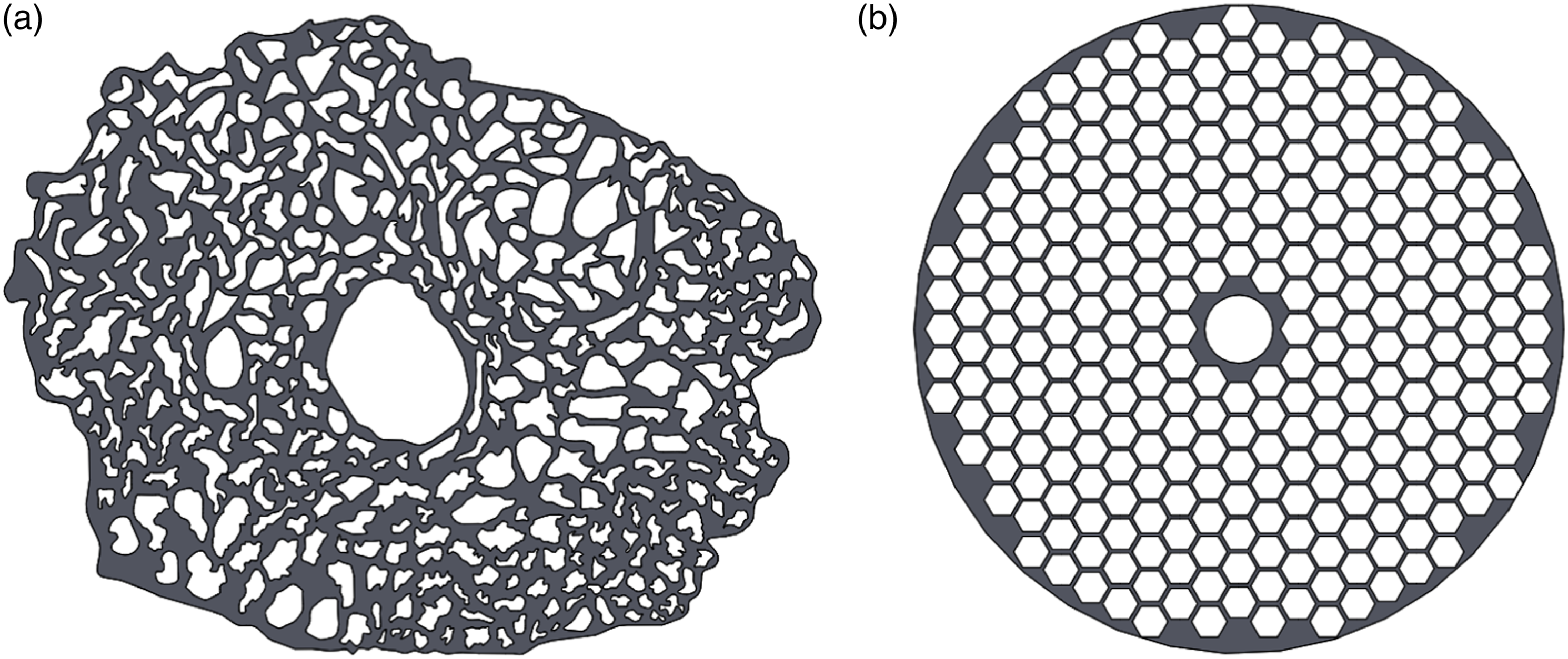
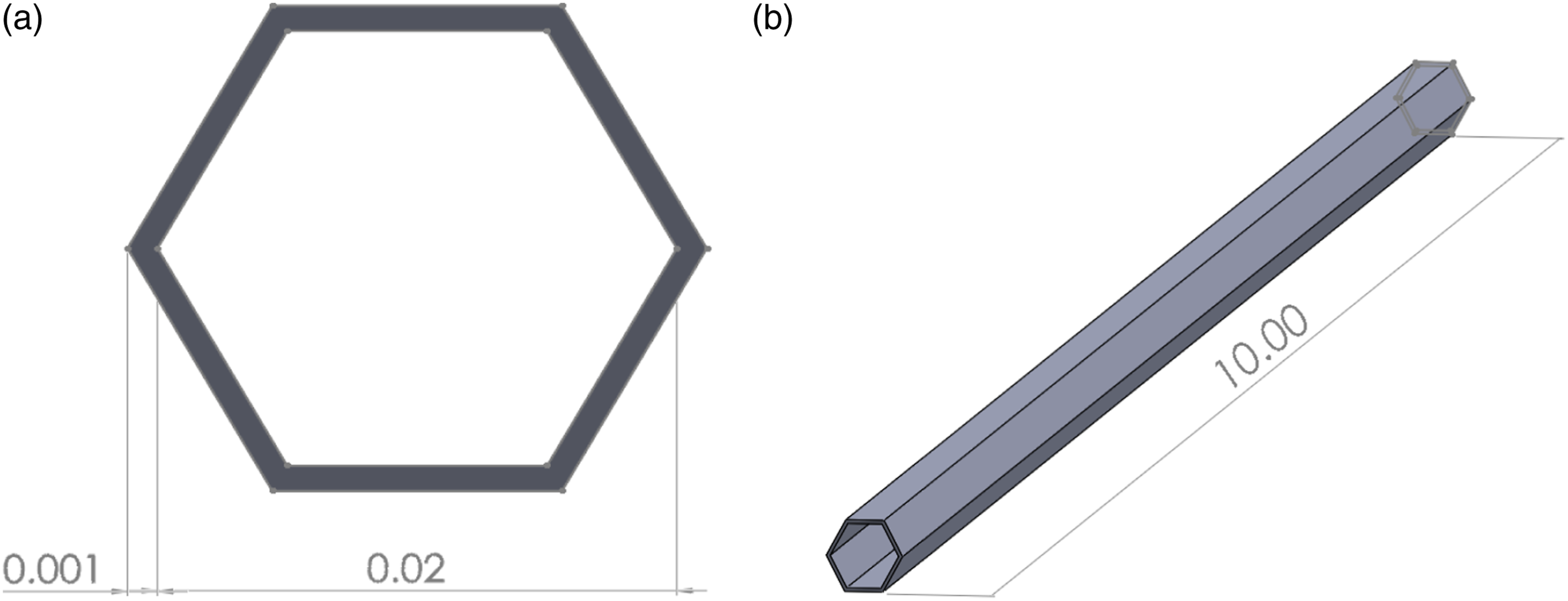
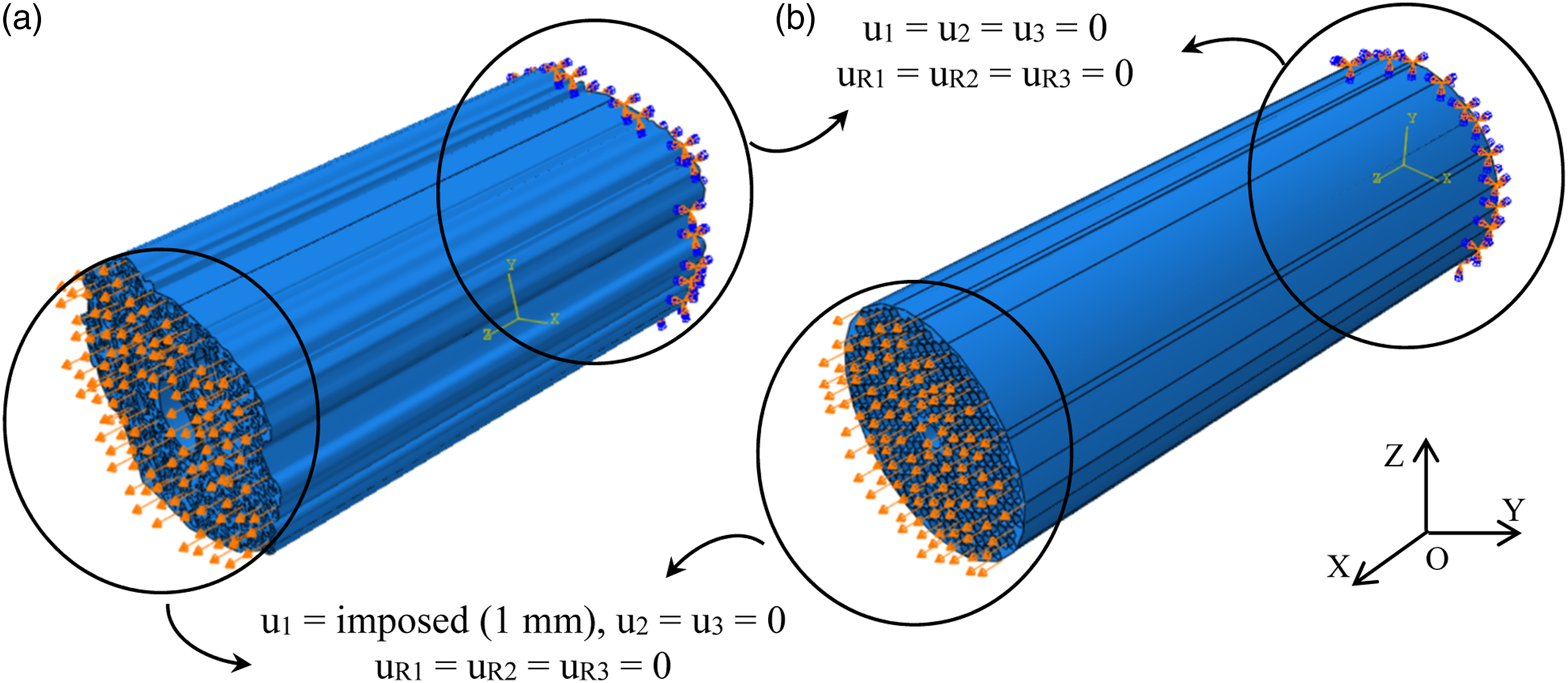
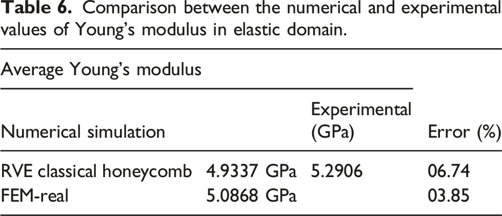
Figure 14 shows the stress-strain plot obtained from the numerical simulation for both sections (FEM-real and RVE classical honeycomb) were compared with the experimental test data. The comparison of numerical simulation obtained for both sections and experimental results of a single DPLSF show that a nearly perfect agreement has been achieved between them in the elastic domain. Table 6 shows the relative error between the Young’s modulus as measured experimentally and by numerical simulation for both sections. The average relative error is below 7% for most of the cases. Comparison between experimental and FE results for tensile loading vs. elastic strain. Comparison between the numerical and experimental values of Young’s modulus in elastic domain.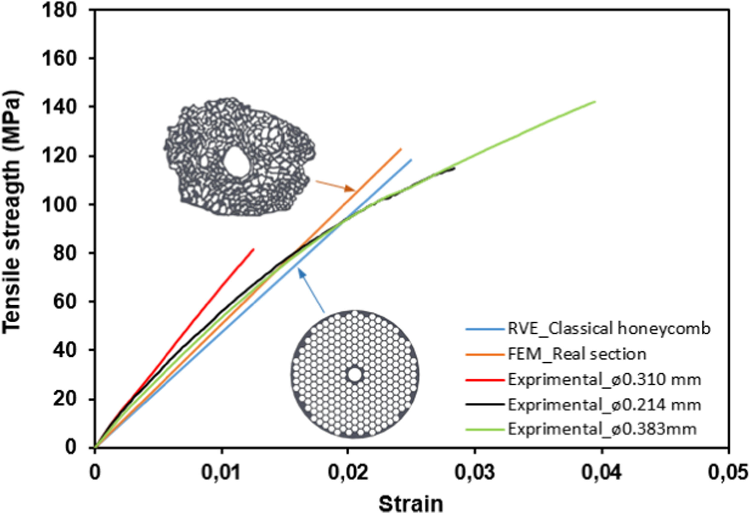
Conclusion
This study focused on the extraction and characterization of a novel class of natural fiber obtained from Doum Palm Leaf Stalks DPLS to evaluate their potential use as reinforcement in polymer matrix composites. Morphological, physiochemical, thermal, and mechanical analyses were performed, and the results are reported in this paper. The following conclusions can be drawn from this work: • Scanning electron microscopy (SEM) images showed that increasing the treatment time removed the hemicellulose, lignin, wax, and other the impurities from the fiber surface and made it rough, resulting in better mechanical bonding between the fiber and polymer matrix in composites. • The raw DPLSF had an average diameter of 379.18 ± 48.14 μm, and exhibited tensile strength, tensile modulus, and elongation of 81.13 MPa, 4.12 GPa, and 2.23%, respectively. • XRD analysis showed that the maximum crystallinity index (C.I.), percentage crystallinity (%Cr), and crystal size (Crsize) values of 81.03%, 70.96%, and 17.33 nm, respectively, were observed for a treatment period of 24 h at 20 wt% concentrations of sodium bicarbonate. • FTIR analysis confirmed the presence of chemical groups (O–H), (C–H), (C = O), (C–O), and (C–OH) in the DPLSF, similar to other lignocellulosic fibers, and also confirmed the partial removal of hemicellulose, lignin, wax, and other impurities during chemical treatments. The position of the peaks showed a slight variation in intensity with increased treatment time. • TG analysis confirmed that the thermal stability of the untreated and NaHCO3-treated DPLSF was improved (from 290°C to 330°C), indicating that these fibers can be used in industrial applications where higher temperatures are involved. • The overall morphological, physiochemical, thermal, and mechanical properties of the extracted fibers are comparable to those of other natural fibers such as date palm fiber, palm leaf fiber, and Raffia palm fiber. The DPLSF could find applications in making ropes, carpet backing, cordages, mats, and as candidates for reinforcement in gypsum and polymer matrix composites.
Footnotes
Declaration of conflicting interests
The author(s) declared no potential conflicts of interest with respect to the research, authorship, and/or publication of this article.
Funding
The author(s) disclosed receipt of the following financial support for the research, authorship, and/or publication of this article: This work was supported by the Ministry of Higher Education and Scientific Research.
Data availability statement
Data sharing not applicable to this article as no datasets were generated or analyzed during the current study.