
Abstract
The analysis of the Hip and Knee (HK) joint angles during single-leg stance (SLS) activity contributes to a great understanding of the bio-mechanical mechanisms and balance maintenance across different age groups. Comprehending how these joints operate in the dynamic state is critical for identifying age-related changes in joint control and stability which can contribute to reducing the risk of falls and improving mobility among people. The possibility of revealing the health of HK joints without resorting to an MRI scan is the significance of this work. By obtaining the data on joint angles during the SLS test, we succeeded in identifying those with a higher risk of HK issues and thus the possibility of early intervention and treatment. Our proposed work is going to use a pose estimation technique that will track the trajectories of HK angles followed by association with instabilities or compromised balance in this way, we can detect the affected joint and evaluate the severity of the issue. The study found stable mean and standard deviation values for HK joints in a young participant, both hips (107.14 ± 5, 96.42 ± 7) and both knees (36.76 ± 7, 44.30 ± 4), these values align with the expected norm (110–120° for the hip and 45–65° for the knee) which indicates stable results. while elderly participants showed high variability and low mean values for both hips (65.42 ± 77, 85 ± 76.67) and both knees (4.15 ± 10.8, 7 ± 18) indicating concerns about joint health and stability. The evaluation of HK joint angles through SLS activity offers new insights.
Introduction
Postural stability, often referred to as balance, is a fundamental aspect of human movement and is essential for performing various activities of daily living, such as standing, walking, and running. 1 It encompasses the ability to maintain equilibrium and stability while stationary or in motion, thereby preventing falls and maintaining functional independence. 2 Postural stability is governed by a complex interplay of sensory, motor neurons, and musculoskeletal systems, which work in unity to maintain an upright posture against the force of gravity and external perturbations. By sorting out the major instruments of postural control and using advanced examination techniques, investigators and clinicians can work on their ability to analyze, diagnose, and treat individuals with balance shortcomings. 3 The human body’s stability is composed of the interaction of three primary systems: the sensory, motor, and central nervous systems. Information from visual, vestibular, and proprioceptive sensors is used by the postural system. 4 The single-leg stance (SLS) test is a widely used clinical assessment tool for evaluating postural stability and lower extremity function in both healthy individuals and those with musculoskeletal or neurological impairments. 5 During the SLS test, an individual stands on one leg while maintaining balance and stability without support from the contralateral limb. 6 This task challenges the body’s neuromuscular control system and requires coordinated activation of muscles around the hip, knee, and ankle joints to maintain an upright posture. 7 The SLS test provides valuable insights into an individual’s ability to control body alignment and weight distribution, which are essential components of postural stability, the complex coordination of neurophysiological (neuromuscular, cerebellum, and vestibular) and biomechanical (knee, hip, and ankle) systems to produce and provide equilibrium while standing on one leg makes the single-leg stance, an essential exercise to analyze the stability of a body. 8 Furthermore, abnormalities observed during the SLS test, such as sway, asymmetrical weight distribution, or inability to maintain balance may indicate underlying deficits in postural control mechanisms. The hip and knee (HK) are fundamental components of the human musculoskeletal system. 9 Assessing posture stability involves the measurement and analysis of various biomechanical parameters to understand how effectively an individual maintains balance and equilibrium during static and dynamic activities. Several techniques have been developed to analyze posture stability, ranging from subjective clinical assessments to objective biomechanical measurements using advanced technologies. 10 Asymmetrical weight distribution and an inability to maintain balance may indicate underlying deficits in postural control mechanisms, particularly involving the hip and knee joints, which are fundamental components of the human musculoskeletal system. Traditional methods for assessing posture stability, such as MRI, CT scans, and marker-based techniques, are often expensive, time-consuming, and resource-intensive, limiting their accessibility in clinical and research settings. Current assessment methods also vary from subjective clinical evaluations to objective biomechanical measurements, but they often lack efficiency and scalability.
To address these limitations, our research aims to enhance the efficiency and accessibility of posture stability assessments by utilizing an open-source AI tool, Media Pipe. By analyzing joint angles, particularly during single-leg stance tasks, we propose a cost-effective and non-invasive solution that leverages pose estimation to evaluate hip-knee health and overall posture stability. This approach has the potential to be widely adopted, providing clinicians and researchers with a practical and scalable method for assessing postural control and mitigating musculoskeletal disorders.
Subjective clinical assessments
Traditional clinical assessments rely on subjective observations and qualitative evaluations of an individual’s posture, balance, and movement patterns. These assessments often involve visual inspection, manual palpation, and functional tests performed by trained clinicians or physical therapists. 11 While subjective assessments provide valuable clinical insights, they may lack objectivity and reproducibility, making it challenging to quantify postural stability accurately.
Instrumented assessments
Instrumented assessments utilize specialized equipment, such as force plates, motion capture systems, and wearable sensors, to obtain objective measurements of posture stability and movement biomechanics. 12 Force plates measure ground reaction forces and center of pressure (COP) sway to assess weight distribution and balance control during static and dynamic tasks. Motion capture systems track the movement of body segments and joints in three-dimensional space, enabling precise kinematic analysis of posture and gait. 13 Wearable sensors, including accelerometers, gyroscopes, and inertial measurement units (IMUs), provide real-time feedback on movement patterns and postural sway during functional activities. 14
Computer vision-based techniques
Computer vision-based techniques utilize image processing and pattern recognition algorithms to analyze posture and movement from video or depth sensor data. 15 These techniques can automatically detect and track anatomical landmarks and joint angles, enabling quantitative assessment of posture stability and movement quality. 16 Media Pipe Pose, a popular framework developed by Google, uses deep learning models to estimate key body landmarks and infer skeletal poses from video input, making it suitable for analyzing posture stability in diverse settings. 17 This study, 18 focuses on the action problem of human activity and posture recognition, which is important for various health and robotics applications. Using the Kinect Activity Recognition Dataset (KARD) and the MSR Pairs dataset, the paper proposes a new way of detecting motion with the help of CNNs. To sum up, to reduce the video influence on training performance, the study extracts the human poses from the video frames removing the unnecessary frames, and then aggregates those frames together into one image format. It also improves the overall classification accuracy and at the same time shortens a lot of time that otherwise would have been used during the training phase. It can be seen that the proposed approach yielded 100% classification of the activities in the two datasets hence showing its effectiveness in human motion detection. The main purpose of this study 19 was to establish the concordance and validity of the Open Pose algorithm, a posture detection tool that measures the hip-knee-ankle (HKA) angle in patients with knee osteoarthritis, compared with radiography. Data from 60 knees belonging to 30 patients with knee osteoarthritis were collected. HKA angle was measured using both Open Pose and radiography, either before or after the total knee arthroplasty. The internal consistency of each technique was assess using the intraclass correlation coefficient (ICC) (1,1). Linear regression analysis was applied to predict radiographic measurement values from Open Pose values, while the ICC (2,1) and the Bland-Altman tests were, used to determine the level of agreement and the error between these two methods, respectively. Due to its non-invasiveness and ease of use, Open Pose may become an indispensable tool for assessing the HKA angle monitoring the progression of OA, and seeing the effectiveness of treatment after joint surgery. In addition, the verified applications of Open Pose increase the possibility of its use outside clinical areas to include self-assessment and monitoring at home or in training gyms. This research aims 20 the usefulness of a deep learning-based markerless motion capture system for kinematic assessment against the conventional marker-based system. Kinematics is commonly used in biomechanics and frequently involves tracking human movement; however, marker-based systems may take considerable time and necessitate skilled personnel. Markerless systems, on the other hand, present some practical benefits that are not offset by decreased precision. For this study, data of thirty healthy individuals who were walking on the treadmill were collected through both systems at the same time. It was ascertained that the markerless system was almost as effective as the marker-based system and had an average RMSD of less than 2. Most joints should be no greater than 5 cm and no more than 5. Thus, these premises indicate that markerless motion capture is a feasible solution, especially in contexts where its operational advantages are required.
The choice of using pose estimation, particularly the Media Pipe Pose framework, in our research is driven by several factors. First, pose estimation offers a non-invasive and cost-effective method for quantifying posture stability using standard video recordings, eliminating the need for specialized equipment or marker-based motion capture systems. 21 Second, Media Pipe Pose provides real-time performance and high accuracy in detecting key body landmarks, including joints and segments, facilitating efficient analysis of posture stability during dynamic tasks such as the single-leg stance test. Additionally, Media Pipe Pose offers robustness to variations in lighting conditions, camera perspectives, and subject characteristics, ensuring reliable and reproducible measurements across different experimental conditions. 22 By leveraging pose estimation with Media Pipe Pose, we aim to obtain objective and quantitative measurements of posture stability during the SLS test, enabling comprehensive analysis of lower extremity function and balance control. This approach allows us to gain valuable insights into the mechanisms underlying postural instability and identify potential risk factors for musculoskeletal injuries and falls. We will be analyzing the posture stability and looking into the lower joint angles (Hip and Knee) and their role in maintaining balance by detecting which lower joint is causing the instability and needs attention because dysfunction in lower joints can compromise posture stability, leading to gait abnormalities and an increased risk of falls.
Study on lower joints assessment
In this study, 23 they sought to understand the association between ankle muscle strength, range of motion (RoM), and body stability during quiet single-leg stance for highly trained athletes. The participants were young athletes who competed in a total of nine different disciplines and the measurements were taken for the center of pressure velocity, amplitude, and frequency, as well as for ankle plantar flexion (PF) and dorsal flexion (DF) rate of torque development (RTD) in select time frames. The experiments demonstrated that the ankle strength index and range of motion did not show a direct relationship with the CoP velocity in different directions but instead had a positive effect on the CoP amplitude in the medial-lateral (ML) direction. More importantly, ankle ML frequency went up as ankle strength went down consequently with reduced ankle RoM. The paper concludes that the range of motion of the ankle influences postural balance during a single-leg stand, especially evoking the ML direction of the CoP. This research result reflects the implication of the limited ankle RoM with a balance in athletes who have been in training. The reported study endeavored to determine the contrasts in hip external and internal rotation ranges of motion between the supine and sitting postures. A study group of 151 student volunteers participated, and hip rotational RoM was measured at each position. Analysis showed that the RoM for internal and external hip rotations was significantly different in the prone and sitting positions, with higher values obtained in the prone position. Furthermore, the study demonstrated that the hip rotation RoM differed between the males and the females which had the males exhibiting more external rotation RoM and the females exhibiting more internal rotation RoM in both positions. This paper 24 aims to develop an automated tool that can be used to quantify and assess postural deviations in Parkinson’s disease (PD) patients. This study aimed to evaluate the anterior flexion angle (AFA) and dropped head angle (DHA), using lateral photos of 28 PD patients based on a deep learning based pose estimation algorithm. Finally, the findings revealed excellent synchrony between the automated measurements and the traditional manual procedures with an intraclass correlation coefficient of more than 0.95 and a mean bias of 3° or less. Based on these findings, it can be stated that the proposed tool is highly accurate and may prove helpful for evaluating pathological posturing in PD patients and for posture analysis as well. This study 25 was designed to assess the comparability of static and dynamic postural stability, as well as the muscle endurance between the chronic ankle instability (CAI) and healthy (HO) controls, and also to investigate the reliability and utility of the single leg heel-raise balance test in the CAI patients. A parallel group design was adopted with 26 people in each of the CAI (computer-assisted instruction) and control groups. Postural stability was examined using a balance stability system, and muscle endurance was assessed through tests conducted on dorsiflexion and plantarflexion using an isokinetic device. Findings showed a reduction in the affected anklés modified static and dynamic postural stability in the case of people who experience chronic ankle instability (CAI) as compared to the participants from the control group. The main risk factor of modified static postural stability during both CAI individuals and healthy controls was endurance in plantarflexion. In the research, it has been found that the modified dynamic postural stability test can be a reliable option for evaluating improvements in muscle endurance and postural stability in CAI patients before as well as after the rehabilitation process. In this research 26 it is devoted to identifying the foot structure and ankle characteristics associated with fall risk among elderly. The study involved 176 retirees living in one of the retirement villages who participated in various tests related to the foot structure and general falls risk factors followed by at least a 12-month period to identify any falls. The research found that those with falls revealed stiffer ankle joint, more severe hallux valgus deformity, less plantar sensitivity, weaker toe plantarflexior strength, and a higher prevalence of disabling foot pain compared to those non-fallers. Discriminant function analysis reveals that reduced toe plantar flexor strength along with the pain that prevents us from standing comes before falls with aging and takes into consideration age and biological factors. This data suggests that the foot and ankle problems of the elderly are just one of the many contributing factors as causes of falls in this group of individuals.
In previous work, either they have compared the results of the AI-based technique with other techniques or they have applied the techniques to patients already having difficulty in maintaining their posture.
Approach to the problem
In our research, assessing Hip-Knee joint angles in flexion movement during single-leg stance activity provides valuable insights into the biomechanical dynamics of lower joints and stability maintenance across different age groups. Understanding how these joints function under dynamic conditions is crucial for identifying age-related changes in joint control and stability, as well as informing interventions aimed at reducing the risk of falls and improving mobility in elderly populations. We have analyzed both healthy, and weak joints to detect knee or hip issues being able to assess hip and knee health without the need for complex MRI, CT scans, or marker-based techniques is the focal point. By simply comparing joint angles and stability between healthy and weakened joints, we can easily identify those at risk of joint issues, paving the way for early intervention and reducing the chances of joint instability.
Methods
In this section, the experimental participants, their procedure, and statistical analysis is described.
Experimental participants
Participants for this study were divided into two groups: young individuals and older adults. The young group consisted of 20 participants (male) with an average age of 20 years (18–22 years), average height of 175.5 cm (167–187 cm), and average weight of 65 kg (55–90 kg). On the other hand, the older group comprised 10 participants (8 males, 2 females) with an average age of 60 years (50–75 years), average height of 165.5 cm (160–175 cm), and average weight of 68 kg (51–85 kg). All thirty (30) participants were informed about the testing procedures in detail and were requested to sign a written consent form prior to the experiment. The activity for young persons was performed at the university (Bahria University) located in Islamabad, while for older persons, it was conducted at local old age homes (Zain ul Abideen Foundation, Najjat Homes) located in Rawalpindi. Before conducting this activity, consent forms or approvals were taken from the respective authorities.
Experimental apparatus
The experimental apparatus utilized in this study comprised a DSLR Camera, specifically the (Nikon D5600) model, along with a tripod. This setup was employed to capture and record the motion of individuals during the experimental sessions. The DSLR camera provided high-quality video recording capabilities, additionally, the tripod ensured stability and consistency in the recording process.
Procedure
To ensure precise and accurate recording of lower body motion, the following procedure was punctiliously implemented. The activity involved a single leg stance, designed to assess posture stability and lower joint motion. Utilizing a DSLR camera, mounted on a tripod, participants were recorded in sagittal plane mode from a side-view perspective. In the result section we have presented the data of young and old participants in two separate graphs for a better understanding of the data. During the single-leg stance activity, participants were instructed to maintain balance on one leg while lifting the opposite leg, with arms crossed over the chest to prevent reliance on upper body stability as shown in Figure 1. Participants were specifically instructed to refrain from using their arms for support and maintain direct eye contact. This rigorous protocol aimed to isolate and assess the motion of the lower joints, particularly the hip, and knee without interference from upper body movements. Participants were gathered for the test in a room where there was a camera attached to the tripod participants were called one by one for the activity. The activity was conducted for 30 s for each leg, allowing sufficient time to capture the full range of motion and stability. 27 However, if the video duration is less than the desired duration for plotting (e.g. 30 s), setting the x-axis limit based on the actual video duration may result in empty space on the plot. To avoid this and ensure consistency across all plots, the x-axis limit was set to a fixed value (e.g. 60 s) in the knee angles calculation. Analysis of the bending angle of the lifted leg relative to the hip was performed to evaluate joint flexibility and stability. Notably, participants were instructed to maintain a 90° angle between the lifted leg and hip, providing a standardized metric for assessing joint function. The single-leg stance activity holds paramount importance in analyzing posture and detecting diseases related to lower joints. By challenging participants to maintain balance on one leg, this activity offers valuable insights into posture stability, proprioception, and neuromuscular control. Moreover, large deviations from the prescribed 90° angle can indicate potential joint abnormalities or musculoskeletal imbalances, facilitating early detection and intervention for lower body conditions. Incorporating the single-leg stance activity into the experimental procedure enhances the comprehensiveness and accuracy of lower body motion analysis. Through meticulous recording and analysis, this protocol enables researchers to gain valuable insights into lower body biomechanics, contributing to advancements in sports science, rehabilitation, and clinical assessment methodologies.
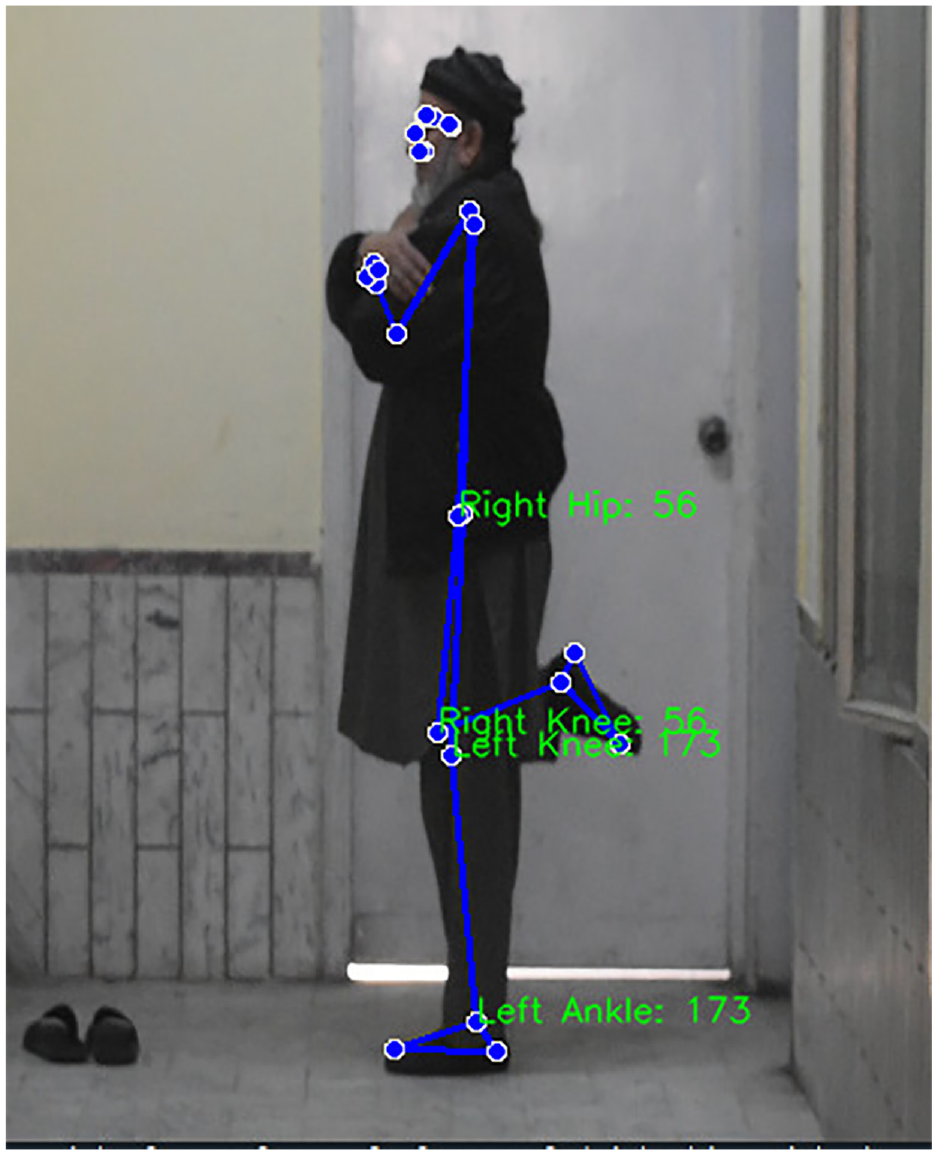
Shows an individual performing single leg stance activity upon which Media Pipe technique is being used for key points.
Theoretical framework of the proposed method
Figure 2 illustrates the research framework, divided into two main blocks. The first block represents the musculoskeletal model, which extracts lower joint data from the human body. This data is then analyzed by the AI model (Media Pipe), utilized in our study for joint analysis. The results are compared against standardized references of healthy joints to assess joint function and stability. Based on this comparison, we can determine whether an individual exhibits a stable posture or may be at risk for joint instability.
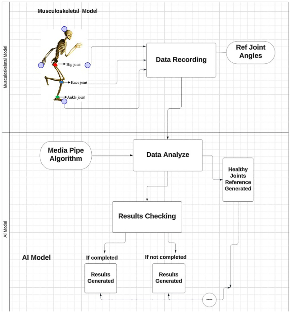
Schematic diagram of the work.
Statistical analysis
The study employed a comprehensive statistical analysis to investigate the effects of age on posture stability and lower joint motion during SLS activity. Descriptive statistics revealed distinct demographic profiles between the young (average age: 20 years; average height: 175.5 cm; average weight: 65 kg) and old (average age: 60 years; average height: 165.5 cm; average weight: 68 kg) participant groups. We have used Media Pipe Pose estimation model, which detects 33 landmarks (key points) for various body parts, including the upper and lower body joints. These landmarks include points such as the nose, eyes, ears, shoulders, elbows, wrists, hips, knees, and ankles but for our research, we were more focused on lower joint landmarks. Media Pipe is an open-source framework developed by Google that provides a comprehensive suite of tools and solutions for building real-time perception pipelines. It is designed to enable developers to efficiently integrate machine learning-based models into various multimedia applications, including video analysis, gesture recognition, facial recognition, and body pose estimation. One of the key features of Media Pipe is its robustness and versatility in handling complex tasks involving multiple modalities such as image, video, and audio data. 28 The range of motion for hip angles during single-leg stance can vary depending on factors such as age, fitness level, and the presence of any musculoskeletal conditions. However, in general, the expected range of motion for hip flexion during single-leg stance is typically between 0° and 150°, while for hip extension, it is usually between 0° and 15°. 29 These ranges may slightly vary among individuals and may also be influenced by specific activities or tasks being performed during single-leg stance. It’s essential to consider individual variability and factors such as muscle flexibility, joint health, and overall biomechanics when assessing hip angles during single-leg stance. In knee angles during normal activities like walking or standing typically fall within a range of 0°–90°, 30 depending on the individual’s posture, gait, and activity level. Setting this range of angles ensures that variations in knee angles can be easily observed and analyzed. However, the specific range may vary depending on the context of the analysis and the expected range of motion for the individuals being studied. For angle calculation between the Hip, Knee, and ankle we used trigonometric functions to calculate the distance between these angles and then applied the cosine angle list of formulas given in the 2.6 section. If a person is unable to maintain a consistent Hip-knee joint angle over a period, it may indicate instability in their posture. In Figure 3 we can see the flow chart for this activity where posture analysis for stability involves some steps for analysis first, is the time mark which is essential in analyzing the stability of a person the ability to maintain proper alignment and control of body segments during the activity is the objective. If an individual, can perform the activity within time without losing their balance then the AI model will analyze and generate its results which then will be compared with healthy or balanced joint results, if a person is unable to complete his/her activity and then it’s obvious there is an issue with his/her joints. Specifically, the results will be verified by comparing the data from individuals with unhealthy conditions to a baseline of healthy individuals, focusing on the trends in their joint angles. Now posture instability could be due to factors such as muscle weakness, joint stiffness, impaired proprioception (awareness of body position), damaged neurons, or impaired balance. For our research, during SLS activity, hip and knee joint pose estimation is one of the critical factors in assessing their health and stability. In this technique, we don’t have to use markers or sensors like in the marker-based motion capture technique this makes the present technique advantageous over the past technique because it does not require the use of markers or sensors that limit the motion.
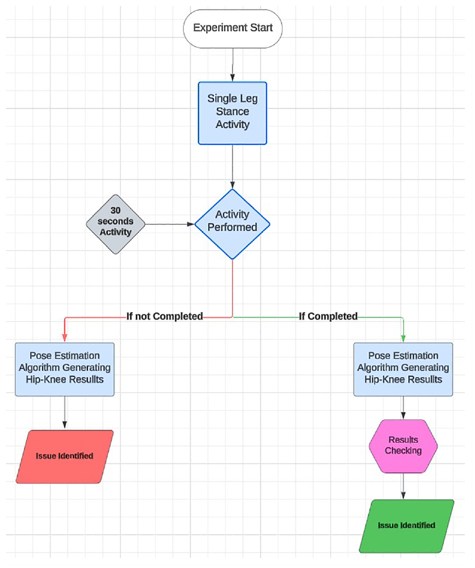
Flow chart of the experiment.
Trigonometric functions
For the calculation of angles between three lower joints multiple trigonometric functions were used in the pose estimation technique.


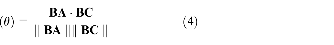

where ab is the angle between Hip and Knee, ac is the angle between Hip and Ankle and bc is the angle between Knee and ankle.
Results
The result section consists of two parts first for Hip Results and second for Knee Results.
HIP results
The results of the left hip in flexion movement of twenty young participants are presented in two separate graphs Figures 4 and 5. In Figure 4 the first 10 participants’ data is depicted which shows, the starting range of their hip angles ranging from 75° to 120°. However, these angles are close to each other indicating less deviation and showing consistency. This constancy means they are likely to possess stable and upright posture; which in turn indicates good posture and overall stability. In addition, their left hip angles do not show any major variations which feature proves the absence of any obvious problems with their hip. Similarly, Figure 5 shows the data of the other ten young participants. Among participants 1, 2, 3, 5, 9, and 10, the angles observed similar ranging and consistency like Figure 4, meaning that the posture is stable and shows minimal deviations. However, individuals 4, 6, 7, and 8 whose hip angles alternate and deviate from the baseline show a lot of inconsistency and deviation in their angles. This inconsistency indicates difficulty in maintaining their upright posture and hints at potential issues with their left hip and would be advised to see a medical expert for further diagnosis and treatment.
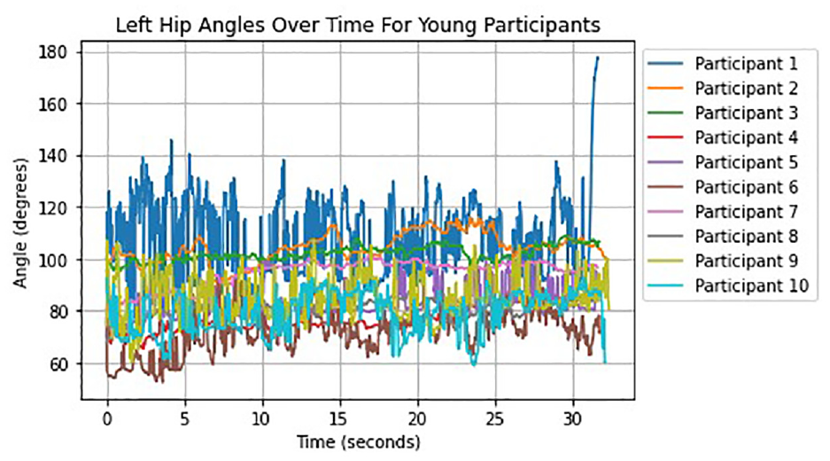
Left hip angle results for young participants.
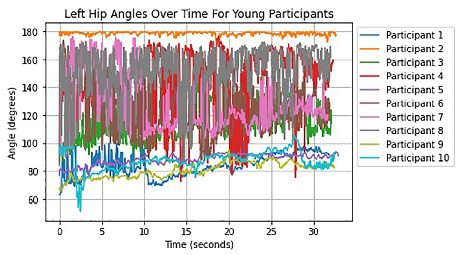
Left hip angle results for young participants.
The results for ten older participants’ left hip angles are depicted in Figures 6 and 7. Figure 6 shows that every one of the individuals had failed to keep a grip on their stance before the 30-second imprint which is the activity time. The results show that it is difficult to maintain posture stability and that there are obvious differences and vacillations. This shows left hip dysfunction and necessitates clinical consideration. The only participant in the second graph of Figure 7, who is unable to maintain an upright posture after 20 s is participant number 5. Nonetheless, others showed better results with less movement yet for certain deviations. As a result, older people may suffer from left hip joint dysfunction and require additional medical care.
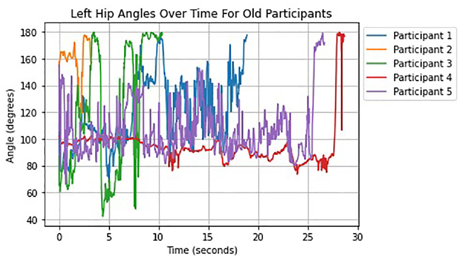
Left hip results for old participants (1).
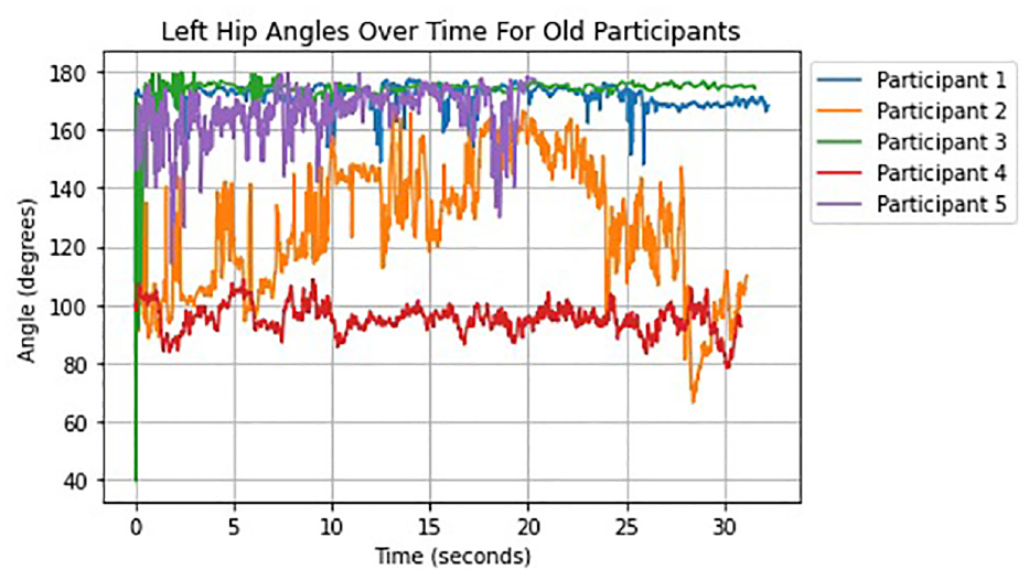
Left hip results for old participants (2).
In Figures 8 and 9, we analyze the right angle joint in flexion movement of twenty young participants whose left hip joint was measured before. In Figure 8, we can notice that angles are stable, no posture loss happens and there is less movement in their results which shows stability. Figure 9 shows that only participants 1, 9, and 10 maintain within the normal line the rest of the participants show very high unsteadiness and deviations in their outcomes although they manage to keep their posture balance throughout the activity which means their right hip joint should be addressed medically as soon as possible.
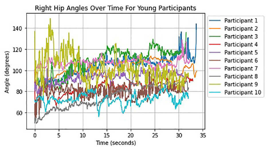
Right hip results for young participants (1).
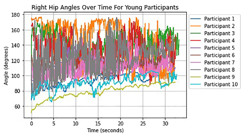
Right hip results for young participants (2).
Figures 10 and 11 show the right hip angles of ten older participants. In Figure 10, we can see the data of five individuals in which participants 1 and 5 are the only ones to keep their posture still throughout the activity otherwise participants 2,3 and 4 loss their mark before completing their task which shows that these individuals having a severe right hip issue which needs medical attention as soon as possible. The remaining five participants were able to maintain an upright posture, as shown in Figure 11, but their results showed significant variation, indicating that they were having difficulty maintaining an upright posture. Their joint points are frail and they additionally need clinical evaluation.
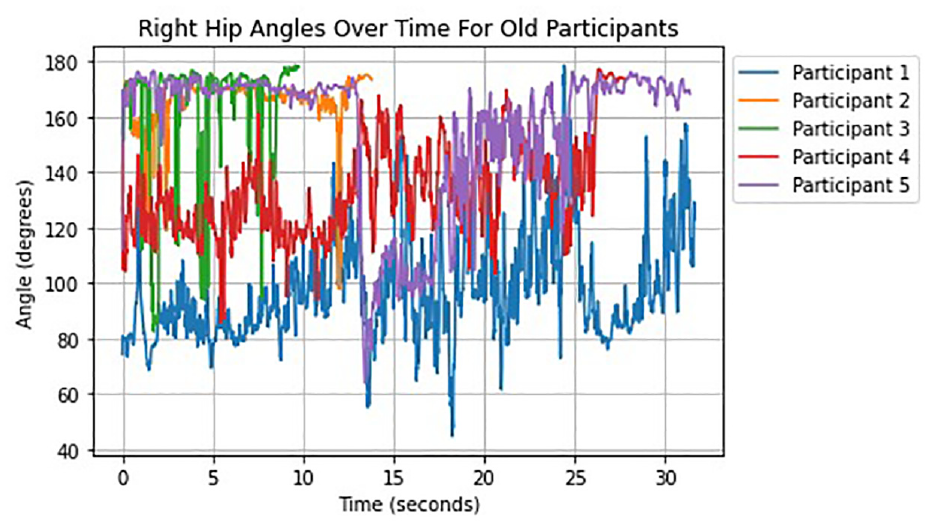
Right hip results for old participants (1).
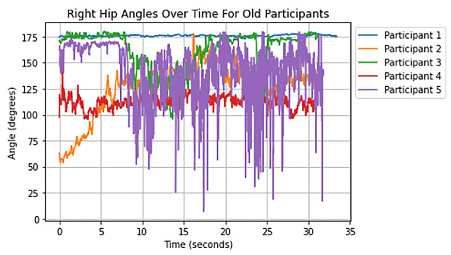
Right hip results for old participants (2).
KNEE results
This section discusses knee results as the angles of the left knee of twenty young participants, which are shown in Figures 12 and 13. However, due to the video being shorter than the desired duration for plotting (e.g. 30 s), setting the x-axis limit based on the actual video duration may have resulted in empty space on the plot. To ensure consistency across all plots, the x-axis limit in knee angle results was fixed at a value of 60 s. The data of the first 10 young people are presented in Figure 12, which shows consistency in their angles with less movement and stability in their results. There was no loss in posture stability observed. Similarly, Figure 13 also demonstrates a similar level of consistency with fewer changes and no loss in stance, indicating that these individuals do not possess any issues with their left knee.
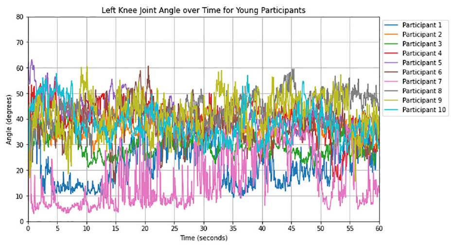
Left knee results for young participants (1).
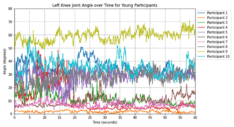
Left knee results for young participants (2).
In Figures 14 and 15, the 10 older participants’ data for left knee angles is shown. In Figure 14 we can see the data of the first five individuals which shows that none of them was able to maintain their posture and showed huge fluctuations in their knee results before losing their posture. Their results show huge deviations in their knee angles and indicate that their left knee requires medical attention before fracturing it completely. Figure 15 shows that the primary member, number five, failed to complete his activity and had a significant increase in outcomes before losing his posture. The other four members completed their tasks. Member two and four had significant fluctuations in their outcomes, but they were able to complete the activity.
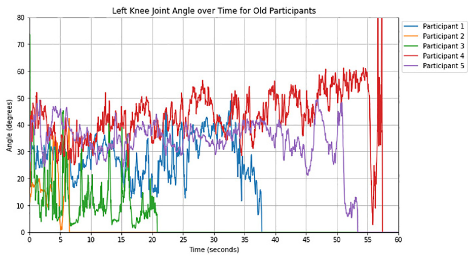
Left knee results for old participants (1).
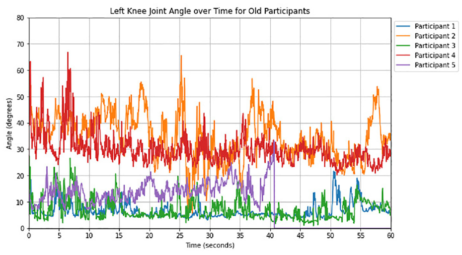
Left knee results for old participants (2).
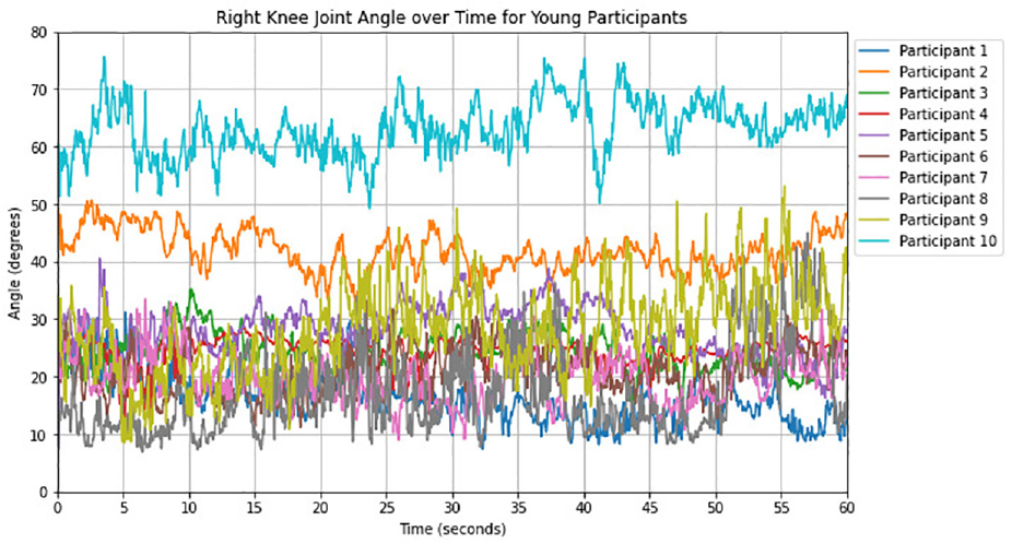
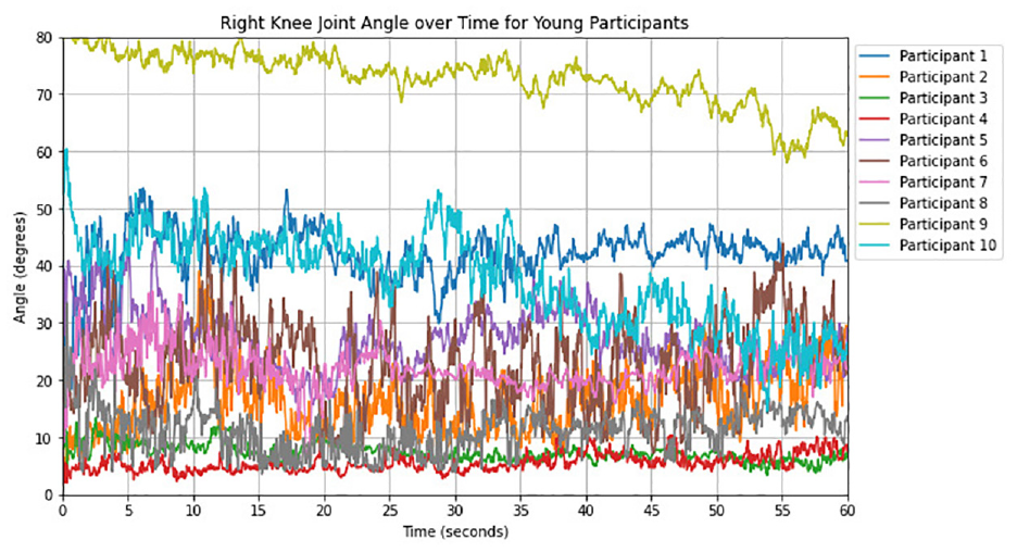
Figures 16 and 17 present the measurements of right knee angles for twenty young participants. Figure 16 displays the data for the first 10 individuals, showing a high level of consistency in their angles with minimal fluctuations. These results demonstrate stability in their posture with no loss of stability. In Figure 17, we can see the same level of consistency with even fewer fluctuations and no loss of posture stability. These findings indicate that all participants have no issues with their right knee angles.
Right knee results for young participants (1).
Right knee results for young participants (2).
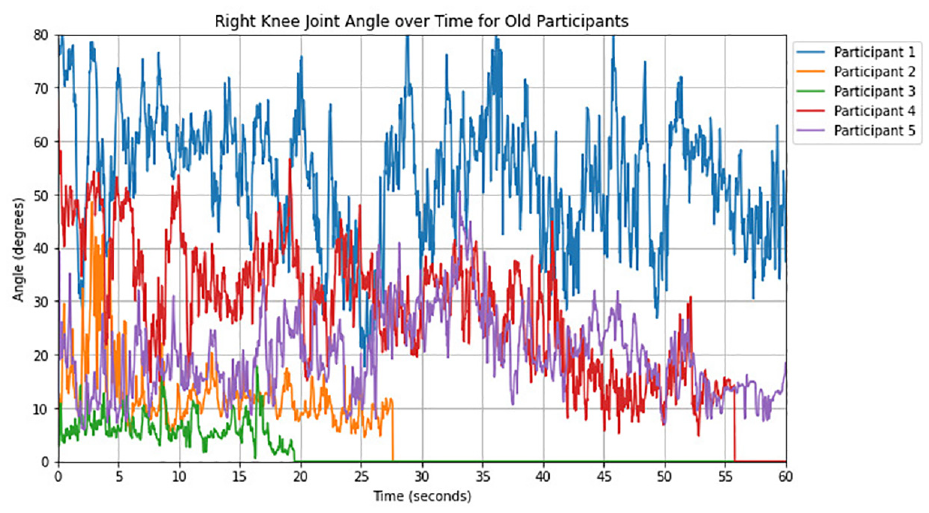
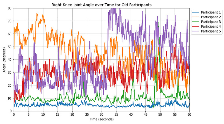
Figures 18 and 19 illustrate the right knee angle results of ten older participants. In Figure 18, the data of the first five participants showed significant spikes in their results, indicating that participants 2, 3, and 4 have problems with both of their knees. They lost their posture stability, which is a clear sign that they require medical attention for both knees. The other two participants’ results were also abnormal, showing spikes, which is a strong indication that they also need medical attention. In Figure 19, participants 2, 4, and 5 showed significant spikes in their results, indicating that their knees require medical attention.
Right knee results for old participants (1).
Right knee results for young participants (2).
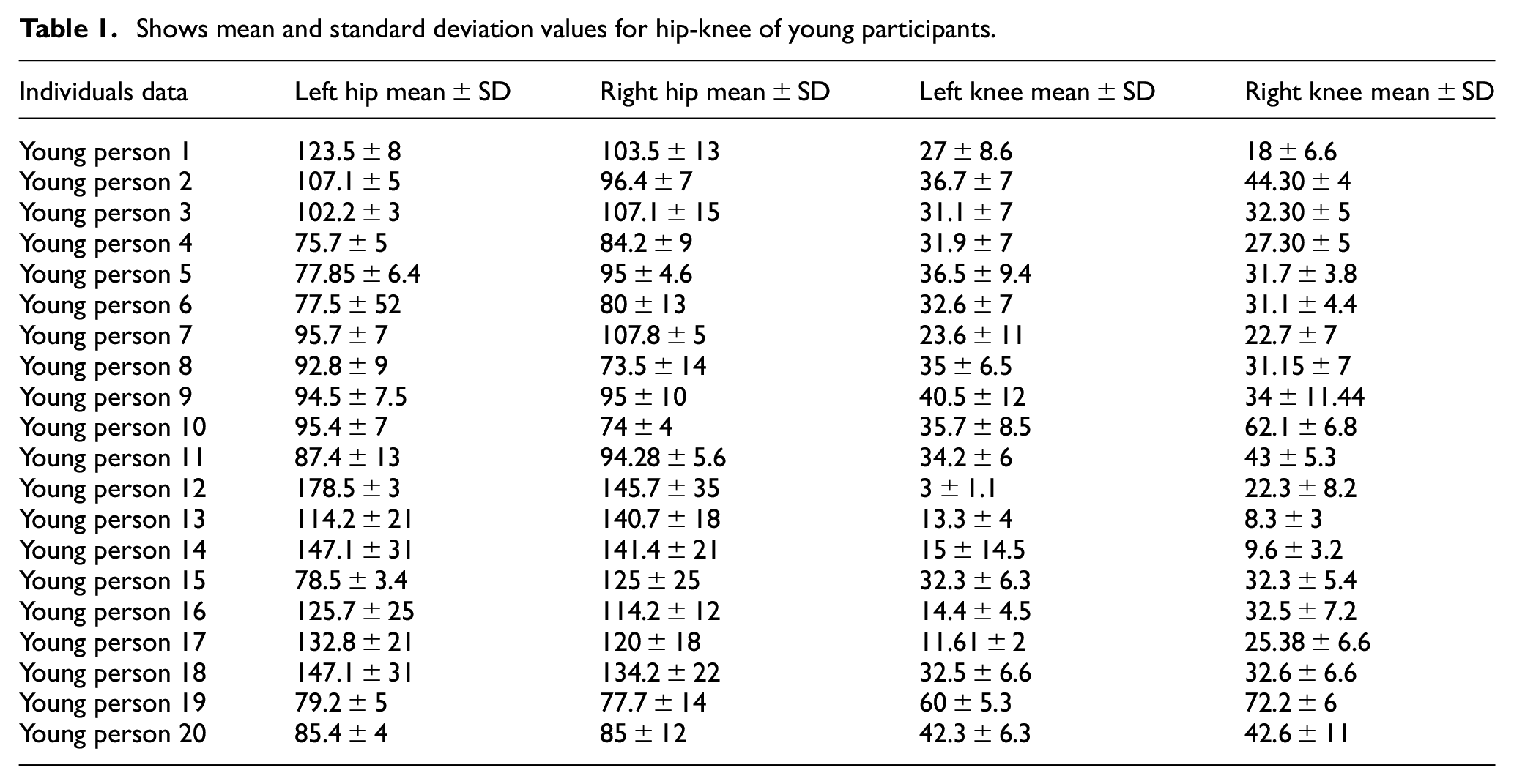
Tables 1 and 2 display the mean and standard deviation values extracted from graphs of the HK joints. The first table shows data gathered from twenty young subjects, with their mean and standard deviations of measurements of both hip and knee joints. The results indicate satisfactory joint performance among the young subjects, with stability in posture observed. Although some concerns were seen in the HK results from young participants, the remainder of the results were satisfactory. Further research showed that most of the subjects had good posture with excellent results. These facts testify to the strong state of participants’ joint health and show that their upright posture is balanced. The following mean and standard deviation values were obtained: (107.14 ± 5, 96.42 ± 7) for both hips and (36.76 ± 7, 44.30 ± 4) for both knees from one of the young participants. These values are in line with the expected joint angles (110°–120° for the hip and 45°–60° for the knee) and show stability in results.
Shows mean and standard deviation values for hip-knee of young participants.
Shows mean and standard deviation values for hip-knee of old participants.
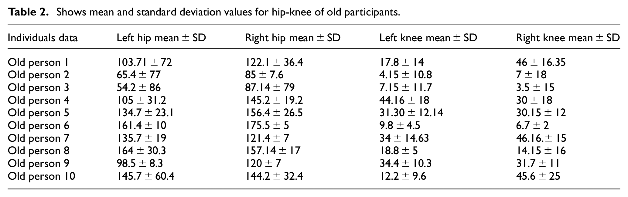
On the other hand, the second table shows data obtained from 10 older participants, revealing an alarming situation for the participants. Indeed, as many as 60% of them displayed symptoms of balance loss, combined with other concerning posture and joint measurements. The results from one of the elderly participants show a high level of variability and low mean values for both hips and knees: (65.42 ± 77, 85 ± 76.67) and (4.15 ± 10.8, 7 ± 18), respectively. This is indicative of deviations from the expected norm, this locates particular worries pertaining to the general health and stability of the older participants’ joints, which may suggest immediate medical intervention. These worrying findings necessitate immediate action to allay any possible risks that could manifest due to joint wear and tear and poor posture. Such findings suggest the importance of considering individual differences and variability when analyzing biomechanical data, and how early detection in the lower joints not only helps maintain proper posture but also prevents undue stress and strain from burdening other joints. Based on the results obtained from the analysis of hip and knee joint angles in both older and younger individuals, several key findings can be drawn. It was demonstrated that there were various kinds of hip and knee joint angles between older ones and younger individuals when they carried out functional tasks. The elderly group, in general, seemed to display higher averages of joint angles and more variability compared to their young counterparts. These experiments show that age-associated changes in joint biomechanics, muscle strength, and neuromuscular control can impact posture stability, movement coordination, and balance during daily activities. Thus, the levels of individual joint angle fluctuation assessed via standard deviations allow differentiating between the abilities of the subjects to maintain stable postures and movements. The greater range observed in elderly people indicates possible issues with balance and posture. Seniors may sometimes experience problems with joint position, which can lead to greater instability and the risk of falls and injuries. The findings underscore the fact that precise rehabilitation measures to improve mobility, stability, and balance, especially in older people, are vital. Interventions such as training muscles, improving proprioception, and enhancing balance can efficiently prevent joint biomechanics triggered by age, thereby reducing the risk of falls and injuries. The findings also show an opportunity for early detection of joint problems without the need for invasive methods such as MRI scans or CT scans. Deviations in joint angles and fluctuations can potentially be used to track musculoskeletal conditions or joint mobility.
Discussion
The assessment of Hip-Knee joint angles in flexion movement during single-leg stance activity provides valuable insights into the biomechanical dynamics and stability maintenance across different age groups. Understanding how these joints function under dynamic conditions is crucial for identifying age-related changes in joint control and stability, as well as informing interventions aimed at reducing the risk of falls and improving mobility in elderly populations. The ability to detect knee or hip issues without the need for MRI scans represents a significant innovation in this research. By leveraging data on joint angles and stability during single-leg stance activity, we were able to identify individuals at risk of hip or knee issues, thereby enabling early intervention and targeted treatment strategies. For individuals experiencing instability or deviations in knee or hip angles during the SLS task, our analysis provided valuable insights into potential joint issues or muscular imbalances. By carefully monitoring the trajectories of hip and knee angles and correlating them with instances of instability or compromised balance, we could pinpoint the affected joint and assess the severity of the issue. For example, if an individual consistently exhibited fluctuations or spikes in left knee angles accompanied by moments of instability during the SLS task, it could indicate potential issues with the left knee joint or surrounding musculature. Similarly, deviations in hip angles or loss of stability on one side could suggest asymmetrical loading or weakness in the corresponding hip joint. By identifying these early signs of joint dysfunction or instability, healthcare professionals can initiate targeted treatment approaches to address the underlying issues and prevent further deterioration. This may include targeted strength training exercises, proprioceptive training, gait correction techniques, or other rehabilitation interventions tailored to the specific needs of the individual. Furthermore, the ability to detect hip or knee issues non-invasively and without the need for costly imaging scans represents a significant advantage in terms of accessibility and cost-effectiveness. By utilizing simple yet effective bio mechanical assessments during functional tasks such as single-leg stance, we can streamline the diagnostic process and ensure timely intervention for individuals at risk of joint-related issues. Firstly, let’s delve into the analysis of knee joint in flexion movement among young and elderly individuals. In young participants, the knee joint exhibits remarkable stability and minimal deviation in angles throughout the SLS task. This consistency suggests robust lower limb strength, joint stability, and proprioceptive control, which are essential for maintaining balance and stability during dynamic movements. The smooth trajectory of knee angles reflects efficient neuromuscular coordination and effective load distribution across the lower limb joints, contributing to optimal posture maintenance. In contrast, elderly participants display distinct patterns characterized by notable fluctuations and spikes in knee angles. These fluctuations often precede instances of compromised balance and stability, indicating challenges in maintaining joint control and posture integrity during dynamic tasks. Age-related changes such as muscle weakness, reduced proprioception, and impaired neuromuscularcoordination may contribute to these observed differences in knee joint dynamics. As a result, older individuals are more susceptible to falls and mobility impairments, emphasizing the importance of targeted interventions to address these age-related deficits. Moving on to the analysis of hip joint angles in flexion movement, similar contrasts emerge between age groups regarding posture stability and joint control. Among older individuals, hip angles exhibit pronounced fluctuations and deviations, often leading to moments of instability and compromised posture. These fluctuations may stem from age-related changes in muscle strength, joint flexibility, and sensory feedback, which can affect the ability to maintain balance and control joint motion effectively. Moreover, factors such as osteoarthritis and musculoskeletal degeneration may further exacerbate these challenges, contributing to increased fall risk and mobility limitations in elderly populations. Conversely, younger participants demonstrate smoother trajectories with minimal deviations in hip angles for some of the participants, indicative of superior posture stability and joint control. The consistent maintenance of hip angles reflects efficient neuromuscular coordination, optimal muscle activation patterns, and effective load distribution across the lower limb joints. These factors collectively contribute to enhanced balance control and stability during SLS task, reducing the risk of falls and injury.
Conclusion with future work
In general, the study of the angles of Hip-Knee joint in flexion movement during single-leg stance activity can indicate aging-related changes in joint dynamics and posture stability. Young people show remarkable stability, while elderly participants struggle to maintain posture integrity. This may result from age-related changes such as muscle weakness, reduced proprioception, and damaged neuromuscular system. Incorporating joint angle analysis in our assessment protocol can detect hip or knee problems early on and enable positive interventions for patients. Detecting hip or knee issues without MRI scans represents a significant innovation in this research. This new approach may provide a basis for personalized rehab programs for musculoskeletal disorders. In future work, we can add certain disease detection techniques for these lower joint angles like the application of image processing, and deep learning plus, we can also use this technique for elbow or shoulder joint abnormalities and detection of disease or dislocation in them as well.
Footnotes
Acknowledgements
AI tools have been used for rephrasing and correction of sentences. I want to express my sincere gratitude to Bahria University for providing me with the invaluable resources, support, and opportunities that have significantly contributed to the completion of this research.
Declaration of conflicting interests
The author(s) declared no potential conflicts of interest with respect to the research, authorship, and/or publication of this article.
Funding
The author(s) received no financial support for the research, authorship, and/or publication of this article.
Data availability statement
The data that support the findings of this study are available from the corresponding author upon reasonable request.