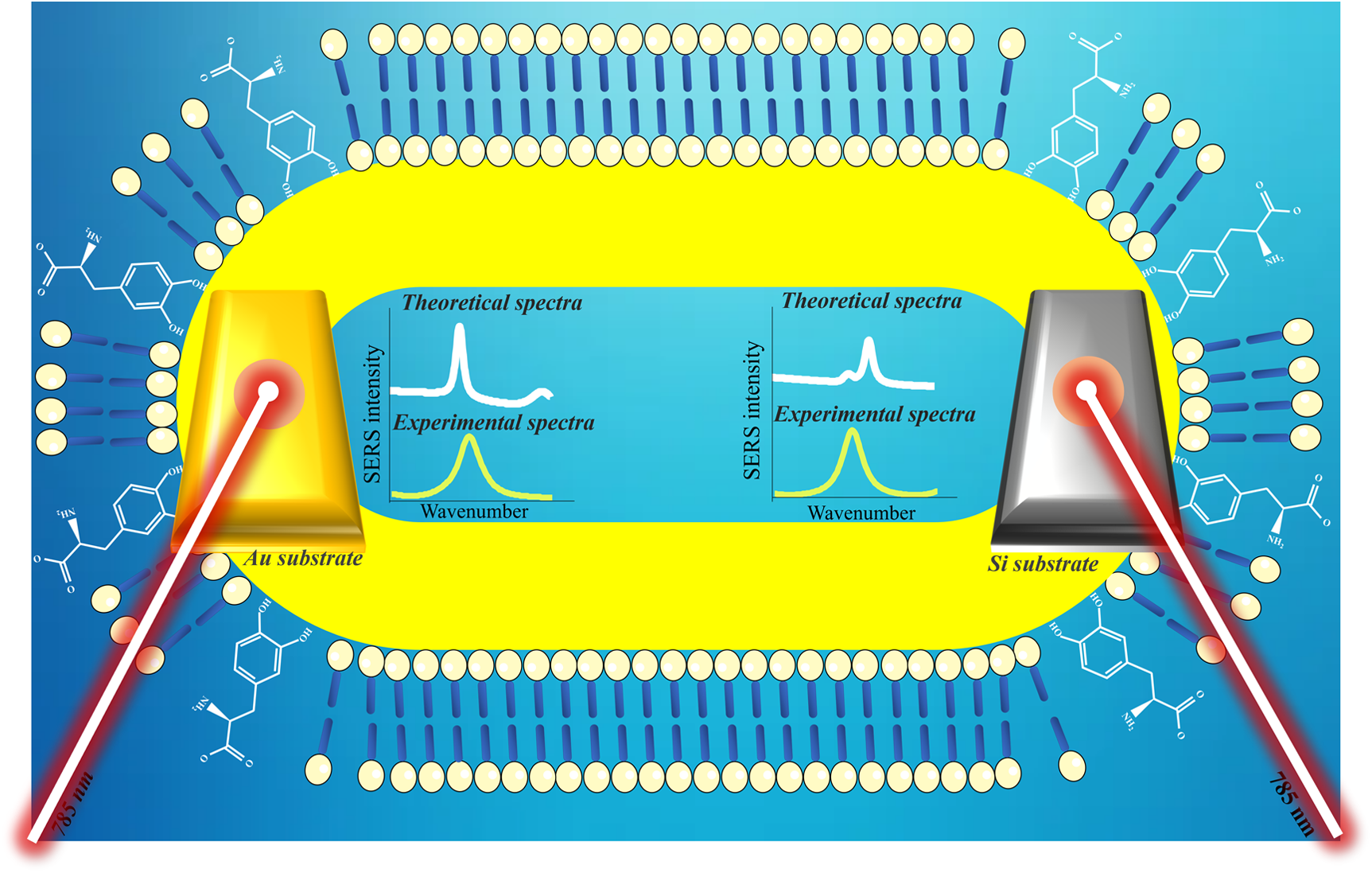
Experimental efforts aimed at detecting levodopa (l-DOPA) using surface-enhanced Raman scattering (SERS) face a persistent challenge in obtaining a SERS signal with negatively charged nanoparticles. This challenge stems from the repulsion between deprotonated l-DOPA in aqueous solution and the charged surface of the nanoparticles, revealing dependencies on time and concentration to achieve the SERS signal. This study explores the adsorption mechanism of l-DOPA on the surface of gold nanorods (AuNRs) covered with a cetrimonium bromide (CTAB) bilayer as a colloidal solution, subsequently dried onto a solid substrate such as glass, silicon, and Au substrate. Experimental findings are supported by density functional theory theoretical calculations. The comparison between experimental and theoretical results highlights that the SERS profile can be attributed to the adsorption of l-DOPA via the catechol ring, leading to the formation of anionic and dianionic species.
FahnS.OakesD.ShoulsonI.KiebertzK., et al. “Levodopa and the Progression of Parkinson’s Disease”. N. Engl. J. Med. 2004. 351(24): 2498–2508. https://doi.org/10.1056/nejmoa033447
2.
KimW.H.KarimM.M.LeeS.H.. “Simultaneous Determination of Levodopa and Carbidopa by Synchronous Fluorescence Spectrometry Using Double Scans”. Anal. Chim. Acta. 2008. 619(1): 2–7. https://doi.org/10.1016/j.aca.2008.01.006
ZaimM.KumarY.HallanV.ZaidiA.A.. “Velvet Bean Severe Mosaic Virus: A Distinct Begomovirus Species Causing Severe Mosaic in Mucuna pruriens (L.) DC”. Virus Genes. 2011. 43(1): 138–146. https://doi.org/10.1007/S11262-011-0610-Z
5.
LoganathanP.. “Production of Dl-DOPA from Acellular Slime-Mould Stemonitis Herbatica”. Bioprocess Eng. 1998. 18(4): 307–308.
6.
TakedaH.H.SilvaT.A.JanegitzB.C.VicentiniF.C., et al. “Electrochemical Sensing of Levodopa or Carbidopa Using a Glassy Carbon Electrode Modified with Carbon Nanotubes Within a Poly(Allylamine Hydrochloride) Film”. Anal. Methods. 2016. 8(6): 1274–1280. https://doi.org/10.1039/C5AY03041B
7.
Hormozi-NezhadM.R.MoslehipourA.BigdeliA.. “Simple and Rapid Detection of l-Dopa Based on In Situ Formation of Polylevodopa Nanoparticles”. Sens. Actuators B. 2017. 243: 715–720.
8.
RubiraR.J.G.CamachoS.A.MartinC.S.Mejía-SalazarJ.R., et al. “Designing Silver Nanoparticles for Detecting Levodopa (3,4-Dihydroxyphenylalanine, l-DOPA) Using Surface-Enhanced Raman Scattering (SERS)”. Sensors. 2019. 20(1): 15.
9.
OliveiraT.A.RubiraR.J.G.Da Silva MartinC.De BarrosA., et al. “The Plasmonic Effect of Gold Nanorods on Charged Molecules: SERRS and SEF Effects”. Mater. Res. 2021. 24: 1–11. https://doi.org/10.1590/1980-5373-MR-2021-0029
10.
JabbarM.L.. “Computational Studies on Electronic and Optical Properties of Dopamine Derivatives Structure: A DFT Study”. J. Mech. Behav. Mater. 2021. 30(1): 279–284. https://doi.org/10.1515/jmbm-2021-0030
11.
ZhuangZ.RuanW.JiN.ShangX., et al. “Surface-Enhanced Raman Scattering of 4,4′-Bipyridine on Silver by Density Functional Theory Calculations”. Vib. Spectrosc. 2009. 49(2): 118–123. https://doi.org/10.1016/j.vibspec.2008.05.007
12.
LiR.JiW.ChenL.LvH.ChengJ.ZhaoB.. “Vibrational Spectroscopy and Density Functional Theory Study of 4-Mercaptophenol”. Spectrochim. Acta, Part A. 2014. 122: 698–703. https://doi.org/10.1016/j.saa.2013.11.109
13.
WuX.GaoS.WangJ.-S.WangH., et al. “The Surface-Enhanced Raman Spectra of Aflatoxins: Spectral Analysis, Density Functional Theory Calculation, Detection, and Differentiation”. Analyst. 2012. 137(18): 4226. https://doi.org/10.1039/c2an35378d
14.
JuniorB.R.A.SoaresF.L.F.ArdilaJ.A.DurangoL.G.C., et al. “Determination of B-Complex Vitamins in Pharmaceutical Formulations by Surface-Enhanced Raman Spectroscopy”. Spectrochim. Acta, Part A. 2018. 188: 589–595. https://doi.org/10.1016/J.Saa.2017.07.049
15.
Montes-GarcíaV.Rodal-CedeiraS.Cordero-FerradásM.J.GómezB., et al. “Pillar[5]arene-Stabilized Plasmonic Nanoparticles as Selective SERS Sensors”. Isr. J. Chem. 2018. 58(11): 1251–1260. https://doi.org/10.1002/ijch.201800041
16.
OliveiraT.A.MartinC.S.RubiraR.J.G.De BarrosA., et al. “Influence of l-DOPA Adsorption Time and Concentration on Au Nanorods Aggregation for SERS Quantitative Analysis”. Curr. Appl. Phys. 2023. 54: 31–37.
17.
XuD.MaoJ.HeY.YeungE.S.. “Size-Tunable Synthesis of High-Quality Gold Nanorods Under Basic Conditions by Using H2O2 as the Reducing Agent”. J. Mater. Chem. C. 2014. 2(25): 4989–4996. https://doi.org/10.1039/c4tc00483c
18.
AlloucheA.. “Gabedit: A Graphical User Interface for Computational Chemistry Softwares”. J. Comput. Chem. 2011. 32(1): 174–182. https://doi.org/10.1002/jcc.21600
19.
SchaftenaarG.NoordikJ.H.. “The Effect of Isodensity Surface Sampling on ESP Derived Charges and the Effect of Adding Bondcenters on DMA Derived Charges”. J. Comput. Aided. Mol. Des. 2000. 14(3): 233–242. https://doi.org/10.1023/A:1008163129031
20.
Batagin-NetoA.OliveiraE.F.GraeffC.F.O.LavardaF.C.. “Modelling Polymers with Side Chains: MEH-PPV and P3HT”. Mol. Simul. 2013. 39(4): 309–321. https://doi.org/10.1080/08927022.2012.724174
21.
WangJ.WolfR.M.CaldwellJ.W.KollmanP.A.CaseD.A.. “Development and Testing of a General Amber Force Field”. J. Comput. Chem. 2004. 25(9): 1157–1174. https://doi.org/10.1002/jcc.20035
22.
StewartJ.J.P.. “Optimization of Parameters for Semiempirical Methods V: Modification of NDDO Approximations and Application to 70 Elements”. J. Mol. Model. 2007. 13(12): 1173–1213. https://doi.org/10.1007/S00894-007-0233-4
23.
KlamtA.SchüürmannG.. “COSMO: A New Approach to Dielectric Screening in Solvents with Explicit Expressions for the Screening Energy and Its Gradient”. J. Chem. Soc., Perkin Trans. 1993. 2(5): 799–805. https://doi.org/10.1039/P29930000799
24.
StewartJ.J.P.. “MOPAC: A Semiempirical Molecular Orbital Program”. J. Comput. Aided. Mol. Des. 1990. 4(1): 1–103. https://doi.org/10.1007/BF00128336
25.
StewartJ.J.P.. “An Examination of the Nature of Localized Molecular Orbitals and Their Value in Understanding Various Phenomena that Occur in Organic Chemistry”. J. Mol. Model. 2019. 25(1): 7. https://doi.org/10.1007/S00894-018-3880-8
26.
BeckeA.D.. “Density-Functional Thermochemistry. I. The Effect of the Exchange-Only Gradient Correction”. J. Chem. Phys. 1992. 96(3): 2155–2160. https://doi.org/10.1063/1.462066
27.
FrischM.J.TrucksG.W.SchlegelH.B.ScuseriaG.E., et al.Gaussian 09, Rev. B.01. Wallingford, CT: Gaussian, Inc., 2009. Pp. 1–20.
28.
CossiM.BaroneV.CammiR.TomasiJ.. “Ab Initio Study of Solvated Molecules: A New Implementation of the Polarizable Continuum Model”. Chem. Phys. Lett. 1996. 255(4–6): 327–335. https://doi.org/10.1016/0009-2614(96)00349-1
29.
YangW.MortierW.J.. “The Use of Global and Local Molecular Parameters for the Analysis of the Gas-Phase Basicity of Amines”. J. Am. Chem. Soc. 1986. 108(19): 5708–5711. https://doi.org/10.1021/Ja00279a008
RoyR.K.PalS.HiraoK.. “On Non-Negativity of Fukui Function Indices”. J. Chem. Phys. 1999. 110(17): 8236–8245. https://doi.org/10.1063/1.478792
32.
De ProftF.Van AlsenoyC.PeetersA.LangenaekerW.GeerlingsP.. “Atomic Charges, Dipole Moments, and Fukui Functions Using the Hirshfeld Partitioning of the Electron Density”. J. Comput. Chem. 2002. 23(12): 1198–1209. https://doi.org/10.1002/jcc.10067
33.
ChirlianL.E.FranclM.M.. “Atomic Charges Derived from Electrostatic Potentials: A Detailed Study”. J. Comput. Chem. 1987. 8(6): 894–905. https://doi.org/10.1002/jcc.540080616
34.
Jmol.org. “Jmol: An Open-Source Java Viewer for Chemical Structures in 3D”. https://jmol.sourceforge.net/ [accessed Jan 8 2025].
35.
BarrosC.L.De OliveiraP.J.P.JorgeF.E.Canal NetoA.CamposM.. “Gaussian Basis Set of Double Zeta Quality for Atoms Rb Through Xe: Application in Non-Relativistic and Relativistic Calculations of Atomic and Molecular Properties”. Mol. Phys. 2010. 108(15): 1965–1972. https://doi.org/10.1080/00268976.2010.499377
36.
ArslanH.AlgülÖ.. “Synthesis and Ab Initio/DFT Studies on 2-(4-Methoxyphenyl)benzo[D]thiazole”. Int. J. Mol. Sci. 2007. 8(8): 760–776. https://doi.org/10.3390/I8080760
37.
EdwinB.Hubert JoeI.. “Vibrational Spectral Analysis of Anti-Neurodegenerative Drug Levodopa: A DFT Study”. J. Mol. Struct. 2013. 1034: 119–127. https://doi.org/10.1016/j.molstruc.2012.09.004
38.
JaberM.BouchouchaM.DelmotteL.MéthivierC.LambertJ.-F.. “Fate of l-DOPA in the Presence of Inorganic Matrices: Vectorization or Composite Material Formation?” J. Phys. Chem. C. 2011. 115(39): 19216–19225.
39.
YuL.LiuX.YuanW.BrownL.J.WangD.. “Confined Flocculation of Ionic Pollutants by Poly(l-DOPA)-Based Polyelectrolyte Complexes in Hydrogel Beads for Three-Dimensional, Quantitative, Efficient Water Decontamination”. Langmuir. 2015. 31(23): 6351–6366.
40.
LiuY.AiK.LuL.. “Polydopamine and Its Derivative Materials: Synthesis and Promising Applications in Energy, Environmental, and Biomedical Fields”. Chem. Rev. 2014. 114(9): 5057–5115. https://doi.org/10.1021/cr400407a
41.
LeeN.S.HsiehY.Z.PaisleyR.F.MorrisM.D.. “Surface-Enhanced Raman Spectroscopy of the Catecholamine Neurotransmitters and Related Compounds”. Anal. Chem. 1988. 60(5): 442–446. https://doi.org/10.1021/ac00156a014
42.
Estrada-SalasR.E.BarrónH.ValladaresA.A.José-YacamánM.. “Exploring the Surface Reactivity of Ag Nanoparticles with Antimicrobial Activity: A DFT Study”. Int. J. Quantum Chem. 2012. 112(18): 3033–3038. https://doi.org/10.1002/qua.24207
FigueiredoM.L.B.MartinC.S.FuriniL.N.RubiraR.J.G., et al. “Surface-Enhanced Raman Scattering for Dopamine in Ag Colloid: Adsorption Mechanism and Detection in the Presence of Interfering Species”. Appl. Surf. Sci. 2020. 522: 146466. https://doi.org/10.1016/j.apsusc.2020.146466
45.
QinL.LiX.KangS.-Z.MuJ.. “Gold Nanoparticles Conjugated Dopamine as Sensing Platform for SERS Detection”. Colloids Surf., B. 2015. 126: 210–216. https://doi.org/10.1016/j.colsurfb.2014.12.028
46.
McCreeryR.L.. Raman Spectroscopy for Chemical Analysis. New York: John Wiley and Sons, 2000.
47.
KuzminA.PlissA.PrasadP.. “Ramanomics: New Omics Disciplines Using Micro Raman Spectrometry with Biomolecular Component Analysis for Molecular Profiling of Biological Structures”. Biosensors. 2017. 7(4): 52. https://doi.org/10.3390/bios7040052
48.
MatousekP.MorrisM.D.. Emerging Raman Applications and Techniques in Biomedical and Pharmaceutical Fields. Berlin; Heidelberg: Springer, 2010. https://doi.org/10.1007/978-3-642-02649-2
49.
NarayananK.B.ParkH.H.. “Unnatural Amino Acid-Mediated Synthesis of Silver Nanoparticles and Their Antifungal Activity Against Candida Species”. J. Nanoparticle Res. 2014. 16(8): 2523. https://doi.org/10.1007/s11051-014-2523-y
50.
Lopez-TobarE.AntalikM.JancuraD.CañamaresM.V., et al. “Adsorption and Detection of Amyloid Marker Thioflavin T on Ag Nanoparticles by Surface-Enhanced Raman Scattering”. J. Phys. Chem. C. 2013. 117(8): 3996–4005. https://doi.org/10.1021/Jp310619c
51.
MorrisB.. “The Components of the Wired Spanning Forest are Recurrent”. Probab. Theory Relat. Fields. 2003. 125(2): 259–265. https://doi.org/10.1007/s00440-002-0236-0
52.
YuC.VargheseL.IrudayarajJ.. “Surface Modification of Cetyltrimethylammonium Bromide-Capped Gold Nanorods to Make Molecular Probes”. Langmuir. 2007. 23(17): 9114–9119. https://doi.org/10.1021/la701111e
53.
QinM.LiP.ZhouX.ZhuJ.WangH.YangL.. “Cationic Surfactant Regulated Synthesis of Au Nanorods for Sensitive Detection of Negatively Charged Colorants by Surface-Enhanced Raman Spectroscopy”. J. Raman Spectrosc. 2019. 50(6): 809–817. https://doi.org/10.1002/jrs.5575
Supplementary Material
Please find the following supplemental material available below.
For Open Access articles published under a Creative Commons License, all supplemental material carries the same license as the article it is associated with.
For non-Open Access articles published, all supplemental material carries a non-exclusive license, and permission requests for re-use of supplemental material or any part of supplemental material shall be sent directly to the copyright owner as specified in the copyright notice associated with the article.