Green Textile Materials for Surface Enhanced Raman Spectroscopy Identification of Pesticides Using a Raman Handheld Spectrometer for In-Field Detection
Restricted accessResearch articleFirst published online October, 2022
Green Textile Materials for Surface Enhanced Raman Spectroscopy Identification of Pesticides Using a Raman Handheld Spectrometer for In-Field Detection
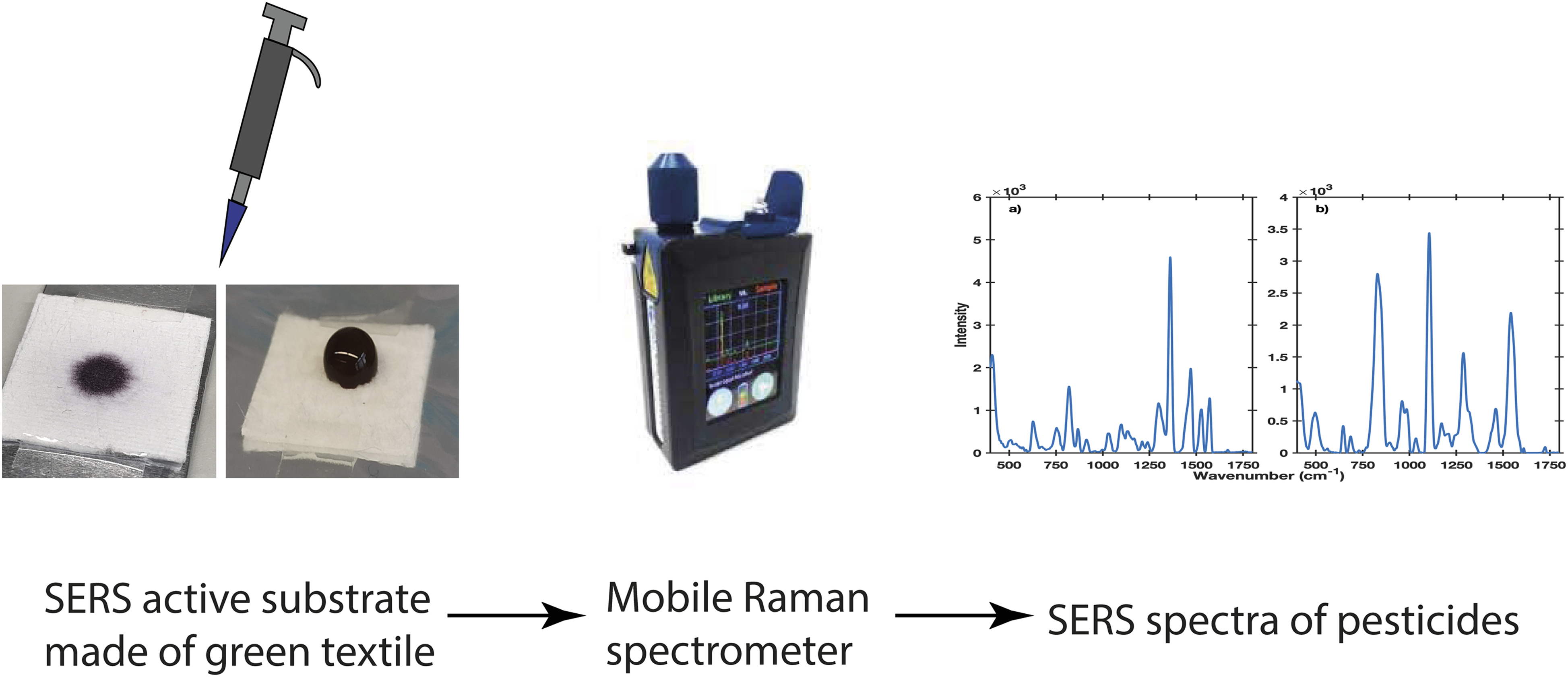
Surface enhanced Raman spectroscopy (SERS) has evolved into a powerful analytical method in food and environmental analytical sciences due to its high sensitivity. Pesticide analysis is a major discipline therein. Using sustainable materials has become increasingly important to adhere to Green Chemistry principles. Hence, the green textiles poly-(L-lactic acid) (PLA) and the mixed fabric polyethylene terephthalate polyamide (PET/PA) were investigated for their applicability as solid supports for gold nanoparticles to yield SERS substrates. Gold nanoparticle solutions and green textile supports were prepared after preparation optimization. Particle size, dispersity, and particle distribution over the textiles were characterized by absorption spectroscopy and transmission electron imaging. The performance of the SERS substrates was tested using the three pesticides imidacloprid, paraquat, and thiram and a handheld Raman spectrometer with a laser wavelength of 785 nm. The resulting SERS spectra possessed an intra-substrate variation of 7–8% in terms of the residual standard deviation. The inter-substrate variations amounted to 15% for PET/PA and to 27% for PLA. Substrate background signals were smaller with PLA but more enhanced through PET/PA. The pesticides could be detected at 1 pg on PET/PA and at 3 ng on PLA. Hence, PET/PA woven textile soaked with gold nanoparticle solution provides green SERS substrates and might prove, in combination with fieldable Raman spectrometers, suitable for in-field analytics for pesticide identification.
PilotR.SignoriniR.DuranteC.OrianL., et al. “A Review on Surface-Enhanced Raman Scattering”. Biosensors. 2019. 9(2): 57‐155. doi:10.3390/bios9020057
4.
YanZ.X.ZhangY.L.WangW.FuX.Y., et al. “Superhydrophobic SERS Substrates Based on Silver-Coated Reduced Graphene Oxide Gratings Prepared by Two-Beam Laser Interference”. ACS Appl. Mater. Interfaces. 2015. 7(49): 27059‐27065. doi:10.1021/acsami.5b09128
5.
YaoC.GaoX.LiuX.ShenY.XieA.. “In-Situ Preparation of Ferrero® Chocolate-Like Cu2O@Ag Microsphere as SERS Substrate for Detection of Thiram”. J. Mater. Res. Technol. 2021. 11: 857‐865. doi:10.1016/j.jmrt.2021.01.069
6.
DingQ.KangZ.HeX.WangM., et al. “Eggshell Membrane-Templated Gold Nanoparticles as a Flexible SERS Substrate for Detection of Thiabendazole”. Microchim. Acta. 2019. 186(7): 453‐462. doi:10.1007/s00604-019-3543-1
7.
HuangD.ZhaoJ.WangM.ZhuS.. “Snowflake-Like Gold Nanoparticles as SERS Substrates for the Sensitive Detection of Organophosphorus Pesticide Residues”. Food Control. 2020. 108(July 2019): 106835. doi:10.1016/j.foodcont.2019.106835
8.
ZhuC.ZhaoQ.MengG.WangX., et al. “Silver Nanoparticle-Assembled Micro-Bowl Arrays for Sensitive SERS Detection of Pesticide Residue”. Nanotechnology. 2020. 31(20): 205303‐205321. doi:10.1088/1361-6528/ab7100
9.
GaoM.LinX.LiZ.WangX., et al. “Fabrication of Highly Sensitive and Reproducible 3D Surface-Enhanced Raman Spectroscopy Substrates through In Situ Cleaning and Layer-By-Layer Assembly of Au@Ag Nanocube Monolayer Film”. Nanotechnology. 2019. 30(34): 345604‐345616. doi:10.1088/1361-6528/ab1ff2
Dias SoaresJ.M.de OliveiraH.P.. “Silver-Based Surface Enhanced Raman Spectroscopy Devices for Detection of Organophosphorus Pesticides Traces”. Biotechnol. Prog. 2019. 35(4): E2809. doi:10.1002/btpr.2809
12.
YuW.W.WhiteI.M.. “A Simple Filter-Based Approach to Surface Enhanced Raman Spectroscopy for Trace Chemical Detection”. Analyst. 2012. 137(5): 1168–1173. doi:10.1039/C2AN15947C
13.
SunJ.GongL.WangW.GongZ., et al. “Surface-Enhanced Raman Spectroscopy for On-Site Analysis: A Review of Recent Developments”. Luminescence. 2020. 35(6): 808‐820. doi:10.1002/bio.3796
14.
JiangJ.ZouS.MaL.WangS., et al. “Surface-Enhanced Raman Scattering Detection of Pesticide Residues Using Transparent Adhesive Tapes and Coated Silver Nanorods”. ACS Appl. Mater. Interfaces. 2018. 10(10): 9129‐9135. doi:10.1021/acsami.7b18039
15.
ZhangY.HuangY.ZhaiF.DuR., et al. “Analyses of Enrofloxacin, Furazolidone and Malachite Green in Fish Products with Surface-Enhanced Raman Spectroscopy”. Food Chem. 2012. 135(2): 845‐850. doi:10.1016/j.foodchem.2012.04.082
16.
FikietM.A.KhandasammyS.R.MistekE.AhmedY., et al. “Surface Enhanced Raman Spectroscopy: A Review of Recent Applications in Forensic Science”. Spectrochim. Acta, Part A. 2018. 197(2017): 255‐260. doi:10.1016/j.saa.2018.02.046
17.
LiuY.LiX.ChengJ.ZhouN., et al. “SERS Devices with ‘Hedgehog-Like’ Nanosphere Arrays for Detection of Trace Pesticides”. J. Innov. Opt. Health Sci. 2021. 14(4): 2141005. doi:10.1142/S1793545821410054
18.
YuH.LyuQ.ChenX.GuoD., et al. “Nylon Membranes Modified by Gold Nanoparticles as Surface-Enhanced Raman Spectroscopy Substrates for Several Pesticides Detection”. RSC Adv. 2021. 11(39): 24183‐24189. doi:10.1039/D1RA03490A
19.
HanC.WeiY.LeiF.ZhaoS., et al. “Heterostructured CuO@ZnO@Ag Biomimetic Setaria as Wettability-Switchable Difunctional SERS Substrate for Trace Pesticide and DNA Detections”. Nanophotonics. 2021. 10(10): 2671‐2682. doi:10.1515/nanoph-2021-0179
European Food Safety Authority (EFSA). “Evaluation of the Data on Clothianidin, Imidacloprid, and Thiamethoxam for the Updated Risk Assessment to Bees for Seed Treatments and Granules in the EU”. 2018. https://efsa.onlinelibrary.wiley.com/doi/pdf/10.2903/sp.efsa. 2018.EN-1378 [accessed Apr 11 2022].
22.
ChenJ.HuangM.KongL.LinM.. “Jellylike Flexible Nanocellulose SERS Substrate for Rapid In-Situ Non-Invasive Pesticide Detection in Fruits/Vegetables”. Carbohydr. Polym. 2019. 205(2): 596‐600. doi:10.1016/j.carbpol.2018.10.059
23.
WoodT.J.GoulsonD.. “The Environmental Risks of Neonicotinoid Pesticides: A Review of the Evidence Post 2013”. Environ. Sci. Pollut. Res. 2017. 24(21): 17285‐17325. doi:10.1007/s11356-017-9240-x
24.
TsvetkovN.SoodK.PatelH.S.MalenaD.A., et al. “Chronic Exposure to Neonicotinoids Reduces Honey Bee Health Near Corn Crops”. Science. 2017. 356(6345): 1395‐1397. doi:10.1126/science.aam7470
25.
BabazadehS.MoghaddamP.A.KeshipourS.MollazadeK.. “Analysis of Imidacloprid and Penconazole Residues during Their Pre-Harvest Intervals in the Greenhouse Cucumbers by HPLC–DAD”. J. Iran. Chem. Soc. 2020. 17(6): 1439‐1446. doi:10.1007/s13738-020-01868-4
26.
HermsenA.LamersD.SchoettlJ.MayerC.JaegerM.. “In-Field Detection Method for Imidacloprid by Surface Enhanced Raman Spectroscopy”. Toxicol. Environ. Chem. 2021: 1‐13. doi:10.1080/02772248.2021.1991929
27.
FrensG.. “Controlled Nucleation for the Regulation of the Particle Size in Monodisperse Gold Suspensions”. Nat. Phys. Sci. 1973. 241(105): 20‐22. doi:10.1038/physci241020a0
28.
KimlingJ.MaierM.OkenveB.KotaidisV., et al. “Turkevich Method for Gold Nanoparticle Synthesis Revisited”. J. Phys. Chem. B. 2006. 110(32): 15700‐15707. doi:10.1021/jp061667w
29.
TurkevichJ.StevensonP.C.HillierJ.. “A Study of the Nucleation and Growth Processes in the Synthesis of Colloidal Gold”. Discuss. Faraday Soc. 1951. 11(0): 55‐75. doi:10.1039/DF9511100055
30.
BohrenC.F.HuffmanD.R.. Absorption and Scattering of Light by Small Particles. Weinheim, Germany: Wiley-VCH Verlag Gmbh and Co., 1983. doi:10.1002/9783527618156
31.
MunroC.H.SmithW.E.GarnerM.ClarksonJ.WhiteP.C.. “Characterization of the Surface of a Citrate-Reduced Colloid Optimized for Use as a Substrate for Surface-Enhanced Resonance Raman Scattering”. Langmuir. 1995. 11(10): 3712‐3720. doi:10.1021/la00010a021
32.
TashiroK.KounoN.WangH.TsujiH.. “Crystal Structure of Poly(Lactic Acid) Stereocomplex: Random Packing Model of PDLA and PLLA Chains as Studied by X-ray Diffraction Analysis”. Macromolecules. 2017. 50(20): 8048‐8065. doi:10.1021/acs.macromol.7b01468
33.
YunkerP.J.DurianD.J.YodhA.G.. “Coffee Rings and Coffee Disks: Physics on the Edge”. Phys. Today. 2013. 66(8): 60‐61. doi:10.1063/PT.3.2093
34.
JosephV.MatschulatA.PolteJ.RolfS., et al. “SERS Enhancement of Gold Nanospheres of Defined Size”. J. Raman Spectrosc. 2011. 42(9): 1736‐1742. doi:10.1002/jrs.2939
35.
ChenQ.HassanM.M.XuJ.ZareefM., et al. “Fast Sensing of Imidacloprid Residue in Tea Using Surface-Enhanced Raman Scattering by Comparative Multivariate Calibration”. Spectrochim. Acta, Part A. 2019. 211: 86‐93. doi:10.1016/j.saa.2018.11.041
36.
DowgialloA.M.GuentherD.A.. “Determination of the Limit of Detection of Multiple Pesticides Utilizing Gold Nanoparticles and Surface-Enhanced Raman Spectroscopy”. J. Agric. Food Chem. 2019. 67(46): 12642‐12651. doi:10.1021/acs.jafc.9b01544
37.
TangJ.ChenW.JuH.. “Rapid Detection of Pesticide Residues Using a Silver Nanoparticles Coated Glass Bead as Nonplanar Substrate for SERS Sensing”. Sens. Actuators, B. 2019. 287: 576‐583. doi:10.1016/j.snb.2019.02.084
38.
QiuH.GuoJ.WangM.JiS., et al. “Reduced Graphene Oxide Supporting Ag Meso-Flowers and Phenyl-Modified Graphitic Carbon Nitride as Self-Cleaning Flexible SERS Membrane for Molecular Trace-Detection”. Colloids Surf., A. 2019. 560: 9‐19. doi:10.1016/j.colsurfa.2018.09.059
39.
OliveiraM.J.QuaresmaP.De AlmeidaM.P.AraújoA., et al. “Office Paper Decorated with Silver Nanostars-An Alternative Cost-Effective Platform for Trace Analyte Detection by SERS”. Sci. Rep. 2017. 7(1): 1‐14. doi:10.1038/s41598-017-02484-8
40.
De OliveiraV.E.CastroH.V.EdwardsH.G.M.De OliveiraL.F.C.. “Carotenes and Carotenoids in Natural Biological Samples: A Raman Spectroscopic Analysis”. J. Raman Spectrosc. 2010. 41(6): 642‐650. doi: 10.1002/jrs.2493
Supplementary Material
Please find the following supplemental material available below.
For Open Access articles published under a Creative Commons License, all supplemental material carries the same license as the article it is associated with.
For non-Open Access articles published, all supplemental material carries a non-exclusive license, and permission requests for re-use of supplemental material or any part of supplemental material shall be sent directly to the copyright owner as specified in the copyright notice associated with the article.