
Abstract
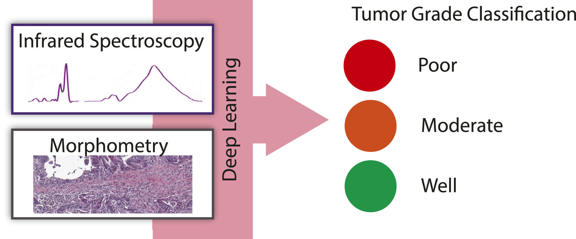
Tumor grade assessment is critical to the treatment of cancers. A pathologist typically evaluates grade by examining morphologic organization in tissue using hematoxylin and eosin (H&E) stained tissue sections. Fourier transform infrared spectroscopic (FT-IR) imaging provides an alternate view of tissue in which spatially specific molecular information from unstained tissue can be utilized. Here, we examine the potential of IR imaging for grading colon cancer in biopsy samples. We used a 148-patient cohort to develop a deep learning classifier to estimate the tumor grade using IR absorption. We demonstrate that FT-IR imaging can be a viable tool to determine colorectal cancer grades, which we validated on an independent cohort of surgical resections. This work demonstrates that harnessing molecular information from FT-IR imaging and coupling it with morphometry is a potential path to develop clinically relevant grade prediction models.
Keywords
Get full access to this article
View all access options for this article.
References
Supplementary Material
Please find the following supplemental material available below.
For Open Access articles published under a Creative Commons License, all supplemental material carries the same license as the article it is associated with.
For non-Open Access articles published, all supplemental material carries a non-exclusive license, and permission requests for re-use of supplemental material or any part of supplemental material shall be sent directly to the copyright owner as specified in the copyright notice associated with the article.