
Abstract
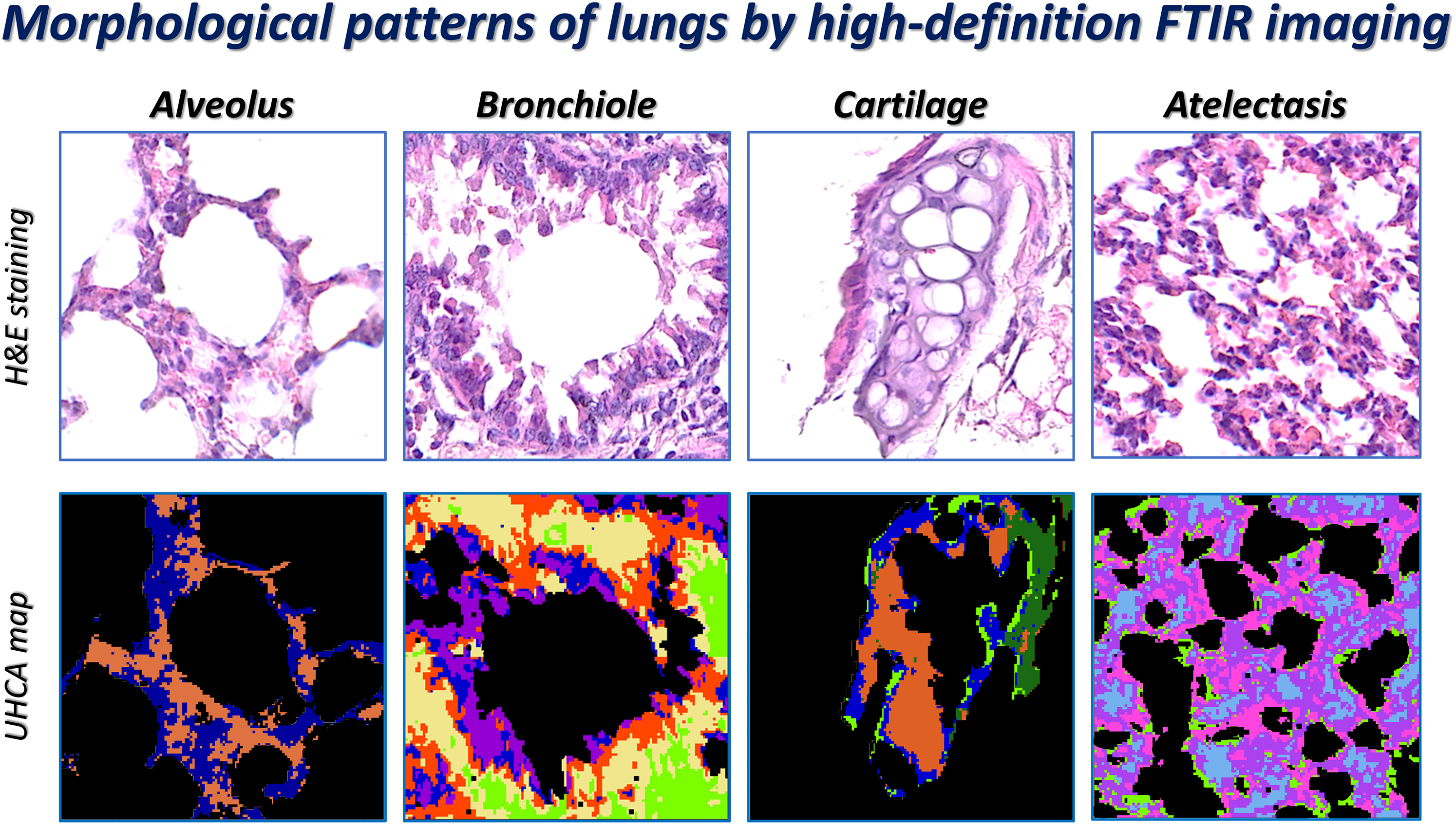
Label-free molecular imaging is a promising utility to study tissues in terms of the identification of their compartments as well as chemical features and alterations induced by disease. The aim of this work was to assess if higher magnification of optics in the Fourier transform infrared (FT-IR) microscope coupled with the focal plane detector resulted in better resolution of lung structures and if the histopathological features correlated with clustering of spectral images. FT-IR spectroscopic imaging was performed on paraffinized lung tissue sections from mice with optics providing a total magnification of 61× and 36×. Then, IR images were subjected to unsupervised cluster analysis and, subsequently, cluster maps were compared with hematoxylin and eosin staining of the same tissue section. Based on these results, we observed minute features such as cellular compartments in single alveoli and bronchiole, blood cells and megakaryocytes in a vessel as well as atelectasis of the lung. In the case of the latter, differences in composition were also noted between the tissue from the non-cancerous and cancerous specimen. This study demonstrated the ability of high-definition FT-IR imaging to evaluate the chemical features of well-resolved lung structures that could complement the histological examination widely used in animal models of disease.
Keywords
Get full access to this article
View all access options for this article.
References
Supplementary Material
Please find the following supplemental material available below.
For Open Access articles published under a Creative Commons License, all supplemental material carries the same license as the article it is associated with.
For non-Open Access articles published, all supplemental material carries a non-exclusive license, and permission requests for re-use of supplemental material or any part of supplemental material shall be sent directly to the copyright owner as specified in the copyright notice associated with the article.