
Abstract
Loading cells with magnetic nanoparticles, and tracking their fate in vivo by high resolution MRI, is an attractive approach for enhancing the efficacy of cell-based therapies including those utilizing hematopoietic stem cells, neuroprogenitor cells, and T cells. The transfection agent (internalization agent) assisted loading with the Feridex IV® nanoparticle is an attractive method of loading because of the low cost of materials, and possible low regulatory barriers for eventual clinical use. We therefore explored the interaction between Feridex IV® and three internalization agents protamine (PRO), polylysine (PLL), and lipofectamine (LFA). Feridex reacted with internalization agents to form aggregates, except when either the internalization agent or Feridex was present in large excess. When Jurkat T cells were incubated with Feridex/LFA or Feridex/PRO mixtures, and washed by centrifugation, nanoparticle aggregates co-purified with cells. With C17.2 cells large iron oxide particles adhered to the cell surface. At 30 μg/mL Feridex and 3 μg/mL LFA, internalization was largely mediated by LFA and was largely cytoplasmic. However, we found that the conditions used to label cells with Feridex and transfection agents need to be carefully selected to avoid the problems of surface adsorption and nanoparticle precipitation.
Introduction
Loading cells with magnetic nanoparticles, and tracking their fate in vivo by high-resolution MRI, is an attractive approach for enhancing the efficacy of novel cell-based therapies, including those utilizing hematopoietic stem cells [1], neuroprogenitor cells [2,3], and immune-specific T cells [4]. There are three general methods of labeling cells with magnetic nanoparticles. In the first, cells internalize magnetic particles through general mechanisms such as fluid-phase pinocytosis or phagocytosis [2,5,6]. In the second, nanoparticles are designed with a surface that promotes internalization, such as through the attachment of a membrane translocating signal of the HIV tat peptide (tat-CLIO) [1,4,7]. In the third, a nanoparticle is used in conjunction with a transfection agent (internalization enhancing agent) to increase the uptake by a cell [8–10]. An attractive version of the third method utilizes Feridex IV®, an approved MR contrast agent with internalization agents such as protamine sulfate (PRO), polylysine (PLL), and lipofectamine (LFA), to minimize the costs and potential regulatory barriers associated with designing a magnetic nanoparticle for cell loading in a clinical setting. When Feridex IV® is used in conjunction with PRO, two key materials in the labeling protocol are approved drugs [10]. It would seem, however, that if this approach is to be successful, the interaction between Feridex and the internalization agent must be fully understood to properly design cell-labeling protocols. Magnetic nanoparticles, like colloidal particles generally, feature a very high surface area-to-weight ratio, and the correct surface charge is crucial for maintaining a dispersed state for these types of materials[11]. This report began as an effort to develop a method of loading cells using Feridex and internalization agents, particularly of the Feridex/PRO combination, that might make this the method of choice both for the research and clinical applications because of cost, availability, and lack of regulatory barriers. We therefore explored the interaction between Feridex IV®, an approved nanoparticle-based MR contrast agent, and three internalization-enhancing materials PRO, PLL, and LFA under a wide range of conditions, measuring changes in surface properties as changes in nanoparticle size determined by photon correlation spectroscopy. We found that the internalization enhancing agents could cause Feridex agglomeration and precipitation, as might be expected from the addition of positively charged materials to the Feridex nanoparticle which exhibits a negative Zeta potential [12]. A full understanding of the complex changes in Feridex induced by its reaction with internalization agents is essential to obtain nanoparticle-loaded cells with the desired level and cytoplasmic disposition of iron.
Materials and Methods
Two superparamagnetic iron oxide nanoparticles, Feridex IV® and MION-47, were employed. Feridex IV® (Advanced Magnetics, Cambridge, MA) is an approved Kupffer cell directed MR contrast agent whose physical properties have been described [13,14]. (Feridex IV is a registered trademark of Advanced Magnetics. We refer to the iron nanoparticle from Feridex IV as Feridex.) MION-47 is a monodisperse nanoparticle, similar to the drug Combidex (USAN name ferumoxtran or referred to as AMI-227), whose physical properties have been described [13–15]. MION/Combidex has completed Phase III studies as an MR contrast agent for lymph node imaging [16–18].
Three internalization-promoting (transfection) agents were used. Protamine sulfate (PRO) was the injectable drug (American Pharmaceutical Partners, Los Angeles, CA), poly-
Incubations of internalizing agents and Feridex were in PBS (Figures 1 and 2). Size was determined with a Zetasizer HS1000 (Malvern, Marlboro, MA) as the effective diameter. C17.2 cells (from Dr. Even Snyder, Boston, MA) are a mouse neuroprogenitor cell line and were cultured in DMEM supplemented with 10% FBS, 5% HS, 2 mM
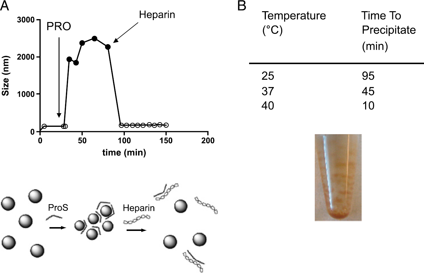
Interaction between Feridex IV and PRO. (A) Aggregation of Feridex IV in response to PRO monitored by light scattering. To Feridex IV, with a diameter of 170 nm, was added 3 μg/mL PRO, which caused the formation of materials of about 2200 nm. Heparin (3000 μg/mL) antagonized this effect of PRO and returned Feridex to its original size. (B) Effect of temperature on the time required for PRO to induce precipitation of Feridex. PRO/Feridex: 30 μgFe/mL, 3 μg/mL.

Interaction between Feridex and transection agents determined by light scattering. Values are in units of nanometers and were gathered after 2 hr at 37°C. (A) PRO and Feridex, (B) PLL and Feridex, (C) LFA and Feridex. PRO and PLL are too small to detect, whereas Feridex without a transfection agent has a size between 162 and 173 nm (n = 4). LFA (C) showed a concentration-dependent size (see data at 0 μgFe/mL). (D) The size of MION-47 was unaffected by PRO. Size of MION-47 at 10 μgFe/mL could not be determined. The formation of aggregates or precipitates depends critically on the nature of the nanoparticle (Feridex vs. MION) and concentrations of iron and transfecting agents.
Jurkat T cells were from the American Tissue Culture Collection (ATCC, Manassas, VA, USA) and were cultured according to the manufacturer's instructions in RPMI 1640 medium supplemented with 10% FBS, 10.6 mM
Results
The addition of PRO to Feridex caused a rapid increase in the effective diameter by light scattering of the nanoparticle from 170 to 2000–2500 nm (Figure 1A). The formation of aggregates caused by the positively charged PRO was reversed by negatively charged heparin, similar to the reaction that occurs when protamine reacts with heparin to reverse its anticlotting action after surgery. The rate of precipitate formation after addition of PRO to Feridex was temperature sensitive, with visible precipitation requiring as much as 95 min (25°C) or as little as 10 min at 40°C (Figure 1B).
We next examined the interaction between Feridex and three internalization agents, PRO, PLL, and LFA at 37°C over a range of iron and internalization agent concentrations using laser light scattering (Figure 2). PRO reacted with Feridex to form suspended aggregates or visible precipitates over a wide range of conditions, the exception being when there was a high ratio of Feridex to PRO or a high ratio of PRO to Feridex (Figure 2A). Similar results were obtained with PLL, which like PRO is a highly positively charged protein (Figure 2B). LFA, a positively charged lipid, exhibited a concentration-dependent size, increasing from 410 to 2257 nm (in the absence of Feridex) (Figure 2C). A reaction between LFA and Feridex was evident at 1 μg/mL, where a particle of 1760 nm was obtained that was larger than either of the two reactants (Feridex at 170 nm and LFA at 410 nm). Only at high concentrations of Feridex and LFA was a precipitate formed. Unlike Feridex, MION-47 did not to react any of the three internalization agents (Figure 2D).
Based on Figure 2, we selected concentrations of Feridex (30 and 100 μgFe/mL) and LFA (3 μg/mL) that were very different from conditions where precipitation occurred and incubated the combination with C17.2 cells. These adherent cells were washed by gentle agitation and stained for iron and HE, or washed followed by lysis for determination of cell-associated iron (Figure 3). A brief exposure to Feridex (30 μg Fe/mL) at 4°C, where endocytosis is blocked, caused an occasional iron-staining particle to be adsorbed to the surface of cells. A longer exposure to Feridex at 37°C under these conditions produced an increase in cell-associated iron from 31 to 167 ng (compare 30 μgFe at 2 hr vs. 30 μgFe/mL at zero minutes), which could be either surface adsorption or endocytosis. Addition of LFA increased cell-associated iron to 388 ng (30 μg Fe/mL, 2 hr at 37°C), which was present as a large number of small iron deposits throughout the cell. At 100 μgFe/mL, substantial uptake of iron occurred without LFA (462 ng) but was increased to 831 ng by LFA.

Interaction of Feridex and LFA with C17.2 cells. Cells on chamber sides were incubated with Feridex or Feridex plus LFA, stained for iron and HE, and examined by microscopy. Identical slides were incubated with lysis buffer and assayed for iron and cell-associated iron given. There were approximately 50,000 cells per slide.
We next incubated Jurkat T cells with Feridex IV with and without LFA and PRO. However, the conditions used to precipitate the suspended cells (2000 rpm, 5 min) resulted in precipitation of Feridex/LFA or Feridex/PRO complexes in a cell free blank (Figure 4). With PRO, 11 μg Fe of the 25 μg Fe in the tube (50 μgFe/mL at 0.5 mL) precipitated. As was expected based on Figure 2D, MION-47 showed almost no interaction with LFA.
Discussion
The use of transfection agents with Feridex as a method to induce cells to internalize nanoparticles was highly sensitive to the concentrations of both materials. With the two highly positively charged proteins, PLL (15–30 kDa) and PRO (4–5 kDa), the reaction with Feridex was similar to equivalence-type reactions seen, for example, when proteins and antibodies react in solution (Figure 2A and B). In equivalence reactions, two materials with multiple binding sites on each materials form aggregates over a wide range of conditions, the exception being when one material is in vast excess. Consistent with this model, the size of Feridex was unaffected with PLL in excess (10 μg Fe/mL Feridex/30 μg/mL PLL) or Feridex in excess (300 μgFe/mL/1 μg/mL PLL) (Figure 2B). The equivalence model for the reaction of Feridex and PLL is shown schematically in Figure 5. The model indicates that a lack of precipitation occurs when an excess of free PLL is used to completely coat Feridex, or when an excess of Feridex is incompletely coated with PLL. However, avoiding precipitation by using an excess of PLL may result in cytotoxicity due to free PLL [19], whereas use of Feridex excess can result in nonspecific adsorption to the cell surface (Figure 3). The interaction between LFA and Feridex was less prone to precipitate formation (Figure 2C), perhaps because nanoparticle/lipid complexes are less dense than nanoparticle/PLL complexes. Phospholipids like LFA have specific gravities less than 1, whereas proteins and iron oxides have specific gravities greater than 1.

Interaction of Feridex IV and LFA or PRO with Jurkat T cells. Media with or without cells were incubated (4 hr, 37°C) with 50 μgFe/mL iron and 5 μg/mL LFA or PRO as indicated. Pellets were washed by centrifugation, lysis buffer was added, and iron was determined by relaxometry. There were approximately 2,500,000 cells per tube.

Interactions between Feridex and PLL. When Feridex is in excess over PLL, some coating of Feridex occurs but the size of Feridex is unchanged. When PLL is in excess, it coats Feridex and free PLL exists. At equivalence, a precipitate forms.
It appears that a small subpopulation of large particles within the Feridex mixture was prone to adsorb to the surface of C17.2 cells even in the absence of LFA. Feridex is a polydisperse colloid consisting of iron oxide crystals (4.8 nm in diameter) present in clusters ranging from 20 to 1000 nm and contains a small number of particles as large as a micron (see Figure 9 of Ref. [13]). After 2 hr at 37°C, 462 ng of iron was cell-associated which corresponded to only 0.92% of the total iron (total iron 50 μg/well). Smaller particles within the Feridex mixture seem to interact with LFA and eventually become cytoplasmic.
The selection of a magnetic nanoparticle and method of loading cells, from any of the three general loading methods outlined above, will likely use a variety of cytotoxicity and cell functional assays, to demonstrate the normal function of nanoparticle loaded cells. Of particular concern are potential complications of using nanoparticle loaded cells as therapeutic agents in a clinical setting where complications might arise that are difficult to model using in vitro assays. The adsorption of nanoparticles to the cell surface is particularly undesirable because this modification of cells might induce an unwanted host immune response to those cells, a response that cannot be modeled with current in vitro assays.
A useful negative control for the reaction of Feridex with internalization agents was provided by MION-47, which did not form aggregates with these materials either in PBS (Figure 2D) or in the more complex media used with Jurkat T cells (Figure 4). Unlike Feridex, which is polydisperse and consists of aggregates ranging from 10 to 1000 nm, MION-47 is monodisperse with a narrow size distribution of 20–25 nm (Figure 2D). Although both materials have a dextran coating, the coating of MION-47 is thicker [14]. MION-47 has a nearly neutral Zeta potential of–2.0 mV, whereas Feridex has considerable more negative charge with a Zeta potential of–41 mV [12]. This combination of factors, polydispersity, a thinner coating of dextran, and more negative charge, causes Feridex to react with positively charged transfection agents far more strongly than MION-47.
With adherent C17.2 cells, there were two types of cell-associated iron apparent from the micrographs shown in Figure 3. Iron staining particles on the order of a micron in size were adsorbed to the cell surface of cells, an example of which is shown in the absence of LFA at 4°C, where endocytosis does not occur, and the exposure time of cells to Feridex was less than a minute. Increasing the temperature to 37°C and exposure time to 2 hr increased this form of iron so that with 100 μgFe/mL and 37°C numerous large particles could be seen. With the addition of LFA, a smaller, cytoplasmic iron was seen, and this was a predominant form of iron at 30 μgFe/mL and 3 μg/mL LFA. At 100 μgFe/mL, the smaller, cytoplasmic iron could be seen, but it appeared there were some large iron particles as well. With Jurkat T cells, Feridex/LFA aggregates co-purified with cells by centrifugation (Figure 4), and provided a high background that precluded an analysis of cell loading.
The reaction between Feridex and internalization agents alters the surface chemistry of the nanoparticle, and through changes in surface chemistry causes the formation of micron-sized aggregates. The alternative possibility, that internalization agents might induce Feridex internalization by affecting the cell rather than interacting with the nanoparticle surface, does not seem to be the case. Important variables in the formation of Feridex/internalization agent complexes included: (i) the time and temperature of the Feridex/internalization agent incubation (Figure 1), (ii) the presence of internalization agent binding materials like heparin (Figure 1), (iii) the nature of the transfecting agent (Figure 2), (iv) the concentration of transfecting agent and the concentration of Feridex (Figure 2), and (v) the nature of nanoparticle (Feridex vs. MION-47). A wide range of substances present in cell culture media but absent in PBS, substances like serum proteins or divalent cations, could modify results from those we obtained in PBS and are shown in Figures 1 and 2. However, with the media used for Jurkat T cells (RPMI 1640 medium supplemented with 10% FBS, 10.6 mM glutamine and penicillin/streptomycin), results were qualitatively similar to those obtained with PBS (compare Figures 2 and 4). With both RPMI and PBS, PRO caused greater Feridex precipitation than LFA, and in both types of media Feridex was far more prone to precipitate than MION-47.
We did find conditions (30 μg/mL Feridex) where the addition of LFA (3 μg/mL) did not produce aggregate formation (Figure 2), where LFA increased cell-associated iron with C17.2 cells (Figure 3), and where the predominant form of cell-associated iron was a large number of small iron oxide particles (Figure 3). However, in our hands, the use of Feridex and internalization agents for loading cells suffered from two problems, the adsorption of large aggregates within the Feridex mixture to the cell surface (in the absence of an internalization agent), and the formation of aggregates of Feridex (in the presence of an internalization agent). In light of the complexity of the interaction between Feridex and internalization agents, conditions used to label cells for tracking by MRI need to be carefully examined if the adsorption of nanoparticles onto cells or precipitation with cells are to be avoided.
Footnotes
Acknowledgments
We thank Dr. Evan Snyder for supplying C17.2 cells. This research was funded in part by R01-EB00662-A01 and R01 EB004626.