
Abstract
Numerous macromolecular MRI contrast agents prepared employing relatively simple chemistry may be readily available that can provide sufficient enhancement for multiple applications. These agents operate using a ~100-fold lower concentration of gadolinium ions in comparison to the necessary concentration of iodine employed in CT imaging. Herein, we describe some of the general potential directions of macromolecular MRI contrast agents using our recently reported families of dendrimer-based agents as examples. Changes in molecular size altered the route of excretion. Smaller-sized contrast agents less than 60 kDa molecular weight were excreted through the kidney resulting in these agents being potentially suitable as functional renal contrast agents. Hydrophilic and larger-sized contrast agents were found better suited for use as blood pool contrast agents. Hydrophobic variants formed with polypropylenimine diaminobutane dendrimer cores created liver contrast agents. Larger hydrophilic agents are useful for lymphatic imaging. Finally, contrast agents conjugated with either monoclonal antibodies or with avidin are able to function as tumor-specific contrast agents, which also might be employed as therapeutic drugs for either gadolinium neutron capture therapy or in conjunction with radioimmunotherapy.
Introduction
There is a requirement of only a 1/100-fold concentration of Gd(III) atoms for an MRI contrast agent to provide adequate contrast enhancement comparable to that obtained using the required concentration of iodine atoms for a CT contrast agent. The result of this physical phenomenon has been the development of a multitude of diverse small molecular weight chemical reagents for use as MRI contrast agents. Somewhat in contrast to the extensive efforts pertaining to the creation of novel small molecules has been the now burgeoning interest in macromolecular MR contrast agents. Considerable advantages resulting from clearance and targeting properties in conjunction with enhanced molar relaxivities make this area of research attractive. In this review, we discuss the relationship between chemical characteristics and pharmacological properties of macromolecular MRI contrast agents with dendrimer cores to provide some basis for strategies to synthesize and to use optimal agents for specific purposes.
Three fundamental types of macromolecular contrast agents for MRI have been reported. One employs serum albumin conjugated with forms of diethylene-triaminepentaacetic acid (DTPA) to chelate the gadolinium metal ions [albumin-(DTPA-Gd)30–34], originally described by Ogan et al. [1]. Other researchers have made extensive use of these reagents to investigate vascular anatomy and physiology especially in cancer [2–4]. However, the use of a protein platform or core makes it difficult to synthesize routinely and consistently a variety of agents that possessed similar chemical characteristics. The other two basic types of agents have generally been linear polyamine-conjugated chelates [DTPA derivatives or 1,4,7,10-tetraazacyclododecane tetraacetic acid (DOTA) derivatives]. The coupling methods of cores with those chelates to form MRI contrast agents are similar and either chelating agent could substitute for the other. DOTA is a slightly larger molecule and is noteworthy for forming highly stable metal complexes, somewhat more so than DTPA. Margerum et al. [5] studied the physical and phamacokinetic properties of dendrimers coupling with DOTA chelate, which behaved similarly to our dendrimer-1B4M agents. However, while neither free chelate exhibited any immunogenecity, when conjugated with a macromolecule such as an antibody, DOTA was found to be more immunogenic than DTPA [6]. Thus, in all of our studies for dendrimer-based contrast agents, a DTPA derivative was employed to chelate Gd(III) ions. However, those agents termed gadomers were commercially prepared with DOTA on the molecule to complex the Gd [7,8].
Numerous synthetic molecules such as polylysine [9–12], dextran [13–15], polyethylene glycol [16,17], a brush-like co-polymer of polylysine and dextran [18], synthetic peglated linear co-polymer with polylysine [19], have been investigated for their suitability as a core of the contrast agent rather than albumin. Thus, the vast majority of polymers, which were employed as cores, have been molecules of linear, branched linear, or circular chains. While convenient and reasonably available, limitations to the use of linear polyamines include difficulties in the synthesis, characterization, and purification of the agent to produce a final product of a single physical size with consistent chemical characteristics. Therefore, since polyamidoamine (PAMAM) or diaminobutane (DAB) dendrimers and gadomers, which are based on a different type of synthetic polymers made by Schering (Berlin, Germany) [7,8], are spherical molecules, the advantage of their use as cores or platforms for forming contrast agents is the ability to better control the physical sizes of the molecules.
Dendrimers are a class of highly branched synthetic spherical polymers consisting of a vast array of types, chemical structures, and functional groups [20]. Two types of dendrimers, PAMAM [21] and DAB dendrimers, are commercially available [22]. They are highly soluble in aqueous solution and both have a unique surface topology of primary amino groups [21,23] (Figure 1). The defined structure and large number of available surface amino groups of these dendrimers have led to their use as substrates for the attachment of large numbers of chelating agents to a single antibody molecule [23–27]. These dendrimers have enabled the synthesis of numerous MRI contrast agents that possess very similar chemical structures but also cover a wide range of molecular weights. From this point of view, gadomers have similar properties to the dendrimer-based agents. The internal structures of gadomers, which possess aromatic rings, are simpler, resulting in having smaller size than polyamine dendrimer-based agents of same generations [7]. Therefore, although we have not had a chance to perform direct comparison studies between dendrimer-based agents and gadomers so far, even Gadomer-17, which had the largest size in gadomers, seemed to clear much more rapidly, perfuse greater into extravascular spaces, and excrete greater into the urine than dendrimer-based agents of same generations (G3) [7,28–31].
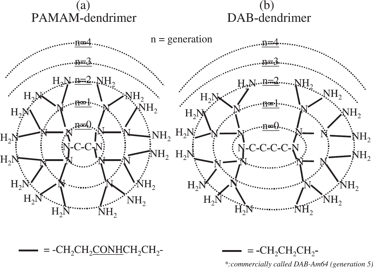
Schema of the dendrimer used as cores of contrast agents.
We have recently synthesized novel macromolecular MRI-contrast agents with different molecular weights ranging from 29 to 3850 kDa to change blood retention, tissue perfusion, and excretion rate and pathway [30,32–34]. All of these were prepared using generation −2, −3, −4, −5, −6, −7, −8, −9, and −10 PAMAM dendrimers (Figure 1a) conjugated with a bifunctional DTPA derivative, the 1B4M-DTPA. This type of bifunctional DTPA derivative, which has been extensively studied for radioimmunoimaging and radioimmunotherapy, is a component of the recently approved radiolabeled monoclonal antibody non-Hodgkin's therapeutic, Zevalin [35,36]. In addition, these MRI agents have been chemically modified with elements of polyethylene glycol [37]. The dendrimer core unit has been replaced with the generation −2, −3, −4 polypropylenimine DAB dendrimer (Figure 1b) [38,39]. Furthermore, these dendrimers have also been conjugated to specific target molecules (monocloncal antibody or avidin) to evaluate the potential for molecular targeting of these imaging agents [27,40] (Tables 1 and 2).
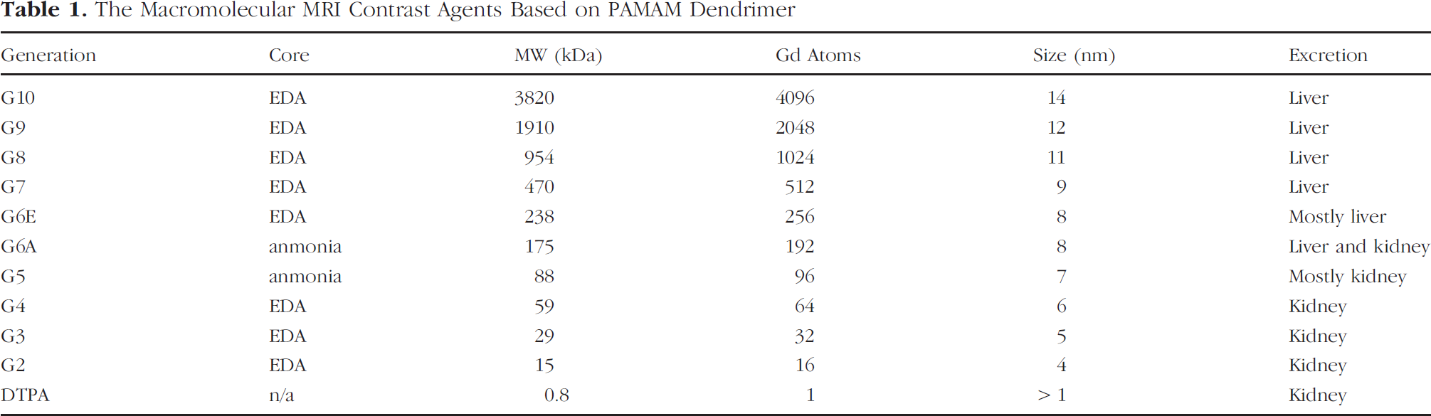
The Macromolecular MRI Contrast Agents Based on PAMAM Dendrimer
Methods
Preparation of Dendrimer-Based Contrast Agents
Typically, dendrimers were obtained from commercial sources, concentrated for use, and thereafter reacted at 40°C with molar equivalent amounts of the 2-(p-isothiocyanatobenzyl)-6-methyl-diethylenetriamine-pentaacetic acid (1B4M-DTPA) [23,33] equal to the number of surface amine residues on the dendrimer molecule. Additional amounts of 1B4M equal to this initial reaction ratio were added as a solid after 24 hr to each reaction. Routinely, over 98% of the amine groups on the dendrimers were reacted with the 1B4M thereby providing relatively consistent products for comparative evaluation [41].
Dendrimer-1B4M conjugates were mixed with Gd(III) citrate and after complex formation, the excess Gd(III) in each preparation was removed by diafiltration. A replacement assay that employed 153Gd as a tracer demonstrated that the number of 1B4M chelators of the dendrimer-1B4M conjugates chelating Gd(III) atoms ranged from 75% to 90% [41]. The purified contrast agents were obtained by diafiltration with appropriate filtration membrane and analyzed by size-exclusion HPLC (SE-HPLC) using a TSK G2000SW column.
The Variations of Macromolecular MRI Contrast Agents

Chemical linkage between the dendrimer-1B4M species and either PEG, antibodies or avidin molecules was performed by reaction between a maleimide residue introduced by a heterogenous cross-linker, sulfosuccinimidyl 4-[N-maleimidomethyl] cyclohexane-1-carboxylate (Sulfo-SMCC), on the dendrimer, while a sulfhydryl group was introduced on the protein using 2-iminothiolane [27,37]. Linkage of the two components was performed employing routine protocols and the desired conjugates were obtained by purification via diafiltration and SE-HPLC.
Characteristics for the chemically conjugated variations of all of these macromolecular contrast agents are shown in Table 2.
MRI Methods
A 1.5-tesla superconductive magnet unit (Signa Horizon or Signa LX GE, Milwaukee, WI) and either a commercially available high-resolution wrist coil or dual 3-in. phased array surface coils with a custom mouse holder or a custom 1-in. bird-cage surface coil were employed for all MRI studies. The mice were anesthetized with 1.15 mg of sodium pentobarbital (Dainabot, Osaka, Japan) and placed in the center of the coils. The T1-weighted 3-D fast spoiled gradient-echo technique [frequency encoding × phase encoding steps 256–512 × 192–512; two to four excitations; slice encoding steps 12–48] with fat-suppression technique and serial 3-D data acquisition were used for all mice studied. The coronal images were usually reconstructed with 0.6–2 mm thick sections with 0.15–1 mm overlap. The field of view was 6–8 × 3–4 cm and the size of each voxel was 0.1–0.3 × 0.1–0.4 × 0.6–2 mm3. The serial dynamic data were analyzed using Advantage Windows (GE). In addition, whole body 3-D MRIs were reconstructed with the maximum intensity projection (MIP) method with an Advantage Windows (GE). Details of individual experiments can be found in the respective listed references.
Applications
Blood Pool MR Contrast Agents
Macromolecular contrast agents for MRI have been reported to be excellent agents for evaluating microvasculature and histological capillary density in tumor tissues [3,42,43]. Although postcontrast signal changes in the tumors showed better correlation with histological capillary mass, there are no reports that evaluated the lower limit of vessel size that can be visualized by MRI using macromolecular contrast agents.
Hydrophilic macromolecules of the size and molecular weight range of albumin or antibodies are retained in the circulation and do not leak out from the vessels rapidly. To assess relative pharmacokinetics and in vivo enhancement characteristics, the generation −2, −3, −4, −5, −6, −7, −8, −9, and −10 PAMAM dendrimers conjugated with 1B4M were synthesized and were evaluated as blood pool MRI contrast agents via biodistribution studies and dynamic micro-MRI using normal mice [30,32,33].
Renal excretion
The G2, G3, and G4 dendrimer-based agents were quickly excreted via the kidney primarily during the first pass as determined by biodistribution and excretion studies. The G5 and G6 dendrimer-based agents were more slowly excreted via the kidney (Figure 2). The G7, G8, G9, and G10 dendrimer-based agents showed minimal excretion from the kidney (Figure 3) [30,32].
Extravasation
The G2 and G3 dendrimer-based agents were quickly distributed from the circulation into the soft tissue similarly to Gd-DTPA resulting in compromised contrast quality between the blood vessels and the surrounding background tissue [30]. Therefore, molecules smaller than 5 nm diameter can quickly leak out of the vasculature into surrounding tissues, even from normal vessels. In contrast, somewhat larger molecules can leak out from the tumor vasculature than from normal vessels. However, molecules larger than 8 nm diameter (i.e., the G6 and larger dendrimer agents), showed minimal leakage even from tumor vessels into surrounding tumor tissue [44–46]. The G4 and G5 agents showed intermediate characters between the larger and smaller dendrimer-based contrast agents. The G4 and G5 agents did not exhibit measurable leakage from the normal vessels before those agents were cleared from the blood pool [30]. However, the G4 leaked out of the tumor vessels much more rapidly than both the G6 and G8 [34,44,47].
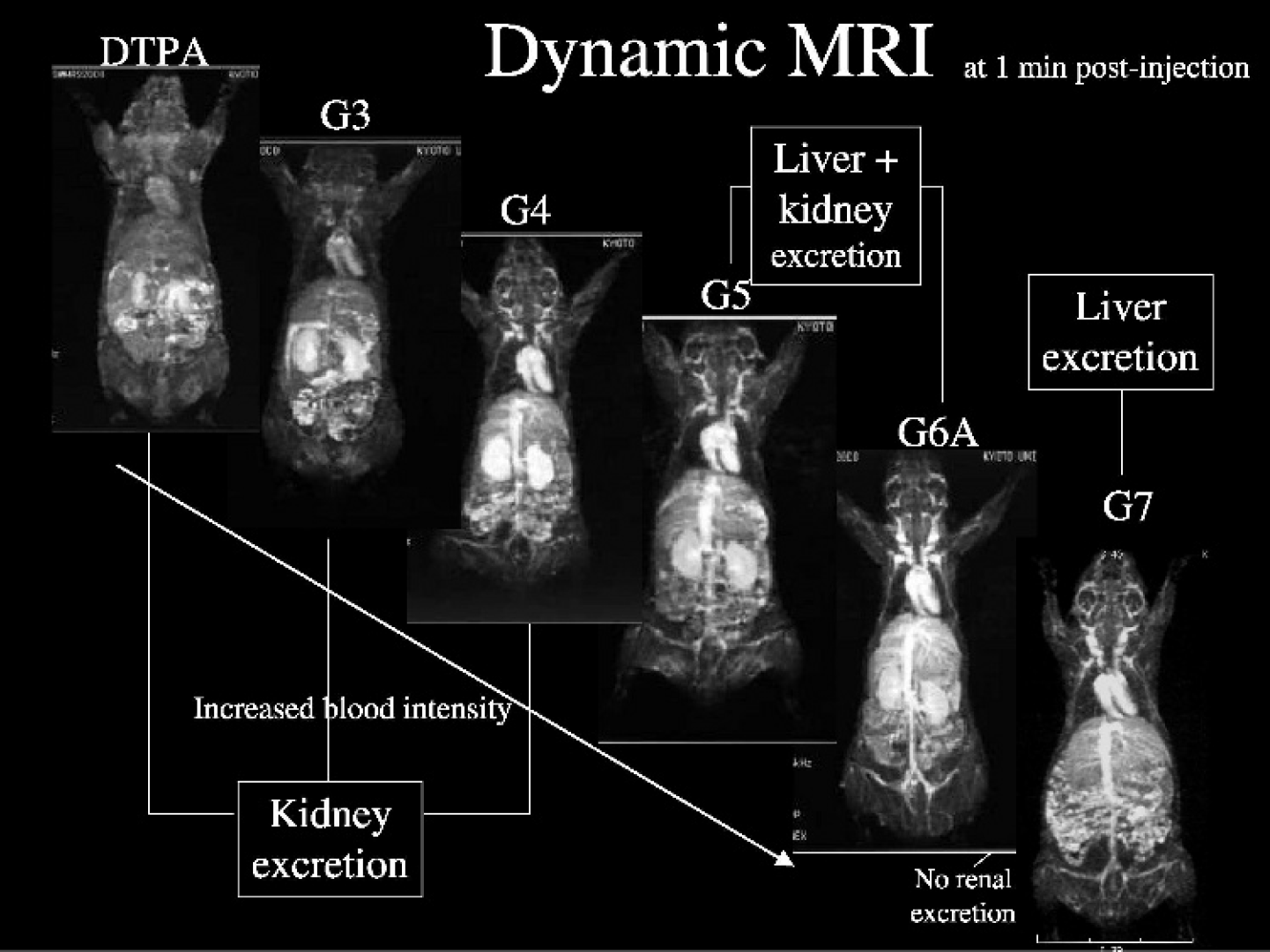
Whole body 3-D MRI (MIP) obtained with 0.03 mmol Gd/kg of PAMAM G3, 4, 5, 6A, and 7 contrast agents at 1 min postinjection.
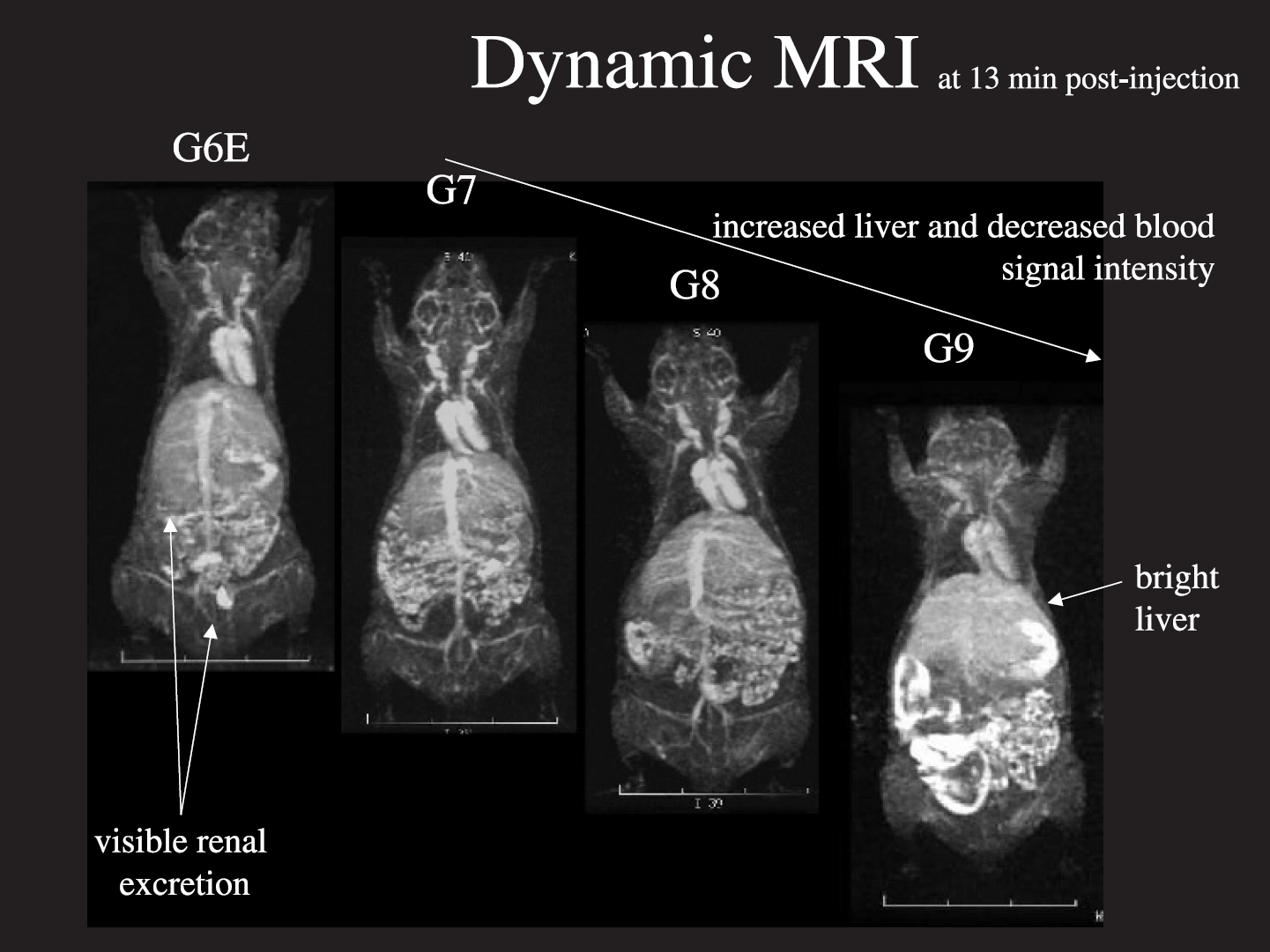
Whole body 3-D MRI (MIP) obtained with 0.03 mmol Gd/kg of PAMAM G6E, 7, 8, and 9 contrast agents at 13 min postinjection.
Entrapment by the reticuloendothelial system
In contrast to the somewhat smaller G8 dendrimer agent, the G9 and G10 dendrimer-based agents were quickly taken up and trapped in the reticuloendothelial system (liver and spleen) resulting in rapid clearance from the circulation (Figure 3) [34]. This result was attributed principally to the size of these charged molecules, which are large enough to be recognized by the reticuloendothelial system.
Increased hydrophilicity
The G4 dendrimer-based agent conjugated with either one or two polyethyleneglycol (PEG) chains of MW = 20,000 had decreased liver accumulation and kidney excretion as compared to the base macromolecule. This resulted in lengthy survival of the contrast agent in circulation and good visualization of the blood vessels [37,39] as compared to the unmodified base agent.
Effect of lysine co-injection
The use of a co-injection of lysine have been employed to minimize renal uptake of smaller molecules such as radiolabeled peptides [48–50]. Co-injection of lysine with the G4 dendrimer-based agents was also found to accelerate urinary excretion of this contrast agent in its intact form [47]. This was interpreted as being caused by neutralizing or saturating the negative charge on the membrane of the proximal tubules with positively charged lysine molecules.
In summary, the G7 and G8 dendrimer-based agents were found to be the best candidates for visualizing blood vessels clearly among all of the dendrimer-based agents described above. PEG conjugation appeared to improve the ability of these agents to visualize blood vessels clearly. In addition, excretion of these PEG conjugated contrast agents was increasing in spite of their increased molecular sizes. Co-injection of lysine with the G4 dendrimer-based agents can accelerate the urinary excretion of this contrast agent in its intact form [47]. However, in terms of realistic potential general clinical application, the G4 dendrimer-based agent is probably a viable candidate because of a reasonable balance between the ability for visualization of vessels and a respectable excretion rate from the kidney.
Liver MRI Contrast Agents
Hydrophobic macromolecules generally tend to accumulate rapidly in the liver. In accordance to this concept, [Gd(BOPTA)(H2O)]2− (MultiHance) and [Gd(EOB-DTPA)(H2O)]2− (Eovist) were synthesized and applied for use in clinical studies as liver-specific MRI contrast agents. DAB dendrimers have a pure aliphatic polyamine core in contrast to the PAMAM dendrimers with an amide functional group core component and also have recently become commercially available. Based on the general principle that hydrophobicity targets the liver, the DAB-Am64-(1B4M-Gd)64, generation 5 with 64 surface amines, was synthesized and evaluated as a liver MR contrast agent [38]. DAB-Am64-(1B4M-Gd)64 homogeneously enhanced the liver parenchyma and was excreted more rapidly through both the liver and kidneys than the analogous PAMAM dendrimer of similar molecular size [38]. Furthermore, dynamic micro-MRIs employing DAB-Am64-(1B4M-Gd)64 were able to visualize liver metastatic tumors of colon cancer cells (LS174T) as small as 0.3 mm in diameter in mice via a reverse contrast image wherein the tumor tissue appeared as a dark area and the normal liver tissue was a bright image (Figure 4) that persisted well past 24 hr and over time provided detailed structural details of the liver itself [38].

The delayed 2-D reconstructed micro-MR images of hepatic micrometastatic LS174T tumors in a nude mouse using 0.03 mmol Gd/kg of DAB-G4 contrast agents at 20 min postinjection (a) and the corresponding sectional surface on the stereoscopic microscopy (b). The scale indicates 1 mm. * = metastatic tumors; ST = stomach; D = duodenum; I = inferior vena cava; S = spine.
Renal Functional MR Contrast Agents
Renal functional MRI have been done previously only with Gd-DTPA [51–53]. Evaluation of the G2, G3, and G4 of both the PAMAM and the DAB dendrimer-based agents revealed that all of these contrast agents were quickly excreted from the kidney. However, these dendrimer-based agents also behaved very differently from the small size and molecular weight Gd-[DTPA]-like agents (Magnevist or Omniscan) in the kidney. After filtering through the glomerulus, the dendrimer-based agents were concentrated and the formation of a high-intensity band at the layer of the proximal tubules was observed. The absence or delayed formation of this high-intensity band correlated very well to the renal tubular function in an acute tubular necrosis model mice generated by the injection of cisplatin (Figure 5) [54].
Lymphatic MRI Contrast Agents
Large contrast agents injected intracutaneously and absorbed into the lymphatic system have been able to clearly visualize lymphatic vessels and lymph nodes [55]. A few groups have recently reported some success in experimental studies of magnetic resonance lymphangiography (MRL) using Gd-DTPA, liposomes, and Gd(III) macromolecular chelates in normal pig, rabbit, and rat models [8,56–59]. In addition, other groups have used intravenous injections of iron oxide particles, including ultra-small particles of ion oxide (USPIO) and ultra-small monocrystalline superparamagnetic iron oxide (MION), which negatively enhanced normal lymph nodes in rabbit and rat models on MR lymphography [60–66]. In contrast to those efforts, highly detailed positive image micro-MRL of both normal and lymphatic disease model mice with G8 dendrimer agents have recently been reported [55]. To date, this has been the only available method for visualizing the deep lymphatic system in mouse model (Figure 6). In addition, this method was able to distinguish the appearance of infectious expansion of lymphocytes from either chronic lymphoproliferative or neoplastic conditions [55]. The enhanced resolution of this method should have wide applicability to the study of immunology and cancer in both experimental animals and clinical medicine.

Normal and cisplatin induced acute renal tubular necrosis kidney disease model with various grades; functional kidney 2-D MRIs obtained with PAMAM-G4 agents at 7 min postinjection.

Whole body 3-D MRL (MIP) of an IL-15 transgenic, lymphoproliferative/lymphoma model mouse obtained with 0.01 mmol Gd/kg of PAMAM G8 contrast agents at 45 min post-intracutaneous injection (arrows indicate lymph nodes swelling).
Tumor-specific MRI Contrast Agents
In order to generate a specific signal from tumor tissue by MRI contrast agents, the signal needs to be effectively amplified by carrying large numbers of Gd(III) atoms on a ligand molecule or targeting receptors to antigens expressed in high numbers per single cell. Several successful studies have been reported with Gddendrimer or Gd-liposome conjugated with monoclonal antibodies, peptides, or folate [67–70]. Antibody- and avidin-dendrimer-1B4M-Gd contrast agents were synthesized to specifically target tumor cells and enhance MRI contrast [27,40]. An antibody was able to carry as many as 49 ions of Gd(III) per single molecule by this strategy [27]. Avidin-dendrimer contrast agents successfully targeted and were internalized into SHIN3 ovarian adenocarcinoma cells in vitro. In addition, the avidin-dendrimer contrast agents successfully accumulated in the intraperitoneally disseminated ovarian tumor (SHIN3) in a mouse model in vivo [40]. The concentration of Gd(III) ions was theoretically adequate to also perform gadolinium-neutron capture therapy by irradiation with epithermal neutrons. However, internalization of Gd(III) ions into the tumor cells compromised the T1-shortening effects as compared with the same concentration of avidin-dendrimer-1B4B-Gd in solution [40]. Dynamic MRI scanning might be useful for assessing the level of accumulation and internalization of the contrast agent in the tumor cells to determine the optimal timing for neutron bean irradiation. Nevertheless, to synthesize successful target-specific MRI contrast agents, optimal target selection in conjunction with full amplification of the signal should be pursued.
Summary
A brief description of the preparation and applications to date of recently synthesized possible variations of dendrimer-based MRI contrast agents has been provided. These agents will have numerous additional applications as MRI contrast agents by themselves as experimental tools for imaging functional anatomy of tumor vessels [34,44–46], specific organs [38,39,54], and the lymphatic system [55], especially to investigate the molecular basis of diseases by analyzing various transgenic and knockout mice as disease models. Alternatively, these chemical products would be useful as molecular probes for signal amplification of the tumor-targeting contrast agents using antibodies or receptor-targeted ligands [27,40,67] for noninvasive detection of tumor localization and possible MRI-guided Gd-neutron capture therapy for cancer.