
Abstract
Significance:
Wound dressing based on naturally derived polymer provides a useful platform for treatment of skin injuries. Owing to the high mechanical strength and tunable structural and physicochemical properties of human elastin-like polypeptides (ELPs), they may be used as excellent materials for fabricating biocompatible scaffolds and other products for wound management.
Recent Advances:
Designing recombinant ELPs mimicking natural elastin to fabricate synthetic polymers suitable for human health care has generated significant interest. ELP-based cell-adhesive biopolymers have been used as an alternative for successful sutureless wound closure due to the physicochemical characteristics of the extracellular matrix.
Critical Issues:
Different systems of ELPs are being developed in the form of scaffolds, films, hydrogels, photo-linkable sheets, and composites linked with various types of growth factors for wound healing application. However, optimizing the quality and safety attributes for specific application needs designing of recombinant ELPs with structural and functional modifications as needed for the intervention.
Future Direction:
Chronic wounds are difficult to treat as the wound repair process is interrupted by conditions such as excessive inflammation, impaired extracellular matrix formation, and persistent infections. Conventional therapies such as skin substitutes or autologous skin grafts, in many cases, are unable to reestablish tissue homeostasis and proper healing. The development of innovative materials could induce a better regenerative healing response. In this study, we are reviewing different types of elastin-based materials for wound care application and their future prospects in regenerative medicine.

Scope and Significance
Current strategies of chronic dermal wound treatments result in permanent deformities of the tissue owing to formation of an aberrant elastic fiber network. 1 The vital presence of elastin in wound healing helps in restoration of an intact and functional elastic fiber network to regain complete skin function after injury. 2 Elastin-based scaffolds have been shown to accelerate wound closure, improve the strength and flexibility of the healed area, enhance dermal regeneration, and reduce wound contraction. 3 Elastin-based materials are easily reproducible protein scaffolds with inherent biological cues that can enhance the surrounding tissue to augment the wound healing process. 4 –7 The highly biocompatible nonimmunogenic properties make the elastin-based biopolymer highly suitable for clinical application in wound management. 8 –11 The multifunctionality of elastin derivatives that benefit in reestablishing the native assembly and cell interaction during tissue repair is elaborated in this review (Table 1). 12 –14
List of the elastin-like polypeptide-based materials utilized for wound healing
AMP, antimicrobial peptide; EL, elastin; ELP, elastin-like polypeptide; ELR, elastin-like recombinamer; GelMA, gelatin methacryloyl; KGF, keratinocyte growth factor; MeTro, methacryloyl-substituted recombinant human tropoelastin; REP, RGD-containing ELP; SDF1, stromal cell-derived factor-1; SF, silk–fibroin.
Translational Relevance
Many elastin-based protein polymers with remarkable elasticity and self-assembling properties were constructed for application in chronic wounds. These smart, elastin biomimetic polymers were formulated into various systems such as porous scaffolds, hydrogels, and fibrous structures according to the application microenvironment and stimuli such as temperature, pH, and enzymatic activities in different pathobiological situations. 15 –21 The mechanical and biological properties were improved by cross-linking or by constituting elastin derivatives with other functional peptides or proteins. Elastin-like polypeptides (ELPs) in association with other self-assembly sequences derived from nonhuman sources such as silk, collagen, and resilin were reported to fulfill the requirement of an ideal support system for skin regeneration. 22
Clinical Relevance
Different types of bioactive or inert elastin-based systems have been exploited in various platforms for multiple tissue regeneration purposes. Many studies have demonstrated that fabrication of bioactive ELPs of diverse composition facilitates growth and spreading of a broad range of cell types, such as dermal fibroblasts and vascular endothelial and smooth muscle cells. 3 ELP-based biomaterials offer a versatile cell-organizing platform for regeneration with the mechanical support for repair of cutaneous wounds. 14,16
Background
Overview of chronic wounds
Chronic wounds are common in pathological conditions such as diabetes, vascular insufficiency, infections, local pressure areas, and trauma and burn injuries. Management of these wounds is considered to be one of the most difficult challenges with limited treatment options in the past decades. 23 Over 2% of the U.S. population is affected with chronic nonhealing wounds, characterized by prolonged and excessive inflammation, 24 and are prone to recurrent infections. 25 Approximately 25 billion dollars are spent on treatment of chronic skin wounds in the United States every year. 26 To minimize mortality and morbidity, appropriate therapeutic management of chronic wounds is most essential. Repair of these wounds is inhibited by various factors such as continuous recruitment of inflammatory cells in the wound area, which secrete serine proteases and matrix metalloproteinases that degrade extracellular matrix (ECM) proteins and inactivate growth factors required for the healing process. 24,25,27 One of the commonly used treatment strategies is the application of skin substitutes or autologous skin grafts over the area. These grafts and skin substitutes in several cases fail to reestablish tissue homeostasis, leading to significant complications. 23,28 Defective closure/repair of the wound may lead to microbial infection in the wound area delaying the process of healing, leading to necrosis, sepsis, and even death. 29 Therefore, an appropriate strategy or product that can facilitate wound healing is in high demand to improve the quality of life with reduction of morbidity and mortality.
Many different biomaterials (synthetic or natural) have been investigated for use as dermal substitutes and most of them can achieve partial restoration of tissue. It has been suggested that wound closure obtained by means of skin regeneration rather than repair would be the appropriate approach to wound management. 30 Due to ease of manipulation, synthetic materials such as polylactic acid, polyglycolic acid, polyethylene glycol, and polylactic-co-glycolic acid are being used in wound repair. 31 Although synthetic materials are suitable for various clinical applications, they have drawbacks such as alteration of cell adhesion, migration, cell-mediated degradation, and immunogenicity, while naturally derived biomaterials such as collagen, gelatin, chitosan, fibrin, and elastin provide many benefits for regenerative therapy because of low toxicity, nonimmunogenicity, and retention of biological activity. 32
Application of elastin-based materials on wound healing
Role of elastin in skin regeneration
Elastin is an important constituent of elastic fibers that maintain resilience and provide mechanical strength for many tissues such as skin, heart, blood vessels, and connective tissues. 33 In adults, elastin does not regenerate sufficiently following significant tissue loss after trauma or skin damage, leading to scar formation. 34,35 These scar tissues are different from original skin as they lack elasticity and are stiffer due to overproduction of collagen fibers by fibroblasts during the wound healing process. 36 Hence, supplementing elastin to the damage tissue may be highly advantageous, which can be achieved through ELPs. It was reported that elastin derivatives accelerate wound closure and improve the strength and flexibility of the healed region when applied to chronic wounds. They also accelerate the healing process essential for restoration of skin's barrier function in the short period after injury. The sol–gel transition of elastin derivatives allows complete coverage of the wound area, protecting it from external pathogens, and incorporation of antibacterial components further accelerates the healing process. 37,38
ELPs
ELPs are genetically engineered polypeptides based on the sequence found in the hydrophobic domain of human tropoelastin. They consist of a pentapeptide amino acid repeat, VPGXG, where X can be any amino acid except proline. 38 Numerous bioinspired materials have been generated by substituting the guest residue X, thereby precisely controlling their physical and functional properties to match the intended applications. 39 These polypeptides can undergo reversible inverse temperature phase transition at a certain temperature indicated as the transition temperature (Tt). These ELP molecules form coacervates when the temperature rises above Tt and remain soluble below Tt (Fig. 1). 13,14 ELPs can be easily expressed from a plasmid-borne gene in Escherichia coli and their phase transition behavior enables rapid purification on a large scale using inverse temperature cycling. 39 For regenerative medicine or tissue engineering where large quantities of polymers are essential, these ELPs may provide an approach to eliminate the expensive mode of purification by chromatography and replace it with simplified large-scale purification using the ITC method with relatively high yields (∼500 mg/L growth). 40
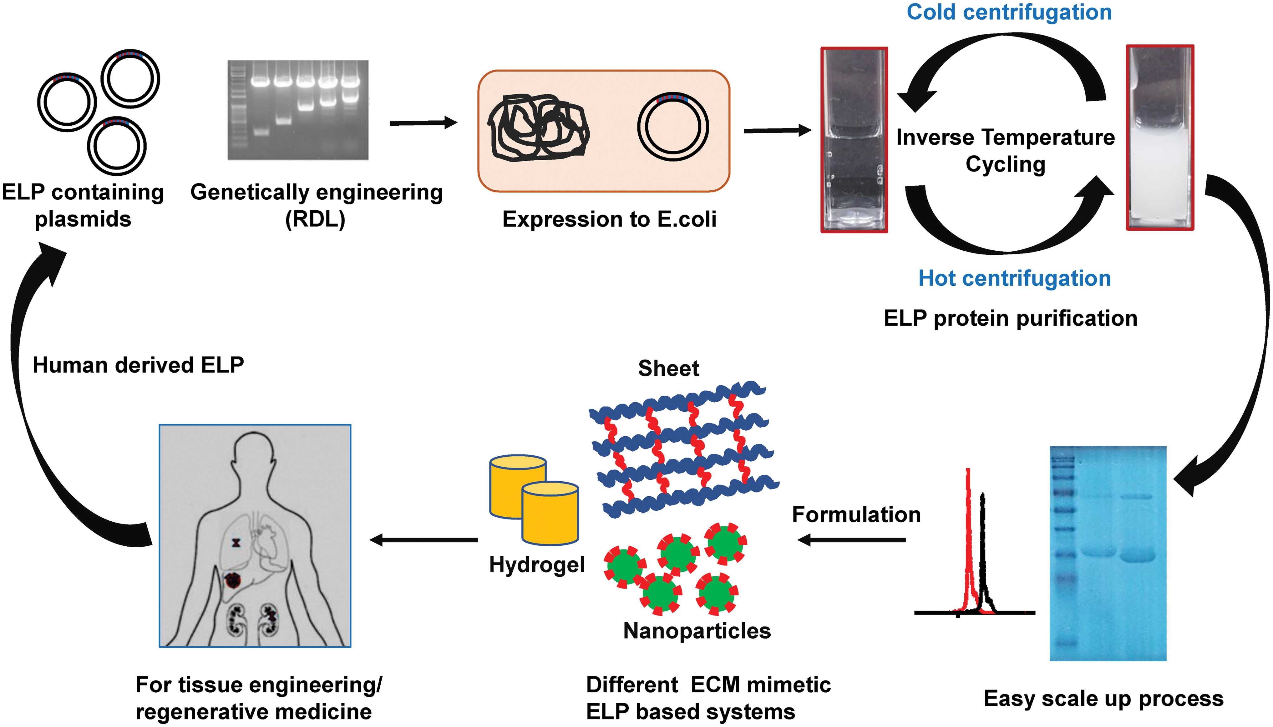
ELPs are recombinant proteins consisting of a pentapeptide repeat such as (VPGXG)n (where X can be any amino acid except proline) derived for human tropoelastin. They are synthesized using the recursive directional ligation method of cloning. ELPs are easily expressed from a plasmid-borne gene in Escherichia coli and purified on a large scale by exploiting their ITC. Thus, ELPs provide an approach to eliminate the expensive mode of purification by chromatography. The excellent architectural features and mechanical properties of ELPs allow generation of different systems for regenerative medicine for human health care. ECM, extracellular matrix; ELP, elastin-like polypeptide; ITC, inverse transition cycling. Color images are available online.
ELPs and ELP composites for wound application
The flexibility of ELPs allows easy association with additional functional proteins or reactive groups in distinct positions without loss of thermal sensitivity, which conveniently enables the conjugation of many growth factors involved in the process of healing. Fusion of growth factors (stromal cell-derived factor [SDF], keratinocyte growth factor [KGF], and epidermal growth factor [EGF], etc.) and wound healing components with ELPs allows formation of self-assembled nanoparticles at body temperature while retaining their biofunctional activity, thereby increasing their stability and prolonging exposure at the wound site, which are crucial for accelerating the healing process (Fig. 1). At the same time, inclusion of cell recognition sequences (RGD or REDV) along with ELPs can enhance cell adhesion, proliferation, and reepithelialization at the wound site. 41 –43
Furthermore, owing to stimulus-responsive self-assembly and desirable mechanical properties of ELPs, they may be utilized for development of a wide range of different matrices for wound management. Unlike electrospun collagen sheets that are highly degradable in nature, 44 electrospun ELPs or ELP composites are very stable, retain thermal sensitivity, and share functional resemblance with the native component of the ECM. 45,46 In association with other peptides, proteins, polysaccharides, or biopolymer residues, ELPs assist in fabrication of more complex architectures with enhanced functional properties. They have immense application potential for treatment of chronic wounds because of the easy maneuverability, which is otherwise difficult in case of other synthetic polymers. Various applications of ELP-based hydrogels formed by thermal-responsive coacervation, 47 physical cross-linking, 48 or chemical cross-linking of ELP solutions 49 have been developed for regenerative medicine (Table 2).
List of the elastin-like polypeptide-based materials exploited for regenerative medicine and tissue engineering
EGDE, ethylene glycol diglycidyl ether; EGF, epidermal growth factor; pAzF, para-azidophenylalanine; SELPs, silk–elastin-like transition cycling; ZELR, zipper–elastin-like recombinamer.
Discussion
With the knowledge of properties of recombinant elastin or ELP-based polymers, such as thermo-responsive self-assembly, 40 mechanical strength, 4 and biocompatibility as in human tropoelastin, 5,50 many researches have designed new materials suitable for various applications in tissue engineering and regenerative medicine. 4,5,51 ELPs have been tailored or processed into various forms such as hydrogels, 4,10,49,52 –54 fibers, 55,56 and nanoparticles 13,14 to meet the requirements of specific applications (Fig. 2). In this study, we have discussed the processes and applications of various ELP-based systems utilized for successful treatment of wound injury (Tables 1 and 2).
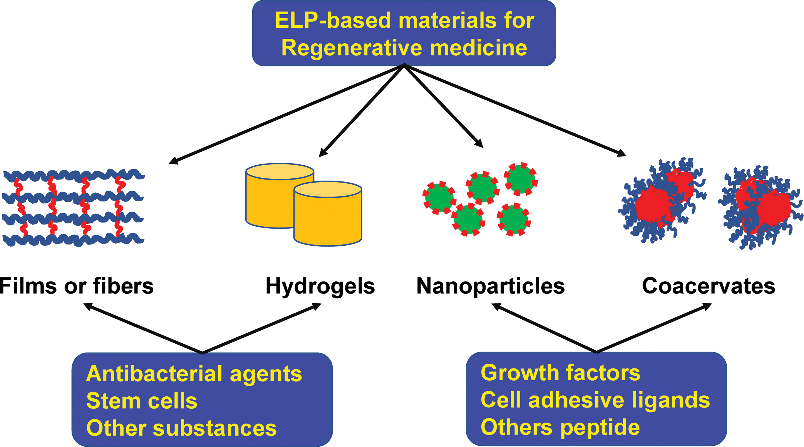
The feasibility and versatile nature of ELPs allow easy formulation into various bioinspired materials such as coacervates, films, hydrogels, and nanoparticles depending on the desired application. Other antibacterial agents, growth factors, stem cells, and peptides, etc., can be incorporated easily with ELP biopolymers to enhance activity in wound healing and can be utilized for the multifunctional approach. Color images are available online.
ELP-based hydrogel
Researchers have generated ELP-based hydrogels through physical, chemical, enzymatic, or photocrosslinking methods for application in wound care. The highly controllable nature of ELPs makes them attractive for preparing bioactive or functionalized hydrogels. Physical cross-linking exploits relatively weak forces such as hydrophobic interaction, electrostatic interaction, and hydrogen bonds for stabilization, yet these weak forces could control the structural properties of the resulting hydrogel. 57 The versatility of the ELP also allows incorporating other protein sequences such as silk-like domains or leucine zippers for β-sheet formation to produce a more stable and reversible hydrogel system. 58,59 However, these physically linked ELP-based hydrogels lack the desired strength essential for regenerative medicine applications. 43 Chemical cross-linking methods, on the other hand, fabricate hydrogels with desirable mechanical properties and tunability depending on the cross-linking amino acid (K) present in the ELP and extendibility of the chemical cross-linkers. The chemical cross-linking mediated by different compounds such as glutaraldehyde, bis (sulfosuccinimidyl) suberate, β-[tris(hydroxymethyl)phosphino] propionic acid, genipin, and pyrroloquinoline quinone was well investigated for biocompatibility. 60 –63 Although these hydrogels carry favorable characteristics and mechanical properties, their applications are limited due to the potential toxicity from cross-linkers.
Interestingly, Zhang et al. have reported photocrosslinkable systems, which can overcome the limitation raised from chemical and enzymatic cross-linking (Fig. 3). 64 They have introduced the technique of incorporation of thiols (KCTS) from a pair of cysteine residues in the ELP sequence to allow disulfide bond formation. The cysteine-containing ELPs, KCTS-E31-KCTS, could be photocrosslinked and form elastic gels after exposure to UV light in the presence of a photoinitiator, Irgacure® 2959 (Fig. 3B). A highly elastic stable hydrogel was formed between 30 s and 3 min of irradiation in relation to its volume based on the inversion test. In an in vivo preclinical study, photocrosslinked ELPs with NP solution promoted hemostasis in standardized liver wounds with acute hemorrhage. The authors further revealed that by controlling ELP concentrations, the physical properties such as mechanical stiffness and swelling behavior of the resulting hydrogel could be easily tuned. These ELP constructs showed extended structural stability in vivo and advanced host integration without triggering the host immune response, thus highlighting their potential for wound repair. Thus, the photocrosslinkable ELP-based system can be preferred for other biomedical applications as a sealant for soft, flexible tissue injuries such as in blood vessels, skin, lungs, or cardiac tissue.
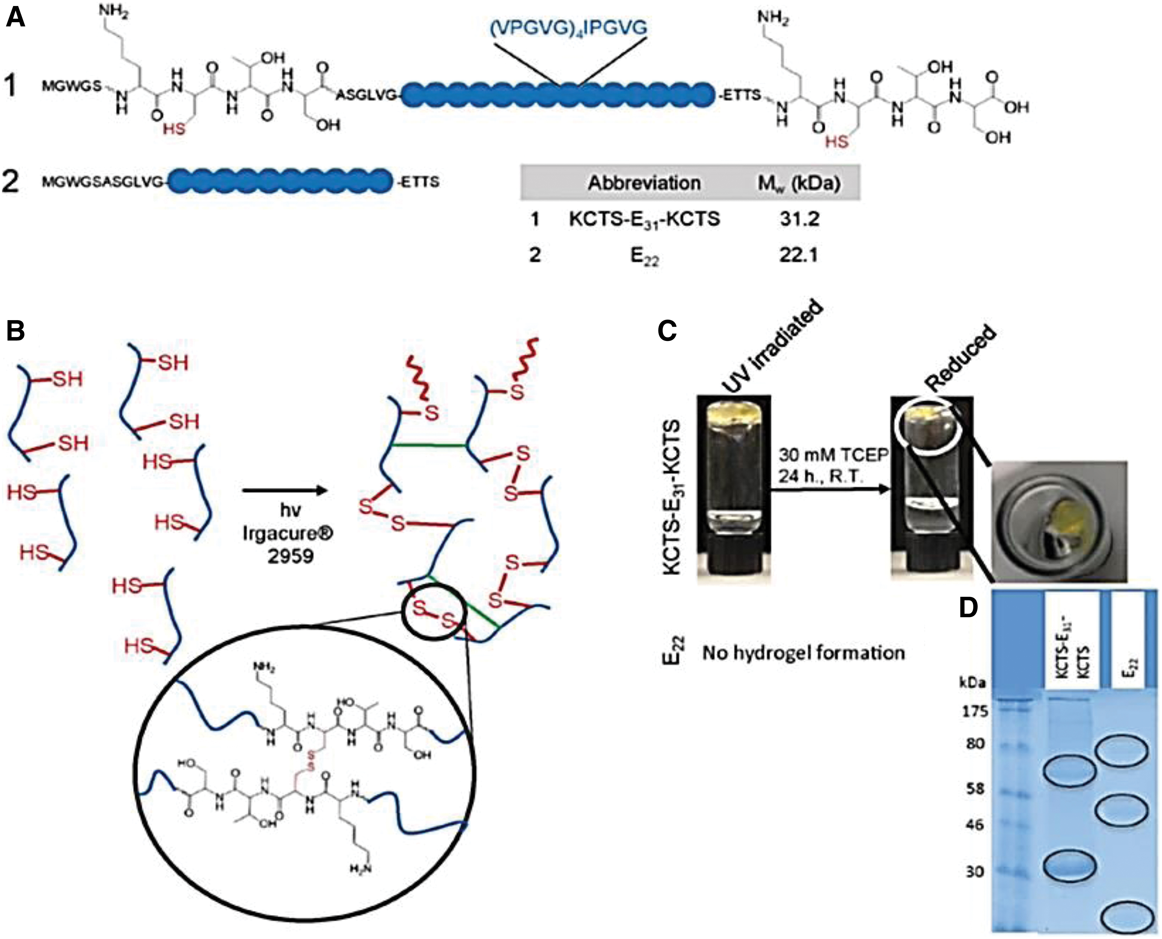
Design and photocrosslinking of ELPs.
Staubli et al. proposed the utilization of bioactive or inert hydrogel to deliver cells of the adipose tissue-derived stromal vascular fraction that holds the ability to directly interact with the host tissue for regeneration. 65 The functionalized elastin-like recombinamer (ELR) hydrogels containing cell adhesion sequences (RGD), endothelial cell-adhesive sequence (REDV), and VGVAPG sequences with elastase sensitivity or chemotactic activity for monocytes help in vascularization and showed greater cell adhesion and induction of vascularization. The VGVAPG sequence favors cell-guided degradation of the hydrogel by recruitment of monocytes that assist in matrix colonization and facilitates the infiltration of endothelial cells as well as fibroblasts. Successful tissue and organ regeneration/repair based on angiogenesis and the immune reaction was controlled accordingly for specific applications.
Likewise, elastin-based hydrogels in combination with other natural protein domains can produce bioinspired materials with mechanical properties (extending from hundreds to millions of Pascals) and cytocompatibility. For example, genetic fusion of ELP with a domain derived from silk–fibroin (SF) renders high tensile strength of silk–fibroin 66 and elasticity and resilience of elastin. 67 The first silk–fibroin and elastin scaffolds (SF/EL) were produced by Vasconcelos et al. for treatment of burn injuries. 63 These scaffolds were cross-linked by genipin, resulting in conformational transition from a random coil to β-sheet of SF chains. Their physical properties were tailored to fabricate hydrogels with different pore sizes, swelling ratios, and degradation and release rates by altering the ratio of SF/EL. SF/EL scaffolds support proliferation of human skin fibroblasts, thereby accelerating reepithelialization and wound closure.
Zhou et al. introduced novel, redox-sensitive protein-based hydrogels with desirable mechanical properties, comprising recombinant silk–elastin-like protein polymers (SELPs). 68 Cysteine-containing SELPs with different ratios with silk blocks rapidly formed hydrogels at body temperature under physiologically relevant, mild oxidative conditions. Redox-sensitive features and tunable mechanical properties of these injectable SELP hydrogels will make them applicable for various purposes in tissue engineering.
Kawabata et al. have described development of a flexible silk–elastin sponge for a clinical setting using the recombinant silk–elastin protein polymer consisting of the SF-derived sequence (GAGAGS) and human elastin-derived sequence (GVGVP). 69 It was found that postadministration, the silk–elastin sponge was absorbed and dissolved by the exudate. A silk–elastin sponge, 12.5 or 25 mg/cm3 in density, could be easily applied to a full-thickness skin wound in guinea pigs to promote stable wound healing. Kawabata et al. further proved that when applied on a full-thickness skin wound of a diabetic mouse in aqueous condition, silk–elastin spread on the wound area, forming a hydrogel coating and maintaining a moist condition without inflammation. 70 This helped in protecting the wound from external bacterial exposure, enhancing granulation tissue formation and epithelialization.
Generally, chronic wounds are highly susceptible to microbial infections, which can delay the healing process, leading to necrosis, sepsis, and even mortality. 29 Thus, incorporation of biocidal agents, cationic polymers, 71 and antimicrobial peptides (AMPs) 72 with a biopolymer may be very useful for prevention of wound infections. Annabi et al. have reported a new composite hydrogel for the treatment of chronic wounds, which is biocompatible, elastic, and sprayable with antimicrobial activity. 73 This composite hydrogel was synthesized using two ECM-derived biopolymers, gelatin methacryloyl (GelMA) and methacryloyl-substituted recombinant human tropoelastin (MeTro), through visible light-induced cross-linking. Antimicrobial activity against Gram-positive and -negative bacteria was provided to the hydrogel network through conjugation of the AMP Tet213. They demonstrated that the system could support proliferation and spreading of fibroblast cells in in vitro two-dimensional or three-dimensional (3D) culture systems. In vivo biocompatibility and biodegradation were confirmed upon subcutaneous implantation of hydrogels in Wistar rats. They further documented the potential of MeTro/GelMA-AMP hydrogels for sutureless wound closure, which could prevent infections and promote healing of chronic wounds.
Delivery of a growth factor and other functional peptides using ELPs
Chronic nonhealing wounds in diabetes and other conditions usually show increased levels of proteases, which degrade the essential growth factors needed for dermal regeneration. 74,75 Therefore, the delivery of a growth factor such as the EGF or inclusion with other materials has shown improvement in healing of such wounds, 14,76 –79 specially diabetic ulcers. 14,80 Using ELP in such conditions has added advantages as it can withstand the fusion of any complex protein without losing the biofunctional property of the fused protein as well as the ELP itself. The ELP provides a shield in the wound area, thereby increasing the half-life of growth factors to support the reepithelialization process. Thus, various viable and effective ELP-based delivery techniques of these growth factors have been generated by many researchers. Recently, Koria et al. have published the strategy of fabricating a fusion protein comprising recombinant human KGF and ELPs for enhanced reepithelialization in the dermal wound. 13 The fusion of KGF with the ELP polymer triggered self-assembly into a nanoparticle-like structure and sustained the functional activity of KGF, which in turn enhanced keratinocyte and fibroblast proliferation. Devalliere et al. showed that the codelivery of two bioactive molecules, KGF and the cellular protective peptide (ARA290), using the ELP-based approach was effective in healing of diabetic wounds. 81 KGF-ELP stimulated proliferation and migration of keratinocytes, while ARA290-ELP protected cells from apoptosis, thereby accelerating the healing process (Fig. 4).
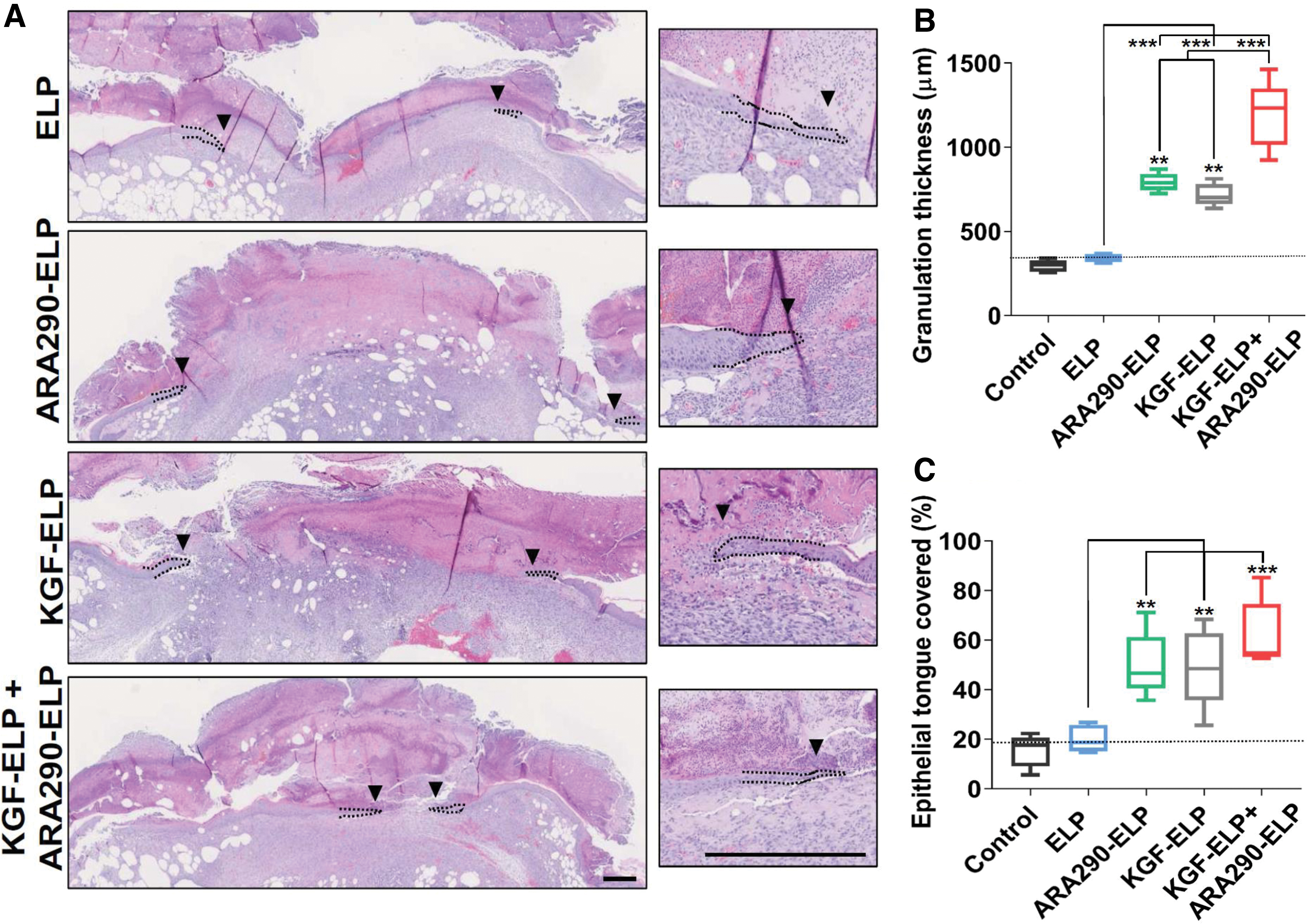
Histological sections and analysis during wound closure. Two, full-thickness, 10-mm diameter punch wounds were created on the dorsum of genetically diabetic mice, and wound analysis was performed 12 days postinjury.
Similarly, Yeboah et al. documented that the fusion of stromal cell-derived factor-1 (SDF1) with ELP increased its stability and it was retained in the chronic wound for a longer time, promoting maximum revascularization. 82 When applied to excisional wounds on diabetic mice, the SDF1-ELP nanoparticles significantly enhanced wound closure, whereas wounds treated with only SDF1 did not accelerate reepithelialization due to loss of its biological activity in the wound bed. Wounds treated with SDF1-ELP nanoparticles were healed with a significantly thicker epidermis and achieved complete closure of the dermal layer in 21 days post-treatment, while SDF required up to 42 days for complete closure. Another growth factor often used for acute wound healing is the EGF. Leonard and Koria reported that the biologically active EGF-ELP fusion protein could overcome the drawback encountered with application of the exogenous EGF. 14 The rapid degradable nature of EGF and relatively easier dispersion from the application site compromised the effective doses of EGF with minimal improvements during skin regeneration. The biological activity of EGF was sustained even after fusion with ELPs, and thermal sensitivity of the ELP led to self-assembled particles at its Tt. These EGF-ELPs promote fibroblasts and epithelial cell proliferation and migration, which are important for granulation and epithelialization in complete wound repair. Not only the growth factor but also more complex ELP composites were fabricated by researchers. Choi and his group have investigated the potential of an RGD-containing ELP (REP), TGPG[VGRGD(VGVPG)6]20WPC, where the cell-adhesive ligand RGD was decorated at regular intervals of the ELP backbone to improve attachment and proliferation of various cells in the treatment of a chronic wound. 83 When implanted into full-thickness excisional wounds in mice along with mouse adipose stem cells (ASCs), it significantly augmented wound closure and reepithelialization (Fig. 5). REP, when mixed with ASCs, triggered formation of a hydrogel-like ASC/REP composite under physiological conditions, enhancing neovascularization, and did not provoke any inflammation as well as immune response. Therefore, REP holds the therapeutic potential for improved stem cell-based therapy with enhanced ability of tissue repair and regeneration.
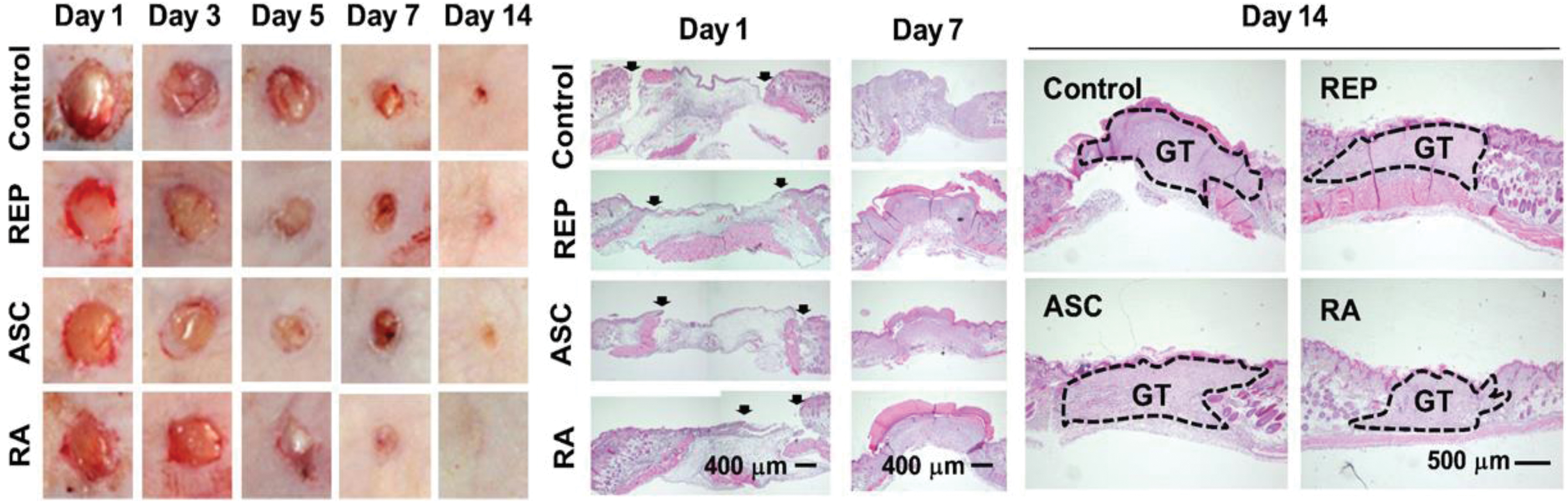
Comparison of wound healing progression among the sham control, REP, ASC, and RA groups over a 2-week period. Sixty nude mice were randomly divided into four groups of 15 mice each. On each mouse, four dorsal wounds were made with an 8-mm biopsy punch. The wounds were filled with 50 μL of PBS, 50 μM REP, and ASCs (1 × 10⁶ cells) or 50 μM REP and ASCs (1 × 10⁶ cells). For histological analysis, tissues were excised with a 10-mm diameter punch. (Left panel) Macroscopic monitoring of wound healing. (Middle and right panels) Microscopic pictures of H&E-stained tissue specimens. Arrows mark the ends of epithelial tongues at day 1. ASC, adipose stem cell; GT, granulation tissue; RA, RGD-containing ELP and adipose stem cells; REP, RGD-containing ELP; (Choi et al. 83 : to be reproduced with permission, Licence no. 4871160258960). Color images are available online.
Electrospun elastin-based scaffolds
ELPs have drawn much attention to create micro- and nanofibers by various methods of fabrication to generate diverse structures with desirable properties. Electrospinning of elastin or elastin composite allows synthesis of nanofibrous, 3D structure scaffolds with accurate porosity, high pore interconnectivity, and biocompatibility mimicking the architecture of native extracellular matrix. 84 –86 Electrospinning is a useful and accessible nanofabrication procedure that has been practiced to manufacture several biomedical systems. 87,88 Blending with polycaprolactone before spinning, 89 silk-like cross-linking domain incorporation at the sequence level, 90 and incorporation of methacrylate for radical-mediated photocrosslinking have been tried to stabilize ELP fibers. 53 Benitez et al. have proposed a novel method of using a two-step cross-linking process that consisted of vapor-phase initiation, followed by aqueous-phase completion of cross-links. 91 The glutaraldehyde vapor was employed to stabilize electrospun fibers and avoid distortion while directing the sequences that could withstand the modification without damaging mechanical or bioactive sequences. Blending of an RGD-containing ELP (ELP-RGD) with another ELP variant that includes a noncell-adhesive scrambled ligand sequence (ELP-RDG) increased cell adhesion and interaction with fibers, which were vital for tissue engineering.
Costa et al. described the application of CM4-A200, a genetically engineered polymer containing AMP ABP-CM4 (isolated from the Chinese silkworm) and the ELR (VPAVG)200, with 200 repetitions of the pentamer VPAVG. 92 CM4-A200 electrospun fiber mats were shown to be more stable with slow degradation. The electrospun CM4-A200 fibers possess high antimicrobial activity with no cytotoxicity effects against normal human skin fibroblasts and keratinocytes. Thus, these materials have a great potential for application in wound healing and skin tissue engineering. De Torre et al. reported the synthesis of bioinspired ELP fibers that were cross-linked in situ using an orthogonal click reaction between azide and cyclooctyne groups present in two different ELR molecules. 93 These bioactive fibers were able to support cell growth due to the presence of RGD motifs and remained completely stable under in vitro conditions. These ELR-click fibers allowed proper adhesion and proliferation of keratinocytes and fibroblasts, the two important cells of the dermis and epidermis layers. This study provides the concept of biomaterial scaffolds based on ELR-click fibers, which can be used directly for wound dressing immediately after the electrospinning process to overlay the damaged skin areas.
Summary
Development of a perfect skin substitute that can effectively repair the wound without scar formation is a great challenge. Different forms of genetically engineered ELPs or ELP fusion proteins with various functional peptides may have great potential in this regard for development of an array of wound care products. These ELP composites with the thermal specific phase transition property have the mechanical strength of elastin and biocompatibility of the extracellular matrix. Coacervation at physiological temperature in the wound site offers temporary or quick wound protection by providing a mechanical barrier to infections. These polymers can be employed as a dermal substitute such as Integra (as a template) for the repair of a partial- or full-thickness wound. These ELPs can be modified relatively easily to combine with other biomolecules, such as growth factors, antibacterial peptides, and stem cells, to accelerate the healing process with prevention of wound infection. A large array of products, including injectable hydrogels, nanoformulations, scaffolds, and electrospun fiber mats, can be made with these novel materials. Thus, the ELP-based system can create a new platform for generation of bioinspired, clinically relevant, cost-effective wound care products suitable for both acute and chronic wound management.
Take-Home Messages
Chronic wound management (with reduction of subsequent scar formation) to avoid functional impairment of tissues remains a major challenge in health care.
Elastin-based biomaterials, due to their unique properties, including their reversible thermoresponsiveness, mechanical strength, and nonimmunogenic and biocompatibility properties, are suitable for various applications in regenerative medicine.
Because ELPs are genetically encoded, their amino acid sequence can be modified for specific functional properties needed for the application.
ELPs are readily expressed in E. coli and purified at high yields (∼500 mg/L growth) by exploiting their inverse temperature phase transition without the need for chromatography.
The easy scale-up process of ELP creates a new platform for low-cost readily available treatment of a wide range of wounds.
ELPs can be used for creation of diverse systems (hydrogels, fibers, sheets, or scaffolds) for clinical application as next-generation skin substitutes for the treatment of chronic wounds.
The highly flexible nature of ELPs allows fabrication of ECM mimic materials, which can be combined with stem cells for regeneration of skin and other tissues.
Footnotes
Acknowledgment and Funding Sources
The authors acknowledge the financial assistance from the Department of Science & Technology (DST) Grant No. SR/WOS-A/LS-136/2018 (G), Govt. of India, provided as a fellowship to Dr. Vijaya Sarangthem.
Author Disclosure and Ghostwriting
The content of this article was mainly written by the authors listed. No ghostwriters were involved in the writing of this article. The authors declared no competing financial interests.
About the Authors