
Abstract


Introduction:
Laparoscopic adrenalectomy is the gold standard for benign adrenal lesions. 1 –3 Successful excision requires proper positioning and meticulous dissection. This video depicts a left adrenalectomy that was removed for an enlarging cystic lesion.
Methods:
A 58-year-old male with a history of hypertension presented to the emergency department with left flank pain while urinating. He denied gross hematuria. He had a remote history of an exploratory laparotomy for a stab wound. Physical exam showed an obese abdomen with a well-healed midline incision, without masses or hernia defects, and without tenderness. A CT scan documented a septated 7 cm left adrenal lesion. The adrenal lesion was biochemically inactive. Based on the size and heterogeneity, an adrenalectomy was recommended. The patient was placed in a full right lateral decubitus position and flexed to open the lateral space. An extensive lysis of adhesions was required due to previous surgery. The adrenalectomy began by incising the peritoneal layer inferior to the spleen to mobilize the distal aspect of the transverse colon. The patient’s anatomy did not require a release of the splenic flexure. The dissection extended lateral to the spleen to rotate the spleen medially until the stomach was visualized superiorly. The dissection was performed 1 cm from the splenic capsule to avoid any injuries and bleeding. The pancreatic tail was then rotated medially also to expose the medial border of the adrenal gland, which was then dissected up towards the diaphragm, mainly through blunt dissection. Following the superior dissection, the lateral dissection of the gland was performed from the kidney. Next, the adrenal vein was exposed, clipped, and divided. The remaining attachments of the lesion to the retroperitoneum were divided. Hemostasis was verified, and the specimen was placed in a bag and extracted.
Results:
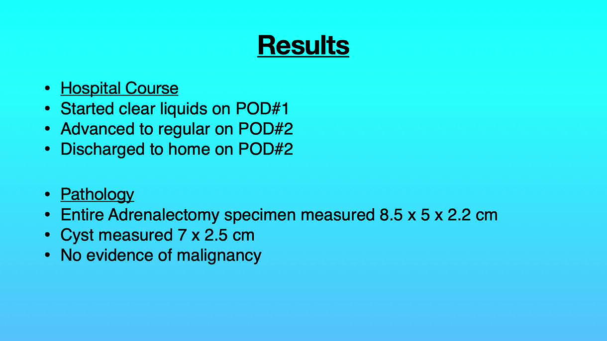
The patient was started on a clear liquid diet on postoperative day 1 and advanced as tolerated. He was discharged home on postoperative day 2. Pathology showed an entire specimen that measured 8.5 cm in largest diameter with a cystic component that measured 7 cm and contained a minimal amount of debris and septa. There was no evidence of malignancy.
Conclusion:
Laparoscopy remains the gold standard for adrenalectomies for benign etiologies. With experience and meticulous dissection, laparoscopic excision may be performed for larger adrenal lesions, even those lesions that may be suspicious for malignancy. If a malignant lesion is encountered, surgeons should have a low threshold to convert to an open procedure.
Disclosures and Patient Consent:
All authors have no commercial, financial, or personal interests to disclose. Authors have received and archived patient consent for video recording/publication in advance of video recording of the procedure.
Runtime of video:
7 mins 47 secs
Get full access to this article
View all access options for this article.