
Abstract
Introduction:
Use of laparoscopy has been described for repair of congenital anomalies, and in patients with anorectal malformations (ARMs), its use is most beneficial when an abdominal approach is required to complete the repair. 1 Cloacal malformations represent the most complex form of ARM, and reports on the use of laparoscopy to fully repair the cloacal anomaly are lacking. 2,3 Our video aims to present a complete laparoscopic repair of a long-channel cloacal malformation through the case presentation of a 13-month-old girl.
Methods/Case Presentation:
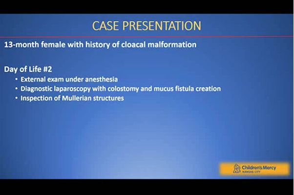
The authors have received and archived patient consent for video recording/publication in advance of video recording of the procedure. Our patient was born at 37 weeks gestational age and found to have a single perineal orifice, consistent with a cloacal malformation. Diagnostic laparoscopy, colostomy creation, and mucus fistula formation were performed on day of life 2; she was discharged after demonstrating appropriate weight gain on oral feedings. A 3D cloacagram was performed at 11 months of age and revealed a common channel length of 4 cm, a urethral length of 1.5 cm, a high insertion of the rectum into the common channel, a duplicated left renal collecting system, and a sacral ratio of 0.70.
To begin the laparoscopic repair, a camera was inserted into the umbilicus, and three 5 mm trocars were placed: one in the left upper quadrant and two in the right lower quadrant. Dissection began by mobilizing the distal rectum off of the urogenital sinus, with the fistula divided sharply at its insertion. The fistula was closed with using Vicryl Endoloop. Dissection of the vagina off of the urogenital sinus was also performed laparoscopically, which included mobilization of the lateral peritoneal attachments to provide a tension-free vaginoplasty. After vaginal mobilization, the posterior urethral defect was reconstructed utilizing 3-0 Vicryl sutures. Inspection of the Mullerian structures revealed two normal cervices and a vaginal septum, which was excised.
A laparoscopic assisted anorectoplasty was performed, pulling the distal rectum into a well-defined sphincter complex. A vaginoplasty and urethroplasty were also performed, incorporating a posterior omega-shaped flap for reconstruction. Indocyanine green fluorescence angiography confirmed tissue perfusion to the rectum, vagina, and urethra. The child was discharged on postoperative day 4 and has since undergone colostomy closure after anorectal examination under anesthesia, cystoscopy, and vaginoscopy, all revealed a healed repair.
Results:
This patient was the first in a series of patients in which an increasing component of the cloacal repair has been performed laparoscopically. Repair was utilized for anorectal, urologic, and gynecologic reconstruction of a long channel cloacal malformation.
Conclusion:
Laparoscopy can be a useful tool for the repair of cloacal malformations as it allows for good observation and complete dissection of the urogenital structures through the same incisions that are required for the anorectoplasty. Understanding candidates for repair are those requiring abdominal mobilization of cloaca components. Further studies are needed to confirm the known benefits of laparoscopy and functional outcomes in the cloacal population.
Patient Consent Statement:
The authors have received and archived patient consent for video recording/publication in advance of video recording of the procedure.
No competing financial interests exist.
Funding:
No funding was received for this article.
Runtime of video: 4 mins 58 secs
This video was presented as a video presentation at the 2021 International Pediatric Endosurgery Group (IPEG) Annual Meeting (Virtual).


Keywords
Get full access to this article
View all access options for this article.