
Abstract
Introduction:
Adenomyoma and leiomyoma are benign uterine lesions commonly encountered by gynecologic surgeons. While often confused with one another, these clinical entities may present with different symptomatology and imaging findings. Pelvic MRI is a key tool to distinguish these two lesions. Although both are amenable to laparoscopic resection, differences in surgical approach are required. 1
Methods:
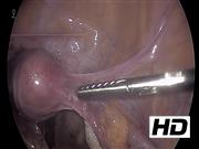
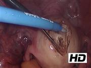
In this video, two separate cases are presented to illustrate the clinical factors and surgical principles. Patient and lesion characteristics at time of presentation are compared. Technical considerations for laparoscopic adenomyomectomy are compared with laparoscopic myomectomy.
Results:
Adenomyomas are characterized by a focus of endometrial glands and stroma, most commonly within the myometrium of the uterus. Symptoms may include heavy menstrual bleeding and pelvic pain as well as potential impact on fertility. Blood products associated with adenomyoma present as T2 hypointensity with corresponding T1 hyperintensity on pelvic MRI. 2,3 In contrast, leiomyomas are benign smooth muscle tumors that may present with heavy menstrual bleeding or bulk-type symptoms with an impact on fertility that is dependent on size and location. On pelvic MRI, leiomyoma typically appears as T2 hypointense lesions without corresponding T1 hyperintensity. 4,5 Laparoscopic resection is a possible treatment option for both lesion types, 2 –5 however, alterations in surgical technique are required. Although both uterine lesions are amenable to laparoscopic resection, the surgical approach differs with significant differences in the surgical planes around the pathology, the tissue characteristics of the pathology itself, and the potential increased difficulty of reapproximation of the adenomyoma resection bed. 1
Conclusion:
Careful assessment of clinical presentation and pelvic MRI findings may help differentiate adenomyoma and leiomyoma, which is important for both patient counseling and surgical planning.
No competing financial interests exist.
Patient Consent: The authors have received and archived patient consent for video recording/publication in advance of video recording of procedure.
Prior presentation: This study was previously presented as a video poster at the 2021 Annual Scientific Meeting of the Society of Gynecologic Surgeons in June 2021.
Runtime of video: 6 mins 43 secs

Keywords
Get full access to this article
View all access options for this article.