
Abstract
Introduction:
Leiomyomas are the most common benign neoplasm of the esophagus. 1 They are extremely rare in children. Surgical enucleation has been the primary mode of treatment, and with the advancement of minimally invasive procedures, laparoscopic and thoracoscopic techniques can provide better alternatives to open surgery, especially in children.
Materials and Methods:
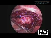
An 11-year-old girl presented with dysphasia of solids and no other symptoms. Contrast swallow revealed a lower esophageal obstruction. CT scan (shown hereunder) and esophagoscopy revealed a lower esophageal and upper gastric mass. She underwent a combined laparoscopic and thoracosopic resection of the mass. In the laparoscopic portion, four ports were employed: one camera port, two working ports, and one port for liver retraction. An esophagoscope was used during the entire procedure to prevent injury to the mucosa. During esophageal mobilization, the posterior vagus nerve was identified and preserved. Esophageal myotomy gradually revealed the leiomyoma and the mass was excised from the esophagus and stomach, but a portion of the mass was inaccessible by laparoscopy. In the thoracoscopic portion, three ports were employed: one camera port and two working ports. The leiomyoma in the lower esophagus was identified and excised after opening the thoracic pleura and performing an esophageal myotomy. The myotomy was closed with interrupted sutures and the pleura was approximated over it.
Results and Conclusions:
There were no apparent complications. Afterward, an upper gastrointestinal contrast study revealed no leaks. CT scan at 3-month follow-up revealed complete removal of lower esophageal and gastric leiomyoma. Minimally invasive procedures are safe and effective options for the excision of benign esophageal masses in children.
No competing financial interests exist.
Runtime of video: 4 mins 44 secs


Get full access to this article
View all access options for this article.