
Abstract
Introduction:
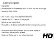
Indocyanine green (ICG) has been in clinical use since 1956 for testing liver and heart function. It has also been used since the 1970s in ophthalmology. Over the past few years, near infrared (NIR) technology has been developed that allows its use as a fluorescence agent during minimally invasive surgery. ICG's safety and excretion profiles have been well established with the summary of product characteristics (SmPCs) documenting a risk of allergic reaction as <1 in 10,000. It is not metabolized in the human body and, is therefore, safe. It is approved by both the Federal Drug Authority and Medicines and Healthcare products Regulatory Agency for intravenous injection for diagnostic purposes. Maximum daily doses have been calculated for ages 0 to 2 years, 2 to 11 years, and >11 years and are defined in the SmPCs. It is licensed for tissue perfusion diagnostics such as hepatic and cardiac function and there are many studies assessing bowel perfusion before and after anastomosis. In adults, clinical studies have evaluated the use of ICG in esophagectomy and colorectal cancer surgery to select a well-perfused anastomotic site along with lymph node harvest. Other studies have suggested a decrease in the rate of anastomotic leak for colonic and colorectal anastomosis from 8.5% to 3.3% when using ICG. In pediatric practice, the use of ICG is still in its infancy with the majority of publications looking at its use in pediatric oncology surgery. This is the first case in the literature documenting its use for colonic anastomosis during emergency surgery.
Description of Video and Patient Details:
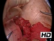
The patient was a 4-year-old girl with a recent diagnosis of acute lymphoblastic leukemia. She had become unwell after administration of the first round of chemotherapy and required admission to the pediatric intensive care unit for respiratory and inotropic support. After clinical improvement, there was noted to be a pneumoperitoneum on abdominal kidney, ureter, and bladder radiograph that was confirmed on CT scanning. The CT scan also showed a suspicion of left colon necrosis. The patient was taken for a midline laparotomy and was found to have complete necrosis of the splenic flexure of the colon. This was resected and a primary anastomosis performed with a covering loop ileostomy. ICG perfusion assessment was undertaken using the ICG VITOM and Rubina OPAL 1 hardware (Karl Storz, Germany) before and after the anastomosis was completed. ICG dose was 0.3 mg/kg in line with product characteristics and license (Renew Health, ROI). She recovered well and was discharged to complete her chemotherapy.
Conclusions:
To our knowledge this is the first report of the use of ICG and NIR for colonic anastomosis assessment in children. More work is required on colonic and colorectal anastomoses with ICG assessment to understand whether this technique results in changes in operative management and whether these subsequently relate to better clinical outcomes in the pediatric age group.
There are no conflicts of interest to disclose.
Patient consent:
Authors have received and archived patient consent for video recording/publication in advance of video recording of the procedure.
Runtime of video:
3 mins 43 secs

Get full access to this article
View all access options for this article.