
Abstract
Introduction:
Primary leiomyosarcoma of the seminal vesicle is an extremely rare disease. There are only 12 total cases of which nine are available in the English language. 1 –11 The 13th case of primary leiomyosarcoma of the seminal vesicle is presented. This is also the first case of a leiomyosarcoma of the seminal vesicle treated using a hand-assisted laparoscopic approach.
Materials and Methods:
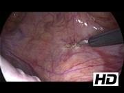
A 62-year-old man was found to have an incidental pelvic mass measuring 9 cm × 6 cm × 7.5 cm by magnetic resonance imaging. The tumor was adjacent to the bladder, prostate, and sigmoid colon, and the seminal vesicles could not be identified. The tumor markers, including alpha fetal protein, carcinoembryonic antigen, cancer antigen 19-9, and prostate-specific antigen were all within normal limits. The differential diagnoses included a seminal vesical adenocarcinoma or sarcoma. He underwent transrectal ultrasound-guided biopsy and leiomyoscarcoma was confirmed by biopsy. He underwent hand-assisted laparoscopic tumor excision and partial cystectomy. A transabdominal laparoscopic approach was conducted for this patient in supine position. One 7-cm periumbilical incision was made for the hand-assisted device application. A 10-mm camera trocar and another 12-mm working trocar were inserted and intraperitoneal insufflation to 15 mm Hg was achieved. The tumor was dissected from the urinary bladder and a partial cystectomy was performed for an adequate surgical margin. The bladder wall was reapproximated in a two-layer running manner with 2-0 chromic catgut suture.
Results:
Total operative time was 105 minutes and total intraoperative blood loss was 100 mL. Time to flatus was 36 hours. His postoperative course was uneventful and total hospital stay was 9 days. Immunohistochemical stains showed positive results for smooth muscle actin, desmin, and Ki-67, whereas negative results were seen for CD34, CD117, S-100, and DOG-1. Pathology analysis confirmed a primary leiomyosarcoma of the seminal vesicle. Adjuvant radiotherapy was conducted 2 months after surgery. Abdominal CT was performed 3 months after radiation therapy and multiple metastatic hepatic lesions were identified and confirmed with biopsy. The patient received adjuvant chemotherapy using ifosfomide followed by oral pazopanib and demonstrated excellent performance at 30-month follow-up.
Discussion:
Primary malignant tumors of seminal vesicle are extremely rare, and most reported cases underwent open surgery for radical tumor removal.7 In this case, we employed a transabdominal approach with a hand-assisted device. A hand-assisted laparoscopic approach affords the advantage of laparoscopic surgery and the tactile feedback of open surgery. For a large tumor in the pelvic cavity, an incision was obviously required to remove the specimen even if a pure laparoscopic approach could have been performed. We feel that this hybrid method reduced operating time compared with a pure laparoscopic approach. The prognosis of a leiomyosarcoma of the seminal vesicle is poor because of the nonspecific presentation, delay in diagnosis, and difficulty in complete surgical excision. In this case, the surgical margin was free from tumor and the follow-up was 30 months. Metastatic disease was controlled by intravenous chemotherapy and oral tyrosine–kinase inhibitor. Our report suggests that a hand-assisted laparoscopic approach is feasible and effective for the treatment of primary leiomyosarcoma of the seminal vesicle.
No competing financial interests exist.
Runtime of video: 7 mins 25 secs
Presented at Scientific Program of 36th World Congress of Endourology Program Book.


Get full access to this article
View all access options for this article.