
Abstract
Background:
Laparoscopic hepaticojejunostomy for choledochal cyst in children is one of the most technically challenging operative procedures. 1 Recent advances in high-definition imaging modalities have improved intraperitoneal visibility, extending indications to meticulous anastomosis for small hepatic ducts. 2 Cases with small hepatic ducts carry a risk of anastomotic stricture after hepaticojejunostomy. Bile duct stenting is sometimes performed for traumatic or iatrogenic injury. 3,4 We herein report a laparoscopic retrograde biliary drainage tube stenting technique of hepaticojejunostomy for a small hepatic duct that prevented anastomotic stenosis.
Case and Operative Procedure:
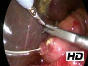
A 1-month-old boy showed obstructive jaundice and infrahepatic cyst on ultrasonography. The patient was suspected of having cystic-type biliary atresia or choledochal cyst and was transferred to our institution. We planned to perform laparoscopic cholangiography followed by a radical operation. Under general anesthesia, the patient was placed in a broad base position, and a 10-mm 30° laparoscope was inserted through an umbilical incision using the open 12-mm Hasson trocar method. Three additional trocars were inserted into the right upper abdomen (3 mm: operator's left hand), right side of the umbilicus (5 mm: operator's right hand), and left lateral abdomen (3 mm: assistant's left hand). Laparoscopic cholangiography revealed choledochal cyst type I with pancreaticobiliary maljunction. We, therefore, performed laparoscopic choledochal cyst excision and hepaticojejunostomy. After cyst excision and preparation of the jejunal limb and Roux-en-Y anastomosis outside of the umbilicus, hepaticojejunostomy was performed. The hepatic duct was 3 mm in size and was enlarged by cutting the bilateral sides of the small hepatic duct. At the start of hepaticojejunostomy, we usually lay down stay sutures at the left side of the hepatic duct (in the 3 o'clock direction). After laying stay sutures using 5-0 monofilament (PDS® Plus; Johnson & Johnson, New Brunswick, NJ, USA), we performed intracorporeal knot tying at the left side of the hepatic duct, and the long tail of the suture was suspended with a needle device (LAPA-HER-CLOSURE; Hakko, Co., Ltd., Tokyo, Japan) inserted at the left upper abdomen. On completing hepaticojejunostomy of the posterior wall, it became evident that the hepaticojejunostomy lumen was small. We, therefore, performed stent insertion as retrograde biliary drainage. A 6F pancreatic tube was inserted from outside of the abdomen, and a small hole was made on the jejunal limb above the transverse colon to insert the tube. After inserting the tube from the jejunal limb, the tip was inserted into the intrahepatic duct as an extra stent before finishing the anterior wall anastomosis. This stent was fixed on the jejunal limb using 6-0 monofilament (PDS Plus; Johnson & Johnson), and the anterior wall hepaticojejunostomy was performed.
Conclusion:
The postoperative course was uneventful. Cholangiography using a stenting tube was performed 3 weeks after the operation. No anastomotic stricture was recognized, and the tube was removed. Four years after surgery, ultrasonography showed no intrahepatic bile duct dilation or intrahepatic ductal gallstones. The liver function remained normal. This laparoscopic retrograde biliary drainage tube stenting technique of hepaticojejunostomy is useful for preventing postoperative anastomotic stricture of a small hepatic duct. Careful long-term follow-up is required.
Acknowledgment:
We thank Brian Quinn for his comments and help with the article.
Funding:
This study was supported by Grants-in-Aid for Scientific Research from the Japan Society for the Promotion of Science (JSPS, Nos. 16K11350, 19K09150), a research grant from JFE (The Japanese Foundation for Research and Promotion of Endoscopy), research grant from J-CASE (Japanese Consortium of Advanced Surgical Endoscopy), research grant from Kyushu Society of Endoscopic and Robotic Surgery, research grant of Karl Storz Award from Japan Society for Endoscopic Surgery, research grant from The Mother and Child Health Foundation, and research grant from the Kawano Masanori Memorial Public Interest Incorporated Foundation for Promotion of Pediatrics.
No competing financial interests exist.
Runtime of video: 6 mins 13 secs
This case report was presented at the 30th Annual Congress of Japanese Society of Endoscopic Surgeons, Kyoto, Japan, 2017.


Get full access to this article
View all access options for this article.